Embed Size (px)
Citation preview

Fysikkens rolle i medisinen og biologien
Bergen, 15. mai, 2009Johan Moan

Nearly all progress in biology and medicine made after Hippocrates is based on application
og physical instruments and methods!!
• Morphology on cell- and tissue level• Understanding of mechanisms• Investigational manipulations
• Diagnosis of diseases• Therapy of diseases

Physics in medicine and biologyThe Italian anatomist and physician LuigiGalvani was one of the first to investigateexperimentally the phenomenon of whatcame to be named "bioelectrogenesis".
Luigi Galvani In a series ofexperiments around1780 he found that theelectric current causedcontractions of themuscles in the leg of a frog.

Microscopy• Transmission, res. 200 nm,Ibn al-Haytam,
(1029); Galilei, (1609); Chr. Huygen, (1680), biology.
• Fluorescence, res. 200 nm.• Confocal laser scanning, spatial modulation
gives res. 10 nm.• Electron microscopy, res. 0.05 nm;trans.,
scanning, freeze fracture.• Scanning tunnelling, res. 0.1 nm or less (1981).

X-ray chrystallography
• Gave us the structure of DNA!!!!• Can someone beat that???

Aakus & Poppe. Medisinsk radiologi i Norge 1995
W. C. Røntgen
X-ray diagnostics

L
K
Elektron hνK
Ionization Elektron transfer Emission of characteristicphoton
X-ray spectrum
hν
T0
T0-hν
Z Z
Bremsstralung
Characteristic radiation
X-ray diagnostics

X-ray diagnosticsX ray tube
patient
detector
X-ray immage

Computer-tomography-CT

COMPUTED MEDICAL IMAGING
”Computed tomography measuresthe attenuation of X-ray beams passing through sections of thebody from hundreds of differentangles, and then a computer is ableto reconstruct pictures of thebody’s interior.”
GODFREY N. HOUNSFIELD,Nobel Lecture, 8 December, 1979
Sir Godfrey N. Hounsfield
Allan M. Cormack

Computer tomography (CT)

Positron emission
tomography
PET

TIME No. 48, 2000
PETPositron emisjons tomografi
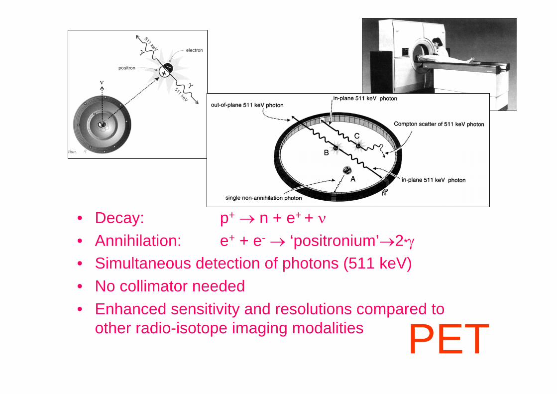
• Decay: p+ → n + e+ + ν• Annihilation: e+ + e- → ‘positronium’→2*γ• Simultaneous detection of photons (511 keV)• No collimator needed• Enhanced sensitivity and resolutions compared to
other radio-isotope imaging modalities PET

Positron emission tomography (PET)
Boellaard (2007) Heart Metab. 34

Positron emission tomography (PET)
http://upload.wikimedia.org/wikipedia/commons/c/c1/PET-schema.png

Nuclear imaging

Nuclear imaging
Gamma camera
• Apply radioactive material, photon emitter, iv, oraly, injection
• Collimated detection

Magnetic resonnance-
MR

B0
Siemens AG. Magnets, spins and resonances 1992
Magnetic resonance imaging
fLarmour~B

• T1 relaxation – how fast do the spins line up with the applied field B0
Magnetic resonanceimaging
• T2 relaxation – how fast do the spins randomize in the XV plane after a pulse coordinating them in one plane

• T1 relaxation• T2 relaxation• T2* relaxation• magnetisation transfer
contrast (MTC)• diffusion• perfusion• blood flow• oxygenation• temperature• spectroscopy of different
atomic species (carbon, hydrogen, phosphor)
• ….
Effective alveolar sizehistogram
3He Diffusion mapping of a 69-year old COPD patient
Courtesy of MD Anderson Cancer Center
Magnetic resonance imaging

Magnetic resonance imaging
vs

MR-spektroskopi - MRSSpektra of tissue in an exact position of the body (tumor, tissue) give a lot of information about composition and reactions taking place
– Energy metabolism:– Lipid metabolism:– pH
Magnetic resonance imaging

Ultrasound
BushbergBushberg, et al. The Essential Physics of , et al. The Essential Physics of Medical Imaging, 2Medical Imaging, 2ndnd ed., p. 509.ed., p. 509.
A-mode “amplitude”mode: displays echo amplitude vs. time (depth) One “A-line” of data per pulse repetition
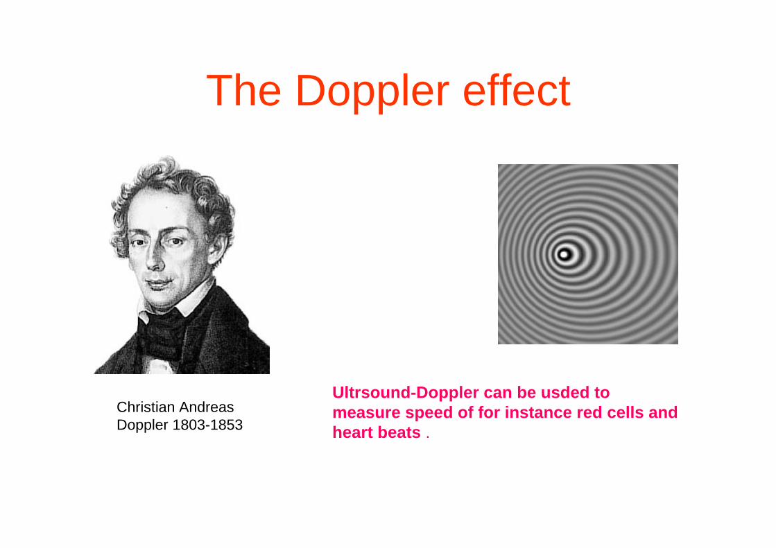
Christian Andreas Doppler 1803-1853
The Doppler effect
Ultrsound-Doppler can be usded to measure speed of for instance red cells and heart beats .

( )2 cosi r id
t
vf f f f
cθ
= − =
BushbergBushberg, et al. The Essential Physics of , et al. The Essential Physics of Medical Imaging, 2Medical Imaging, 2ndnd ed., p. 532.ed., p. 532.
Ultrasound

“Finally, in the biological sciences the rays of radium and its emanation produce interesting effects which are being studied at present. Radium rays have been used in the treatment of certain diseases like lupus, cancer, nervous diseases.”
P I E R R E C U R I ERadioactive substances, especially radiumNobel Lecture, June 6, 1905
Radiation therapy

• Photoelectric effect• Compton scattering• Pair production
Shung et al. Principles of medical imaging 1992
Medical radiation physics

Radiation therapy of cancer
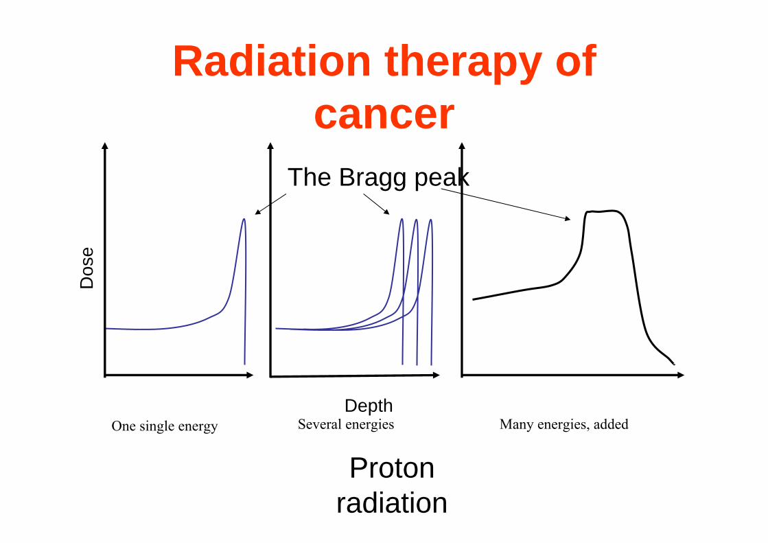
Depth
Dos
e
Proton radiation
Radiation therapy of cancer
The Bragg peak
One single energy Several energies Many energies, added

Dose distribution for protons and photons
Depth (cm)
Dos
e de
posi
tion
photons
protons
tumor
Radiation therapy

Proton therapy
Ordinary radiation therapy
Radiation therapyTumor behind the eye. Proton therapy saves normal tissue

Lasers in Therapy of Human diseases
A new and promising option.Many modes of action.
Many possibilities of optimalization


Excimer - Ar:F 193 nm, Kr:F 248 nm nm (microscopic surgery)Ar - 488, 514 nm (retinal and ear surgery, birthmarks, facial veins)Ruby - 694 nm (freckles, naevus, hair removal)Alexandrite - 755 nm (hair and tattoo removal)Dye - 577-585 nm(vascular lesions, PWS)Diode - 800-900 nm (hair removal, dental surgery, treatment of veins)Nd:YAG - 1064 (tissue cut, hair, tattoo removal), 532 nm (vascular lesions)Ho:YAG - 2070 nm (bone and cartilage ablation, urology, dental fields)Er:YAG - 2940 nm (cosmetic skin resurfacing, dental drill)CO2 - 10600 nm (first laser used by surgeons)
- (moles, warts, keratoses; tumours; wrinkles)


Photothermal damage
R.R.Anderson and J.A.Parish (1983) Science 220, 524-527.
Selective photothermolysis (SP))confine damage to specific tissue structuresby regulating pulse duration and repetition
rate
target 0.1 1-10 100 1000 µmpulse 10-9 10-6 10-3 10-1 s

P.Rol et al. (2000) Graefe’s Arch. Clin. Exp. Ophthalmol. 238, 249-272.A.Pirracchio et al. (2001) J. Bombay Ophthalmol. Assoc. 11, 135-144.
Photothermal therapy in Ophthalmology
Pupillary and retinal melanomasRetinal detachment

C.Gorman (1999) Time 154(18), 60-65
LASIK- laser assisted
in situ keratomileusis
Correction of nearsightedness and
farsightedness
Excimer lasers
Photothermal therapy in Ophthalmology

T.S.Alster et al. (1998) South. Med. J. 91, 806-814.
Vascular diseases
Photocoagulation or PHD ofhemorrhage and bleeding
Photothermal destruction or PDTof atherosclerotic plaques
E.B.Diethrich (2002)

Hair RemovalSelective Absorption in the folliclesDestruction of the follicle structure
K.G.Klavuhn and D.Green (2002) Lasers Surg. Med. 31, 97-105.

Skin rejuvenation
Wrinkle removal
Lasers emittingin µm region(CO2, Er:YAG)
K.Karpowicz (2002)

Action spectroskopyAction spectroskopyis our best tool in is our best tool in
evaluating evaluating
HEALTH EFFECTS OF HEALTH EFFECTS OF SUN AND SUN BED SUN AND SUN BED
EXPOSUREEXPOSURE
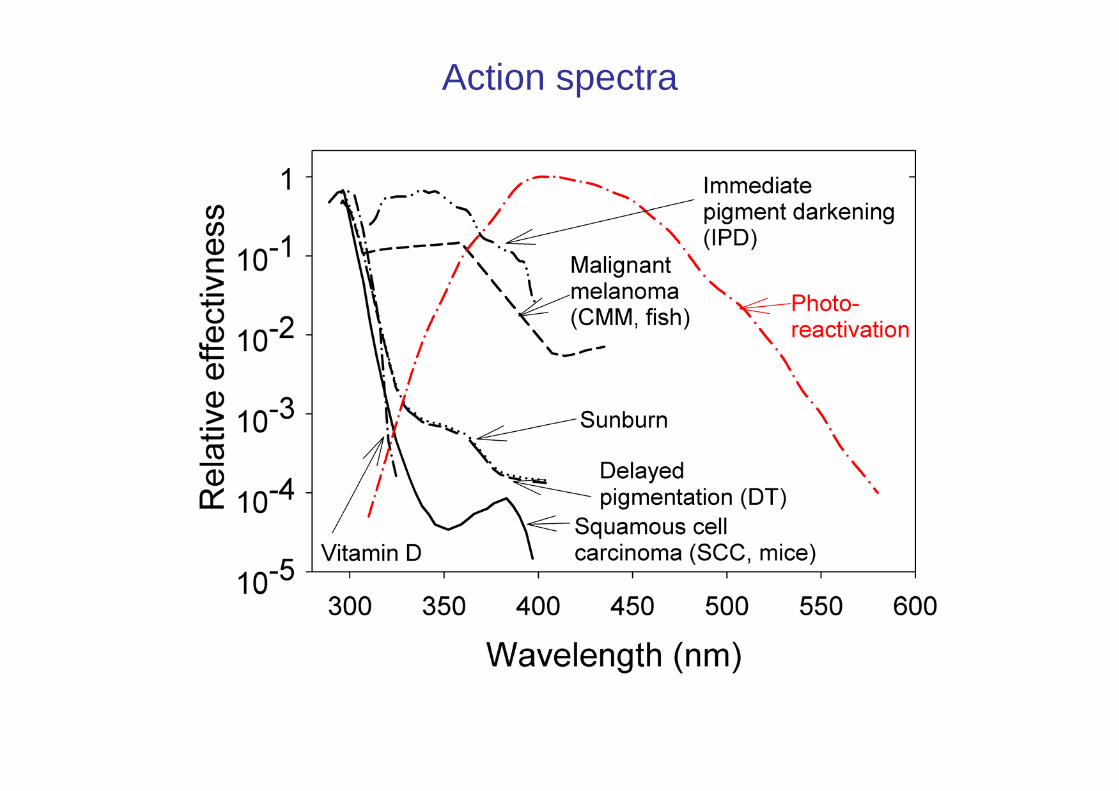
Action spectra

Tid på dagen
4 6 8 10 12 14 16 18 20 22
Føflekk-kreftD vitamin
1.0
0,5
0,0

In vivo Raman Spectroscopy
• Can be used for:
• Diagnosis, for instance of skin cancer• Real time investigations of chemical
reactions in vivo

Fluorescence diagnostics
• The use of fluorescing and localizing dyes for diagnosis amd delineation of malignant or abnormal tissue (Skin, brain, bladder, lung).
• The tissue must be reached by light.


PDT articles published in 1950-2006
http://www.ncbi.nlm.nih.gov/entrez/query.fcgi?DB=pubmed


Peng Q. et al. Cancer (1997) 79:2282-308.
• Topically applied ALA acts deeper and goes systemically in mice.
• Ester derivatives of ALA induce PpIX only in the site of cream application.
• ALA goes to the blood and high contents of ALA in the blood may be neuro- and livertoxic.
Juzeniene A. et al. Photochem. Photobiol. (2002) 76:329-334.
ALA and ALA methylester (MAL) induced PpIX: Penetration depth

PHOTOREJUVENATION

Optical coherence tomography (OCT)
http://en.wikipedia.org/wiki/File:HautFingerspitzeOCT.gif
OCT tomogram of a fingertip
Steiner and Rapp (2007)DOI: 10.1117/2.1200702.0685

Photoacoustic spectroscopy
http://www.ehponline.org/realfiles/docs/1999/107-10/innovations.html
Haisch and Niessner (2002)Spectr. Europe 14

Evanescent wave microscopy(within ≤ 100 nm)
Axelrod (2001) Traffic 2

Evanescent wave microscopy

Optical tweezers
Kimura and Bianco (2006) Analyst 131

Laser microsurgeryof a chromosome
Before After
Liang et al. (1993) Exp. cell res. 204.

Electric Cell-Substrate Impedance Sensing (ECIS)
Ivar Giæver and Cahrles R. Keese (1984)

http://www.nature.com/nnano/journal/v2/n8/full/nnano.2007.223.html
Nanoparticles

Electroporation

Electroporation

Electroporation applies brief electrical pulses, inducing pores to open in the cell membrane and dramatically increasing uptake of useful drugs, genes & DNA vaccines
Therapeutic Platform Based on ElectroporationTherapeutic Platform Based on ElectroporationTherapeutic Platform Based on Electroporation
Cell MembraneBefore Pulse
Cell MembraneDuring Pulse
Cell MembraneAfter Pulse
(Cell returns to original state)
A simple and effective system of delivering drugs or genes into cells
ElectroporationDC

Cancer Therapy Procedure Utilizing EPTCancer Therapy Procedure Utilizing EPTCancer Therapy Procedure Utilizing EPT
Very effective treatment requiring low level of technical skills
DC
Electroporation
Cell Poration, drug enters cells
Injection ofBleomycin
Pulsing theTumor withapplicator
Cells ResealAnd Die
Drug SurroundsTumor Cells
Electroporation Therapy (EPT)

Patient 002: EPT
Electroporation Therapy (EPT) is applied with the applicator.
Electroporation Therapy in Head and Neck Cancer
Electroporation Therapy in Head and Neck Electroporation Therapy in Head and Neck CancerCancer