Embed Size (px)
Citation preview
Rice et al.
Gain-of-function mutations in IFIH1 cause a spectrum of human disease phenotypes associated with upregulated type I interferon signaling Gillian I Rice1,46, Yoandris del Toro Duany2,3,46, Emma M Jenkinson1, Gabriella MA Forte1, Beverley H Anderson1, Giada Ariaudo4,5, Brigitte Bader-Meunier6, Eileen M Baildam7, Roberta Battini8, Michael W Beresford7,9, Manuela Casarano8, Mondher Chouchane10, Rolando Cimaz11, Abigail E Collins12, Nuno JV Cordeiro13, Russell C Dale14, Joyce E Davidson15, Liesbeth De Waele16, Isabelle Desguerre17, Laurence Faivre18, Elisa Fazzi19, Bertrand Isidor20,21, Lieven Lagae16, Andrew R Latchman22, Pierre Lebon23, Chumei Li24, John H Livingston25, Charles M Lourenço26, Maria Margherita Mancardi27, Alice Masurel-Paulet18, Iain B McInnes28, Manoj P Menezes29, Cyril Mignot30, James O’Sullivan1, Simona Orcesi4, Paolo P Picco31, Enrica Riva32, Robert A Robinson33, Diana Rodriguez34-36, Elisabetta Salvatici32, Christiaan Scott37, Marta Szybowska24, John L Tolmie38, Adeline Vanderver39, Catherine Vanhulle40, Jose Pedro Vieira41, Kate Webb37, Robyn N Whitney42, Simon G Williams1, Lynne A Wolfe43, Sameer M Zuberi44,45, Sun Hur2,3,47, Yanick J Crow1,47 1Manchester Academic Health Science Centre, University of Manchester, Genetic Medicine, Manchester, UK 2Department of Biological Chemistry and Molecular Pharmacology, Harvard Medical School, Boston, MA 02115, USA 3Department of Medicine, Boston Children’s Hospital, Boston, MA 02115, USA 4Child Neurology and Psychiatry Unit, C. Mondino National Neurological Institute, Pavia, Italy 5Department of Brain and Behavioral Sciences, Unit of Child Neurology and Psychiatry, University of Pavia, Pavia, Italy
6Department of Pediatric Immunology and Rheumatology, INSERM U 768, Imagine Foundation, APHP, Hôpital Necker, Paris, France 7Department of Paediatric Rheumatology, Alder Hey Children's National Health Service (NHS) Foundation Trust, Liverpool, UK 8Department of Developmental Neuroscience, Istituto di Ricovero e Cura a Carattere Scientifico Stella Maris, Pisa, Italy 9Institute of Translational Medicine, University of Liverpool; Department of Paediatric Rheumatology, Alder Hey Children's NHS Foundation Trust, Liverpool, UK 10Service de Pédiatrie 1, Centre Hospitalier Universitaire (CHU) de Dijon, Dijon, France 11Department of Pediatrics. Azienda Ospedaliero Universitaria Meyer and University of Florence, Italy 12Department of Pediatrics, Division of Pediatric Neurology, University of Colorado, Denver, School of Medicine, USA 13Department of Paediatrics, Rainbow House NHS Ayrshire & Arran, Irvine, Scotland, UK 14Neuroimmunology group, the Children's Hospital at Westmead, University of Sydney, Australia 15Department of Paediatric Rheumatology, Royal Hospital for Sick Children, Glasgow, UK 16Department of Paediatric Neurology, University Hospitals Leuven, Leuven, Belgium 17Department of Pediatric Neurology, INSERM U 768, Imagine Foundation, APHP, Hôpital Necker, Paris, France 18Centre de Génétique, Hôpital d'Enfants, CHU de Dijon et Université de Bourgogne, Dijon, France 19Child Neurology and Psychiatry Unit. Civil Hospital. Department of Clinical and Experimental Sciences, University of Brescia, Italy 20Service de Génétique Médicale, Inserm, CHU Nantes, UMR-S 957, Nantes, France 21INSERM, UMRS 957, Nantes, France 22Division of General Pediatrics, Department of Pediatrics, McMaster Children’s Hospital, McMaster University, Hamilton, Canada 23Université et Faculté de Medecine Paris Descartes, Paris, France
Nature Genetics: doi:10.1038/ng.2933
2
24Department of Pediatrics, Clinical Genetics Program, McMaster Children's Hospital, McMaster University, Hamilton, Canada 25Department of Paediatric Neurology, Leeds Teaching Hospitals NHS Trust, Leeds, UK 26Clinics Hospital of Ribeirao Preto, University of São Paulo, Brasil 27Operative Unit Child Neuropsychiatry, Department of Neuroscience, Giannina Gaslini Institute, Genoa, Italy 28Institute of Infection Immunity and Inflammation, University of Glasgow, Glasgow, UK 29Institute for Neuroscience and Muscle Research, the Children's Hospital at Westmead, University of Sydney, Australia 30AP-HP, Department of Genetics, Groupe Hospitalier Pitié Salpêtrière, F-75013, Paris, France 31Paediatric Rheumatology, Giannina Gaslini Institute, Genoa, Italy 32Clinical Department of Pediatrics, San Paolo Hospital, University of Milan, Italy 33Department of Neurology, Great Ormond Street Hospital for Children, London, UK 34Service de Neuropédiatrie, Centre de Référence de Neurogénétique, Hôpital A. Trousseau, Hôpitaux Universitaire Est Parisien, F-75012 Paris, France 35Université Pierre & Marie Curie Université Paris 06, Paris, France 36Inserm U676, Paris, France 37Department of Paediatric Rheumatology, University of Cape Town, Red Cross War Memorial Children's Hospital, Republic of South Africa 38Department of Clinical Genetics, Southern General Hospital, Glasgow, Scotland, UK 39Department of Paediatric Neurology, Children's National Medical Center, Washington DC, USA 40Service de Néonatalogie et Réanimation, Hôpital Charles Nicolle, CHU Rouen, F-76031 Rouen, France 41Neurology Department. Hospital Dona Estefânia, Centro Hospitalar de Lisboa Central, Portugal 42Division of Pediatric Neurology, Department of Pediatrics, McMaster Children’s Hospital, McMaster University, Hamilton, Canada 43US National Institutes of Health Undiagnosed Diseases Program, Common Fund, Office of the Director, US National Institutes of Health, Bethesda, MD, USA 44Paediatric Neurosciences Research Group, Fraser of Allander Neurosciences Unit, Royal Hospital for Sick Children, Glasgow, UK 45School of Medicine, College of Medical, Veterinary & Life Sciences, University of Glasgow, UK 46These authors contributed equally to this work. 47These authors jointly directed this work. * Corresponding authors: E-mail: [email protected] & [email protected]
Nature Genetics: doi:10.1038/ng.2933
3
Supplementary Note F102. This male child was born to non-consanguineous European Italian parents with no family history of note. He was delivered at 34 weeks gestation by caesarean section indicated because of intrauterine growth retardation (birth weight 1.5 kg; head circumference 29 cm; length 29 cm). In the neonatal period he demonstrated congenital microcephaly, hepatosplenomegaly and thrombocytopenia. He was irritable and fed poorly in the first months of life, and was recognized to have profound developmental delay with spastic-dystonic tetraparesis. Cranial imaging at age 8 months revealed cerebral atrophy with basal ganglia calcifications and white matter disease. At the age of 12 months, interferon activity was recorded as 150 IU/ml and 25 IU/ml in the CSF and serum respectively. At 13 months of age he presented with seizures which were difficult to control. He was also recorded to have hypertension of unknown cause. Autoimmune screening (including ANA, ANCA and ENA antibody testing) at 22 months of age was negative. He died of pneumonia at the age of 2 years. His phenotype is characteristic of Aicardi-Goutières syndrome (AGS). F163. This male child was born to non-consanguineous European French parents with no family history of note. After an unremarkable pregnancy and delivery at 39 weeks gestation (birth weight 2.9 kg; head circumference 34 cm), he presented before the age of 1 month with irritability, feeding difficulties, axial hypotonia and abnormal limb posturing. Cranial CT and MRI scanning around the age of 2 years demonstrated cerebral and cerebellar atrophy with periventricular calcification and abnormal high T2 signal of the deep white matter. At age 52 months his CSF interferon activity was 37 IU/ml. Two months later the level was 25 IU/ml, with serum interferon activity recorded as 50 IU/ml. Further serum samples at the ages of 58 months and 8 years 6 months were 25 and 37 IU/ml respectively. At age 12 years he presented with generalized urticaria and associated oedema which was treated with steroids and antihistamines. Autoimmune investigations revealed only a mildly positive antinuclear factor. Now, at the age of 13 years, he demonstrates a severe quadriplegia, with no head control, no speech, abnormal ocular movements and microcephaly. He is fed by gastrostomy. His phenotype is characteristic of AGS. F237. This male child was born to non-consanguineous white North American parents with no family history of note. His development was considered normal until the age of 15 months, at which time he was riding a push car and had 6 - 10 words. After this point he developed intermittent posturing and rigidity of his legs, and then of the upper extremities. He also developed exaggerated startles which have persisted. He subsequently experienced a relentless loss of motor and intellectual skills, so that by age 24 months he was unable to sit unsupported and had lost the ability to swallow. Between 15 months and 4 years of age he demonstrated a fluctuating pattern of poor sleep, with persistent whining and crying. CSF neopterin was 4 x and 3 x the upper limit of normal at 2 years and 7 years of age respectively. Calcification of the basal ganglia and white matter were observed on cranial CT imaging at age 2 years, with abnormal high signal of the deep white matter seen on T2 weighted MRI. Extensive immunological testing at age 3 years was normal. Now, at the age of 12 years, he has no useful hand function, cannot sit independently and has limited words, although his understanding is relatively preserved. His phenotype conforms to one of rapid onset of neuro-regression with spasticity and dystonia beginning at age 15 months.
Nature Genetics: doi:10.1038/ng.2933
4
F259_1. This male child was born to non-consanguineous European Italian parents with no family history of note. After an unremarkable pregnancy and delivery at 40 weeks gestation (birth weight 3.1 kg; head circumference 34 cm), and an apparently normal early neonatal history, at age 6 months he was noted to demonstrate axial hypotonia and lower limb spasticity. Cranial CT scan at 2 years 6 months of age demonstrated cerebral and cerebellar atrophy with periventricular calcification. MRI at the same age revealed abnormal high T2 signal of the deep white matter. At age 31 months his CSF showed a normal white cell count, but a raised level of interferon activity (50 IU/ ml: serum level of <2IU/ml). He developed chilblains aged 6 years. Now, at the age of 8 years, he is severely delayed with a head circumference of 48 cm. He has never acquired the ability to sit, has minimal hand function (he will reach out for some objects), feeds via gastrostomy, and cannot communicate – although he can smile and cry when apparently content / upset. He has abnormal eye movements with nystagmus, but seems to have some useful vision. He demonstrates marked dystonic posturing of the limbs with axial hypotonia. His phenotype is characteristic of AGS. F259_2. At the age of 48 years, the father of AGS259_1 is completely asymptomatic with no recognized clinical signs. He has not undergone any neuro-imaging. F259_3. Up to the age of 79 years, the paternal grandmother of AGS259_1 has remained healthy throughout her life apart from a squint recognized in infancy, and a left hip replacement for osteoarthritis at age 78 years. She has not undergone any neuro-imaging. F376. This male child was born to non-consanguineous white British parents with no family history of note. He was delivered at 35 weeks gestation indicated because of significant oligohydramnios and intra-uterine growth retardation (birth weight 1.9 kg). The child was irritable, fed poorly, and experienced recurrent chest infections. Developmental delay was obvious by age 4 months, with central hypotonia, peripheral hypertonia and poor head growth. He presented at age 29 months with a 4 week history of recurring fevers, irritability, extensor spasms, developmental regression, weight loss and diarrhea. On examination, in addition to the preexisting neurological signs, he exhibited a florid ulcerative photosensitive vasculitic rash over the face and trunk, hepatosplenomegaly, generalized lymphadenopathy, and arthritis of the knees and ankles. He had serositis with pericardial effusion, a pronounced inflammatory response with raised ESR and CRP, haemolytic anaemia, and markedly abnormal autoantibody profile (ANA >1:640, dsDNA 1200 IU/ml, raised anti-cardiolipin antibodies, low complement C30.53, C4 0.11 g/l, and strongly positive pANCA). An infection and malignancy screen, including for HIV and hepatitis, was negative. At age 30 months his CSF interferon activity was 6 IU/L, with a serum level of 9 IU/L (normal <2). Cranial CT at the same age demonstrated gross cerebral atrophy with basal ganglia and white matter calcification, together with abnormal high signal in the periventricular and deep white matter on T2 weighted imaging. Treatment with a variety of immunosuppressive drugs (steroids, Cyclophosphamide, Infliximab, Rituximab, immunoglobulins and Azathioprine) was associated with an improvement in the quality of his social interactions, skin rash, lymphadenopathy, and blood indices. However, therapeutic weaning was impossible due recurrent flares, particularly of gastrointestinal vasculitis with rectal bleeds. He subsequently died at age 3 years 6 months after possible hemophagocytic lymphohistiocytosis with severe liver derangement (pancytopenia, ferritin of 3700, low fibrinogen). His phenotype is characteristic of AGS associated with a severe lupus-like disease. F524_1. This female child was born to non-consanguineous white British parents. There was no other family history of note beyond the problems affecting her father (see below). She was delivered at term (birth weight 3 kg). Her motor development was delayed, so that at age 21 months she was only just able to maintain a stable sitting position, and she
Nature Genetics: doi:10.1038/ng.2933
5
demonstrated markedly increased tone in her legs. Intellectually she was considered to be normal. In light of her father’s condition a working diagnosis of hereditary spastic paraparesis was suggested. At this point her MRI brain scan revealed non-specific high T2 signal in a periventricular distribution, with no atrophy. Spinal cord MRI was normal. After this time she became non-specifically unwell, with bouts of fever, irritability, vomiting, general lethargy and a suggestion of motor regression. A repeat MRI brain revealed obvious cerebral atrophy and accentuation of white matter changes. At age 3 years and 7 months, she suffered an episode of urinary retention and loss of upper limb function. She was treated empirically with steroids for a diagnosis of transverse myelitis confirmed on MRI which demonstrated the new appearance of increased T2 signal in her cervical and thoracic cord, leading to a diagnosis of transverse myelitis, and obvious cerebral atrophy. She was positive for Aquaporin 4, antinuclear (ANA) and anti dsDNA antibodies. Her CSF neopterin was 15 x the upper limit of normal with no increase in white cells. Further assessment identified evidence of a multisystem inflammatory process with a history of hair loss, marker bilateral palmar erythema, a livedo rash, raised ESR with normal CRP, mildly elevated transaminases, positive lupus anticoagulant and a raised urinary protein/creatinine ratio. She was diagnosed as having a lupus-like illness. She was treated with high dose immunosuppression (pulsed steroids followed by Rituximab) to which she had a marked positive response, and was subsequently maintained on oral prednisolone and mycophenolate mofetil. A repeat MRI brain showed some resolution of the atrophic changes associated with her deterioration. Now, at the age of 5 years, she can commando crawl across the floor but has not acquired the ability to walk independently. She demonstrates increased tone with spasticity in all 4 limbs and clawing of the hands. She has good understanding, but her speech remains dysarthric, and she is considered to have a degree of intellectual delay. Her phenotype conforms to one of congenital lower-limb spasticity, with acute neurological deterioration beginning at age 3 years, and an associated lupus-like disease. F524_2. This male, the father of F524_1, was born to non-consanguineous white British parents. The birth and perinatal period were uneventful, and he acquired all of his early milestones appropriately – sitting at age 6 months and walking independently at just under 1 year old. At age 2 years it was noted that he was toe-walking, and he started to fall more than previously. By the age of 3 years he had been given a diagnosis of cerebral palsy, and between the ages of 4 and 15 years he underwent multiple tendon lengthening operations. His disorder has been very slowly progressive, so that in his teens he was able to play as a goal-keeper for a local football team, whilst he can now only walk with the aid of sticks. At the age of 33 years he demonstrates significant lower limb spasticity, with no involvement of the upper limbs. He is cognitively fully intact, and has experienced no other problems with his health. At this time, he had a borderline positive ANA result (titre of 1 in 160), with normal anti-DNA, anti-Ro, La, Sm, RNP, Scl-70, Jo-1, centromere and ANCA antibody titres. Cardiolipin Ab (IgG) and (IgM), Rheumatoid Factor, ESR and CRP were also normal. He had a normal cranial and spinal MRI at age 29 years. His phenotype conforms to one of childhood-onset lower-limb spasticity which has been very slowly progressive. F626. This male child was born to non-consanguineous European Italian parents with no family history of note. He was delivered at term by elective caesarean section (birth weight 3.3 kg; head circumference 34 cm; length 52 cm). Initial concerns were raised at age 3 months because of poor feeding, for which he was admitted to hospital and noted to have a mild transaminitis. Microvesicular steatosis was seen on a liver biopsy taken at 1 year of age. Serological and infection-related investigations were consistently negative. Transaminase values gradually normalized by the age of 4 years. He sat at age 7 months, started to babble at age 8 - 9 months, and could walk with support by 1 year of age. At age 13 months he experienced a rapid loss of motor and intellectual skills over a period of 1
Nature Genetics: doi:10.1038/ng.2933
6
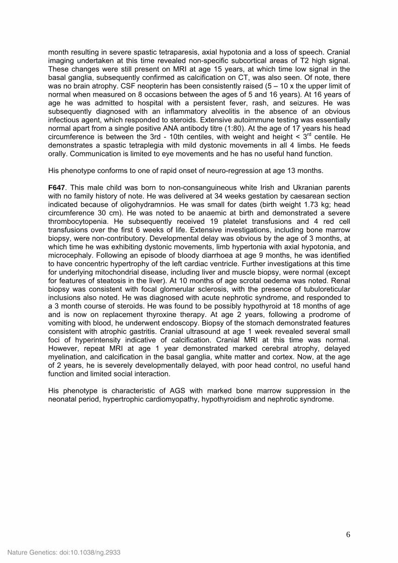
month resulting in severe spastic tetraparesis, axial hypotonia and a loss of speech. Cranial imaging undertaken at this time revealed non-specific subcortical areas of T2 high signal. These changes were still present on MRI at age 15 years, at which time low signal in the basal ganglia, subsequently confirmed as calcification on CT, was also seen. Of note, there was no brain atrophy. CSF neopterin has been consistently raised (5 – 10 x the upper limit of normal when measured on 8 occasions between the ages of 5 and 16 years). At 16 years of age he was admitted to hospital with a persistent fever, rash, and seizures. He was subsequently diagnosed with an inflammatory alveolitis in the absence of an obvious infectious agent, which responded to steroids. Extensive autoimmune testing was essentially normal apart from a single positive ANA antibody titre (1:80). At the age of 17 years his head circumference is between the 3rd - 10th centiles, with weight and height < 3rd centile. He demonstrates a spastic tetraplegia with mild dystonic movements in all 4 limbs. He feeds orally. Communication is limited to eye movements and he has no useful hand function. His phenotype conforms to one of rapid onset of neuro-regression at age 13 months. F647. This male child was born to non-consanguineous white Irish and Ukranian parents with no family history of note. He was delivered at 34 weeks gestation by caesarean section indicated because of oligohydramnios. He was small for dates (birth weight 1.73 kg; head circumference 30 cm). He was noted to be anaemic at birth and demonstrated a severe thrombocytopenia. He subsequently received 19 platelet transfusions and 4 red cell transfusions over the first 6 weeks of life. Extensive investigations, including bone marrow biopsy, were non-contributory. Developmental delay was obvious by the age of 3 months, at which time he was exhibiting dystonic movements, limb hypertonia with axial hypotonia, and microcephaly. Following an episode of bloody diarrhoea at age 9 months, he was identified to have concentric hypertrophy of the left cardiac ventricle. Further investigations at this time for underlying mitochondrial disease, including liver and muscle biopsy, were normal (except for features of steatosis in the liver). At 10 months of age scrotal oedema was noted. Renal biopsy was consistent with focal glomerular sclerosis, with the presence of tubuloreticular inclusions also noted. He was diagnosed with acute nephrotic syndrome, and responded to a 3 month course of steroids. He was found to be possibly hypothyroid at 18 months of age and is now on replacement thyroxine therapy. At age 2 years, following a prodrome of vomiting with blood, he underwent endoscopy. Biopsy of the stomach demonstrated features consistent with atrophic gastritis. Cranial ultrasound at age 1 week revealed several small foci of hyperintensity indicative of calcification. Cranial MRI at this time was normal. However, repeat MRI at age 1 year demonstrated marked cerebral atrophy, delayed myelination, and calcification in the basal ganglia, white matter and cortex. Now, at the age of 2 years, he is severely developmentally delayed, with poor head control, no useful hand function and limited social interaction. His phenotype is characteristic of AGS with marked bone marrow suppression in the neonatal period, hypertrophic cardiomyopathy, hypothyroidism and nephrotic syndrome.
Nature Genetics: doi:10.1038/ng.2933
7
Supplementary Figures
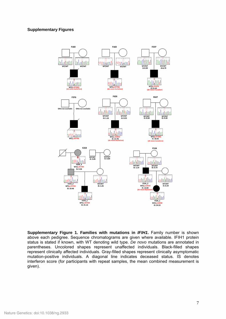
Supplementary Figure 1. Families with mutations in IFIH1. Family number is shown above each pedigree. Sequence chromatograms are given where available. IFIH1 protein status is stated if known, with WT denoting wild type. De novo mutations are annotated in parentheses. Uncolored shapes represent unaffected individuals. Black-filled shapes represent clinically affected individuals. Gray-filled shapes represent clinically asymptomatic mutation-positive individuals. A diagonal line indicates deceased status. IS denotes interferon score (for participants with repeat samples, the mean combined measurement is given).
Nature Genetics: doi:10.1038/ng.2933
8
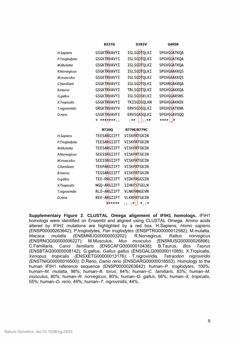
Supplementary Figure 2. CLUSTAL Omega alignment of IFIH1 homologs. IFIH1 homologs were identified on Ensembl and aligned using CLUSTAL Omega. Amino acids altered by IFIH1 mutations are highlighted by a red box. H.Sapiens, Homo sapiens (ENSP00000263642); P.troglodytes, Pan troglodytes (ENSPTRG00000012582); M.mulatta, Macaca mulatta (ENSMMUG00000003202); R.Norvegicus, Rattus norvegicus (ENSRNOG00000006227); M.Musculus, Mus musculus (ENSMUSG00000026896); C.Familiaris, Canis familiaris (ENSCAFG00000010438); B.Taurus, Bos Taurus (ENSBTAG00000008142); G.gallus, Gallus gallus (ENSGALG00000011089); X.Tropicalis, Xenopus tropicalis (ENSXETG00000013176); T.nigroviridis, Tetraodon nigroviridis (ENSTNIG00000016500); D.Rerio, Danio rerio (ENSDARG00000018553). Homology to the human IFIH1 reference sequence (ENSP00000263642): human–P. troglodytes, 100%; human–M. mulatta, 98%; human–B. torus, 84%; human–C. familiaris, 83%; human–M. musculus, 80%; human–R. norvegicus, 80%; human–G. gallus, 56%; human–X. tropicalis, 55%; human–D. rerio, 49%; human–T. nigroviridis, 44%.
Nature Genetics: doi:10.1038/ng.2933
9
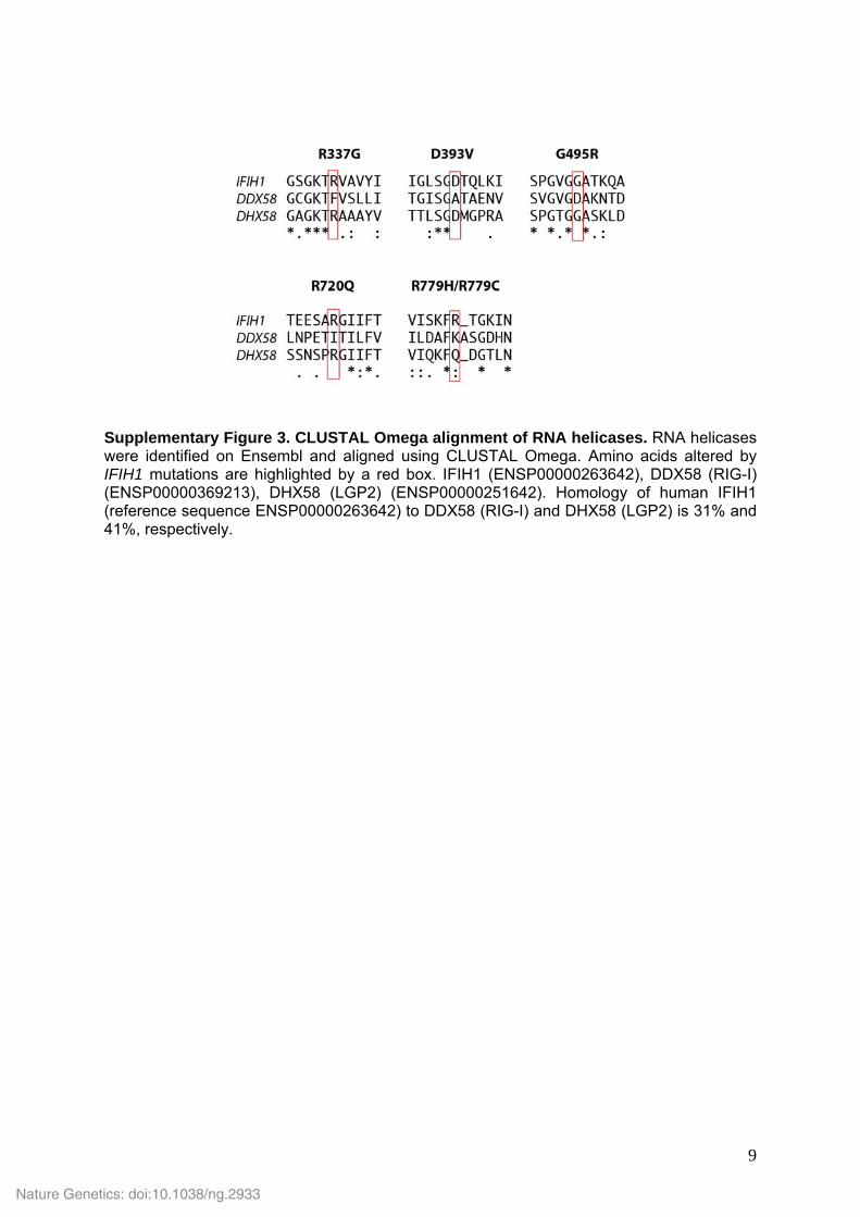
Supplementary Figure 3. CLUSTAL Omega alignment of RNA helicases. RNA helicases were identified on Ensembl and aligned using CLUSTAL Omega. Amino acids altered by IFIH1 mutations are highlighted by a red box. IFIH1 (ENSP00000263642), DDX58 (RIG-I) (ENSP00000369213), DHX58 (LGP2) (ENSP00000251642). Homology of human IFIH1 (reference sequence ENSP00000263642) to DDX58 (RIG-I) and DHX58 (LGP2) is 31% and 41%, respectively.
Nature Genetics: doi:10.1038/ng.2933
10
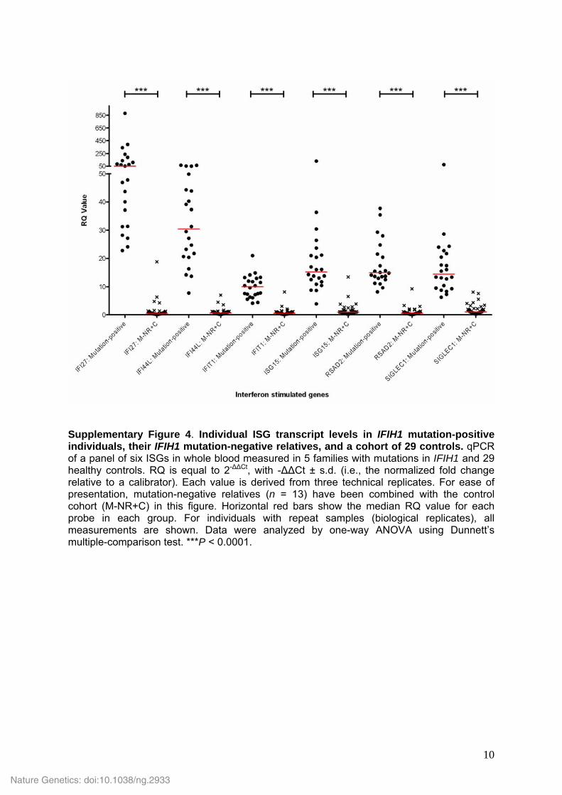
Supplementary Figure 4. Individual ISG transcript levels in IFIH1 mutation-positive individuals, their IFIH1 mutation-negative relatives, and a cohort of 29 controls. qPCR of a panel of six ISGs in whole blood measured in 5 families with mutations in IFIH1 and 29 healthy controls. RQ is equal to 2-ΔΔCt, with -ΔΔCt ± s.d. (i.e., the normalized fold change relative to a calibrator). Each value is derived from three technical replicates. For ease of presentation, mutation-negative relatives (n = 13) have been combined with the control cohort (M-NR+C) in this figure. Horizontal red bars show the median RQ value for each probe in each group. For individuals with repeat samples (biological replicates), all measurements are shown. Data were analyzed by one-way ANOVA using Dunnett’s multiple-comparison test. ***P < 0.0001.
Nature Genetics: doi:10.1038/ng.2933
11
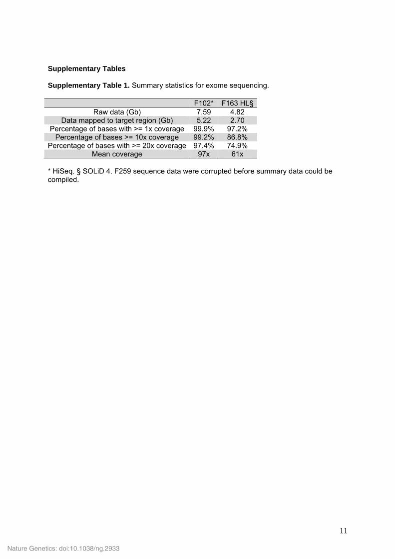
Supplementary Tables Supplementary Table 1. Summary statistics for exome sequencing.
F102* F163 HL§Raw data (Gb) 7.59 4.82
Data mapped to target region (Gb) 5.22 2.70 Percentage of bases with >= 1x coverage 99.9% 97.2%
Percentage of bases >= 10x coverage 99.2% 86.8% Percentage of bases with >= 20x coverage 97.4% 74.9%
Mean coverage 97x 61x * HiSeq. § SOLiD 4. F259 sequence data were corrupted before summary data could be compiled.
Nature Genetics: doi:10.1038/ng.2933
Rice et al.
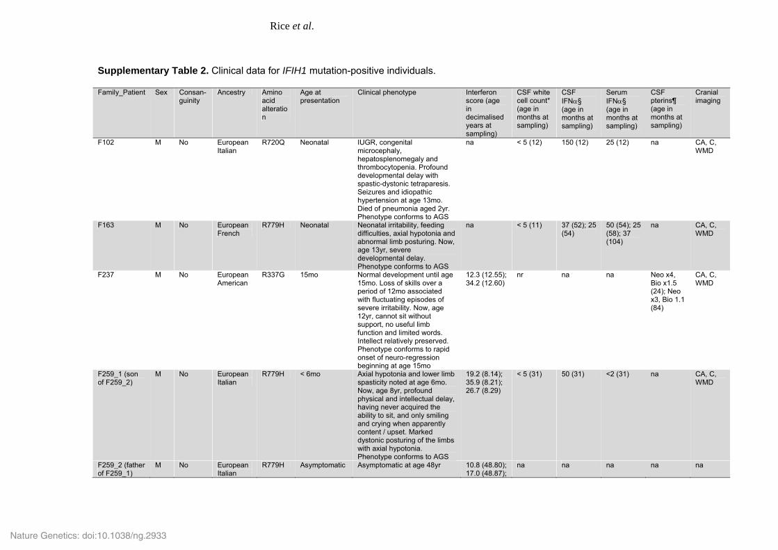
Supplementary Table 2. Clinical data for IFIH1 mutation-positive individuals. Family_Patient Sex Consan-
guinity Ancestry Amino
acid alteration
Age at presentation
Clinical phenotype Interferon score (age in decimalised years at sampling)
CSF white cell count* (age in months at sampling)
CSF IFN᧠(age in months at sampling)
Serum IFN᧠(age in months at sampling)
CSF pterins¶ (age in months at sampling)
Cranial imaging
F102 M No European Italian
R720Q Neonatal IUGR, congenital microcephaly, hepatosplenomegaly and thrombocytopenia. Profound developmental delay with spastic-dystonic tetraparesis. Seizures and idiopathic hypertension at age 13mo. Died of pneumonia aged 2yr. Phenotype conforms to AGS
na < 5 (12) 150 (12) 25 (12) na CA, C, WMD
F163 M No European French
R779H Neonatal Neonatal irritability, feeding difficulties, axial hypotonia and abnormal limb posturing. Now, age 13yr, severe developmental delay. Phenotype conforms to AGS
na < 5 (11) 37 (52); 25 (54)
50 (54); 25 (58); 37 (104)
na CA, C, WMD
F237 M No European American
R337G 15mo Normal development until age 15mo. Loss of skills over a period of 12mo associated with fluctuating episodes of severe irritability. Now, age 12yr, cannot sit without support, no useful limb function and limited words. Intellect relatively preserved. Phenotype conforms to rapid onset of neuro-regression beginning at age 15mo
12.3 (12.55); 34.2 (12.60)
nr na na Neo x4, Bio x1.5 (24); Neo x3, Bio 1.1 (84)
CA, C, WMD
F259_1 (son of F259_2)
M No European Italian
R779H < 6mo Axial hypotonia and lower limb spasticity noted at age 6mo. Now, age 8yr, profound physical and intellectual delay, having never acquired the ability to sit, and only smiling and crying when apparently content / upset. Marked dystonic posturing of the limbs with axial hypotonia. Phenotype conforms to AGS
19.2 (8.14); 35.9 (8.21); 26.7 (8.29)
< 5 (31) 50 (31) <2 (31) na CA, C, WMD
F259_2 (father of F259_1)
M No European Italian
R779H Asymptomatic Asymptomatic at age 48yr 10.8 (48.80); 17.0 (48.87);
na na na na na
Nature Genetics: doi:10.1038/ng.2933
13
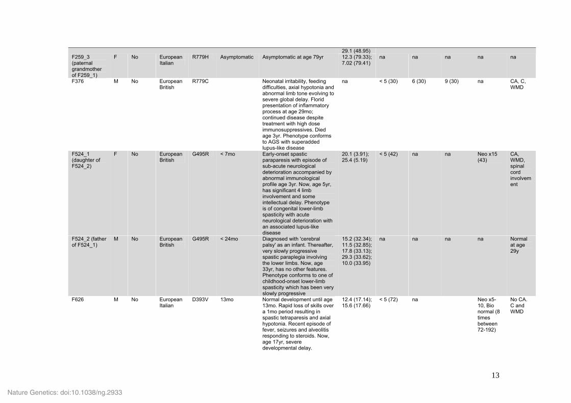
29.1 (48.95) F259_3 (paternal grandmother of F259_1)
F No European Italian
R779H Asymptomatic Asymptomatic at age 79yr 12.3 (79.33); 7.02 (79.41)
na na na na na
F376 M No European British
R779C Neonatal irritability, feeding difficulties, axial hypotonia and abnormal limb tone evolving to severe global delay. Florid presentation of inflammatory process at age 29mo; continued disease despite treatment with high dose immunosuppressives. Died age 3yr. Phenotype conforms to AGS with superadded lupus-like disease
na < 5 (30) 6 (30) 9 (30) na CA, C, WMD
F524_1 (daughter of F524_2)
F No European British
G495R < 7mo Early-onset spastic paraparesis with episode of sub-acute neurological deterioration accompanied by abnormal immunological profile age 3yr. Now, age 5yr, has significant 4 limb involvement and some intellectual delay. Phenotype is of congenital lower-limb spasticity with acute neurological deterioration with an associated lupus-like disease
20.1 (3.91); 25.4 (5.19)
< 5 (42) na na Neo x15 (43)
CA, WMD, spinal cord involvement
F524_2 (father of F524_1)
M No European British
G495R < 24mo Diagnosed with 'cerebral palsy' as an infant. Thereafter, very slowly progressive spastic paraplegia involving the lower limbs. Now, age 33yr, has no other features. Phenotype conforms to one of childhood-onset lower-limb spasticity which has been very slowly progressive
15.2 (32.34); 11.5 (32.85); 17.8 (33.13); 29.3 (33.62); 10.0 (33.95)
na na na na Normal at age 29y
F626 M No European Italian
D393V 13mo Normal development until age 13mo. Rapid loss of skills over a 1mo period resulting in spastic tetraparesis and axial hypotonia. Recent episode of fever, seizures and alveolitis responding to steroids. Now, age 17yr, severe developmental delay.
12.4 (17.14); 15.6 (17.66)
< 5 (72) na Neo x5-10, Bio normal (8 times between 72-192)
No CA. C and WMD
Nature Genetics: doi:10.1038/ng.2933
14
Phenotype conforms to rapid onset of neuro-regression beginning at age 13mo
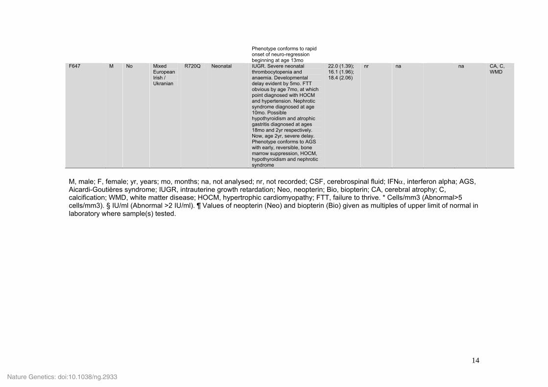
F647 M No Mixed European Irish / Ukranian
R720Q Neonatal IUGR. Severe neonatal thrombocytopenia and anaemia. Developmental delay evident by 5mo. FTT obvious by age 7mo, at which point diagnosed with HOCM and hypertension. Nephrotic syndrome diagnosed at age 10mo. Possible hypothyroidism and atrophic gastritis diagnosed at ages 18mo and 2yr respectively. Now, age 2yr, severe delay. Phenotype conforms to AGS with early, reversible, bone marrow suppression, HOCM, hypothyroidism and nephrotic syndrome
22.0 (1.39); 16.1 (1.96); 18.4 (2.06)
nr na na CA, C, WMD
M, male; F, female; yr, years; mo, months; na, not analysed; nr, not recorded; CSF, cerebrospinal fluid; IFNα, interferon alpha; AGS, Aicardi-Goutières syndrome; IUGR, intrauterine growth retardation; Neo, neopterin; Bio, biopterin; CA, cerebral atrophy; C, calcification; WMD, white matter disease; HOCM, hypertrophic cardiomyopathy; FTT, failure to thrive. * Cells/mm3 (Abnormal>5 cells/mm3). § IU/ml (Abnormal >2 IU/ml). ¶ Values of neopterin (Neo) and biopterin (Bio) given as multiples of upper limit of normal in laboratory where sample(s) tested.
Nature Genetics: doi:10.1038/ng.2933
Rice et al.
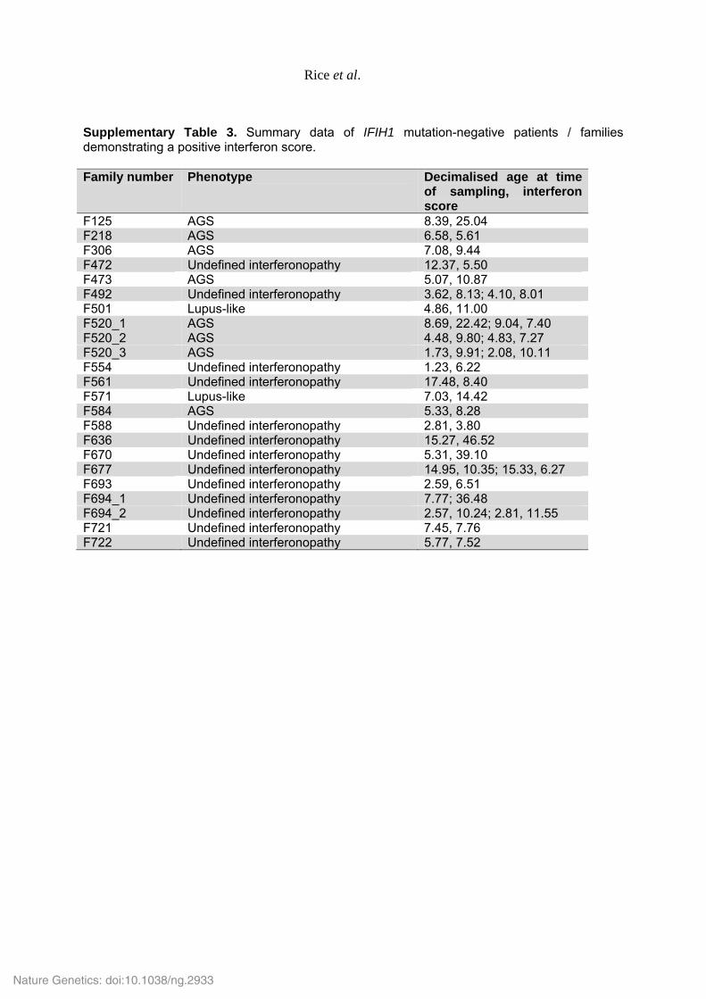
Supplementary Table 3. Summary data of IFIH1 mutation-negative patients / families demonstrating a positive interferon score. Family number Phenotype Decimalised age at time
of sampling, interferon score
F125 AGS 8.39, 25.04 F218 AGS 6.58, 5.61 F306 AGS 7.08, 9.44 F472 Undefined interferonopathy 12.37, 5.50 F473 AGS 5.07, 10.87 F492 Undefined interferonopathy 3.62, 8.13; 4.10, 8.01 F501 Lupus-like 4.86, 11.00 F520_1 AGS 8.69, 22.42; 9.04, 7.40 F520_2 AGS 4.48, 9.80; 4.83, 7.27 F520_3 AGS 1.73, 9.91; 2.08, 10.11 F554 Undefined interferonopathy 1.23, 6.22 F561 Undefined interferonopathy 17.48, 8.40 F571 Lupus-like 7.03, 14.42 F584 AGS 5.33, 8.28 F588 Undefined interferonopathy 2.81, 3.80 F636 Undefined interferonopathy 15.27, 46.52 F670 Undefined interferonopathy 5.31, 39.10 F677 Undefined interferonopathy 14.95, 10.35; 15.33, 6.27 F693 Undefined interferonopathy 2.59, 6.51 F694_1 Undefined interferonopathy 7.77; 36.48 F694_2 Undefined interferonopathy 2.57, 10.24; 2.81, 11.55 F721 Undefined interferonopathy 7.45, 7.76 F722 Undefined interferonopathy 5.77, 7.52
Nature Genetics: doi:10.1038/ng.2933
Rice et al.
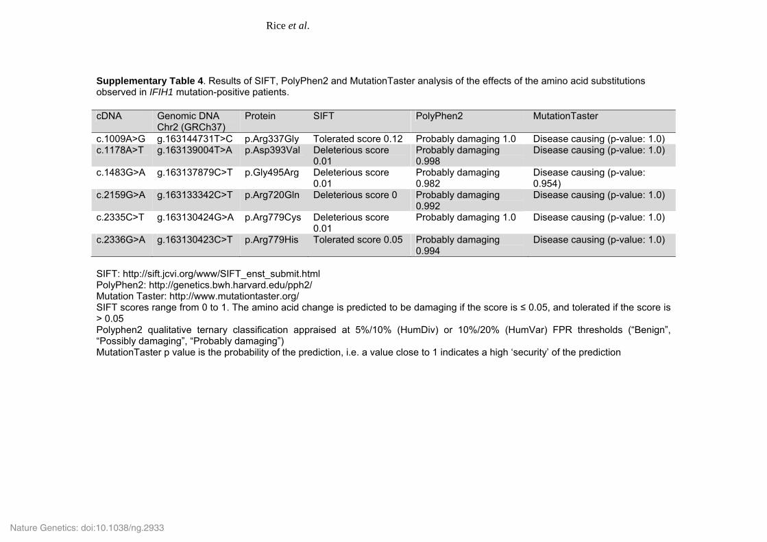
Supplementary Table 4. Results of SIFT, PolyPhen2 and MutationTaster analysis of the effects of the amino acid substitutions observed in IFIH1 mutation-positive patients. cDNA Genomic DNA
Chr2 (GRCh37) Protein SIFT PolyPhen2 MutationTaster
c.1009A>G g.163144731T>C p.Arg337Gly Tolerated score 0.12 Probably damaging 1.0 Disease causing (p-value: 1.0) c.1178A>T g.163139004T>A p.Asp393Val Deleterious score
0.01 Probably damaging 0.998
Disease causing (p-value: 1.0)
c.1483G>A g.163137879C>T p.Gly495Arg Deleterious score 0.01
Probably damaging 0.982
Disease causing (p-value: 0.954)
c.2159G>A g.163133342C>T p.Arg720Gln Deleterious score 0 Probably damaging 0.992
Disease causing (p-value: 1.0)
c.2335C>T g.163130424G>A p.Arg779Cys Deleterious score 0.01
Probably damaging 1.0 Disease causing (p-value: 1.0)
c.2336G>A g.163130423C>T p.Arg779His Tolerated score 0.05 Probably damaging 0.994
Disease causing (p-value: 1.0)
SIFT: http://sift.jcvi.org/www/SIFT_enst_submit.html PolyPhen2: http://genetics.bwh.harvard.edu/pph2/ Mutation Taster: http://www.mutationtaster.org/ SIFT scores range from 0 to 1. The amino acid change is predicted to be damaging if the score is ≤ 0.05, and tolerated if the score is > 0.05 Polyphen2 qualitative ternary classification appraised at 5%/10% (HumDiv) or 10%/20% (HumVar) FPR thresholds (“Benign”, “Possibly damaging”, “Probably damaging”) MutationTaster p value is the probability of the prediction, i.e. a value close to 1 indicates a high ‘security’ of the prediction
Nature Genetics: doi:10.1038/ng.2933
17
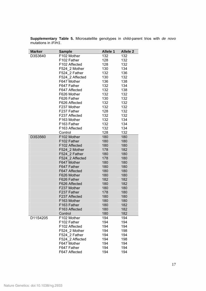
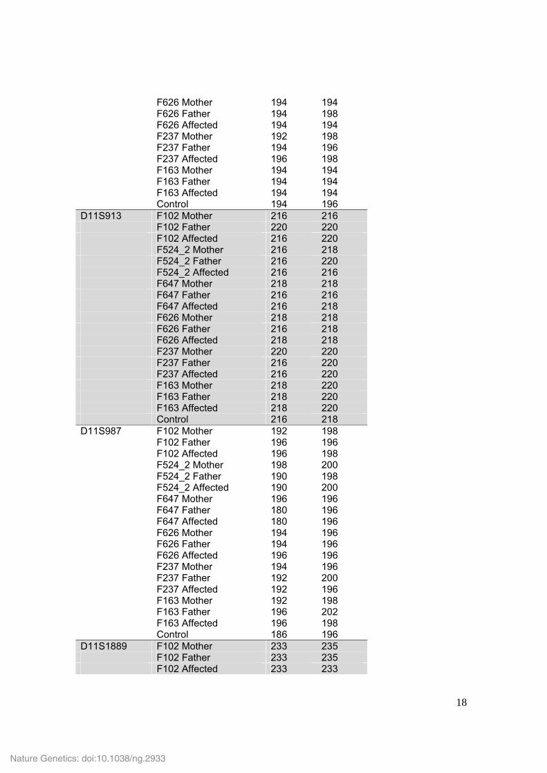
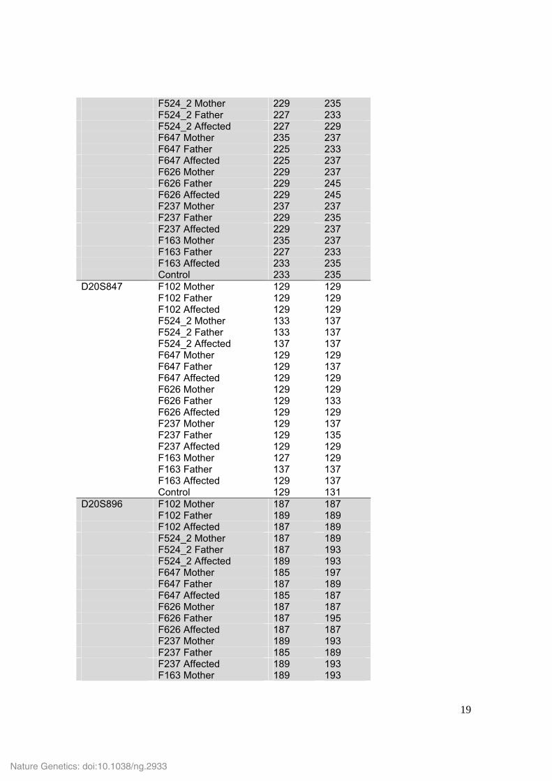
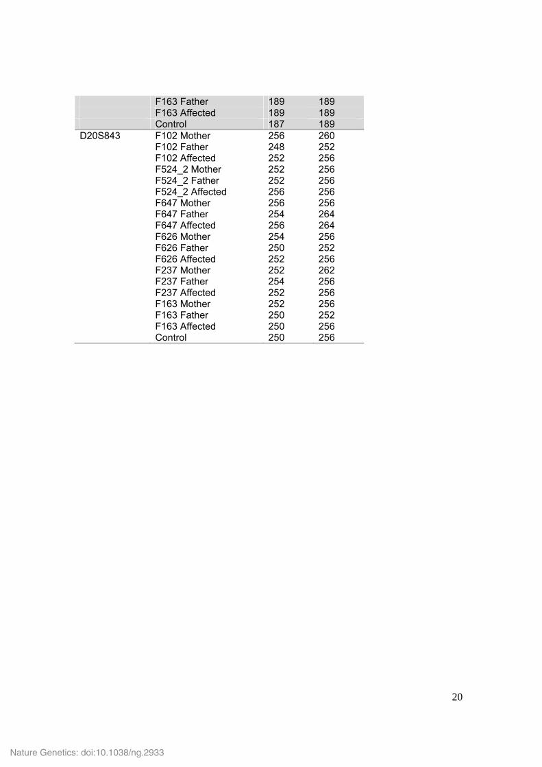
Supplementary Table 5. Microsatellite genotypes in child-parent trios with de novo mutations in IFIH1. Marker Sample Allele 1 Allele 2 D3S3640 F102 Mother 132 132 F102 Father 128 132 F102 Affected 128 132 F524_2 Mother 130 134 F524_2 Father 132 136 F524_2 Affected 130 132 F647 Mother 136 138 F647 Father 132 134 F647 Affected 132 138 F626 Mother 132 132 F626 Father 130 132 F626 Affected 132 132 F237 Mother 132 132 F237 Father 128 132 F237 Affected 132 132 F163 Mother 132 134 F163 Father 132 134 F163 Affected 132 134 Control 128 132 D3S3560 F102 Mother 180 180 F102 Father 180 180 F102 Affected 180 180 F524_2 Mother 178 182 F524_2 Father 180 180 F524_2 Affected 178 180 F647 Mother 180 180 F647 Father 180 180 F647 Affected 180 180 F626 Mother 180 180 F626 Father 182 182 F626 Affected 180 182 F237 Mother 180 180 F237 Father 178 180 F237 Affected 180 180 F163 Mother 180 180 F163 Father 180 182 F163 Affected 180 182 Control 180 182 D11S4205 F102 Mother 194 194 F102 Father 194 194 F102 Affected 194 194 F524_2 Mother 194 198 F524_2 Father 194 194 F524_2 Affected 194 198 F647 Mother 194 194 F647 Father 194 194 F647 Affected 194 194
Nature Genetics: doi:10.1038/ng.2933
18
F626 Mother 194 194 F626 Father 194 198 F626 Affected 194 194 F237 Mother 192 198 F237 Father 194 196 F237 Affected 196 198 F163 Mother 194 194 F163 Father 194 194 F163 Affected 194 194 Control 194 196 D11S913 F102 Mother 216 216 F102 Father 220 220 F102 Affected 216 220 F524_2 Mother 216 218 F524_2 Father 216 220 F524_2 Affected 216 216 F647 Mother 218 218 F647 Father 216 216 F647 Affected 216 218 F626 Mother 218 218 F626 Father 216 218 F626 Affected 218 218 F237 Mother 220 220 F237 Father 216 220 F237 Affected 216 220 F163 Mother 218 220 F163 Father 218 220 F163 Affected 218 220 Control 216 218 D11S987 F102 Mother 192 198 F102 Father 196 196 F102 Affected 196 198 F524_2 Mother 198 200 F524_2 Father 190 198 F524_2 Affected 190 200 F647 Mother 196 196 F647 Father 180 196 F647 Affected 180 196 F626 Mother 194 196 F626 Father 194 196 F626 Affected 196 196 F237 Mother 194 196 F237 Father 192 200 F237 Affected 192 196 F163 Mother 192 198 F163 Father 196 202 F163 Affected 196 198 Control 186 196 D11S1889 F102 Mother 233 235 F102 Father 233 235 F102 Affected 233 233
Nature Genetics: doi:10.1038/ng.2933
19
F524_2 Mother 229 235 F524_2 Father 227 233 F524_2 Affected 227 229 F647 Mother 235 237 F647 Father 225 233 F647 Affected 225 237 F626 Mother 229 237 F626 Father 229 245 F626 Affected 229 245 F237 Mother 237 237 F237 Father 229 235 F237 Affected 229 237 F163 Mother 235 237 F163 Father 227 233 F163 Affected 233 235 Control 233 235 D20S847 F102 Mother 129 129 F102 Father 129 129 F102 Affected 129 129 F524_2 Mother 133 137 F524_2 Father 133 137 F524_2 Affected 137 137 F647 Mother 129 129 F647 Father 129 137 F647 Affected 129 129 F626 Mother 129 129 F626 Father 129 133 F626 Affected 129 129 F237 Mother 129 137 F237 Father 129 135 F237 Affected 129 129 F163 Mother 127 129 F163 Father 137 137 F163 Affected 129 137 Control 129 131 D20S896 F102 Mother 187 187 F102 Father 189 189 F102 Affected 187 189 F524_2 Mother 187 189 F524_2 Father 187 193 F524_2 Affected 189 193 F647 Mother 185 197 F647 Father 187 189 F647 Affected 185 187 F626 Mother 187 187 F626 Father 187 195 F626 Affected 187 187 F237 Mother 189 193 F237 Father 185 189 F237 Affected 189 193 F163 Mother 189 193
Nature Genetics: doi:10.1038/ng.2933
20
F163 Father 189 189 F163 Affected 189 189 Control 187 189 D20S843 F102 Mother 256 260 F102 Father 248 252 F102 Affected 252 256 F524_2 Mother 252 256 F524_2 Father 252 256 F524_2 Affected 256 256 F647 Mother 256 256 F647 Father 254 264 F647 Affected 256 264 F626 Mother 254 256 F626 Father 250 252 F626 Affected 252 256 F237 Mother 252 262 F237 Father 254 256 F237 Affected 252 256 F163 Mother 252 256 F163 Father 250 252 F163 Affected 250 256 Control 250 256
Nature Genetics: doi:10.1038/ng.2933
21
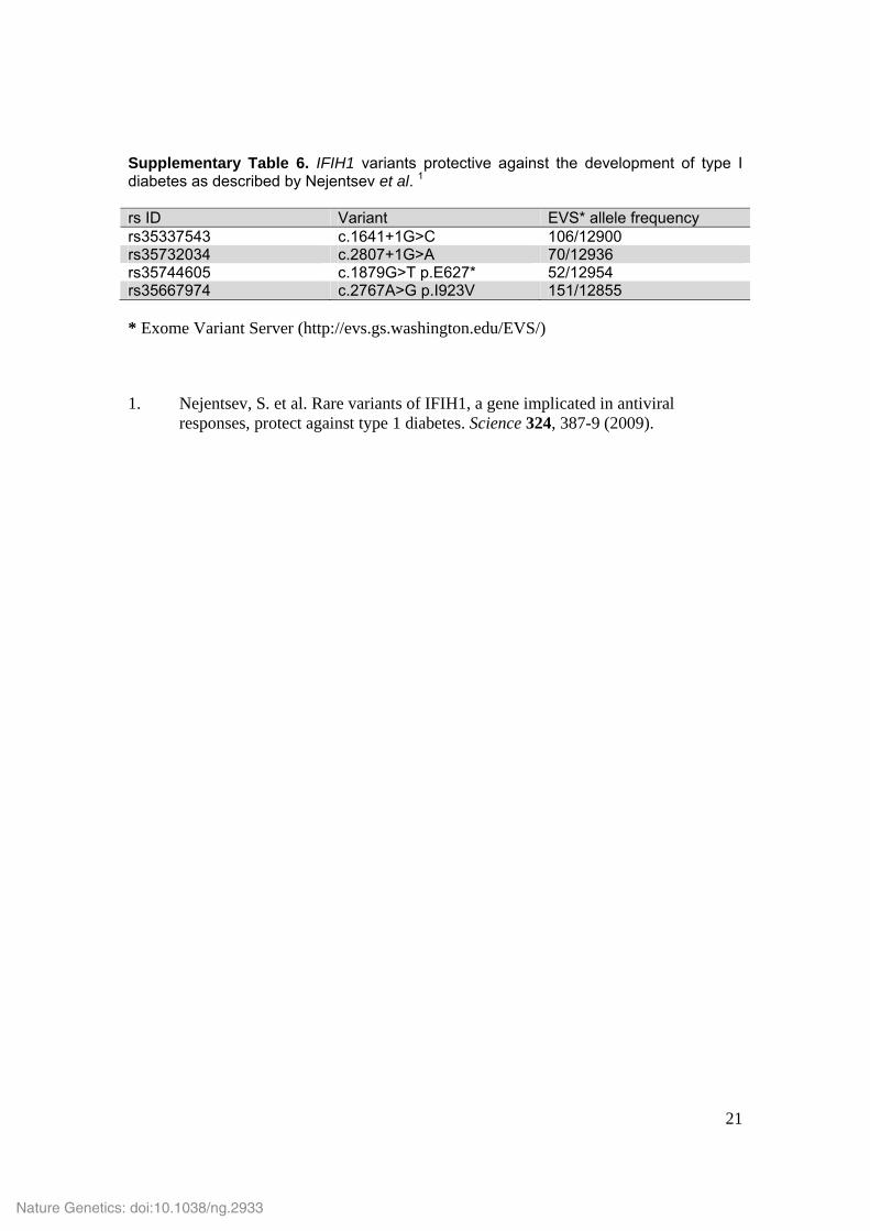
Supplementary Table 6. IFIH1 variants protective against the development of type I diabetes as described by Nejentsev et al. 1 rs ID Variant EVS* allele frequency rs35337543 c.1641+1G>C 106/12900 rs35732034 c.2807+1G>A 70/12936 rs35744605 c.1879G>T p.E627* 52/12954 rs35667974 c.2767A>G p.I923V 151/12855 * Exome Variant Server (http://evs.gs.washington.edu/EVS/) 1. Nejentsev, S. et al. Rare variants of IFIH1, a gene implicated in antiviral
responses, protect against type 1 diabetes. Science 324, 387-9 (2009).
Nature Genetics: doi:10.1038/ng.2933
22
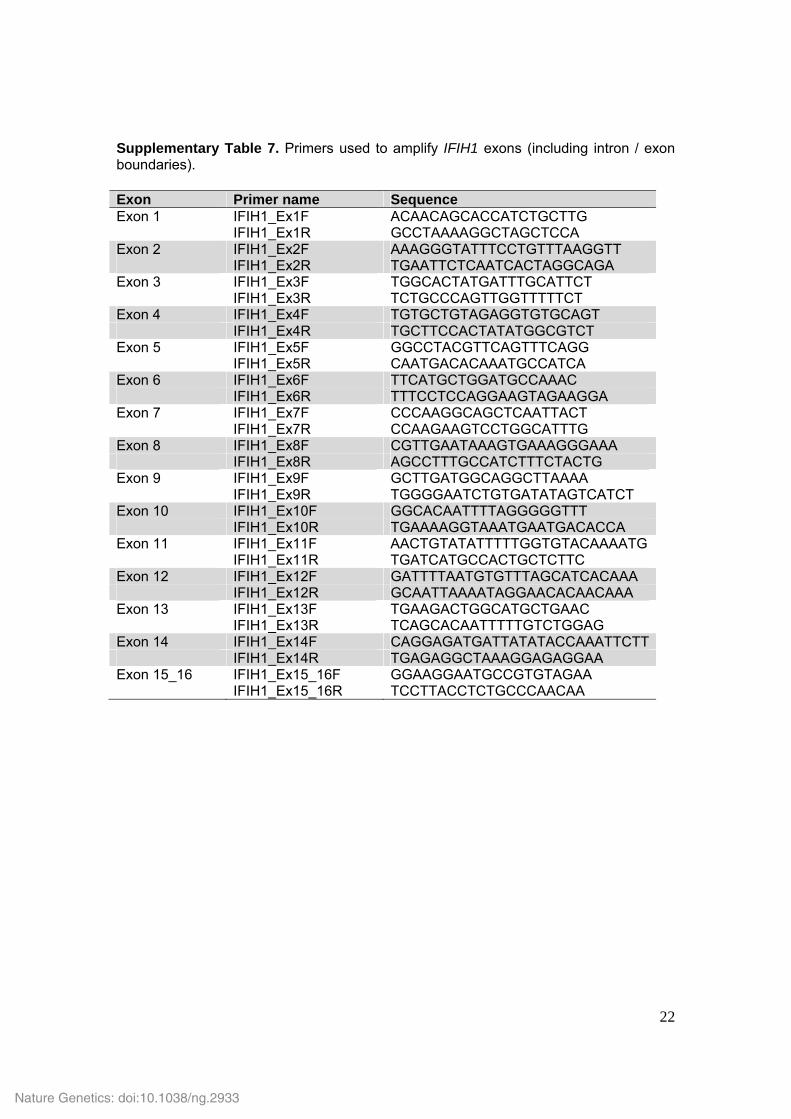
Supplementary Table 7. Primers used to amplify IFIH1 exons (including intron / exon boundaries). Exon Primer name Sequence Exon 1 IFIH1_Ex1F ACAACAGCACCATCTGCTTG IFIH1_Ex1R GCCTAAAAGGCTAGCTCCA Exon 2 IFIH1_Ex2F AAAGGGTATTTCCTGTTTAAGGTT IFIH1_Ex2R TGAATTCTCAATCACTAGGCAGA Exon 3 IFIH1_Ex3F TGGCACTATGATTTGCATTCT IFIH1_Ex3R TCTGCCCAGTTGGTTTTTCT Exon 4 IFIH1_Ex4F TGTGCTGTAGAGGTGTGCAGT IFIH1_Ex4R TGCTTCCACTATATGGCGTCT Exon 5 IFIH1_Ex5F GGCCTACGTTCAGTTTCAGG IFIH1_Ex5R CAATGACACAAATGCCATCA Exon 6 IFIH1_Ex6F TTCATGCTGGATGCCAAAC IFIH1_Ex6R TTTCCTCCAGGAAGTAGAAGGA Exon 7 IFIH1_Ex7F CCCAAGGCAGCTCAATTACT IFIH1_Ex7R CCAAGAAGTCCTGGCATTTG Exon 8 IFIH1_Ex8F CGTTGAATAAAGTGAAAGGGAAA IFIH1_Ex8R AGCCTTTGCCATCTTTCTACTG Exon 9 IFIH1_Ex9F GCTTGATGGCAGGCTTAAAA IFIH1_Ex9R TGGGGAATCTGTGATATAGTCATCT Exon 10 IFIH1_Ex10F GGCACAATTTTAGGGGGTTT IFIH1_Ex10R TGAAAAGGTAAATGAATGACACCA Exon 11 IFIH1_Ex11F AACTGTATATTTTTGGTGTACAAAATG IFIH1_Ex11R TGATCATGCCACTGCTCTTC Exon 12 IFIH1_Ex12F GATTTTAATGTGTTTAGCATCACAAA IFIH1_Ex12R GCAATTAAAATAGGAACACAACAAA Exon 13 IFIH1_Ex13F TGAAGACTGGCATGCTGAAC IFIH1_Ex13R TCAGCACAATTTTTGTCTGGAG Exon 14 IFIH1_Ex14F CAGGAGATGATTATATACCAAATTCTT IFIH1_Ex14R TGAGAGGCTAAAGGAGAGGAA Exon 15_16 IFIH1_Ex15_16F GGAAGGAATGCCGTGTAGAA IFIH1_Ex15_16R TCCTTACCTCTGCCCAACAA
Nature Genetics: doi:10.1038/ng.2933