Embed Size (px)
Citation preview

11/5/2012
1
Glaucoma Surgery – Where Do We Stand ?
ASORN – 2012 (Chicago)
Peter J.G. Maris, Jr., M.D.
Peter J.G. Maris, Jr., M.D.
Private Practice:Long Island Ophthalmic Care, P.L.L.C.230 Hilton Avenue, Suite 118Hempstead, NY
Academic Practice:Assistant Clinical Professor of OphthalmologyEdward S. Harkness Eye InstituteColumbia University Medical CenterNew York, NY
Disclosures
• 1.) I am on the Speakers Bureau for Alcon Laboratories, Inc.
• 2.) I will not be discussing the unlabeled use of commercial products or the investigational use of commercial products not yet approved by the U.S. FDA.
11/5/2012
2
Glaucoma - Definition
• A group of diseases that have in common a characteristic optic neuropathy and associated visual field loss for which elevated intraocular pressure (IOP) is one of the primary risk factors.
Glaucoma as a Medical Science
Classification of Glaucoma
• Open-Angle Glaucomas:– Primary Open-Angle Glaucoma– Normal-Tension Glaucoma– Congenital Glaucoma– Juvenile Open-Angle Glaucoma– Pigmentary Glaucoma– Pseudoexfoliation Glaucoma– Phacolytic Glaucoma– Phacoanaphylactic Glaucoma– Inflammatory Glaucoma (open)– Ghost Cell Glaucoma– Hemolytic Glaucoma– Steroid-Induced Glaucoma– Angle-Recession Glaucoma– Glaucoma Associated w/ Elevated
Episcleral Venous Pressure– Glaucoma Associated w/ Intraocular
Tumors– Glaucomatocyclic Crisis– Fuchs Heterochromic Iridocyclitis
• Angle-Closure Glaucomas:– Acute Angle-Closure Glaucoma– Intermittent Angle-Closure Glaucoma– Chronic Angle-Closure Glaucoma– Plateau Iris Syndrome– Phacomorphic Glaucoma– Aphakic Angle-Closure Glaucoma– Neovascular Glaucoma– Inflammatory Glaucoma (closed)– Malignant (Ciliary Block) Glaucoma– Iridocorneal Endothelial (ICE)
Syndrome – Epithelial Downgrowth

11/5/2012
3
Pseudoexfoliation Glaucoma
Pigmentary Glaucoma
Inflammatory (Uveitic) Glaucoma

11/5/2012
4
Iridocorneal Endothelial (ICE) Syndrome w./ Glaucoma
Essential Iris Atrophy
Glaucoma - Definition
• A group of diseases that have in common a characteristic optic neuropathy and associated visual field loss for which elevated intraocular pressure (IOP) is one of the primary risk factors.
Glaucoma - Epidemiology
• Approximately 3 million Americans suffer from Glaucoma
• Approximately 69 million people worldwide suffer from Glaucoma
• More than 130,000 Americans are legally blind from the disease
• Major Risk Factors:– Older Age (60 + yrs.)– Black and Hispanic Race– Positive Family History

11/5/2012
5
Glaucoma – Mechanism of Disease
Trabecular Meshwork Outflow Pathway
Glaucoma – Mechanism (Trabecular Meshwork Outflow)
Gonioscopy of Anterior Chamber Angle
Glaucoma – Clinical Diagnosis and Monitoring
• Intraocular Pressure (IOP)– Mean Population IOP: 16 mm Hg (SD ± 3)
– Clinical Range of Normal: 8 – 22 mm Hg
• Optic Nerve Appearance
• Visual Field

11/5/2012
6
Intraocular Pressure –Applanation Tonometry
Intraocular Pressure –Applanation Tonometry
Intraocular Pressure - Tonopen
11/5/2012
7
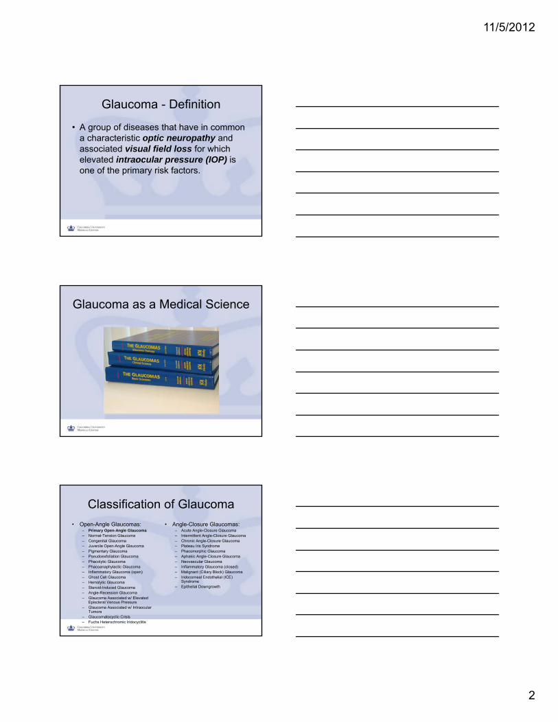
Optic Nerve Appearance
Normal Advanced Glaucoma
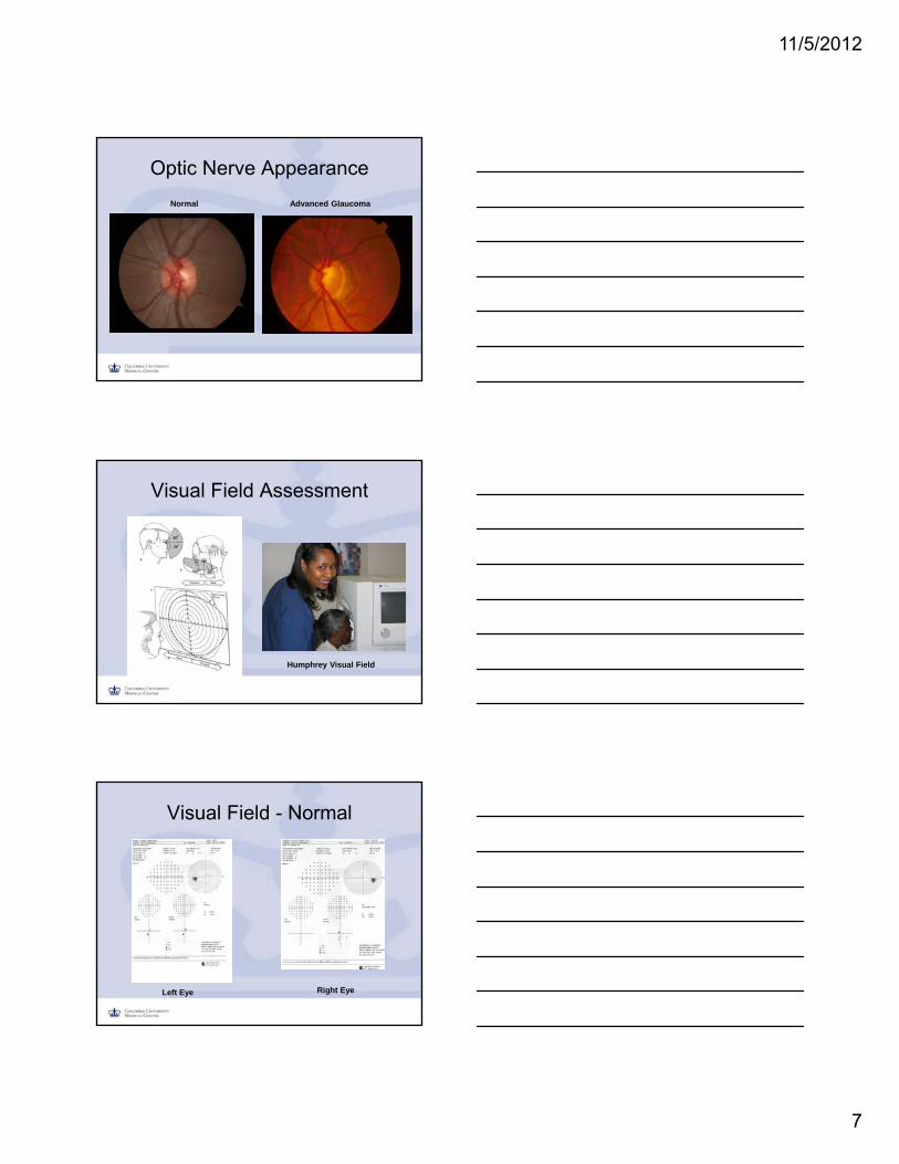
Visual Field Assessment
Humphrey Visual Field
Visual Field - Normal
Left Eye Right Eye

11/5/2012
8
Glaucomatous Field Loss (Mild –Moderate) – Right Eye
Glaucomatous Field Loss (Advanced) – Left Eye
Visual Field Loss – Advanced (End-Stage)
Left Eye Right Eye

11/5/2012
9
Glaucomatous Field Loss – Over Time
How Is Glaucoma Treated ?
• I.) Medical Therapy (Drops and Pills)
• II.) Laser Treatment
• III.) Surgery
Glaucoma - Medical Therapy

11/5/2012
10
Glaucoma – Drop Therapy
• Beta Blockers:– Timolol (Timoptic, Timoptic XE,
Betimol, Istalol)– Betaxolol (Betoptic S)– Metipranolol (OptiPranolol)– Levobunalol (Betagan)
• Miotics:– Pilocarpine (1% - 4%)– Echothiophate iodide
(Phospholine Iodide)
• Carbonic Anhydrase Inhibitors:– Brinzolamide (Azopt)– Dorzolamide (Trusopt)
• Alpha-2 Agonists:– Brimonidine (Alphagan,
Alphagan-P)– Apraclonidine (Iopidine)
• Prostaglandin Analogues:– Latanoprost (Xalatan)– Travoprost (Travatan)– Bimatoprost (Lumigan)
• Combination Agents:– Dorzolamide/Timolol
(Cosopt)– Brimonidine/Timolol
(Combigan)
Laser Treatment
• Argon Laser Trabeculoplasty (ALT)
• Selective Laser Trabeculoplasty (SLT)
Laser Trabeculoplasty

11/5/2012
11
Glaucoma Surgery
When Should Glaucoma Surgery Be Performed ?
• Intraocular Pressure Too High Despite Maximal Medical Therapy
• Progression of Field Loss and/or Optic Neuropathy
• Medical Therapy Not Tolerated (Side Effects)
• Laser Therapy Ineffective (“Burn Out”)
Trabeculectomy (Filtration) Surgery
• Most commonly performed glaucoma surgery
• First Described in 1968 (Cairns JD)
• Objective: to divert aqueous outflow through a surgical fistula into subconjunctival space
• Modulation of Wound Healing:– Mitomycin C (MMC)– 5-Fluorouracil (5-FU)
Bleb

11/5/2012
12
Trabeculectomy
1968 Paper
Trabeculectomy Surgery -Schematic
Cairns Version - 1968 Contemporary Rendition
Trabeculectomy – Surgical Sequence

11/5/2012
13
Conjunctival Dissection (Limbal-Based)
Creation of the Scleral Flap
Internal Sclerostomy

11/5/2012
14
Surgical Iridectomy
Trabeculectomy – Instrument Layout
• Lieberman Lid Speculum
• .12 Forceps
• Conjunctival Forceps (Non-Toothed)– Anatomical Forceps
– Fechner Forceps
• Wescott Tenotomy Scissors (Blunt-Tip)
• Vannas Scissors
• Kelly Descemet’s Punch
• Beaver Blade (# 69)
• Castroviejo Caliper
• Barraquer Needle Holder
• Tying Forceps (straight and curved)
• Eraser Cautery
Trabeculectomy Layout
• Suture Material:– 6-0 Vicryl
• Traction Suture
– 10-0 Nylon• Scleral Flap
– 9-0 Vicryl or 9-0 Nylon• Conjunctival Closure
• Antimetabolite:– Exposure Time = 1.5 – 5 min.– Delivery System:
• cut-up Weckcel sponges• corneal shield (1/2’s)
– Mitomycin C (MMC)• 0.1 – 0.5 mg/ml
– 5-Fluorouracil (5-FU)• 50 mg/ml

11/5/2012
15
Trabeculectomy – Postoperative Care
• Fourth-Generation Fluoroquinolone (Zymar or Vigamox):
– 4x/day for 2 wks.
• Prednisolone Acetate (EconoPred; PredForte):
– 8x/day for 2 wks.
– 6x/day for 2 wks.
– 4x/day for 2 wks.
– 3x/day for 2 wks.
– 2x/day for 2 wks.
– 1x/day for 2 wks.
Trabeculectomy – Postoperative Care
Laser Suture Lysis Bleb Needlings and Injections (5-FU)
Trabeculectomy – Post-Op
Day # 12 Two Years

11/5/2012
16
Trabeculectomy – Postoperative Complications
Flat Anterior Chamber (Overfiltration – Pressure Too
Low)
Requires Intervention Within 24 hrs.
Serous Choroidal Effusions (Overfiltration – Pressure Too
Low)
Fundus Photo – Left Eye Ultrasound (B-Scan) – Left Eye

11/5/2012
17
Hyphema
Cataract Formation
Suprachoroidal Hemorrhage

11/5/2012
18
Late-Onset Bleb Leak
Fluorescein Dye Test: Seidel-Positive LeakThin, Cystic, Ischemic Bleb
Hypotony (Chronically Low IOP) Maculopathy
Fundus Photo
Fluorescein Angiogram
Blebitis (Late-Onset Infection)

11/5/2012
19
Bleb-Related Endophthalmitis (Late-Onset Infection)
Glaucoma Drainage Device (Seton) Surgery
Glaucoma Drainage Device (GDD)
• GDD’s shunt aqueous to a site posterior to the limbus
• Tube placed into the anterior chamber or through pars plana
• Plate acts as an extraocular resevoir
• Indications:– Failed Prior Trabeculectomy– Active Inflammation (Uveitis)– Neovascular Glaucoma– Scarred / Inadequate
Conjunctiva (prior trauma, retinal detachment surgery, etc.)

11/5/2012
20
Ahmed Glaucoma Valve
Other Glaucoma Drainage Devices (GDD’s)
Baerveldt Molteno
Glaucoma Drainage Device (GDD) Implantation
Ahmed Glaucoma Valve

11/5/2012
21
Contemporary Adult Glaucoma Drainage Devices (GDD’s)
Shunt Design:
Year of Introduction
Plate Size
Resistance Mechanism
Molteno 1979 135 mm2 None
Baerveldt 1990 250 mm2
350 mm2
None
Krupin 1990 180 mm2 Slit Valve
Ahmed 1993 184 mm2 Venturi Valve
Ahmed Valve Implantation –Surgical Technique (Anterior)
Tube inserted1.0 – 1.5 mm posterior to corneal limbus
Plate sutured to episclera approx. 9 mm posterior to limbus
Ahmed Glaucoma Valve (Anterior Placement)
Six Months Post Surgery

11/5/2012
22
Ahmed Valve Implantation –Posterior (Pars Plana) Placement
Tube inserted at pars plana (3.5 mm posterior to corneal limbus)
Ahmed Glaucoma Valve (Anterior vs. Posterior Placement)
Ahmed Glaucoma Valve (Anterior vs. Posterior Placement)
• Surgical Success (Final Follow-Up):– Anterior Group = 83.9%
– Posterior Group = 83.9%
• Complication Profile:– Similar in Both Groups

11/5/2012
23
Postoperative Complications –Tube Obstruction
Iris Incarceration
Vitreous Incarceration
Postoperative Complications –Corneal Decompensation
Failed Corneal Graft (PK) Focal Corneal Edema and Folds
Postoperative Complications –Implant Erosion

11/5/2012
24
New Glaucoma Surgical Innovations
Microinvasive Glaucoma Surgery (MIGS)
Microinvasive Glaucoma Surgery (MIGS)
• ExPRESS Glaucoma Filtration Device (Alcon Laboratories, Inc)
• Trabectome (Neomedix Corporation)
• Canaloplasty
• iStent Trabecular Micro-Bypass (Glaukos Corporation)

11/5/2012
25
ExPRESS Glaucoma Device
ExPRESS Glaucoma Device
ExPRESS Device - Implantation
Under Partial-Thickness Scleral Flap

11/5/2012
26
ExPRESS Miniature Glaucoma Implant
Ten Months Post-Op
ExPRESS Trabeculectomy vs. Standard Trabeculectomy
ExPRESS Standard Trab.
ExPRESS Glaucoma Device vs. Standard Trabeculectomy

11/5/2012
27
ExPRESS vs. Standard Trabeculectomy – IOP Control
Intraocular Pressure (IOP) over Time
ExPRESS vs. Standard Trabeculectomy - Complications
Choroidal Effusions
Canaloplasty
11/5/2012
28
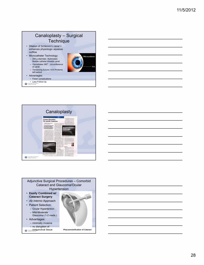
Canaloplasty – Surgical Technique
• Dilation of Schlemm’s canal > enhances physiologic aqueous outflow
• Microcatheter Technology:– 200-μ-diameter, illuminated,
flexible catheter threads canal
– Viscodilates 360° circumference of canal
– Tensioning Suture (10-0 Prolene) left behind
• Advantages:• Fewer complications
• Less Follow-Up
Canaloplasty
Adjunctive Surgical Procedures – Comorbid Cataract and Glaucoma/Ocular
Hypertension• Easily Combined w/
Cataract Surgery
• Ab Interno Approach
• Patient Selection: – Ocular Hypertention
– Mild-Moderate Glaucoma (1-2 meds.)
• Advantages:– minimally invasive
– no disruption of conjunctival tissue Phacoemulsification of Cataract

11/5/2012
29
Trabectome Surgery
• Electrosurgical ablation of Trabecular Meshwork(main site of outflow resistance)
• Ab Interno Approach (gonioscopic guidance)
• Designed to improve aqueous drainage
• Indications:– Mild-Moderate Glaucoma
(pt. taking 1-2 meds.)
Trabectome Surgery
iStent (Trabecular Micro-Bypass)
Trabecular Stent inserted into Schlemm’s canal

11/5/2012
30
iStent
iStent – Surgical Technique
• Ab Interno procedure(gonioscopic visualization)– via 1.5 – 2 mm corneal incision
• self-trephinating tip inserts into trabecular meshwork
• device gently advanced into Schlemm’s canal
• inserter button is depressed and device is disengaged
Adjunctive Glaucoma Procedures – Future Potential
• Examples: – Trabectome (Neomedix Corporation)
– iStent (Glaukos Corporation)
• Cataract Surgery:– Over 3 million cataract procedures performed each
year in U.S.
• Comorbidity: Glaucoma/Ocular Hypertension– More than 650,000 cataract patients
• “Buzz Phrases” (Clever Marketing):– “short learning curve” - “safe” - “minimally invasive”
– “widely adoptable” - “fast recovery” - “easier”

11/5/2012
31
Equation =
$
• Surgery is indicated when medical and laser therapy have been exhausted
• Trabeculectomy and Glaucoma Drainage Device (GDD) surgery remain the most effective IOP-lowering procedures
• Glaucoma surgery can be fraught with sight-threatening postoperative complications
• Surgical innovations and improvements (minimal-risk) remain ongoing
Summary – Glaucoma Surgery
Acknowledgements
• Photographs:– S. Fabian Lerner and Richard K. Parish II. Glaucoma
Surgery. Lippincott, Williams & Wilkins, 2003
– New World Medical, Inc.
– Alcon, Inc
– Cataract & Refractive Surgery Today
– iScience Interventional, Inc.
– NeoMedix
– Lama A. Al-Aswad, M.D.

11/5/2012
32
Thank You