Embed Size (px)
Citation preview
2786
Abstract. – OBJECTIVE: To explore wheth-er HCP5 participates in the pathogenic progres-sion of colon cancer (CC) and its underlying mechanism.
PATIENTS AND METHODS: HCP5 expres-sion in CC tissues and cell lines was detected by quantitative Real Time-Polymerase Chain Re-action (qRT-PCR). The correlation between the HCP5 expression and tumor stage of CC patients was then analyzed. After CC cells were transfect-ed with HCP5-siRNA, the proliferation and migra-tion capacities were detected by cell counting kit-8 (CCK-8), colony formation and transwell assay, respectively. Cell cycle was examined by flow cy-tometry. Western blot was conducted to detect protein expressions of HCP5, AP1G1 and relative molecules in the PI3K/AKT pathway. Rescue ex-periments were performed by co-transfection of HCP5-siRNA and AP1G1-siRNA into CC cells, fol-lowed by cell function detection.
RESULTS: HCP5 was highly expressed, where-as AP1G1 was lowly expressed in CC tissues and cell lines. Besides, CC patients with stage III-IV presented higher expression of HCP5 than those with stage I-II. The knockdown of HCP5 in CC cells down-regulated proliferation and migration capacities, and arrested cell cycle in the G0/G1 phase, which was reversed by the AP1G1 knock-down. In addition, HCP5 knockdown up-regulat-ed AP1G1 expression, whereas down-regulated the expression of relative proteins in the PI3K/AKT pathway.
CONCLUSIONS: HCP5 was significantly in-creased in CC and enhanced the proliferation and migration of CC cells by inhibiting the AP1G1 expression. HCP5 promoted CC development by activating the PI3K/AKT pathway.
Key Words:CC, HCP5, AP1G1, PI3K/AKT pathway.
Introduction
Colon cancer (CC) is a common malignancy in the digestive tract and frequently occurs at the
junction of the rectum and sigmoid colon1. In re-cent years, the incidence of CC has been on the rise. Although current treatments for CC have been advanced, including surgical resection, ra-diotherapy and chemotherapy, the clinical out-comes are still not satisfactory. It is reported that 50% of CC patients have a poor prognosis and the five-year survival rate of less than 5%2,3.
With the in-depth study of tumor pathogenesis, the signaling pathways related to tumor cell pro-liferation and differentiation have been well rec-ognized4. The PI3K/AKT pathway is thought to be a crucial pathway in regulating a variety of cell activity5. It is reported that the PI3K/AKT path-way is closely related to the occurrence and devel-opment of malignant tumors, such as lung cancer, breast cancer and kidney cancer. The exploration of the significance of key genes involved in the PI3K/AKT pathway in cancer cells will contrib-ute to further revealing the occurrence and devel-opment of malignancies6.
Abnormal expression of lncRNAs exerts im-portant roles in the occurrence and progression of malignant tumors and provides a new promising strategy for tumor treatment7-9. Depending on the transcript size, non-coding RNAs can be divided into long non-coding RNAs (lncRNAs) and small non-coding RNAs (small ncRNAs)10. LncRNAs contain more than two hundred nucleotides and cannot be translated into proteins8. LncRNA HCP5 is mainly expressed in the immune system and often considered to be associated with herpes zoster and other serious skin reactions11. Recent studies have reported differential expression of HCP5 in multiple cancers. For example, HCP5 was significantly downregulated in patients with ovarian cancer12. In addition, the HCP5 gene was also considered to be a susceptibility locus for HCV-associated hepatocellular carcinoma13. However, the role of HCP5 in CC remains un-known.
European Review for Medical and Pharmacological Sciences 2019; 23: 2786-2793
W.-K. YUN1, Y.-M. HU1, C.-B. ZHAO1, D.-Y. YU1, J.-B. TANG2
1Department of Radiotherapy, Harbin Medical University Cancer Hospital, Harbin, China.2Department of Oncology, Harbin Medical University Cancer Hospital, Harbin, China.
HCP5 promotes colon cancer development by activating AP1G1 via PI3K/AKT pathway
Corresponding Author: Jiebing Tang, MD; email: [email protected]

High expression of HCP5 promotes colon cancer development
2787
This study aimed to explore the role of HCP5 in the occurrence and progression of CC. Demon-stration of the regulatory effect of HCP5 on the PI3K/AKT pathway might provide novel thera-peutic targets for CC treatment.
Patients and Methods
Sample CollectionCC tissues and paracancerous tissues were
harvested from patients undergoing surgical re-section in our hospital from June 2015 to March 2018. Enrolled patients had complete clinical data and signed an informed consent before the study. All experimental procedures were approved by the Ethics Committee of Harbin Medical Univer-sity Cancer Hospital. CC tissues were immedi-ately preserved in liquid nitrogen. Based on the tumor stage, 22 CC cases were classified as stage I-II and 16 were stage III-IV.
Cell Culture and Transfection Colonic epithelial cell line (NCM460) and
CC cell lines (LoVo, CaCo2, SW620, SW480 and HCT116) were obtained from the Ameri-can Type Culture Collection (ATCC; Manassas, VA, USA). HCT116 cells were cultured in Ros-well Park Memorial Institute-1640 (RPMI-1640; HyClone, South Logan, UT, USA) containing 10% fetal bovine serum (FBS; Gibco, Grand Is-land, NY, USA) and the others were cultured in Dulbecco’s Modified Eagle’s Medium (DMEM; Gibco, Grand Island, NY, USA) containing 10% FBS. One day prior to cell transfection, cells were seeded into the 6-well plates at a density of 4×10⁴ cells per well. Cells were transfected with corre-sponding plasmids when the confluence was up to 50% following the instructions of Lipofectamine 2000 (Invitrogen, Carlsbad, CA, USA). The cul-ture medium was replaced 6 hours later.
RNA Extraction and Quantitative Real Time-Polymerase Chain Reaction (qRT-PCR)
The total RNA in cells was extracted using TRIzol method (Invitrogen, Carlsbad, CA, USA) for reverse transcription according to the instruc-tions of PrimeScript RT reagent Kit (TaKaRa, Otsu, Shiga, Japan). RNA concentration was de-tected using a spectrometer and those samples with A260/A280 ratio of 1.8-2.0 were selected for the following qRT-PCR reaction. QRT-PCR was then performed based on the instructions of SYBR Premix Ex Taq TM (TaKaRa, Otsu, Shiga, Ja-
pan). The relative gene expression was calculated using the 2-ΔCt method. Primers used in the study were as follows: GAPDH forward: 5′-ATCACT-GCCACCCAGAAGAC-3′, GAPDH reverse: 5′-ATGAGGTCCACCACCCTGTT-3′; HCP5 for-ward: 5′-GACTCTCCTACTGGTGCTTGGT-3’, HCP5 reverse: 5′-CACTGCCTGGTGAGCCT-GTT-3′; AP1G1 forward: 5′-GAGTTTTGCCCT-GGTGAATG-3′, AP1G1 forward: 5′-GCAAG-GAAGATTCCAGATGC-3′.
Western BlotCells were lysed for protein extraction. The
concentration of each protein sample was deter-mined by a BCA (bicinchoninic acid) kit (Abcam, Cambridge, MA, USA). The protein sample was separated by gel electrophoresis and transferred to PVDF (polyvinylidene difluoride) membranes (Millipore, Billerica, MA, USA). After incubation with primary and secondary antibody, the immu-noreactive bands were exposed by enhanced che-miluminescence (ECL) method.
Cell Counting Kit-8 (CCK-8) Assay SW480 and HCT116 cells were seeded into 96-
well plates with 2×10³ cells per well. Totally 10 μL of CCK-8 solution (Dojindo, Kumamoto, Japan) was added in each well. About 4 hours later, the medium was replaced and cells were incubated with the CCK8 solution for 1 h. The absorbance at 450 nm of each sample was measured by a mi-croplate reader (Bio-Rad, Hercules, CA, USA).
Colony Formation AssayCells were digested for preparing cell suspen-
sion at a density of 2×10⁵/mL. A total of 200 cells were seeded in each well of the 6-well plates. Colonies visible to the naked eye were fixed with hematoxylin for 30 min, observed and captured using a microplate.
Transwell AssaySW480 or HCT116 cells were resuspended in
serum-free medium at a density of 2×10⁵/mL. To-tally 100 μL of cells suspension was added in the upper transwell chamber of the 24-well plates for 24 hours. The penetrating cells in the lower cham-ber were fixed and stained with 20% Giemsa. In each well, 5 randomly selected fields were cap-tured for cell counting.
Cell Cycle DetectionCells were washed with phosphate-buffered
saline (PBS) three times, digested with trypsin
W.-K. Yun, Y.-M. Hu, C.-B. Zhao, D.-Y. Yu, J.-B. Tang
2788
and then centrifuged. Subsequently, cells were fixed with 75% ethanol at -20°C for 8 h or longer. Before cell cycle detection, fixed cells were cen-trifuged and washed with PBS, followed by incu-bation with 0.5 mg/mL ribotide for 10 min. Final-ly, cells were incubated with 10 μL of propidium iodide (PI) (1 mg/mL) in the dark for 30 min, and cell cycle was detected using flow cytometry.
Statistical AnalysisGraphPad Prism 5 (La Jolla, CA, USA) was used
for data analysis. Data were expressed as mean ± standard deviation (x– ± s). The difference of the measurement data was analyzed using the t-test. p<0.05 was considered statistically significant.
Results
HCP5 was Highly Expressed in CCWe first detected the expression level of HCP5
in CC and paracancerous tissues by qRT-PCR. It
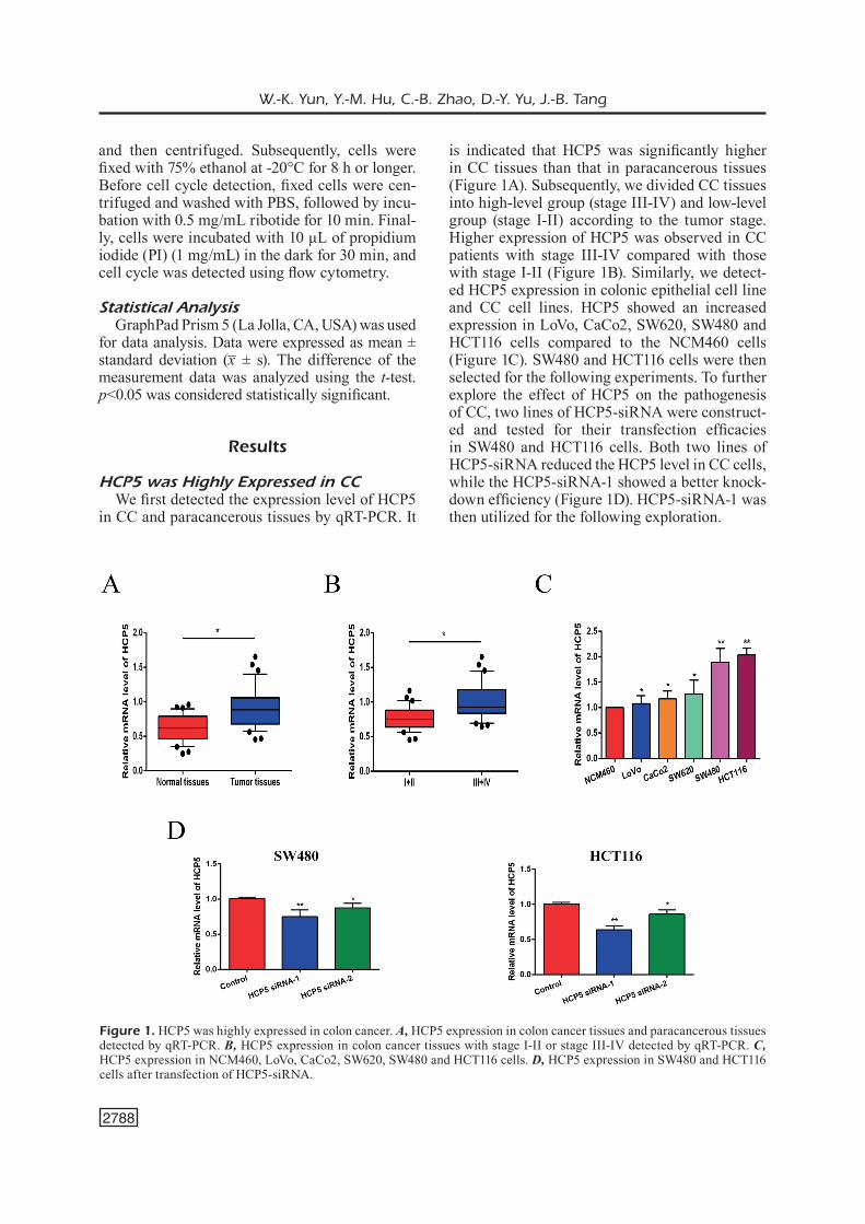
is indicated that HCP5 was significantly higher in CC tissues than that in paracancerous tissues (Figure 1A). Subsequently, we divided CC tissues into high-level group (stage III-IV) and low-level group (stage I-II) according to the tumor stage. Higher expression of HCP5 was observed in CC patients with stage III-IV compared with those with stage I-II (Figure 1B). Similarly, we detect-ed HCP5 expression in colonic epithelial cell line and CC cell lines. HCP5 showed an increased expression in LoVo, CaCo2, SW620, SW480 and HCT116 cells compared to the NCM460 cells (Figure 1C). SW480 and HCT116 cells were then selected for the following experiments. To further explore the effect of HCP5 on the pathogenesis of CC, two lines of HCP5-siRNA were construct-ed and tested for their transfection efficacies in SW480 and HCT116 cells. Both two lines of HCP5-siRNA reduced the HCP5 level in CC cells, while the HCP5-siRNA-1 showed a better knock-down efficiency (Figure 1D). HCP5-siRNA-1 was then utilized for the following exploration.
Figure 1. HCP5 was highly expressed in colon cancer. A, HCP5 expression in colon cancer tissues and paracancerous tissues detected by qRT-PCR. B, HCP5 expression in colon cancer tissues with stage I-II or stage III-IV detected by qRT-PCR. C, HCP5 expression in NCM460, LoVo, CaCo2, SW620, SW480 and HCT116 cells. D, HCP5 expression in SW480 and HCT116 cells after transfection of HCP5-siRNA.

High expression of HCP5 promotes colon cancer development
2789
HCP5 Knockdown Inhibited Proliferative and Migratory Capacities of CC Cells
We transfected HCP5-siRNA-1 into SW480 and HCT116 cells to knock down HCP5 expres-sion, followed by detection of cell proliferation and migration. Cell proliferation was measured by CCK-8 assay at 0, 24 h, 48 h and 72 h after transfection of HCP5-siRNA, respectively. It was found that after HCP5 knockdown, the pro-liferation capacity of CC cells was significantly lower than that of the normal group (Figure 2A). The transfection of HCP5-siRNA in SW480 and HCT116 cells also decreased the number of mi-gratory cells (Figure 2B). The colony formation assay further demonstrated an inhibited cell pro-liferation by knockdown of HCP5 (Figure 2C).
Subsequently, we analyzed the proportion of cells in G0/G1 phase, S phase and G2/M phase by flow cytometry. The results showed that the proportion of cells in the G0/G1 phase was mark-edly increased while decreased in S phase after transfection of HCP5-siRNA (Figure 2D). Above data suggested that the HCP5 knockdown inhib-ited both proliferation and migration capacities of SW480 and HCT116 cells.
HCP5 Regulated the PI3K/AKT Pathway by Inhibiting AP1G1
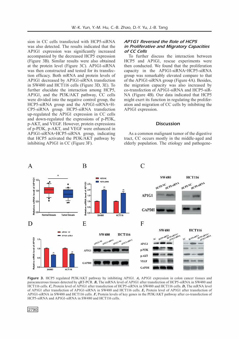
We further examined the AP1G1 expression in CC and paracancerous tissues by qRT-PCR, and found that AP1G1 was significantly decreased in CC tissues (Figure 3A). Next, the AP1G1 expres-
Figure 2. HCP5 knockdown inhibited proliferative and migratory capacities of colon cancer cells. A, Cell proliferation was measured by CCK-8 assay at 0, 24 h, 48 h and 72 h after transfection of HCP5-siRNA in SW480 and HCT116 cells, respective-ly. B, Cell migration after transfection of HCP5-siRNA in SW480 and HCT116 cells. C, Colony formation after transfection of HCP5-siRNA in SW480 and HCT116 cells. D, Cell cycle after transfection of HCP5-siRNA in SW480 and HCT116 cells.
W.-K. Yun, Y.-M. Hu, C.-B. Zhao, D.-Y. Yu, J.-B. Tang
2790
sion in CC cells transfected with HCP5-siRNA was also detected. The results indicated that the AP1G1 expression was significantly increased accompanied by the decreased HCP5 expression (Figure 3B). Similar results were also obtained at the protein level (Figure 3C). AP1G1-siRNA was then constructed and tested for its transfec-tion efficacy. Both mRNA and protein levels of AP1G1 decreased by AP1G1-siRNA transfection in SW480 and HCT116 cells (Figure 3D, 3E). To further elucidate the interaction among HCP5, AP1G1, and the PI3K/AKT pathway, CC cells were divided into the negative control group, the HCP5-siRNA group and the AP1G1-siRNA+H-CP5-siRNA group. HCP5-siRNA transfection up-regulated the AP1G1 expression in CC cells and down-regulated the expressions of p-PI3K, p-AKT, and VEGF. However, protein expressions of p-PI3K, p-AKT, and VEGF were enhanced in AP1G1-siRNA+HCP5-siRNA group, indicating that HCP5 activated the PI3K/AKT pathway by inhibiting AP1G1 in CC (Figure 3F).
AP1G1 Reversed the Role of HCP5 in Proliferative and Migratory Capacities of CC Cells
To further discuss the interaction between HCP5 and AP1G1, rescue experiments were then conducted. We found that the proliferation capacity in the AP1G1-siRNA+HCP5-siRNA group was remarkably elevated compare to that of the AP1G1-siRNA group (Figure 4A). Besides, the migration capacity was also increased by co-transfection of AP1G1-siRNA and HCP5-siR-NA (Figure 4B). Our data indicated that HCP5 might exert its function in regulating the prolifer-ation and migration of CC cells by inhibiting the AP1G1 expression.
Discussion
As a common malignant tumor of the digestive tract, CC occurs mostly in the middle-aged and elderly population. The etiology and pathogene-
Figure 3. HCP5 regulated PI3K/AKT pathway by inhibiting AP1G1. A, AP1G1 expression in colon cancer tissues and paracancerous tissues detected by qRT-PCR. B, The mRNA level of AP1G1 after transfection of HCP5-siRNA in SW480 and HCT116 cells. C, Protein level of AP1G1 after transfection of HCP5-siRNA in SW480 and HCT116 cells. D, The mRNA level of AP1G1 after transfection of AP1G1-siRNA in SW480 and HCT116 cells. E, Protein level of AP1G1 after transfection of AP1G1-siRNA in SW480 and HCT116 cells. F, Protein levels of key genes in the PI3K/AKT pathway after co-transfection of HCP5-siRNA and AP1G1-siRNA in SW480 and HCT116 cells.

High expression of HCP5 promotes colon cancer development
2791
found in multiple tumors, including breast cancer, ovarian cancer, endometrial cancer, nasopharyn-geal cancer and CC15-19.
The occurrence of CC is also accompanied by abnormal expressions of various oncogenes and tumor-suppressor genes. The role of the PI3K/AKT pathway in CC has also been well studied. It is reported that the PI3K/AKT path-way promoted the invasion and metastasis of malignancies by reducing the intercellular ad-hesion, increasing the tumor cell motility, regu-
sis of CC have not been fully elucidated yet. At present, CC development is believed as a dynam-ic process, which involves environmental factors and genetic factors14. The occurrence and pro-gression of the tumor are related to disordered cell metabolism, cell cycle and signaling pathways. The oncogene activation and tumor-suppres-sor gene inactivation are fundamental for tumor pathogenesis.
With the advanced development of tumor biol-ogy, abnormally activated PI3K/AKT pathway is
Figure 4. AP1G1 reversed the role of HCP5 in proliferative and migratory capacities of colon cancer cells. A, Proliferation after co-transfection of HCP5-siRNA and AP1G1-siRNA in SW480 and HCT116 cells. B, Migration after co-transfection of HCP5-siRNA and AP1G1-siRNA in SW480 and HCT116 cells.

W.-K. Yun, Y.-M. Hu, C.-B. Zhao, D.-Y. Yu, J.-B. Tang
2792
lating the growth factor receptors and affecting the expression of the extracellular matrix. Acti-vated PI3K disturbs the normal function of the PI3K/AKT pathway, thus affecting cell prolif-eration, differentiation, apoptosis and transport of intracellular substances. PI3K further stim-ulates the AKT activity to improve tumor cell proliferation and survival.
The crucial roles of lncRNAs in the occurrence and development of tumors have been reported re-cently20,21. LncRNAs function by affecting apopto-sis, metastasis and infiltration of tumor cells, which provide new ideas for tumor therapy. Researches22 have shown that the proportion of tumor-associated lncRNAs (18%) is twice than that of tumor-asso-ciated proteins (9%). HCP5 is located on chromo-some 6q21.3 and has been previously reported to be expressed in immune system-associated cells. The specific role of HCP5 in malignancies, howev-er, has not been fully elucidated23.
In the present work, HCP5 was highly expressed in CC tissues and cell lines. HCP5 knockdown re-duced the proliferation and invasion capacities of CC cells through the PI3K/AKT pathway. In ad-dition, we found that AP1G1 was lowly expressed in CC and might be inhibited by HCP5. Rescue experiments further confirmed that the regulato-ry effect of HCP5 on CC cells were partially re-versed by the AP1G1 knockdown.
Conclusions
We showed that HCP5 was highly expressed in CC and enhanced the proliferation and migration of CC cells by inhibiting the AP1G1 expression. Besides, HCP5 promoted CC development by ac-tivating the PI3K/AKT pathway. Our study pro-vided a new therapeutic target for early diagnosis and treatment of CC.
Conflict of InterestThe Authors declare that they have no conflict of interest.
References 1) Torre LA, BrAy F, SiegeL rL, FerLAy J, LorTeT-TieuLenT J,
JemAL A. Global cancer statistics, 2012. CA Cancer J Clin 2015; 65: 87-108.
2) Vizin T, ChriSTenSen iJ, nieLSen hJ, KoS J. Cathepsin X in serum from patients with colorectal cancer: rela-tion to prognosis. Radiol Oncol 2012; 46: 207-212.
3) VAn CuTSem e, oLiVeirA J. Primary colon cancer: ESMO clinical recommendations for diagnosis, adjuvant treatment and follow-up. Ann Oncol 2009; 20 Suppl 4: 49-50.
4) mCCuBrey JA, ABrAmS SL, FiTzgerALd TL, CoCCo L, mArTeLLi Am, monTALTo g, CerVeLLo m, SCALiSi A, CAndido S, LiBrA m, STeeLmAn LS. Roles of signal-ing pathways in drug resistance, cancer initiating cells and cancer progression and metastasis. Adv Biol Regul 2015; 57: 75-101.
5) mCCuBrey JA, STeeLmAn LS, ABrAmS SL, Lee JT, ChAng F, BerTrAnd Fe, nAVoLAniC Pm, TerriAn dm, FrAnKLin rA, d’ASSoro AB, SALiSBury JL, mAzzArino mC, STi-VALA F, LiBrA m. Roles of the RAF/MEK/ERK and PI3K/PTEN/AKT pathways in malignant transfor-mation and drug resistance. Adv Enzyme Regul 2006; 46: 249-279.
6) mCnAmArA Cr, degTereV A. Small-molecule inhib-itors of the PI3K signaling network. Future Med Chem 2011; 3: 549-565.
7) deng g, Sui g. Noncoding RNA in oncogenesis: a new era of identifying key players. Int J Mol Sci 2013; 14: 18319-18349.
8) PonTing CP, oLiVer PL, reiK W. Evolution and func-tions of long noncoding RNAs. Cell 2009; 136: 629-641.
9) yAng BF, CAi W, Chen B. LncRNA SNHG12 regu-lated the proliferation of gastric carcinoma cell BGC-823 by targeting microRNA-199a/b-5p. Eur Rev Med Pharmacol Sci 2018; 22: 1297-1306.
10) deng g, Sui g. Noncoding RNA in oncogenesis: a new era of identifying key players. Int J Mol Sci 2013; 14: 18319-18349.
11) Liu y, heLmS C, LiAo W, zABA LC, duAn S, gArdner J, WiSe C, miner A, mALLoy mJ, PuLLinger Cr, KAne JP, SACCone S, WorThingTon J, BruCe i, KWoK Py, menTer A, Krueger J, BArTon A, SACCone nL, BoWCoCK Am. A genome-wide association study of psoriasis and psoriatic arthritis identifies new disease loci. PLoS Genet 2008; 4: e1000041.
12) Liu n, zhAng r, zhAo X, Su J, BiAn X, ni J, yue y, CAi y, Jin J. A potential diagnostic marker for ovarian cancer: involvement of the histone acetyltransfer-ase, human males absent on the first. Oncol Lett 2013; 6: 393-400.
13) LAnge Cm, BiBerT S, duFour JF, CeLLerAi C, Cerny A, heim mh, KAiSer L, mALinVerni r, muLLhAuPT B, negro F, SemeLA d, morAdPour d, KuTALiK z, BoChud Py. Comparative genetic analyses point to HCP5 as susceptibility locus for HCV-associated hepato-cellular carcinoma. J Hepatol 2013; 59: 504-509.
14) rAnzAni m, AnnunziATo S, AdAmS dJ, monTini e. Can-cer gene discovery: exploiting insertional muta-genesis. Mol Cancer Res 2013; 11: 1141-1158.
15) ToKunAgA e, KimurA y, mAShino K, oKi e, KATAoKA A, ohno S, moriTA m, KAKeJi y, BABA h, mAehArA y. Activation of PI3K/Akt signaling and hormone resistance in breast cancer. Breast Cancer 2006; 13: 137-144.
16) WAng S, Lei y, CAi z, ye X, Li L, Luo X, yu C. Girdin regulates the proliferation and apoptosis of pan-

High expression of HCP5 promotes colon cancer development
2793
creatic cancer cells via the PI3K/Akt signalling pathway. Oncol Rep 2018; 40: 599-608.
17) PArAmee S, SooKKhee S, SAKonWASun C, nA TAKuAThung m, mungKornASAWAKuL P, nimLAmooL W, PoTiKAnond S. Anti-cancer effects of Kaempferia parviflora on ovarian cancer SKOV3 cells. BMC Complement Altern Med 2018; 18: 178.
18) SLomoViTz Bm, CoLemAn rL. The PI3K/AKT/mTOR pathway as a therapeutic target in endometrial cancer. Clin Cancer Res 2012; 18: 5856-5864.
19) ni J, Peng y, yAng FL, Xi X, huAng XW, he C. Over-expression of CLEC3A promotes tumor progres-sion and poor prognosis in breast invasive ductal cancer. Onco Targets Ther 2018; 11: 3303-3312.
20) CALin gA, CroCe Cm. MicroRNA signatures in human cancers. Nat Rev Cancer 2006; 6: 857-866.
21) giAnnAKAKiS A, CouKoS g, hATzigeorgiou A, SAndALT-zoPouLoS r, zhAng L. miRNA genetic alterations in human cancers. Expert Opin Biol Ther 2007; 7: 1375-1386.
22) KhAChAne An, hArriSon Pm. Mining mammalian transcript data for functional long non-coding RNAs. PLoS One 2010; 5: e10316.
23) Teng h, WAng P, Xue y, Liu X, mA J, CAi h, Xi z, Li z, Liu y. Role of HCP5-miR-139-RUNX1 feedback loop in regulating malignant behavior of glioma cells. Mol Ther 2016; 24: 1806-1822.
![PERITONEUM.ppt [Uyumluluk Modu] - dicle.edu.tr · Hepar Vesica fella Colon transversum Jejenum ve ileum Duodenum Colon ascendens %25-50 Colon descendens %25-50 Colon sigmoideum Pancreas](https://img.pdfslide.net/doc/110x75/5c949dcf09d3f2c7468c79af/uyumluluk-modu-dicleedutr-hepar-vesica-fella-colon-transversum-jejenum.jpg)