Embed Size (px)
Citation preview

HEAD TRAUMA
1
HEAD TRAUMAHEAD TRAUMA
Instructor Name:
Title:
Unit:

OVERVIEWOVERVIEW
• Anatomy of skull and brain• Pathophysiology of head injury• Review of specific head injuries• Assessment of head trauma• Management of head trauma

HEAD INJURYHEAD INJURY
• Cause of death in 25% of trauma patients• Cause of death in 50% of MVCs• Significant long term disability• Prompt recognition and treatment can
improve outcome• All patients with head or facial trauma have
c-spine injury until proven otherwise

ANATOMYANATOMY

BRAIN INJURYBRAIN INJURY
• Brain injury results from:– Direct injury to brain tissue– External forces applied to
outside of skull transmitted to the brain
– Movement of brain inside skull

COUP CONTRACOUPCOUP CONTRACOUP
• “4 collision” concept– Auto strikes tree
– Head strikes windshield
– Brain strikes inside of frontal skull
– Brain rebounds and hits inside of occipital skull

PRIMARY vs. SECONDARY
BRAIN INJURY
PRIMARY vs. SECONDARY
BRAIN INJURY
• Primary injury is immediate from bruising or penetrating objects
• Secondary injury is from hypoxia or perfusion of the brain– Caused by swelling, hypoxia, or
hypotension– May be prevented by good patient care– Hyperventilation decreases perfusion of the
brain tissue– Protect airway, give oxygen, maintain BP

HEAD INJURIESSCALP WOUNDSHEAD INJURIESSCALP WOUNDS
• Very vascular• Bleed briskly• Most scalp bleeding can
be controlled with direct pressure
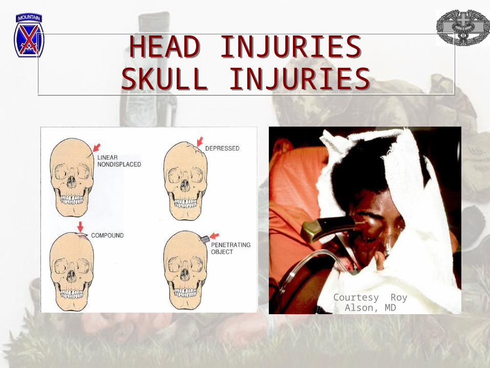
HEAD INJURIESSKULL INJURIESHEAD INJURIESSKULL INJURIES
Courtesy Roy Alson, MD

SIGNS OF BASILAR SKULL FRACTURE
SIGNS OF BASILAR SKULL FRACTURE
Courtesy David Effron, M.D.Courtesy David Effron, M.D.

HEAD INJURIES BRAIN INJURIESHEAD INJURIES BRAIN INJURIES
• Concussion• Cerebral contusion• Diffuse axonal
injury• Anoxic brain injury

HEAD INJURIESEPIDURAL HEMATOMA
HEAD INJURIESEPIDURAL HEMATOMA
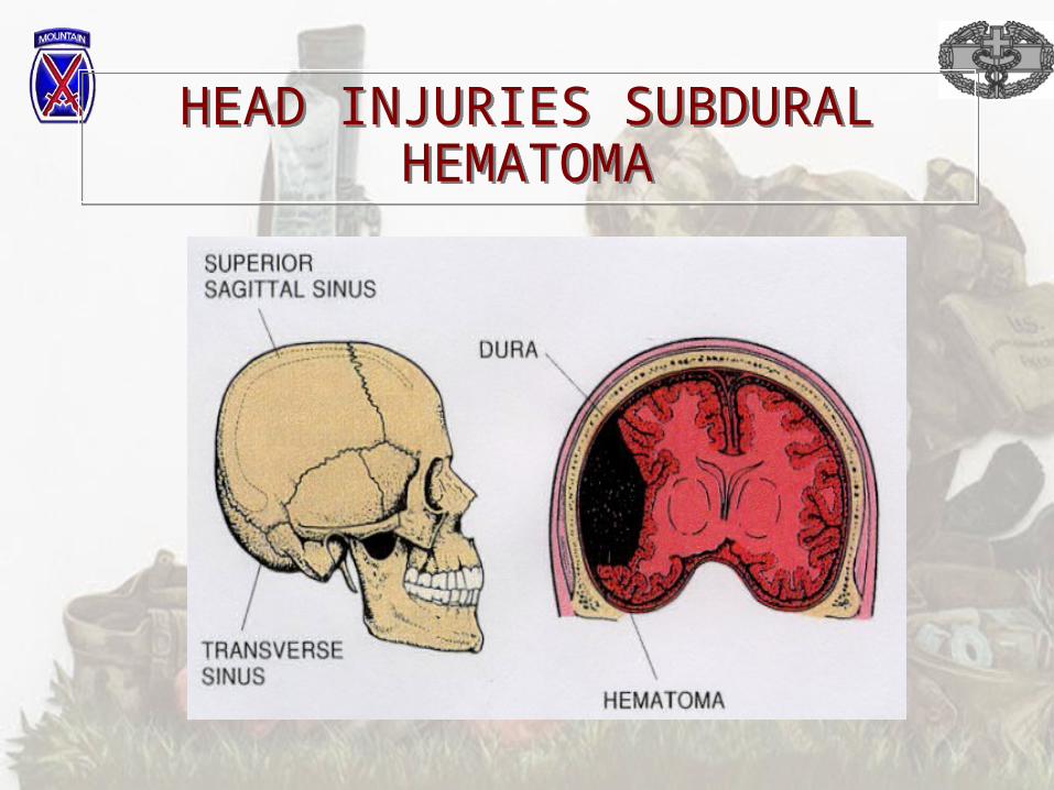
HEAD INJURIES SUBDURAL HEMATOMA
HEAD INJURIES SUBDURAL HEMATOMA

HEAD INJURIES INTRACRANIAL HEMORRHAGE
HEAD INJURIES INTRACRANIAL HEMORRHAGE

ASSESSMENT RAPID TRAUMA SURVEY
ASSESSMENT RAPID TRAUMA SURVEY
• Note LOC (AVPU), secure airway and protect c-spine
• Assess breathing– Do not allow the patient to become hypoxic
• Assess circulation– Control major bleeding– Prevent hypotension
• Transport decision and interventions• Do brief neuro & GCS if altered LOC

ASSESSMENT DETAILED EXAM
ASSESSMENT DETAILED EXAM
• Vital signs• SAMPLE history• Head-to-toe exam, including neurological
and GCS• Further bandaging and splinting• Continuous observation

PUPILSPUPILS

POSTURINGPOSTURING

MANAGEMENT OF THE HEAD TRAUMA PATIENTMANAGEMENT OF THE
HEAD TRAUMA PATIENT
• Stabilize the c-spine• Secure and maintain the airway• Ventilate at about 15 breaths/min.• Prevent hypoxia• Hyperventilate only patients with the
herniation syndrome– Coma, BP, Respiration, bradycardia

HEAD TRAUMA
19
AIRWAY CONTROL CANNOT BE OVEREMPHASIZED
AIRWAY CONTROL CANNOT BE OVEREMPHASIZED

MANAGEMENT MANAGEMENT
• Record baseline exam– Neuro, GCS & pupils– Vital signs
• Maintain good circulation– BP 110-120 systolic
• Continually monitor and record observations
• Prompt transport

PITFALLS & PROBLEMSPITFALLS & PROBLEMS
• Anticipate c-spine injuries• Protect the airway - prevent
aspiration• Prevent hypoxia• Prevent shock
– IV fluids and PASG are OK

PITFALLS & PROBLEMS PITFALLS & PROBLEMS
• Be prepared for seizures• Rapidly deteriorating condition
requires rapid hospital treatment• Assess for other causes of altered
LOC– Hypoglycemia– Alcohol– Drugs

SUMMARYSUMMARY
• Follow patient assessment• Protect c-spine, airway, and circulation• Record frequent vital signs, neuro, pupils,
and GCS• Prompt transport

QUESTIONS?QUESTIONS?


![Definitions of Injury · Immobilization: Traction Summary Procedure ICD-9-CM Summary Procedure ICD-9-CM Skull traction [Tongs, Halo ring] Skull traction Off skull traction Head Halter](https://img.pdfslide.net/doc/110x75/6070dec98da44f13c639a38d/definitions-of-immobilization-traction-summary-procedure-icd-9-cm-summary-procedure.jpg)