Embed Size (px)
Citation preview
International Journal of Pharma Research & Review, Jan 2015; 4(1):34-55 ISSN: 2278-6074
Rasna Gupta et.al, IJPRR 2015; 4(1) 34
Review Article
Hepatoprotective Activities of Triphala and Its Constituents
Rasna Gupta1, Ankit Gupta2, *Ram Lakhan Singh1
1. Nutraceutical Laboratory, Department of Biochemistry, Dr. Ram Manohar Lohia Avadh University, Faizabad-224001, Uttar Pradesh, India.
2. National Institute of Allergy and Infectious Disease, National Institute of Health, 12735 Twinbrook Parkway, Rockville, MD-20852-8132, USA.
ABSTRACT Liver is a vital organ which plays major role in metabolism and excretion of xenobiotics from the body. Liver injury or its dysfunction is a major health problem that challenges not only health care professionals but also the pharmaceutical industry and drug regulatory agencies. Liver cell injury caused by various toxic chemicals, certain chemotherapeutic agents, carbon tetrachloride, excessive alcohol, overloaded iron is well-studied. Some synthetic compounds such as antimicrobials, anticonvulsants, corticosteroids, NSAIDs and analgesic etc. are currently available as hepatoprotective agents. However, such compounds are not totally safe and exert several side effect and disadvantages. In view of severe adverse side effects of synthetic agents, there is growing need to develop more valuable and protected drugs which may be of therapeutic benefits to patients. Hence herbal drugs have become increasingly popular and their use is increasing day by day. A number of herbal preparations are available in the market. Triphala is one of the age old most commonly used polyherbal formulations with known hepatoprotective activities in Indian system of medicine mainly in Ayurveda. This is well known phytomedicine, a combination of three medicinal plants with Phyllanthus emblica (Amlaki, Phyllanthaceae), Terminalia chebula (Haritaki, Combretaceae) & Terminalia bellirica (Baheda, Combretaceae). Present review focuses on mechanism of hepaotoxicity and various scientifically tested hepatoperotective properties of formulation Triphala and its constituents. Keywords: Hepatoprotection, hepatotoxicity, Phyllanthus emblica, Terminalia bellerica, Terminalia chebula, triphala
Received 21 Oct 2014 Received in revised form 19 Dec 2014 Accepted 22 Dec 2014
*Address for correspondence: Dr. Ram Lakhan Singh, Professor and Head Department of Biochemistry, Dr. Ram Manohar Lohia Avadh University, Faizabad – 224001, Uttar Pradesh, India. E-mail: [email protected] ________________________________________________________________________________________________________________
INTRODUCTION Liver is the key organ for detoxification of toxic substances and disposition of endogenous substances. It is continuously and widely exposed to toxins and chemotherapeutic agents that lead to impairment of its function. Chronic Liver diseases stand as one of the foremost health troubles worldwide. Liver disease is one of the major causes of death [1]. According to the National Center for Health Statistics (NCHS) at the Centers for Disease Control and Prevention (CDC), chronic liver disease and cirrhosis is the 12th leading cause of death, claiming 30,000 lives annually
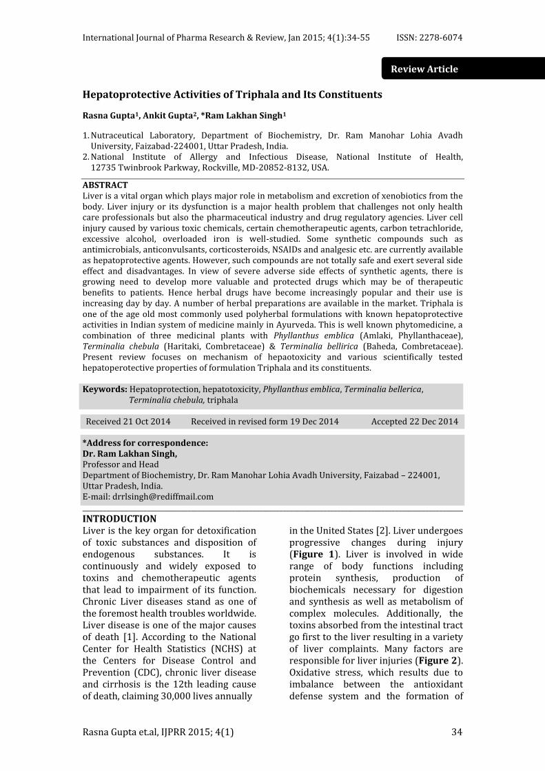
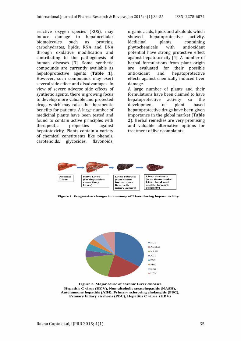
in the United States [2]. Liver undergoes progressive changes during injury (Figure 1). Liver is involved in wide range of body functions including protein synthesis, production of biochemicals necessary for digestion and synthesis as well as metabolism of complex molecules. Additionally, the toxins absorbed from the intestinal tract go first to the liver resulting in a variety of liver complaints. Many factors are responsible for liver injuries (Figure 2). Oxidative stress, which results due to imbalance between the antioxidant defense system and the formation of
International Journal of Pharma Research & Review, Jan 2015; 4(1):34-55 ISSN: 2278-6074
Rasna Gupta et.al, IJPRR 2015; 4(1) 35
reactive oxygen species (ROS), may induce damage to hepatocellular biomolecules such as proteins, carbohydrates, lipids, RNA and DNA through oxidative modification and contributing to the pathogenesis of human diseases [3]. Some synthetic compounds are currently available as hepatoprotective agents (Table 1). However, such compounds may exert several side effect and disadvantages. In view of severe adverse side effects of synthetic agents, there is growing focus to develop more valuable and protected drugs which may raise the therapeutic benefits for patients. A large number of medicinal plants have been tested and found to contain active principles with therapeutic properties against hepatotoxicity. Plants contain a variety of chemical constituents like phenols, carotenoids, glycosides, flavonoids,
organic acids, lipids and alkaloids which showed hepatoprotective activity. Medicinal plants containing phytochemicals with antioxidant potential have strong protective effect against hepatotoxicity [4]. A number of herbal formulations from plant origin are evaluated for their possible antioxidant and hepatoprotective effects against chemically induced liver damage. A large number of plants and their formulations have been claimed to have hepatoprotective activity so the development of plant based hepatoprotective drugs have been given importance in the global market (Table 2). Herbal remedies are very promising and valuable alternative options for treatment of liver complaints.
Figure 1. Progressive changes in anatomy of Liver during hepatotoxicity
Normal
Liver
Fatty Liver
(fat deposition
cause fatty
Liver)
Liver Fibrosis
(scar tissue
forms, more
liver cells
injury occurs)
Liver cirrhosis
(scar tissue make
Liver hard and
unable to work
properly)
Figure 2. Major cause of chronic Liver diseases
Hepatitis C virus (HCV), Non-alcoholic steatohepatitis (NASH),
Autoimmune hepatitis (AIH), Primary sclerosing cholangitis (PSC),
Primary biliary cirrhosis (PBC), Hepatitis C virus (HBV)
HCV
Alcohol
NASH
AIH
PSC
PBC
Drug
HBV
International Journal of Pharma Research & Review, Jan 2015; 4(1):34-55 ISSN: 2278-6074
Rasna Gupta et.al, IJPRR 2015; 4(1) 36
Table 1: Synthetic drugs causing liver injury at higher concentrations Nature of drugs Name of Drugs Anesthetics Desflurane, Enflurane, Halothane, Hyoscyamine, Isoflurane,
Methohexital Antimicrobials Bacitracine, Dapsone, Isoniazid, Ketoconazole, Nalidixic acid,
Penicillin, Pyrazinamide, Rifampsin, Sulfonamides, Trovafloxacin, Vancomycin
Anticonvulsants
Carbamazepine, Felbamate, Halothane, Isoflurane, Nimetazepam, Nitrazepam, Phenytoin, Temazepam, Valproic acid
NSAIDs and analgesic
Bromfenac, Diclofenac, Etodolac, Flurbiprofen, Ibuprofen, Indomethacin, Paracetamol, Piroxicam Oxaprozin, Sulindac
Miscellaneous agents
Disulfuram, Flutamide, Labealol, Nefazodone, Nicotinic acid, Pemoline, Propylthiouracil, Tolcapone, Troglitazone
Table 2: Common hepatoprotective polyherbal formulations and their plant sources
S.N. Formulation Name (manufacture)
Reported Plant sources and (their parts used)
1 Acilvan (Acis Laboratories, Kanpur)
Achillea millefoiliven (flower and leaf), Boerrhaavia diffusa (leaf), Capparis spinosa (fruit), Casearia esculenta (root), Cassia occidentalis (whole plant), Chichorium intybus (whole plant), Eclipta alba (leaf), Ocimum sanctum (whole plant), Picrorrrrhiza kurroa (root), Solanum nigrum (fruit), Tamarix gallica (flower), Terminalia arjuna (fruit), Tinospora cordifolia (stem).
2 Adliv (Abala Drug House, Kolkata)
Aloe barbadensis (leaf), Andropogon muricatus (leaf), Asteracantha longifolia (leaf), Centella asiatica (leaf), Carum copticum (seed), Cassia angustifolia (leaf), Holarrhena antidysentrica (seed and bark), Solanum xanthocarpum (fruit).
3 Amlycure (Aimil Pharmaceuti-cal, Pvt. Ltd., Kolkata)
Achillea millefoiliven (flower and leaf), Aloe barbadensi (leaf), Berberis lyceum (root), Cassia obtusifolia (leaf), Cassytha filliformia (leaf), Chichorium intybus (root), Eclipta alba (leaf), Fumaria officinalis (seed), Helleborus niger (flower), Ipomoea turpethum (root), Ocimum sanctum (leaf), Panicum milliare (seed), Solanum nigru (fruit), Tephrosia purpurea (root), Terminalia belerica (seed).
4 Biligen (Standard Pharmaceu-tical, Kolkata)
Ipomoea turpethum (stem and leaf), Swertia chirata (seed), Trachyspermum ammi (seed), Trigonella foenumgraecum (seed).
5 Hepa-10 (Jupiter Pharmaceuticals Pvt Ltd., Kolkata)
Aloe barbadensis (leaf), Andrographis paniculata (leaf and root), Aphnamixis polystachya (stem bark), Eclipta alba (whole plant), Fumaria officinalis (leaf), Luffa echinata (fruit), Ptychotis ajowan (seed), Solanum nigrum (fruit).
6 Hepex The Anglo French Drug Co. (Eastern) Ltd., Mumbai)
Boerrhaavia diffusa (leaf), Phyllanthus Emblica (fruit), Phyllanthus niruri (fruit).
7 Hipex (H.V. Pharmaceuticals, Rajkot, Gujarat)
Boerrhaavia diffusa (leaf), Cassia occidentalis (leaf and seed), Chichorium intybus (root), Embelia ribes (leaf and root bark), Solanum nigrum (fruit), Terminalia chebula (fruit).
8 Kalmegh (Bengal Chemicals Pharmacecuticals Pvt. Ltd., Kolkata)
Andrographis paniculata (leaf and stem), Apium graveolens (seed), Carum copticum (fruit).
International Journal of Pharma Research & Review, Jan 2015; 4(1):34-55 ISSN: 2278-6074
Rasna Gupta et.al, IJPRR 2015; 4(1) 37
9 Liv-52 (Himalaya Drug Co., Mumbai)
Achillea millefolium (leaf and flower), Andrographis paniculata (leaf and root), Boerhavia diffusa (root), Capparis spinosa (bulb and fruit), Cassia occidentalis (seed), Cinchorium intybus (root), Eclipta alba (seed), Fumaria officinalis (flower), Phyllanthus embilica (fruit), Phyllanthus niruri (fruit), Solanum nigrum (fruit), Tamarix gallica (leaf and bark), Terminalia arjuna (bark), Terminalia chebula (fruit), Tinospora cordifolia (stem).
10 Liv-77 (Gobe Pharmacecuticals, Jalandhar City, Punjab)
Berberis lyceum (leaf and fruit), Boerrhaavia diffusa (leaf and seed), Chichorium intybus (root), Eclipta alba (seed), Hemidesmus indicus (root), Prunus domestica (fruit), Tinospora cordifolia (stem).
11 Liva-16 (Madona Pharmaceutical Research, Kolkata)
Piper nigrum (unripe fruit), Oldenlandia corymbasa (root), Solanum nigrum (fruit), Solanum xanthocarpum (fruit), Tinospora cordifolia (stem).
12 Livarin (Patiala Ayurvedic Pharm., Sirhind)
Picrorrrrhiza kurroa (root), Solanum nigrum (fruit), Tecoma undulate (leaf).
13 Livatone (East India Pharmacecuticals Works Ltd., Kolkata)
Holarrhena antidysentrica (bark), Andrographis paniculata (leaf and root), Apium graveolens (seed), Asteracantha longifolia (seed), Trigonella foenumgraecum (seed).
14 Livergen (Standard Pharmacecutical, Kolkata)
Andrographis paniculata (root), Apium graveolens (seed), Asteracantha longifolia (seed), Cassia angustifolia (fruit), Trachyspermum ammi (seed), Trigonella foenumgraecum (seed).
15 Livex (Bhartiya aushadh Nirmanshala, Rajkot, Gujarat)
Achillea millefoiliven (flower and leaf), Aconitum heterophyllum (root), Cassia occidentalis (leaf), Embelia ribes (fruit), Solanum nigrum (seed), Swertia chirata (seed), Tamarix gallica (leaf).
16 Livin (Araya Aushadhi Pharmaceuticals Works, Indore)
Acorus calamus (root), Andrographis paniculata (leaf), Aphnamixis polystachya (stem bark), Boerrhaavia diffusa (root), Cassia sophera (leaf), Citrullus colocynthis (root), Eclipta alba (seed), Ipomoea turpethum (root), Jatrorrhiza palmate (root), Latsea chinesis (bark), Lawsonia inermis (leaf), Ocimum sanctum (leaf), Plumbago zeylanica (root), Salvandora persica (bark), Tephrosia purpurea (stem), Terminalia chebula (fruit), Tinospora cordifolia (whole plant), Trachyspermum ammi (seed).
17 Livodin (Madona Pharmaceutical Research, Kolkata)
Aloe barbadensis (leaf), Andrographis paniculata (leaf), Aphnamixis polystachya (leaf), Asteracantha longifolia (seed), Carum copticum (seed), Cassia angustifolia (leaf), Embelia ribes (seed), Holarrhena antidysentrica (seed), Piper nigrum (leaf), Solanum xanthocarpum (fruit), Tinospora cordifolia (stem).
18 Livosin (Jupiter Pharmaceuticals Pvt Ltd., Kolkata)
Andrographis paniculata (leaf), Avena sativa (seed), Carica papaya (seed), Cassia angustifolia (leaf), Embelia ribes (seed), Holarrhena antidysentrica (seed), Mentha viridis (leaf), Phyllanthus Emblica (fruit), Solanum nigrum (seed), Terminalia arjuna (fruit), Terminalia belerica (fruit).
19 Livomyn (Charak Pharmaceuticals (India) Pvt. Ltd., Umbargaon, Gujarat)
Aphnamixis polystachya (bark), Capparis spinosa (root bark), Cassia occidentalis (leaf), Embelia ribes (seed), Fumaria officinalis (root), Ipomoea turpethum (root bark), Plumbago zeylanica (root), Swertia decussate (whole plant), Tephrosia purpurea (whole plant), Tinospora cordifolia (whole plant).
International Journal of Pharma Research & Review, Jan 2015; 4(1):34-55 ISSN: 2278-6074
Rasna Gupta et.al, IJPRR 2015; 4(1) 38
20 Livomycin (Charak Pharmaceuticals (India) Pvt. Ltd., Umbargaon, Gujarat)
Boerrhaavia diffusa (root).
21 Livokin (Herbo-Med, Kolkata)
Andrographis paniculata (leaf), Apium graveolens (seed), Asteracantha longifolia (root bark), Berberis lyceum (root bark), Carum copticum (seed), Chichorium intybus (leaf), Embelia ribe (seed), Ipomoea turpethum (root), Oldenlandia corymbasa (whole plant), Picrorrrrhiza kurroa (root), Solanum nigrum (seed), Tephrosia purpurea (root bark), Terminalia arjuna (fruit), Terminalia chebula (fruit), Trigonella foenumgraecum (seed).
22 Neolive-100 (Bharat Pharmaceuticals, Delhi)
Achillea millefoiliven (whole plant), Boerrhaavia diffusa (root), Cassia occidentalis (leaf), Chichorium intybus (leaf), Phyllanthus Emblica (fruit), Solanum nigrum (seed), Tecoma undulate (leaf), Tamarix gallica (leaf), Tephrosia purpurea (root), Terminalia arjuna (bark).
23 Livotone (Gambers Laboratories, Mumbai)
Andrographis paniculata (leaf), Apium graveolens (seed), Asteracantha longifolia (seed), Holarrhena antidysentrica (seed).
24 Triguliv-15 (Triguna Ayurveda Research Lab., New Delhi)
Aphnamixis polystachya (seed), Berberis lyceum (root), Boerrhaavia diffusa (leaf), Carthamus tinctorius (flower), Eclipta alba (seed), Fumaria officinalis (whole plant), Grewia asiatica (fruit), Helleborus niger (flower), Phyllanthus niruri (leaf and stem), Salsola kali (leaf), Solanum nigrum (seed), Tephrosia hirta (root), Tinospora cordifolia (root).
25 Syliv (Systemic Pharmaceuticals, Allahabad)
Achillea millefoiliven (root), Asteracantha longifolia (seed), Capparis spinosa (root bark), Carum copticum (fruit), Cassia occidentalis (leaf), Chichorium intybus (leaf), Oldenlandia corymbasa (whole plant), Solanum nigrum (seed), Tamarix gallica (leaf), Terminalia arjuna (leaf).
26
Livol (Vedic Pharm., Kolkata)
Adhatoda vasica (leaf), Andrographis paniculata (leaf), Berberis lyceum (root), Eclipta alba (seed), Phyllanthus Emblica (fruit), Picrorrrrhiza kurroa (root), Phyllanthus niruri (leaf), Terminalia belerica (fruit), Terminalia chebula (fruit), Tinospora cordifolia (stem).
27 Livomap (Maharishi Ayurveda Corporation Ltd., New Delhi)
Artemisia absinthium (whole plant), Berberis lyceum (root), Boerrhaavia diffusa (leaf), Cedrus Deodara (root), Crataeva religioosa (leaf), Glycyrrhiza glabra (root), Melia azadirachta (leaf), Moringa pterygosperma (stem bark), Phyllanthus niruri (leaf), Picrorrrrhiza kurroa (root), Piper longum (fruit), Tecoma undulate (leaf), Tinospora cordifolia (stem), Tephrosia purpurea (root bark), Terminalia chebula (fruit).
Triphala is an Ayurvedic
herbal Rasayana formula consisting of equal parts of three plants Amlaki, Phyllanthus emblica (syn. Emblica officinalis) of Phyllanthaceae family, Haritaki (Terminalia chebula) & Baheda (Terminalia bellirica) both belonging to Combretaceae family. Triphala is believed to be a well-known
phytomedicine that promotes health, immunity, and longevity. All three constituents of Triphala are used in preparation of many hepatoprotective formulations (Table 3). Available research papers and review articles on the subject give scanty information especially on hepatoprotective activities of Triphala and its constituents which
International Journal of Pharma Research & Review, Jan 2015; 4(1):34-55 ISSN: 2278-6074
Rasna Gupta et.al, IJPRR 2015; 4(1) 39
does not serve the purpose of researchers or pharmaceutical industry. Hence there is need of a review which focuses solely on its hepatoprotective
activities. Present review focuses on the scientifically tested hepatoperotective properties of Triphala and its constituents.
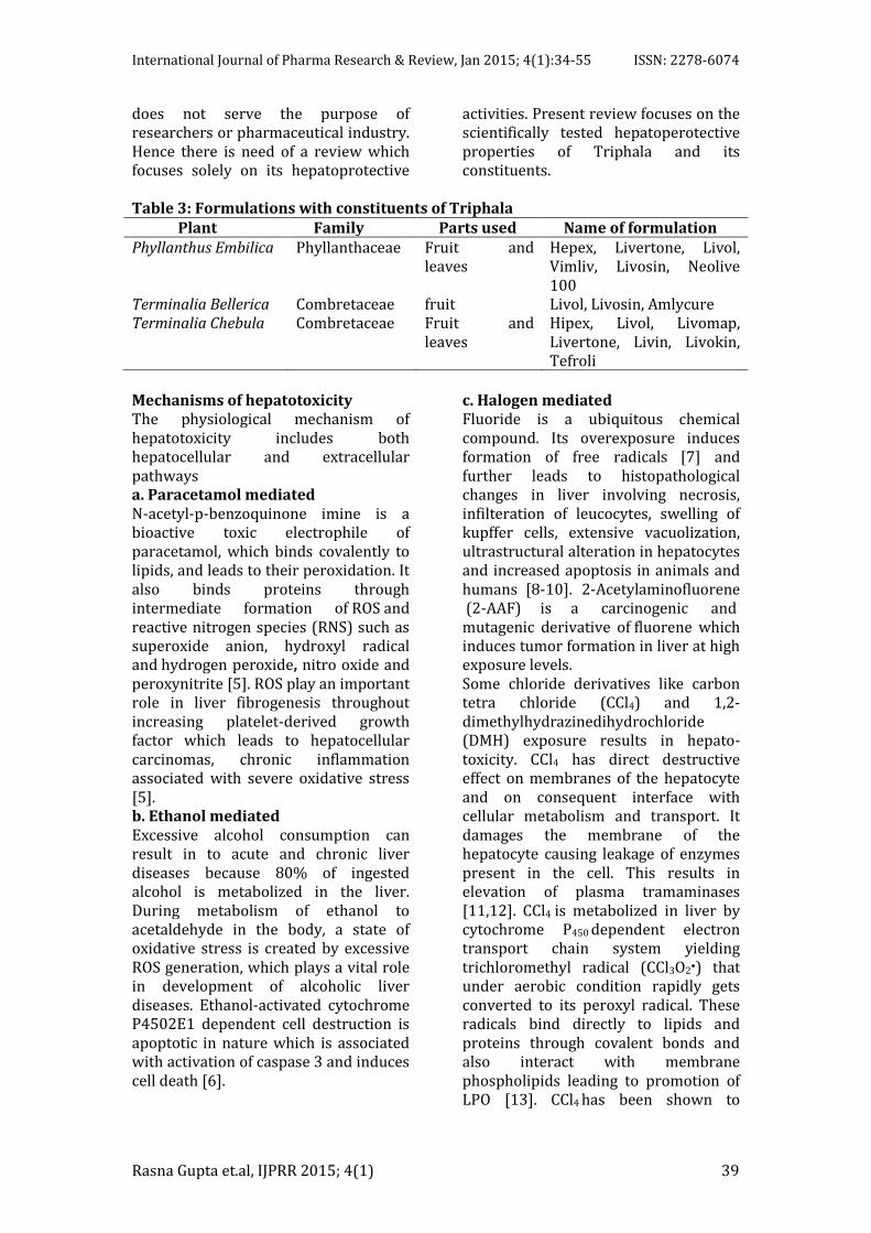
Table 3: Formulations with constituents of Triphala Plant Family Parts used Name of formulation Phyllanthus Embilica Phyllanthaceae Fruit and
leaves Hepex, Livertone, Livol, Vimliv, Livosin, Neolive 100
Terminalia Bellerica Combretaceae fruit Livol, Livosin, Amlycure Terminalia Chebula Combretaceae Fruit and
leaves Hipex, Livol, Livomap, Livertone, Livin, Livokin, Tefroli
Mechanisms of hepatotoxicity The physiological mechanism of hepatotoxicity includes both hepatocellular and extracellular pathways a. Paracetamol mediated N-acetyl-p-benzoquinone imine is a bioactive toxic electrophile of paracetamol, which binds covalently to lipids, and leads to their peroxidation. It also binds proteins through intermediate formation of ROS and reactive nitrogen species (RNS) such as superoxide anion, hydroxyl radical and hydrogen peroxide, nitro oxide and peroxynitrite [5]. ROS play an important role in liver fibrogenesis throughout increasing platelet-derived growth factor which leads to hepatocellular carcinomas, chronic inflammation associated with severe oxidative stress [5]. b. Ethanol mediated Excessive alcohol consumption can result in to acute and chronic liver diseases because 80% of ingested alcohol is metabolized in the liver. During metabolism of ethanol to acetaldehyde in the body, a state of oxidative stress is created by excessive ROS generation, which plays a vital role in development of alcoholic liver diseases. Ethanol-activated cytochrome P4502E1 dependent cell destruction is apoptotic in nature which is associated with activation of caspase 3 and induces cell death [6].
c. Halogen mediated Fluoride is a ubiquitous chemical compound. Its overexposure induces formation of free radicals [7] and further leads to histopathological changes in liver involving necrosis, infilteration of leucocytes, swelling of kupffer cells, extensive vacuolization, ultrastructural alteration in hepatocytes and increased apoptosis in animals and humans [8-10]. 2-Acetylaminofluorene (2-AAF) is a carcinogenic and mutagenic derivative of fluorene which induces tumor formation in liver at high exposure levels. Some chloride derivatives like carbon tetra chloride (CCl4) and 1,2-dimethylhydrazinedihydrochloride (DMH) exposure results in hepato-toxicity. CCl4 has direct destructive effect on membranes of the hepatocyte and on consequent interface with cellular metabolism and transport. It damages the membrane of the hepatocyte causing leakage of enzymes present in the cell. This results in elevation of plasma tramaminases [11,12]. CCl4 is metabolized in liver by cytochrome P450 dependent electron transport chain system yielding trichloromethyl radical (CCl3O2•) that under aerobic condition rapidly gets converted to its peroxyl radical. These radicals bind directly to lipids and proteins through covalent bonds and also interact with membrane phospholipids leading to promotion of LPO [13]. CCl4 has been shown to
International Journal of Pharma Research & Review, Jan 2015; 4(1):34-55 ISSN: 2278-6074
Rasna Gupta et.al, IJPRR 2015; 4(1) 40
activate Kupffer cells by increasing intracellular Ca2+ concentration, causing release of harmful cytokines that contribute to cause death of hepatocytes and oxidative stress [14]. d. Anti-tuberculosis drugs mediated Anti-tuberculosis (anti-TB) drug induced liver injury is a leading cause of liver failure in India and many developed countries. Some most commonly used anti-TB drugs are isoniazid, rifampsin and pyrazinamide. Isoniazid undergoes acetylation by N-acetyl transferase 2 (NAT-2). Acetyl-isoniazid is metabolized mainly to mono-acetyl hydrazine (MAH) and to the nontoxic diacetyl hydrazine. Reactive metabolites of MAH are probably toxic to tissues through free radical generation [15]. Isoniazid inhibits the activity of several cytochrome P450 2E and 2C enzymes, potentially increasing the plasma concentrations of other potentially hepatotoxic drugs, such as phenytoin and carbamazepine [16,18]. Rifampicin appears to enhance a metabolic hepatocellular characteristic reaction in patients getting isoniazid, perhaps by supporting the formation of toxic isoniazid metabolites [19,20]. Rifampicin occasionally can cause hepatocellular injury and potentiate hepatotoxic action of other anti-TB medications [21,22]. Pyrazinamide, a nicotinic acid derivative alters nicotinamide acetyl dehydrogenase levels in rat liver [23], which might results in generation of free radical species. e. Iron mediated Extreme iron deposition increases liver injury which is induced by iron-generated oxyradicals and peroxidation of lipid membranes. LPO results in damage of hepatocellular organelles such as mitochondria and lysosomes, which is thought to contribute to hepatocyte necrosis and apoptosis [24], inflammation [25] and ultimately lead to the development of hepatic fibrogenesis
[26,27] and even to cancer [28]. Some synthetic compounds such as deferoxamine, deferiprone and
deferasirox are currently used as iron chelating agents. However, such compounds are inadequate and exert several side effect and disadvantages
[29,30]. In view of severe adverse side effects of synthetic agents, there is growing focus to develop more valuable and protected drugs [31,32] which may raise the therapeutic benefits for patients. f. D-Galactosamine mediated D-galactosamine (D-GalN) is used as a model hepatotoxin to stimulate experimental liver injury [33,34]. D-GalN induces acute liver injury in mice and has been used to mimic the sequences of events in viral hepatitis [35]. Ingestion of D-GalN depletes several uracil nucleotides and results in formation of uridine-diphosphogalacto-samine (UDP-GalN) [36]. High accumulation of UDP-GalN contributes to disturbance in protein metabolism [37,38]. It is reported that D-GalN may cause loss of the intracellular calcium homeostasis, leading to cell membrane damage [33,39]. It results in hepatocyte necrosis and parenchymal inflammation and also causes imbalance in energy metabolism of hepatic cells [40]. g. Neutrophile mediated Neutrophiles [41] and liver macrophages [42] derived ROS can cause necrotic cell injury by opening the membrane permeability transition pore and collapse of the mitochondrial membrane potential [43]. h. Nitric oxide mediated Hepatocytes express inducible nitric oxide synthase (iNOS). Administration of paracetamol at high doses induces the expression of iNOS in rat hepatocytes [44]. An increase in serum nitrate and nitrite result in increased level of serum marker alanine transferase (ALT) in wild type mice. Based on liver biopsy specimens, ALT elevations in asymptomatic patients have also been connected with fatty liver [45]. In fact, nonalcoholic steatosis has been mentioned as a very common cause of chronic ALT elevations in the general population [46].
International Journal of Pharma Research & Review, Jan 2015; 4(1):34-55 ISSN: 2278-6074
Rasna Gupta et.al, IJPRR 2015; 4(1) 41
i. Non-steroidal anti-inflammatory drugs mediated Non-steroidal anti-inflammatory drugs (NSAIDs) are responsible for roughly 10% of the total cases of drug-induced hepatotoxicity. Aspirin, Nimesulide, Diclofenac and Ibuprofen are commonly used NSAIDs. As epideminological risk is very low, 10 in 100 000, but at high dose it can be serious and can cause diagnostic confusion [47]. Nearly all of the NSAIDs have been implicated in causing liver injury which tends to be hepatocellular in nature [48]. Several NSAIDs have been withdrawn from clinical use because of associated hepatotoxicity [49]. The new more selective COX-2 inhibitors (e.g. celecoxib, rofecoxib, nimesulide) are also associated with hepatotoxicity [50]. Aspirin may uncouple mitochondrial respiration [51] and induces the permeability transition pore [52,53]. Inhibition of mitochondrial β-oxidation of fatty acid causes microvesicular steatosis [54]. Administration of some drugs such as aminodarone, perhexiline concentrates into mitochondria to cause LPO leading to nonalchohlic steatohepatitis (NASH) and ultimately
to liver cell death. Both LPO and ROS-induced cytokine (TGF-β, TNF-α, IL-8) release may contribute to the development of NASH [55]. Mitochondrial apoptosis (MA) is another mechanism of hepatotoxicity caused by opening of permeability transition (PT) pores in the mitochondrial inner membrane. PT pore opening in all mitochondria of a cell causes ATP depletion, which prevents apoptosis (an energy-requiring process) and causes necrosis
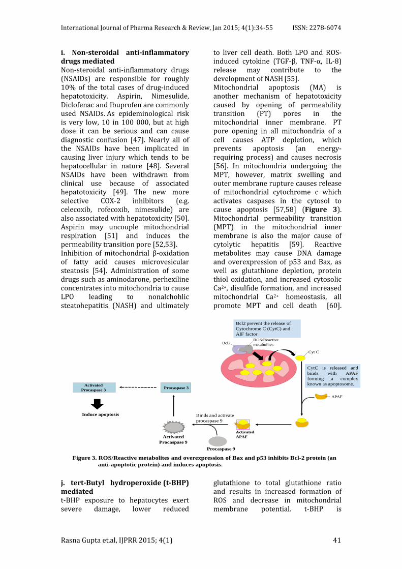
[56]. In mitochondria undergoing the MPT, however, matrix swelling and outer membrane rupture causes release of mitochondrial cytochrome c which activates caspases in the cytosol to cause apoptosis [57,58] (Figure 3). Mitochondrial permeability transition (MPT) in the mitochondrial inner membrane is also the major cause of cytolytic hepatitis [59]. Reactive metabolites may cause DNA damage and overexpression of p53 and Bax, as well as glutathione depletion, protein thiol oxidation, and increased cytosolic Ca2+, disulfide formation, and increased mitochondrial Ca2+ homeostasis, all promote MPT and cell death [60].
Induce apoptosis
Bcl2 prevent the release of
Cytochrome C (CytC) and
AIF factor
Bcl2 ROS/Reactive
metabolites
Cyt C
APAF
Activated
APAF
CytC is released and
binds with APAF
forming a complex
known as apoptosome.
Procaspase 9
Activated
Procaspase 9
Procaspase 3 Activated
Procaspase 3
Binds and activate
procaspase 9
Figure 3. ROS/Reactive metabolites and overexpression of Bax and p53 inhibits Bcl-2 protein (an
anti-apoptotic protein) and induces apoptosis.
j. tert-Butyl hydroperoxide (t-BHP) mediated t-BHP exposure to hepatocytes exert severe damage, lower reduced
glutathione to total glutathione ratio and results in increased formation of ROS and decrease in mitochondrial membrane potential. t-BHP is
International Journal of Pharma Research & Review, Jan 2015; 4(1):34-55 ISSN: 2278-6074
Rasna Gupta et.al, IJPRR 2015; 4(1) 42
metabolized by cytochrome P450, leading to production of peroxyl and alkoxyl radicals [61]. These radicals initiate LPO of membrane phospholipids with subsequent alterations to membrane fluidity and permeability. Other pathway of its metabolism employs glutathione peroxidase (GPx). t-BHP is detoxified to tert-butanol and glutathione (GSH) is depleted by oxidation to its disulphide form (GSSG) [62]. LPO, depletion of GSH and the onset of mitochondrial MPT are general mechanisms involved in cell injury caused by oxidative stress. Hepatoprotective activities of Triphala: a polyherbal formulation: Triphala (equai proportion of Baheda, Harad and Amla in 1:1:1 ratio) is well known phytomedicine as accounted in the Ayurveda. It is widely accepted herbal formulation because of its exclusive capability to gently cleansing and detoxifying the body while at the same time strengthening and nourishing it due to rich source of antioxidants. As per the information available in Ayurvedic literature, Triphala may be used for the treatment of fever, cough, diarrhea, dysentery, skin disease, liver diseases [63] and gastrointestinal tract diseases [64,65]. Triphala is confirmed to have antiviral and antibacterial effects [65]. It is also recommended in myocardial injury, cancer etc. [66,67]. Its antidiabetic [68], antimutagenic [69], pergative [70], radioprotective [71] and cholesterol lowering [72] activities have been reported. It is not only relaxing and regulatory for digestion, but also regulates bowel movement, strengthens and revitalizes body tissues, increases the body’s ability to absorb nutrients, boost life energies and is an entirely natural antioxidant. Charaka Samhita, an 8th century text on Ayurvedic medicine, describes Triphala churna as rejuvenating medicament that can be used alone or in formulation. Charaka, the author of Charaka Samhita, states that “by using Triphala constantly for one year, one can live for a hundred years, free from ageing and diseases”
[73]. Because it works slowly and lightly it can be taken over long periods of time without a problem. a. Protective effect of Triphala against paracetamol The activities of serum enzymes ALT, aspartate amino transaminase (AST), alkaline phosphatase (ALP) were extensively increased in paracetamol treated group as compared to control group. Aqueous extract of Triphala at 100 mg/kg body weight (b.w.) inhibits paracetamol at 900 mg/kg b.w. induced hepatotoxicity in mice as indicated by the decrease in serum AST, ALP, inflammatory mediator TNF-α and liver LPO [74]. Simultaneously, paracetamol administration increases malondialdehyde level (an end product of LPO) whereas levels of antioxidant enzymes superoxide dismutase (SOD), catalase (CAT), GPx, glutathione reductase (GR), glutathione S-transferase (GST) and GSH were found to be decreased when compared with the control group. Aqueous extract of Triphala reversed the above changes by regulating the MDA level and antioxidant enzymes to nearly that of normal levels. Thus Triphala inhibits LPO and prevents oxidative stress [74]. b. Protective effect of Triphala against Ethanol The aqueous and methanolic extract of Triphala exerts significant protection against ethanol-induced toxicity by its ability to improve the oxidative stress enzyme system through free radical scavenging activity, which enhances levels of antioxidant defense system. The study also showed that methanolic extract of Triphala at dose of 100 mg/kg b.w. had greater effect than aqueous extract at the same dose level. Therefore, methanolic extract appears to be more useful in the attenuation of ethanol induced oxidation and showed more prominent effect than aqueous extract. Both the extracts showed significant activity against liver damage when compared with standard drug silymarin [75].
International Journal of Pharma Research & Review, Jan 2015; 4(1):34-55 ISSN: 2278-6074
Rasna Gupta et.al, IJPRR 2015; 4(1) 43
c. Protective effect of Triphala against 1,2-dimethylhydrazinedi-hydrochloride (DMH) DMH treatment causes liver necrosis through changes in the liver microsomal proteins, which reversed back to the normal patterns after treatment with Triphala. It prevents the liver necrosis in DMH treated mice at oral dose of 3 mg/kg b.w. Administration of DMH significantly increased level of serum enzymes ALT, AST and ALP. However, Triphala administration to DMH treated mice led to decreased activation of above enzymes in serum showing the stabilization of plasma membranes as well as the repair of hepatic tissue damage due to DMH exposure. Triphala simultaneously increased the level of antioxidant enzyme GSH and the activity of GST suggesting that it prevents peroxidative damage and also diverts the active metabolites of DMH from their interaction with critical cellular biomolecules which could be responsible for its protective action against DMH [76]. d. Protective effect of Triphala against D-galactosamine (D-GalN) It is reported that D-GalN induced hepatic damage resulted in a significant increase in the levels of ALT, AST, ALP, bilirubin, LPO (MDA level) and TNF-α with a decrease in the levels of anti-oxidant enzymes such as SOD, CAT, GPx, GR, GST and GSH which attained normal levels after the treatment with aqueous Triphala extract at 1000 mg/kg b.w. [77]. Pretreatment of Triphala inhibited LPO, suggesting that Triphala may exert a stabilizing action on liver cell membranes [77]. Hepatoprotective activities of Phyllanthus emblica: P. emblica Linn, or Embilica officinalis Gaertn commonly known as Indian gooseberry or Amla, belongs to family Euphorbiaceae. The plant species, which was originally native to India, is today found growing in Pakistan, Uzbekistan, Sri Lanka, South-East Asia, China, and Malaysia [78,79]. It is well-known that all parts of amla plant are used to treat a range of diseases, but the most
significant is the fruit. Fruit is used either alone or in combination with other plants to treat many ailments such as common cold, fever, peptic ulcer, dyspepsia, and it is also used as a digestive aid. Amla is one of the richest source of vitamin C (478.56 mg/100ml) having more vitamin C than orange and lemon. It contains many active phytochemicals like gallic acid, ellagic acid, emblicanin A, emblicanin B, punigluconin and pedunculagin [78] and flavonoids [79,80] (Table 4). Pharmacological research on amla revealed its analgesic [81], anti-tussive
[82], anti-atherogenic [83], adaptogenic
[84], cardioprotective [85], hepato-protective [86], gastroprotective [87], nephroprotective [88], neuroprotective
[89] and anticancer [90] properties. Amla is also reported to possess chemopreventive [91], radioprotective
[92], wound healing [93], immuno-modulatory [94], free radical scavenging
[95], antioxidant [96], antidiabetic [97], anti-diarrheal [98] and anti-viral [99] properties. These properties are effective in the prevention and treatment of various diseases like cancer, peptic ulcer, atherosclerosis, diabetes, anemia, liver, heart diseases and various other disorders. a. Protective effect of P. embilica against paracetamol Pretreatment of rats with P. embilica fruits extract at oral doses of 100-200 mg/kg b.w. 4 hrs before paracetamol administration, lowered the extent of hepatotoxicity. It has been reported that tannins and flavanoids present in P. embilica are responsible for their dominant antioxidant and hepato-protective activities [100,101]. A polyherbal formulation with P. acidus (30 g), Moringa oleifera (40 g) and P. embilica (30 g) showed activity against paracetamol induced liver toxicity in albino rats [102]. Ethanolic extract of P. embilica leaf was more effective against hepatotoxicity [102]. b. Protective effect of P. embilica against ethanol Alcohol administration in rats showed a significant oxidative stress and ROS
International Journal of Pharma Research & Review, Jan 2015; 4(1):34-55 ISSN: 2278-6074
Rasna Gupta et.al, IJPRR 2015; 4(1) 44
mediated toxicity. P. emblica fruit extract was investigated on ethanol induced rat hepatic injury. Pretreatment of rats with P. embilica at oral dose of 25, 50 and 75 mg/kg b.w. or silymarin (a reference hepatoprotective agent) at 5 mg/kg, 4 h before ethanol treatment, lowered the ethanol induced levels of AST, ALT and interleukine-1 (IL-1). The 75 mg/kg b.w. P. emblica dose showed best results similar to silymarin. Histopathological studies confirmed the beneficial roles of P. embilica and silymarin against ethanol induced liver injury in rats [103]. Administration of P. embilica fruit extract at a dose of 250 mg/kg b.w. per day to alcoholic rats offers protection by simultaneously lowering the carbonyl content and LPO, and elevating antioxidant enzyme activities [104]. c. Protective effect of P. embilica against CCl4
A single dose of CCl4 (1 ml/kg b.w.) elevated the levels of AST, lactate dehydrogenase (LDH), GST and depleted the levels of GSH, glutathione peroxidase (GPx) and glutathione reductase (GR) in Wistar rats. It also enhanced the level of LPO. The pretreatment of P. Embilica for 7 repeated days showed a profound pathological protection to liver cell as depicted by univacuolated hepatocytes. Pretreatment with P. Embilica at doses of 100 and 200 mg/kg b.w., prior to CCl4 intoxication, showed significant reduction in the levels of AST, ALT, LDH, GST, LPO and DNA synthesis [105]. DHC-1, a standardized polyherbal formulation, showed hepatoprotective activity against CCl4 induced liver damage. P. embilica is one of the components of this formulation along with Bacopa monnieri, Glycyrrhiza glabra, Mangifera indica and Syzygium aromaticum [106]. d. Protective effect of P. embilica against anti-TB drugs Many biochemical manifestations of anti-TB drugs such as rifampicin (RIF), isoniazid (INH) and pyrazinamide (PZA) induced hepatotoxicity either alone or in combination. Fifty per cent
hydroalcoholic extract of the fruits of P. embilica was evaluated for their hepatoprotective activity against anti-TB drugs-induced hepatic injury. The result showed significant protection of liver against these drugs [107]. e. Protective effect of P. embilica against Fluoride Increased fluoride exposure leads to fluorosis and further proceeds to damage the cardiovascular system. Fluoride is toxic to almost all the systems and causes oxidative stress in various tissues. Its ingestion also results in hyperlipemia and LPO. Exposure to fluoride resulted in significant elevation of plasma and hepatic lipid levels, bile acid content and reduced plasma HDL-C levels and hepatic HMG-CoA reductase (HMGR) activity. Administration of P. embilica fruit powder (2.5, 5 and 10 gm%) through diet significantly reduced plasma and hepatic lipid levels, tissue LPO and increased plasma HDL-C and fecal cholesterol levels in rats. Both hepatic HMGR activity and the bile acid (hepatic and fecal) production increased on administration of P. embilica fruit powder [108]. f. Protective effect of P. embilica against Nicotine Nicotine is a potent parasympatho-mimetic alkaloid; it is first metabolized in the liver and induces lung and liver damages. Toxicity was induced by oral administration of nicotine at a dose of 5 mg/kg b.w. for 32 days in rats which caused significant decrease in the levels of SOD, CAT, total GSH and GPx in plasma, lung, liver, kidney and brain. The levels of SOD, CAT, GSH and GPx increased on administration of fruit extract of P. embilica (at 250 & 500 mg/kg b.w.) for 7 days. The protective effect was found to be dose dependent
[109]. g. Protective effect of P. embilica against N-nitrosodimethylamine P. embilica at a dose of 100 mg/kg b.w. in N-nitrosodimethylamine-treated rat liver reduced the expression of NOS and CYP2E1 protein, improve manganese SOD and CAT expression as well as supplementation offsets induced liver
International Journal of Pharma Research & Review, Jan 2015; 4(1):34-55 ISSN: 2278-6074
Rasna Gupta et.al, IJPRR 2015; 4(1) 45
injury via its antioxidant, anti-inflammation, anti-apoptosis, and anti-autophagy properties [110]. Administration of aqueous extract of P. embilica at the dose of 2 mg/day for 45 days decreased the level of LPO through its free radical scavenging activity and thus reduced the production of hydroxyl, peroxides and superoxide radicals and subsequently increased the concentration of GSH [111]. h. Protective effect of P. embilica against tert-butyl hydroperoxide (t-BHP) The aqueous and methanolic extracts of leaves and stem of P. embilica were screened for hepatoprotective activity at a concentration of 50 µg/ml againsts t-BHP induced liver toxicity in HepG2 cells. Plant extracts showing hepatoprotective activity were assessed for their 50% effective concentration (EC50) values and their antioxidant activity using a DPPH assay. Methanolic extracts of P. embilica showed significant hepatoprotective activity with EC50 values of 19 µg/ml and 50% inhibitory concentration (IC50) of 3.38 µg/ml for DPPH scavenging activity against an IC50 of 3.69 µg/ml for ascorbic acid [86]. Hepatoprotective activities of Terminalia Chebula T. chebula is also called as “King of Medicines” because of its miraculous power of healing with a wide spectrum of biological activities. It has always been listed first in the Ayurvedic Medica. The fruit of T. chebula is being used for the treatment of verious diseases and disorders since ancient times. It is now considered as a precious source of unique natural products for development of medicines against various diseases and also for the development of industrial products. The phytochemical analysis showed that T. chebula is a rich source of various phenolic and flavonoid compounds
[112] (Table 4) which is well known for their free radical scavenging and iron chelating properties [113]. Seventy percent methanolic extract of T. chebula has been reported to contain some
notable antioxidants [114] (Table 4) and many bioactive constituents [115]. T. chebula possesses a wide variety of scientifically tested biological activities like cytoprotective [116], spasmogenic
[117], NF-κB inhibition in human lymphoblastic T cells [118], antioxidant, neuroprotective [119], antinociceptive
[120], and antiulcerogenic [121] activities. It also showed antiplasmodial activity and cytotoxicity [122], hepato and nephrotoxicity [123], anti-arthritic
[124], anti-aging [125], anti-hyaluronidase effect [126], anticaries
[127] and antidiabetic [128] activities. a. Protective effect of T. chebula against paracetamol Paracetamol is a widely used analgesic and antipyretic drug and is safe when used in therapeutic doses. However, over dosage of paracetamol is known to be hepatotoxic and nephrotoxic in humans and in experimental animals. Treatment with T. chebula leaf extract decreased the elevated serum levels of ALT, AST which may be a consequence of the stabilization of plasma membrane as well as repair of hepatic tissue damage caused by paracetamol [129]. b. Protective effect of T. chebula against CCl4 Carbon tetrachloride increased the levels of serum markers like AST, ALT, ALP, γ-glutamate transpeptidase (GGTP), total bilirubin, total protein and LPO, indicating liver damage. However, treatment with T. chebula ethanol:methanol fraction in 10:10 and 2:18 ratio remarkably recovered the decreased antioxidant levels and prevented CCl4-induced hepatotoxicity when compared to that of standard drug silymarin [130]. c. Protective effect of T. chebula against anti-TB drugs Administration of some anti-TB drugs such as rifampicin, isoniazid and pyrazinamide till 12 weeks caused hepatotoxicity. The 95% ethanolic extract of T. chebula fruit showed hepatoprotective activity against anti-TB drug-induced toxicity which could be attributed to its prominent
International Journal of Pharma Research & Review, Jan 2015; 4(1):34-55 ISSN: 2278-6074
Rasna Gupta et.al, IJPRR 2015; 4(1) 46
antioxidative and membrane stabilizing activities [131]. d. Protective effect of T. chebula against 2-acetylamino-fluorene (2-AAF) Effect of T chebula extract was evaluated on 2-AAF-induced hepatocellular carcinoma in mice. At 25 mg/kg b.w., 2- AAF treatment showed liver injury and up-regulation of multidrug resistance-1 (MDR1) gene, generation of ROS and cyclooxygenase-2 (COX-2) expression via phosphorylation of Akt/PKB-MAPKs (Protein kinase B-mitogen activated protein kinase) and nuclear translocation of NF-κB. Pre-administration of 50 mg/kg b.w. of T. Chebula extract along with 25 mg/kg b.w. 2-AAF inhibited the expression of MDR1 by preventing ROS generation and COX-2 expression through Akt and MAPK signaling pathway. T. chebula may overcome the 2-AAF induced oxidative stress and drug resistance in the hepatic tissue of mice and prevent the possible neoplastic transformation leading to hepatocarcinoma [132]. e. Protective effect of T. chebula against Gentamicin Gentamicin, an aminoglycoside is one of the most broadly used antibiotics. The clinical use of aminoglycosides is limited because of the development of toxic side effects such as hepatotoxicity, nephrotoxicity, ototoxicity etc. at a dose of 80 mg/kg b.w. once daily for seven days. Gentamicin produced significant elevation of serum biochemical parameters like ALT, AST with significant reduction in the level of total protein and albumin. Administration of aqueous extract of T. chebula significantly restored these parameters. The stabilization of these enzyme levels by the crude extract of T. chebula and the standard drug silymarin indicated the improvement of functional status of liver [133]. f. Protective effect of T. chebula against Iron toxicity As iron is an essential element in the body, iron overloaded situation is
coupled with the oxidative stress induced health problems including anemia, heart failure, liver cirrhosis, fibrosis, gallbladder disorders, diabetes, arthritis, depression, impotence, infertility and cancer [134]. Hepatic injury by iron results in the leakage of cellular enzymes into the bloodstream, resulting in augmented levels of serum ALT, AST, ALP and bilirubin. Overloaded iron causes significant increase of hydroxyproline, a marker biomolecule of liver fibrosis. The phytochemical analysis shows that T. chebula is a rich source of various phenolic and flavonoid compounds (Table 4) which are well known for their free radical scavenging and iron chelating properties [135]. The significant dose-dependent reduction in the formation of Fe2+ dependent hydroxyl radical in presence of T. chebula extract (methanolic) reveal its excellent iron chelating capacity. The in vivo experiments showed that methanolic fruit extract of T. chebula administration in iron overloaded mice significantly restored the antioxidant enzyme levels [136]. High level of hydroxyproline content in mice induced hepatic fibrosis. Treatment with methanolic fruit extract of T. chebula significantly reduced hydroxyproline content in iron intoxicated mice, thus demonstrating the hepatic fibrosis inhibitory potency of the fruit extract [136]. Protective effects of T. chebula fruit extract (aqueous) on the t-BHP induced oxidative injury was observed in cultured rat primary hepatocytes and rat liver [137,138]. Hepatoprotective activities of T. bellerica T. bellerica Roxb, one the most essential constituent of Triphala, belongs to family Combretaceace. It is a large deciduous tree generally known as felleric mycobalane and locally as baheda which is found throughout central Asia and some other parts of the world. T. bellerica is used in treating hepatotoxicity due a presence of a lot of phytoconstituents [139,140] and flavons [141] (Table 4). Methanolic
International Journal of Pharma Research & Review, Jan 2015; 4(1):34-55 ISSN: 2278-6074
Rasna Gupta et.al, IJPRR 2015; 4(1) 47
extract of leaves, stem bark and fruit pulp of T. bellerica contains high content of phenolics and flavonoids [142]. Ethanolic leaf extracts of T. belerica and P. amarus exhibited strong and effective in vitro antioxidant activity by chelating metal ions as well as scavenging free radicals [143]. Alcohlic fruits extract of T. bellerica has been reported to possess antiasthmatic [144], hepatoprotective
[145] and antispermicidal [146], antistress [147], hypoglycemic [148], amoebicidal [149], antimicrobial [150] and antifungal properties against the pathogenic yeast, candida albicans and dermatophytes [151]. Its antihypertensive [152], hypolipidemic
[153] and antioxidant activities [154] has also been reported. a. Protective effect of T. bellerica against ethanol Administration of ethanol results in significant elevation of serum marker enzymes AST, ALT, ALP and total bilirubin levels, while albumin and total protein were found to be decreased as compared to control group. Pretreatment with alcoholic and aqueous extract of T. belerica significantly prevented the physical and biochemical changes induced by elevated level of above serum enzymes when compared with standard drug silymarin. The results showed that hepatoprotective activity of extracts were in the order; alcoholic extract (400 mg/kg, p.o.) > Silymarin (50 mg/kg, p.o.) > aqueous extract (400 mg/kg p.o.). From above study it can be concluded that T. belerica fruit extracts possess a protective effect against ethanol-induced hepatotoxicity in Wistar rats, as evidenced by the physical, biochemical and histological parameters. [155]. b. Protective effect of T. bellerica against CCl4
Plasma levels of cholesterol and triglycerides increased significantly after CCl4 treatment [156]. Administration of aqueous fruit extract of T. bellerica significantly reduced plasma levels of cholesterol and triglycerides in rats [157]. Gallic acid
(active principle of T. belerica) was found effective against CCl4 induced liver and kidney damage [158]. Treatment with fruit extract of T. bellerica at different concentrations (200, 400 and 800 mg/kg b.w.) and standard gallic acid (at 50, 100 and 200 mg/kg b.w.) showed dose-dependent recovery in biochemical parameters such as AST, ALT, GSH, lipid peroxidase (LPx) but the effect was more pronounced with gallic acid [159]. c. Protective effect of T. bellerica against iron toxicity High content of polyphenols and flavonoids of T. bellerica are responsible for its iron chelating activity. Methanolic extract of T. bellerica fruit can reduce the toxic level of iron in iron overloaded mice and hence protect liver from oxidative stress and fibrosis. Serum enzyme and ferritin levels, both indicators of severe iron overload, are also effectively lowered owing to its administration [160]. CONCLUSION Triphala and its constituents show valuable hepatoprotective activity. Extracts of Triphala and different plant parts of P. embilica, T. chebula and T. bellerica showed significant protection against acute liver toxicity induced by high doses of drugs and chemicals, which might be due to high levels of phenolic and polyphenolic compounds in these plants. However, more in vitro studies will be beneficial to further understand the mechanism of action of these plants as an antioxidant and hepatoprotective agent. In view of the development of methods for antioxidative and hepatoprotective evaluation, there is immense need to standardize the in vitro and in vivo methods. All the constituents of Triphala provide significant prevention and treatment against various diseases. In comparison to P. embilica and T. bellerica, very little information is reported about hepatoprotective activity of T. bellerica plant and its parts. Further evaluation of T. bellerica needs to be carried out in order to explore the undisclosed areas and their practical
International Journal of Pharma Research & Review, Jan 2015; 4(1):34-55 ISSN: 2278-6074
Rasna Gupta et.al, IJPRR 2015; 4(1) 48
clinical applications, which can be used for the welfare of the mankind. Plant extracts, their mixtures, isolates and concentrates with antioxidant effects and hepatoprotective agents have to
meet all the requirements of human health safety.
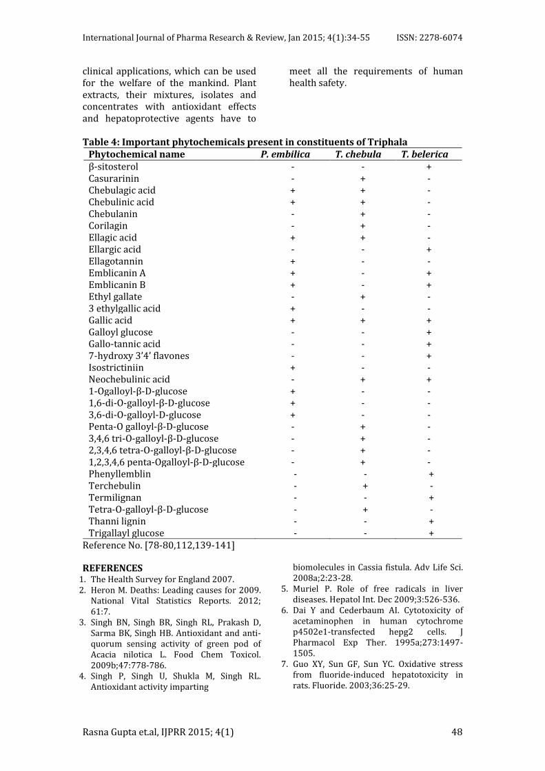
Table 4: Important phytochemicals present in constituents of Triphala
Phytochemical name P. embilica T. chebula T. belerica β-sitosterol - - + Casurarinin - + - Chebulagic acid + + - Chebulinic acid + + - Chebulanin - + - Corilagin - + - Ellagic acid + + - Ellargic acid - - + Ellagotannin + - - Emblicanin A + - + Emblicanin B + - + Ethyl gallate - + - 3 ethylgallic acid + - - Gallic acid + + + Galloyl glucose - - + Gallo-tannic acid - - + 7-hydroxy 3’4’ flavones - - + Isostrictiniin + - - Neochebulinic acid - + + 1-Ogalloyl-β-D-glucose + - - 1,6-di-O-galloyl-β-D-glucose + - - 3,6-di-O-galloyl-D-glucose + - - Penta-O galloyl-β-D-glucose - + - 3,4,6 tri-O-galloyl-β-D-glucose - + - 2,3,4,6 tetra-O-galloyl-β-D-glucose - + - 1,2,3,4,6 penta-Ogalloyl-β-D-glucose - + - Phenyllemblin - - + Terchebulin - + - Termilignan - - + Tetra-O-galloyl-β-D-glucose - + - Thanni lignin - - + Trigallayl glucose - - +
Reference No. [78-80,112,139-141]
REFERENCES 1. The Health Survey for England 2007. 2. Heron M. Deaths: Leading causes for 2009.
National Vital Statistics Reports. 2012; 61:7.
3. Singh BN, Singh BR, Singh RL, Prakash D, Sarma BK, Singh HB. Antioxidant and anti-quorum sensing activity of green pod of Acacia nilotica L. Food Chem Toxicol. 2009b;47:778-786.
4. Singh P, Singh U, Shukla M, Singh RL. Antioxidant activity imparting
biomolecules in Cassia fistula. Adv Life Sci. 2008a;2:23-28.
5. Muriel P. Role of free radicals in liver diseases. Hepatol Int. Dec 2009;3:526-536.
6. Dai Y and Cederbaum AI. Cytotoxicity of acetaminophen in human cytochrome p4502e1-transfected hepg2 cells. J Pharmacol Exp Ther. 1995a;273:1497-1505.
7. Guo XY, Sun GF, Sun YC. Oxidative stress from fluoride-induced hepatotoxicity in rats. Fluoride. 2003;36:25-29.
International Journal of Pharma Research & Review, Jan 2015; 4(1):34-55 ISSN: 2278-6074
Rasna Gupta et.al, IJPRR 2015; 4(1) 49
8. Wang A, Xia T, Ran P, Bai Y, Yang K, Chen X. Effects of selenium and fluoride on apoptosis and lipid peroxidation in human hepatocytes. Zhonghua Yu Fang Yi Xue Za. 2003;36:45-46.
9. Shashi A and Thapar SP. Histopathology of fluoride-induced hepatotoxicity in rabbits. Fluoride. 2001;34:34-42.
10. Dbrowska E and Szynaka B. Effect of fluoride on the ultrastructure of rat liver [abstract]. Fluoride. 2000;33:79-84.
11. Pingale SS. Evaluation of effect of centella asiatica on CCl4 induced rat liver damage. Pharmacologyonline. 2008;3:537-543.
12. Pingale SS. Hepatosuppression by Ricinus communis against CCl4 Induced Liver Toxicity in Rat Journal of pharmacy research. 2010;3:39-42.
13. Comportti M, Biology of disease: Lipidperoxidation and cellular damage in toxic liver injury, Lab Invest. 1985;53:599-623.
14. Decker T, Lohmann-Matthes ML, Karck U, Peters T, Decker K, Comparative study of cytotoxicity, tumor necrosis factor and prostaglandin release after stimulation of rat kupffer cells, murine kupffer cells, and murine inflammatory liver macrophages, J Leukocyte Biol. 1989;45:139-146.
15. Mitchell JR, Thorgeirsson UP, Black M, Timbrell JA, Snodgrass WR, Potter WZ, ‘et al’. Increased incidence of isoniazid hepatitis in rapid acetylators: possible relation to hydrazine metabolites. Clin Pharmacol Ther. 1975;18:70-79.
16. Desta Z, Soukhova NV, Flockhart DA. Inhibition of cytochrome P450 (CYP450) isoforms by isoniazid: potent inhibition of CYP2C19 and CYP3A. Antimicrob Agents Chemother. 2001;45:382-392.
17. Tanaka E, Terada M, Misawa S. Cytochrome P450 2E1: its clinical and toxicological role. J Clin Pharm Ther. 2000;25:165-175.
18. O’Shea D, Kim RB, Wilkinson GR. Modulation of CYP2E1 activity by isoniazid in rapid and slow N-acetylators. Br J Clin Pharmacol. 1997;43:99-103.
19. Dickinson D, Bailey W, Hirschowitz B. The effect of acetylation status on isoniazid (INH) hepatitis. Am Rev Respir Dis. 1977;115:395.
20. Sarma GR, Immanuel C, Kailasam S, Narayana AS, Venkatesan P. Rifampin-induced release of hydrazine from isoniazid: a possible cause of hepatitis during treatment of tuberculosis with regimens containing isoniazid and rifampin. Am Rev Respir Dis. 1986;133:1072-1075.
21. Menzies D, Dion MJ, Rabinovitch B, Mannix S, Brassard P, Schwartzman K. Treatment completion and costs of a randomized trial of rifampin for 4 months versus isoniazid for 9 months. Am J Respir Crit Care Med. 2004;170:445-449.
22. Hong Kong Chest Service, Tuberculosis Research Centre, Madras, British Medical Research Council.Adouble-blind placebo-controlled clinical trial of three anti-tuberculosis chemoprophylaxis regimens in patients with silicosis in Hong Kong. Am Rev Respir Dis. 1992;145:36-41.
23. Shibata K, Fukuwatari T, Sugimoto E. Effects of dietary pyrazinamide, an antituberculosis agent, on the metabolism of tryptophan to niacin and of tryptophan to serotonin in rats. Biosci Biotechnol Biochem. 2001;65:1339-1346.
24. Olynyk J, Hall P, Reed W, Williams P, Kerr R “et al.” A long-term study of the interaction between iron and alcohol in an animal model of iron overload. J. Hepatol. 1995;22:671-676.
25. Deugnier YM, Loreal O, Turlin B, Guyader D, Jouanolle H “et al”. Liver pathology in genetic hemochromatosis: A review of 135 homozygous cases and their bioclinical correlations. Gastroenterology. 1992;102:2050-2059.
26. Gardi C, Arezzini B, Fortino V, Comporti M. Effect of free iron on collagen synthesis, cell proliferation and MMP-2 expression in rat hepatic stellate cells. Biochem. Pharmacol. 2002;64:1139-1145
27. Arezzini B, Lunghi B, Lungarella G, Gardi C. Iron overload enhances the development of experimental liver cirrhosis in mice. Int. J. Biochem. Cell. Biol. 2003;35:486-495.
28. Niederau C, Fischer R, Sonnenberg A, Stremmel W, Trampisch HJ, Strohmeyer G. Survival and causes of death in cirrhotic and in noncirrhotic patients with primary hemochromatosis. N. Engl. J. Med. 1985;313:1256-1262.
29. Al-Refaie FN, Wonke B, Hoffbrand AV, Wickens DG, Nortey P. Efficacy and possible adverse effects of the oral iron chelator 1,2- dimethyl-3-hydroxypyrid-4-one (L1) in thalassemia major. Blood. 1992;80:593-599.
30. Kontoghiorghes GJ. Do we need more iron-chelating drugs? Lancet. 2003;362:495-496.
31. Zhang Y, Li H, Zhao Y, Gao Z. Dietary supplementation of baicalin and quercetin attenuates iron overload induced mouse liver injury. Eur. J. Pharmacol. 2006;535:263-269.
International Journal of Pharma Research & Review, Jan 2015; 4(1):34-55 ISSN: 2278-6074
Rasna Gupta et.al, IJPRR 2015; 4(1) 50
32. Pardo-Andreu GL, Barrios MF, Curti C, Hernandez I, Merino N. Protective effects of Mangifera indica L extract (Vimang) and its major component mangiferin, on iron-induced oxidative damage to rat serum and liver. Pharmacol. Res. 2008;57:79-86.
33. Heekyoung C, Hyun JM, Ki-SJ, Mingoo K, Jungeun Y, Kyung-SK, “et al”. Analysis of differential gene expression profiles on d-galactosamine-induced acute mouse liver injury and regeneration. Toxicology. 2006;227:136-144.
34. Keppler D, Lesch R, Reutter W, Decker K. Experimental hepatitis induced by D-galactosamine. Exp Mol Pathol. 1968;9:279-290.
35. Silverstein R. D-galactosamine lethality model: Scope and limitations. J Endotxin Res. 2004;10:147-162.
36. Keppler DO, Rudigier JF, Bischoff E, Decker KF. The trapping of uridine phosphates by D-galactosamine, D-glucosamine and 2- deoxy-D-galactose. Eur J Biochem. 1970;17: 246-253.
37. Konishi Y, Shinozuka H, Farber JL. The inhibition of rat liver nuclear ribonucleic acid synthesis by galactosamine and its reversal by uridine. Laboratory Investigation. 1974;30: 751-756.
38. Endo Y, Kikuchi T, Nakamura M. Ornithine and histidine decarboxylase activities in mice sensitized to endotoxin, interleukine-1 or tumor necrosis factor by D-galactosamine. British Journal of Pharmacology. 1992;107:888-894.
39. El-Mofty SK, Scrutton MC, Serroni A, Nicolini C, Farber JL. Early reversible plasma membrane injury in galactosamine-induced liver cell death. American journal of Pathology. 1975;79:579-596.
40. Keppler D and Decker K. Studies on the mechanism of galactosamine hepatitis: accumulation of galactosamine-1-phosphate and its inhibition of UDP-glucose pyrophosphorylase. Eur J Biochem. 1969;10:219-25.
41. Jaeschke H, Ho YS, Fisher MA, Lawson JA, Farhood A. Glutathione peroxidase-deficient mice are more susceptible to neutrophilmediated hepatic parenchymal cell injury during endotoxemia: Importance of an intracellular oxidant stress. Hepatology. 1999;29:443-450.
42. Bilzer M, Jaeschke H, Vollmar AM, Paumgartner G, Gerbes AL. Prevention of kupffer cell-induced oxidant injury in rat liver by atrial natriuretic peptide. Am J Physiol. 1999; 276:1137-1144.
43. Nieminen AL, Saylor AK, Tesfai SA, Herman B, Lemasters JJ. Contribution of the mitochondrial permeability transition to lethal injury after exposure of hepatocytes to t-butylhydroperoxide. Biochem J. 1995;307 99-106.
44. Gardner CR, Heck DE, Yang CS, Thomas PE, Zhang XJ, DeGeorge GL, “et al”. Role of nitric oxide in acetaminophen-induced hepatotoxicity in the rat. Hepatology. 1998; 27:748-754.
45. Hultcrantz R, Glaumann H, Lindberg G, Nilsson SL. Liver investigation in 149 asymptomatic patients with moderately elevated activities of serum aminotransferases. Scand. J. Gastroenterol. 1986;21:109-113.
46. Craxi A and Almasio P. Diagnostic approach to liver enzyme elevation. J. Hepatol. 1996;25:47-51.
47. Sgro C, Clinard F, Ouazir K, Chanay H, Allard C, Guilleminet C, “et al”. Incidence of drug-induced hepatic injuries: A frenche population-based study. Hepatology. 2002;36:451-455.
48. Zimmerman HJ. Update on hepatotoxicity due to classes of drugs in common clinical use: non-steroidal drugs, anti-inflammatory drugs, antibiotics, anti-hypertensives and cardiac and psychotropic agents. Semin Liver Dis. 1990;10:322-338.
49. Connor NO, Dargan PI, Jones AL. Hepatocellular damage from non-steroidal anti-inflammatory drugs. QJM. An International Journal of Medicine. 2003;96:787-791.
50. Benichou C. Criteria of drug-induced liver disorders. Report of an International Consensus Meeting. J Hepatol. 1990;11:272-276.
51. Petrescu I and Tarba C. Uncoupling effects of diclofenac and aspirin in the perfused liver and isolated hepatic mitochondria of rat. Biochim Biophys Ada. 1997;1318:385-394.
52. Somasundaram S, Rafi S, Hayllar J, Sigthorsson G, Jacob M, Price AB, “et al”. Mitochondrial damage: a possible mechanism of the topical phase of NSAID induced injury to the rat intestine. Gut. 1997;41:344-353.
53. Al-Nasser IA. Salicylate-induced kidney mitochondrial permeability transition is prevented by cyclosporin A. Toxicol Lett. 1999;105:1-8.
54. Fromenty B and Pessayre D. Inhibition of mitochondrial β-oxidation as a mechanism
International Journal of Pharma Research & Review, Jan 2015; 4(1):34-55 ISSN: 2278-6074
Rasna Gupta et.al, IJPRR 2015; 4(1) 51
of hepatotoxicity. Pharmacol. Ther. 1995;67:101-154.
55. Pessayre D, Berson A, Fromenty B, Mansouri A. Mitochondria in steatohepatitis. Semin. Liver Dis. 2001;21:57-69.
56. Lemasters JJ. Mechanisms of hepatic toxicity. V. Necrapoptosis and the mitochondrial permeability transition: Shared pathways to necrosis and apoptosis. Am. J. Physiol. 1999; 276:1-6.
57. Bradham CA, Qian T, Streetz K, Trautwein C, Brenner DA, Lemasters JJ. The mitochondrial permeability transition is required for tumor necrosis factor a-mediated apoptosis and cytochrome c release. Molec. Cell. Biol. 1998;18:6353-6364.
58. Feldmann G, Haouzi D, Moreau A, Durand-Schneider AM, Bringuier A, Berson A, “et al”. Opening of the mitochondrial permeability transition pore causes matrix expansion and outer membrane rupture in Fas-mediated hepatic apoptosis in mice. Hepatology. 2000;31:674-683.
59. Berson A, Fau D, Fornacciari R, Degove-Goddard P, Sutton A, Descatoire V, “et al”. Mechanism for experimental buprenorphine hepatotoxicity: Major role of mitochondrial dysfunction versus metabolic activation. J. Hepatol. 2001;34:261-269.
60. Haouzi D, Lekehal M, Moreau A, Moulis G, Feldmann G, Robin MA, “et al”. Cytochrome P450-generated reactive metabolites cause mitochondrial permeability transition, caspase activation, and apoptosis in rat hepatocytes. Hepatology. 2000;32:303-311.
61. Davies MJ. Detection of peroxyl and alkoxyl radicals produced by reaction of hydroperoxides with rat liver microsomal fractions. Biochemical Journal. 1989;257:603-606.
62. Crane D, Haussinger D, Graf P, Sies H. Decreased flux through pyruvate dehydrogenase by thiol oxidation during t-butyl hydroperoxide metabolism in perfused rat liver. Hoppe-Seyler's Zeitschrift fur Physiologische Chemie. 1983;364:977-987.
63. Kirtikar KR and Basu BD. Terminalia (L) 2: 2nd Indian medicinal plants, Lalit Mohan Basu, Allahabad, India. 1993:1014-1033.
64. Usmanghani K, Saeed A, Alam MT. Indusyunic Medicine. University of Karachi Press, Karachi. 1997:420-421.
65. Duke JA. Bogenschutz-Godwin MJ, Ducelliar J, Duke PAK. Handbook of Medicinal Herbs,
second ed. CRC Press, Boca Raton. 2002:70-71.
66. Suchalatha S, Srinivasan P, Devi CS. Effect of T. chebula on mitochondrial alterations in experimental myocardial injury. Chem Biol Interact. 2007;169:145-153.
67. Saleem A, Husheem M, Härkönen P, Pihlaja K. Inhibition of cancer cell growth by crude extract and the phenolics of Terminalia chebula retz. Fruit. J Ethnopharmacol. 2002;81: 327-336.
68. Sabu MC and Kuttan R. Antidiabetic activity of medicinal plants and its relationship with their antioxidant property. J. Ethanopharmacol. 2002;81:155-160.
69. Kaur S, Arora S, Kaur K, Kumar S. This is in vitro antimutagenic activity of Triphala-an Indian herbal drug. Food chain Toxicol. 2002;40:527-534.
70. Gaind KN, Mital HC, Khanna SR. A study on the purgative activity of triphala. Indian J Physiol Pharmacol. 1963;18:171-175.
71. Jagetia GC, Baliga MS, Malagi KJ, Kamath MS. The evaluation of the radio protective effect of Triphala (an Ayurvedic rejuvenating drug) in the mice exposed to Gamma radiation. Phytomed. 2002;9:99-108.
72. Mukherjee PK, Rai S, Bhattacharya S, Wahile A, Saha BP. Marker analysis of polyherbal formulation, Triphala-A well known Indian traditional medicine. Indian J Trad Know. 2008;7:379-383.
73. Sharma RK and Dash B, Agnivesa’s Caraka Samhita (Text with English Translation and Critical Exposition based on Cakrapani Datta’s Ayurveda Dipika) Varanasi: Chaukhambha Orientalia; 1988; volume 3.
74. Mahaboob KR, Evan PS, Kumar L, Nithya P. Therapeutic effect of Indian ayurvedic herbal formulation triphala on acetaminophen induced hepatotoxicity in mice. Journal of pharmacology and toxicology. 2007;2:725-731.
75. Nema N and Kharya MD. Impact of Triphala on kupffer Cell Regeneration: A Possible Mechanism. International Journal of Pharmacology and Pharmaceutical Technology (IJPPT). volume-I(1).
76. Sharma A and Sharma KK, Chemoprotective Role of Triphala Against 1,2-Dimethylhydrazine Dihydrochloride Induced Carcinogenic Damage to Mouse Liver. Ind J Clin Biochem. 2011;26:290-295.
77. Sabina EP, Rasool M, Vedi M, Geethanjali A. Protective properties of traditional herbal formulation triphala against D-Galactosamine induced hepatotoxicity in
International Journal of Pharma Research & Review, Jan 2015; 4(1):34-55 ISSN: 2278-6074
Rasna Gupta et.al, IJPRR 2015; 4(1) 52
mice. International Journal of Drug Development & Research. 2013;5:164-173.
78. Zhang LZ, Zhao WH, Guo YJ, Tu GZ, Lin S, Xin LG. Studies on chemical constituents in fruits of Tibetan medicine Phyllanthus emblica. Zhongguo Zhong Yao Za Zhi. 2003;28:940-943.
79. Khan KH. Roles of Emblica officinalis in Medicine-A Review. Botany Research International. 2009;4:218-228.
80. Krishnaveni M and Mirunalini S. Chemopreventive efficacy of Phyllanthus emblica L. (amla) fruit extract on 7,12-dimethylbenz(a)anthracene induced oral carcinogenesis–A dose–response study. Environmental Toxicology and Pharmacology. 2012;34:801-810.
81. Perianayagam JB, Sharma SK, Joseph A, Christina AJ. Evaluation of anti-pyretic and analgesic activity of Emblica officinalis Gaertn. Journal of Ethnopharmacology. 2004;95:83-85.
82. Nosal ova G, Mokry J, Hasan KM. Antitussive activity of the fruit extracts of Emblica officinalis Gaertn, (Euphorbiaceae). Phytomedicine. 2003;10:583-589.
83. Santoshkumar J, Manjunath S, Pranavkumar MS. A study of antihyperlipidemia, hypolipedimic and anti-atherogenic activity of fruit of Emblica officinalis (amla) in high fat fed Albino Rats, International Journal of Medical Researchand Health Sciences. 2013; 2:70-77.
84. Muruganandam AV, Kumar V, Bhattacharya SK. Effect of poly herbal formulation, EuMil, on chronic stress-induced homeostatic perturbations in rats. Indian Journal of Experimental Biology. 2002;40:1151-1160.
85. Baliga MS, Prabhu AN, Prabhu DA, Shivashankara AR, Abraham A, Palatty PL. Antidiabetic and Cardioprotective Effects of Amla (Emblica officinalis Gaertn) and its Phytochemicals: Preclinical Observations. Bioactive Food as Dietary Interventions for Diabetes. 2013:583-600.
86. Srirama R, Deepak HB, Senthilkumar U, Ravikanth G, Gurumurthy BR, Shivanna MB, “et al”. Hepatoprotective activity of Indian Phyllanthus. Pharmaceutical Biology. 2012; 50:948-953.
87. Chatterjee A, Chattopadhyay S, Sandip KB. Biphasic Effect of Phyllanthus emblica L. Extract on NSAID-Induced Ulcer: An Anti-oxidative Trail Weaved with Immunomodulatory Effect. Evidence-Based Complementary and Alternative Medicine. 2011;2010:1-13.
88. Yokozawa T, Kim HY, Kim HJ, Tanaka T, Sugino H, Okubo T, “et al”. Amla (Emblica officinalis Gaertn.) Attenuates Age-Related Renal Dysfunction by Oxidative Stress. Journal of Agricultural and Food Chemistry. 2007;55:7744-7752.
89. Vasudevan M and Parle M. Memory enhancing activity of Anwalachurna (Emblica officinalis Gaertn.): An Ayurvedic preparation. Physiology & Behaviour. 2007;91:46-54.
90. Madhuri S. Studies on estrogen induced uterine and ovarian carcinogenesis and effect of ProImmu in rat, PhD thesis, Jabalpur, MP, RDVV, 2008.
91. Krishnaveni M and Mirunalini S. Chemopreventive efficacy of Phyllanthus emblica L. (amla) fruit extract on 7,12-dimethylbenz(a)anthracene induced oral carcinogenesis-A dose response study. Environmental Toxicology and Pharmacology. 2012;34:801-810.
92. Adil MD, Kaiser P, Satti NK, Zargar AM, Vishwakarma RA, Tasduq SA. Effect of Emblica officinalis (fruit) against UVB-induced photoaging in human skin fibroblasts. Journal of Ethnopharmacology. 2010;132:109-114.
93. Senthil MK, Kirubanandan S, Sripriya R, Sehgal PK. Triphala Promotes Healing of Infected Full-Thickness Dermal Wound. Journal of Surgical Research. 2008;144:94-101.
94. Varadacharyulu N, Reddy D, Padmavathi P, Paramahamsa M. Modulatory role of Em Emblica officinalis against alcohol induced biochemical and biophysical changes in rat erythrocyte membranes. Food and Chemical Toxicology. 2009;47:1958-1963.
95. Prakash D, Upadhyay G, Gupta C, Pushpangadan P, Singh KK. Antioxidant and free radical scavenging activities of some promising wild edible fruits. International Food Research Journal. 2012;19:1109-1116.
96. Hazra B, Sarkar R, Biswas S, Manda N. Comparative study of the antioxidant and reactive oxygen species scavenging properties in the extracts of the fruits of Terminalia chebula, Terminalia belerica and Emblica officinalis. BMC Complementary and Alternative Medicine. 2010;10:1-15.
97. Nain P, Saini V, Sharma S, Nain J. Antidiabetic and antioxidant potential of Emblica officinalis Gaertn. leaves extract in streptozotocin-induced type-2 diabetes mellitus (T2DM) rats. Journal of Ethnopharmacology. 2012;142:65-71.
International Journal of Pharma Research & Review, Jan 2015; 4(1):34-55 ISSN: 2278-6074
Rasna Gupta et.al, IJPRR 2015; 4(1) 53
98. Mehmood MH, Siddiqi HS, Gilani AH. The antidiarrheal and spasmolytic activities of Phyllanthus emblica are mediated through dual blockade of muscarinic receptors and Ca2+ channels. Journal of Ethnopharmacology. 2011;133:856-865.
99. Liu G, Xiong S, Xiang S, Guo CW, Ge F, Yang CR, “et al”. Antiviral activity and possible mechanisms of action of pentagalloylglucose (PGG) against influenza A virus. Archives of Virology. 2011;156:1359-1369.
100. Bhattacharya S, Bhattacharya K, Sairam A, Ghosal S. Antioxidant-antidepressant activity of Withania Somnifera Glycothanolides. Phytomedicine. 2000;7:463-469.
101. Dhir H, Roy AK, Sharma A, Talukder G. Modification of clastogenicity of lead and aluminium in mouse bone marrow cells by dietary ingestion of Phyllanthus embilica fruit extract. Mutat Res.1990;241:305-312.
102. Sabbani V, Alluri R, Mohan B, Pasha MY, Rajkumar M. Evaluation of hepatoprotective activity of polyherbal formulation against paracetamol induced toxicity. Indo American Journal of Pharmaceutical Research. 2013;3:6001-6008.
103. Pramyothin P, Samosorn P, Poungshompoo S, Chaichantipyuth C. The protective effects of Phyllanthus emblica Linn. Extract on ethanol induced rat hepatic injury. J Ethnopharmacol. 2006;107:361-364.
104. Reddy VD, Padmavathi P, Varadacharyulu NCh. Emblica officinalis protects against alcohol-induced liver mitochondrial dysfunction in rats. J Med Food. 2009;12:327-333
105. Sultana S, Ahmad S, Khan N, Jahangir T. Effect of Emblica officinalis (Gaertn) on CCl4 induced hepatic toxicity and DNA synthesis in Wistar rats. Indian Journal of Experimental Biology. 2005;43:430-436.
106. Bafna PA and Balaraman R. Protective effect of DHC-1, a Polyherbal formulation, against CCL4 induced Liver damage. Hygeia. Journal for drugs and medicines. 2013; 5:10-18.
107. Tasduq SA, Kaisar P, Gupta DK, Kapahi BK, Maheshwari HS, Jyotsna S. Protective effect of a 50% hydroalcoholic fruit extract of Emblica officinalis against anti-tuberculosis drugs induced liver toxicity. Phytother Res. 2005;19:193-197.
108. Rupal A, Vasant A, Narasimhacharya VRL. Alliviatory effects of Embilica officinalis G.
As a food supplement in Fluoride induced hyperlipidemia and oxidative stress. International journal of pharmacy and pharmaceutical science. 2012;4:404-408.
109. Vadivelu J and Dawood SS. Free radical scavenging activity Embilica offinalis during nicotine induced toxicity in rats (Rattus norvegicus). Global Journal of Traditional Medicinal Systems. 2013;2:9-13.
110. Chen KH, Lin BR, Chien CT, Ho CH. Emblica officinalis Gaertn. Attenuates N-nitrosodiethylamine‑induced apoptosis, autophagy and inflammation in rat livers. J Med Food. 2011;14:746-755.
111. Chakraborty D and Verma R. Ameliorative effect of Emblica officinalis aqueous extract on ochratoxin-induced lipid peroxidation in the kidney and liver of mice. Int J occup Med and Env Health. 2010;23:63-73.
112. Grover IS and Bala S. Antimutagenic activity of T. chebula (myroblan) in Salmonella typhimurium. Ind J Exp Biol. 1992;30:339-341.
113. Cook NC and Samman S. Flavonoids-chemistry, metabolism, cardioprotective effects, and dietary sources. J Nutr Biochem. 1996;7:66-76.
114. Saleem A, Husheem M, Harkonen P, Pihlaja K. Inhbition of cancer cell growth by crude extract and the phenolics of Terminalia chebula Retz. Fruit. J Ethnopharmacol. 2002;81:327-336.
115. Mahajan A and Pai N. Simultaneous isolation and identification of phytoconstituents from Terminalia chebula by preparative chromatography. J. Chem. Pharm. Res. 2010;2:97-103.
116. Tayal S, Duggal S, Bandyopadhyay P, Aggarwal A, Tandon S, Tandon C. Cytoprotective role of the aqueous extract of Terminalia chebula on renal epithelial cells. Int Braz J Urol. 2012;38:204-213.
117. Mard SA, Veisi A, Naseri MK, Mikaili P. Spasmogenic Activity of the Seed of Terminalia chebula Retz in Rat Small Intestine: In Vivo and In Vitro Studies Malays. J Med Sci. 2011;18:18-26.
118. Das ND, Jung KH, Park JH, Mondol MA, Shin HJ, “et al”. Terminalia chebula extract acts as a potential NF-κB inhibitor in human lymphoblastic T cells. Phytother Res. 2011;25:927-934.
119. Chang CL and Lin CS. Phytochemical Composition, Antioxidant Activity, and Neuroprotective Effect of Terminalia chebula Retzius Extracts. Evid Based Complement Alternat Med. 2012;125:247.
International Journal of Pharma Research & Review, Jan 2015; 4(1):34-55 ISSN: 2278-6074
Rasna Gupta et.al, IJPRR 2015; 4(1) 54
120. Kaur S and Jaggi RK. Antinociceptive activity of chronic administration of different extracts of terminalia bellerica roxb. and terminalia chebula retz. Fruits. Indian J Exp Biol. 2010;48:925-930.
121. Sharma P, Prakash T, Kotresha D, Ansari MA, “et al”. Antiulcerogenic activity of Terminalia chebula fruit in experimentally induced ulcer in rats. Pharm Biol. 2011;49:262-268.
122. Pinmai K, Hiriote W, Soonthornchareonnon N, Jongsakul K, Sireeratawong S. In vitro and in vivo antiplasmodial activity and cytotoxicity of water extracts of Phyllanthus emblica, Terminalia chebula, and Terminalia bellerica. J Med Assoc Thai. 2010;7:120-126.
123. Gopi KS, Reddy AG, Jyothi K, Kumar BA. Acetaminophen-induced Hepato and Nephrotoxicity and Amelioration by Silymarin and Terminalia chebula in Rats. Toxicol Int. 2010;17:64-66.
124. Nair V, Singh S, Gupta YK. Anti-arthritic and disease modifying activity of Terminalia chebula Retz. in experimental models. J Pharm Pharmacol. 2010;62:801-806.
125. Manosroi A, Jantrawut P, Akihisa T, Manosroi W, Manosroi J. In vitro anti-aging activities of Terminalia chebula gall extract. Pharm Biol. 2010;48:469-481.
126. Srivastav A, Chandra A, Singh M, Jamal F, Rastogi P, “et al”. Inhibition of hyaluronidase activity of human and rat spermatozoa in vitro and antispermatogenic activity in rats in vivo by Terminalia chebula, a flavonoid rich plant. Reprod Toxicol. 2010;29:214-224.
127. Carounanidy U, Satyanarayanan R, Velmurugan A. Use of an aqueous extract of Terminalia chebula as an anticaries agent: a clinical study. Indian J Dent Res. 2007; 18:152-156.
128. Shadia AHF, Fatma FAH, Osama AA, Asrar MH, Monira MEY. Effect of Terminalia chebula Extract on induced Diabetic Rats, International journal of pharmaceutical science and health care. 2012;4:47-58.
129. Vidya S, Verma E, Kumar JS, Kumar VTA, Ramesh A. Hepatoprotective activity of Terminalia chebula in paracetamol induced hepato-toxicity rats. International Journal of Advances in Pharmaceutical Research. 2011;2:127-132.
130. Boominathan M. Comparative study on antimicrobial, hepatoprotective activity of Terminalia Chebula different fraction.
International Journal of Institutional Pharmacy and Life Sciences. 2012;16-22.
131. Lee HS, Won NH, Kim KH, Lee H, Jun W, Lee KW. Antioxidant effects of aqueous extract of Terminalia chebula in vitvo and in vitro. Biol Pharm Bull. 2005;28:1639-1644.
132. Reddy PN, Tammineni P, Radhika GJ, Reddy PK, Pallu R. Hepatoprotective Effects of Terminalia chebula Fruit Extract against 2-AAF–Induced Hepatic Damage in Albino Mice: Role of MDR1 and COX-2. Journal of Herbs, Spices & Medicinal Plants. 2014; 20:402-420.
133. Sivachandran S and Hariharan P. Hepatoprotective Effect of Terminalia Chebula on Gentamicin Induced Toxicity in Rats. International Journal of Veterinary Science. 2012; 1:31-33.
134. Goodman LS and Gilman A. The Pharmacological Basis of Therapeutics. 11th edition. McGraw-Hill: New York; 2006.
135. Tasduq SA, Singh K, Satti NK, Gupta DK, Suri KA. Terminalia chebula (fruit) prevents liver toxicity caused by sub-chronic administration of rifampicin, isoniazid and pyrazinamide in combination. Hum Exp Toxicol. 2006;25:111-118.
136. Sarkar R, Hazra B, Mandal N. Reducing power and iron chelating property of Terminalia chebula (Retz.) alleviates iron induced liver toxicity in mice. BMC Complementary and Alternative Medicine. 2012,12:134-144.
137. Lee HS, Jung SH, Yun BS, Lee KW. Isolation of chebulic acid from Terminalia chebula Retz. and its antioxidant effect in isolated rat hepatocytes. Arch Toxicol. 2007;81:211-218.
138. Singh A. Medicinal Plants of the World, Published by Mohan Primlani for Oxford and IBH Co. Pvt, New Delhi.2006.
139. Singh AS. Herbalism phytochemistry and Ethanopharmacology. Science Publishers. 2011;357-361
140. The Ayurvedic Pharmacopoiea of India, 1st edition, Published by The controller of Publications, Civil Lines. New Delhi. 2001;101:252.
141. Indian Herbal Pharmacopoeia Revised New Edition. Published by Indian Drug Manufacturer’s Association, Mumbai. 2002:429-438.
142. Mety SS and Mathad P. Antioxidative and free radical scavenging activities of terminalia species. International Research Journal of Biotechnology. 2011;2:119-127.
International Journal of Pharma Research & Review, Jan 2015; 4(1):34-55 ISSN: 2278-6074
Rasna Gupta et.al, IJPRR 2015; 4(1) 55
143. Singh P, Vishwakarma SP, Singh U, Shukla M, Singh R, Singh RK, “et al”. Quantification and Evaluation of Antioxidant Activity of Some Bioactive Phytochemicals in Different Medicinal Plants. The Open Nutraceuticals Journal. 2012;5:179-186.
144. Trivedi VP, Nesamany S, Sharma VK. A clinical study of the antitussive and antiasthmatic effects of Vibhitakphal churna (Terminalia bellerica Roxb.) in case of kapha swasa. J Res Ayurveda Siddha. 1992;3:1-8.
145. Anand KK, Singh B, Saxena AK. Chandan BK, Gupta VN. Hepatoprotective studies of a fraction from the fruits of Terminalia Bellerica Roxb. on experimental liver injury in rodents. Phytother Res. 1994;8:287-292.
146. Setty BS, Kamboj VP, Garg HS and Khanna NM. Spermicidal potential of saponins isolated from Indian medicinal plants, Contraception. 1976;14:571-578.
147. Anand R, Sukla R and Dhawan BN. Evaluation of antistress activity of some Indian medicinal plants. Indian J Pharmacol. 1999;31:74.
148. Kar A, Choudhary BK, Bandyopadhyay NG. Comparative evaluation of hypoglycemic activity of some Indian medicinal plants in alloxan diabetic rats. J Ethnopharmacol. 2003;84:105-108.
149. Bhutani KK, Kumar V, Kaur R, Sarin AN. Amoebicidal activity of some Indian medicinal plants. Indian Drug. 1988;25:220.
150. Ahmad I, Mehmood Z, Mohammad F. Screening of some Indian medicinal plants for their antimicrobial properties. J Ethnopharmacol. 1998;62:183-193.
151. Mahmood Z, Ahmad I, Mohammad F, Ahmad S. Indian medicinal plant: A potential source for anticandidal drugs. Pharm Biol. 1999;37:237-242.
152. Srivastava RD, Dwivedi S, Sreenivasan KK and Chandrashekhar CN. Cardiovascular
effect of Terminalia species of plants. Indian Drugs. 1992;29:144-149.
153. Shiala HP, Udupa AL and Udupa SL. Preventative actions of Terminalia bellerica in experimentally induced atherosclerosis. Int J Cardiol. 1995;49:101-106.
154. Naik GH, Priyadarsini KI, Bhagirathi RG, Mishra B, “et al”. In vitro antioxidant studies and free radical reaction of Triphala, an Ayurvedic formulation and its constituents. Phytother Res. 2005;19:582-586.
155. Jain R, Nandakumar K, Srivastava V, Kumar VS, Patet S, Kumar PM. Hepatoprotective activity of ethanolic and aqueous extract of Terminalia Bellerica in rats. Pharmacologyonline. 2008;2:411-427.
156. Pingale SS. Hatoprotection by fresh juice of Leucas Aspera leaves. Der Pharmacia Sinica. 2010;1:136-140.
157. Pingale SS. Hepatoprotective action of Terminalia belerica on CCl4 induced hepatic disorders. Der Pharma Chemica. 2011;3:42-48.
158. Tasduq SA, Singh K, Satti NK, Gupta DK, Suri KA, Johri RK. Terminalia chebula (fruit) prevents liver toxicity caused by sub-chronic administration of rifampicin, isoniazid and pyrazinamide in combination. Hum Exp Toxicol. 2006;25:111-118.
159. Shukla S, Jadon A, Bhadauria M. Protective effect of Terminalia belerica Roxb, and gallic acid agaist carbontetra chloride induced damage in albino rats. Journal of Ethanopharmacology. 2006;109:214-218.
160. Hazra B, Sarkar R, Mandal N. Protection of Terminalia Bellerica Roxb against iron overload induced liver toxicity, an account of its reducing and iron chelating capacity. American Journal of Pharmacology and Toxicology. 2012;7:109-122.