Embed Size (px)
Citation preview

Tissues - Learn and Understand
• The composition of a tissue helps us understand what
functions it is capable of.
• Concepts of cell surface area, attachment of tissues to
others, mucus production, role of particular cell types within
tissues, glandular secretion are discovered or reviewed.
• Various forms of proteins (mainly) and polysaccharides
(secondarily) and inorganic materials form the critical, non-
living substances of the body.
• All organs are made up of two or more tissues (less than four
tissues is uncommon) and the presence of particular tissues
helps us understand what functions an organ is capable of.
• Inflammation is a natural, step-wise response to tissue
damage that begins the healing process.

Tissue: The Living Fabric
• Individual body cells specialized
– Each type performs specific functions that
maintain homeostasis
• Tissues
– Groups of cells similar in structure that
perform common or related function
• Histology
– Study of tissues

Tissues and Histology
Tissue classification based on
• structure of cells
• composition of extracellular matrix
• cell function
4 Types
• Epithelium – ‘cover’
• Connective – ‘support’
• Muscle – ‘movement’
• Nervous – ‘control’

Epithelial Tissue - Epithelium
Functions
• Protection• Covering and lining epithelia
• On external and internal surfaces
• Absorption – primary way substances enter/exit
body
• Filtration
• Excretion
• Secretion• Glandular epithelia
• Sensory reception – linked to nervous tissues
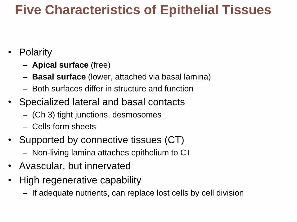
Five Characteristics of Epithelial Tissues
• Polarity
– Apical surface (free)
– Basal surface (lower, attached via basal lamina)
– Both surfaces differ in structure and function
• Specialized lateral and basal contacts
– (Ch 3) tight junctions, desmosomes
– Cells form sheets
• Supported by connective tissues (CT)
– Non-living lamina attaches epithelium to CT
• Avascular, but innervated
• High regenerative capability
– If adequate nutrients, can replace lost cells by cell division

Cell Surface Modifications
May be ciliated or
possess microvilli or
smooth
ciliated surface
“Brush border”
Consider increase in surface area
for absorption, exchange, enzymatic
function

Epithelial
layer
Basement
membrane
Underlying
connective
tissueTubule lumen
Basement membrane = Basal lamina +
reticular laminaReinforces epithelial sheet and defines epithelial boundary
Resists stretching and tearing
Foundation for repair

Classification of Epithelial TissueNumber of layers of cells
• Simple
• Stratified
• Pseudostratified
Shape of cells at surface
• Squamous
• Cuboidal
• Columnar

Simple squamous epithelium
Description: Single layer offlattened cells with disc-shapedcentral nuclei and sparsecytoplasm; the simplest of theepithelia.
Air sacs oflung tissue
Nuclei ofsquamousepithelialcells
Function: Allows materials topass by diffusion and filtrationin sites where protection is notimportant; secretes lubricatingsubstances in serosae.
Location: Kidney glomeruli;air sacs of lungs; lining of heart,blood vessels, and lymphaticvessels; lining of ventral bodycavity (serosae).
Photomicrograph: Simple squamousepithelium forming part of the alveolar(air sac) walls (140×).

Simple cuboidal epithelium
Description: Single layerof cubelike cells with large,spherical central nuclei.
Simplecuboidalepithelialcells
Nucleus
Function: Secretion andabsorption. Basement
membrane
Location: Kidney tubules;ducts and secretory portionsof small glands; ovary surface.
Photomicrograph: Simple cuboidalepithelium in kidney tubules (430×).
Connectivetissue

Simple columnar epithelium
Description: Single layer of tall
cells with round to oval nuclei; manycells bear microvilli, some bear cilia;layer may contain mucus-secretingunicellular glands (goblet cells).
Microvilli
Simplecolumnarepithelialcell
Function: Absorption; secretion
of mucus, enzymes, and othersubstances; ciliated type propelsmucus (or reproductive cells) byciliary action.
Location: Nonciliated type lines
most of the digestive tract (stomachto rectum), gallbladder, and excretoryducts of some glands; ciliated varietylines small bronchi, uterine tubes,and some regions of the uterus.
Mucus ofgoblet cell
Photomicrograph: Simple columnarepithelium of the small intestine mucosa(640×).

Pseudostratified columnar epithelium
Description: Single layer of cells
of differing heights, some notreaching the free surface; nucleiseen at different levels; maycontain mucus-secreting cells andbear cilia.
Goblet cell(containsmucus)
Cilia
Pseudo-stratifiedepitheliallayer
Function: Secrete substances,
particularly mucus; propulsionof mucus by ciliary action.
Location: Nonciliated type in
males’ sperm-carrying ducts andducts of large glands; ciliatedvariety lines the trachea, most ofthe upper respiratory tract.
Basementmembrane
Photomicrograph: Pseudostratified ciliatedcolumnar epithelium lining the human trachea(780×).
Trachea

Stratified squamous epithelium
Description: Thick membrane
composed of several cell layers;basal cells are cuboidal or columnarand metabolically active; surfacecells are flattened (squamous); in thekeratinized type, the surface cells arefull of keratin and dead; basal cells
are active in mitosis and produce thecells of the more superficial layers.
Stratifiedsquamousepithelium
Function: Protects underlying
tissues in areas subjected toabrasion.
Location: Nonkeratinized type forms
the moist linings of the esophagus,mouth, and vagina; keratinized varietyforms the epidermis of the skin, a drymembrane.
Nuclei
Basementmembrane
Connectivetissue
Photomicrograph: Stratified squamousepithelium lining the esophagus (285×).

Transitional epithelium
Description: Resembles bothstratified squamous andstratified cuboidal; basal cellscuboidal or columnar; surfacecells dome shaped orsquamouslike, depending ondegree of organ stretch.
Transitionalepithelium
Function: Stretches readily,permits stored urine to distendurinary organ.
Location: Lines the ureters,bladder, and part of the urethra.
Basementmembrane
Photomicrograph: Transitional epithelium
lining the bladder, relaxed state (360×); notethe bulbous, or rounded, appearance of thecells at the surface; these cells flatten andelongate when the bladder fills with urine.
Connectivetissue

Epithelium
classification
Function Location
Simple
squamous
Allows materials to pass by
diffusion and filtration in sites
where protection is not important;
secretes lubricating substances in
serosae
Kidney glomeruli; air sacs of lungs;
endothelium - lining of heart, blood
vessels, and lymphatic vessels; lining of
ventral body cavity
Simple cuboidal Secretion and absorption Kidney tubules; ducts and secretory
portions of small glands; ovary surface
Simple
columnar
Absorption; secretion of mucus,
enzymes, and other substances;
ciliated type propels mucus (or
reproductive cells) by ciliary action
Nonciliated type lines most of the
digestive tract, gallbladder, and excretory
ducts of some glands; ciliated variety
lines small bronchi, uterine tubes
Stratified
squamous
Protects underlying tissues in
areas subjected to abrasion
Nonkeratinized type forms the moist
linings of the esophagus, mouth, and
vagina; keratinized variety forms the
epidermis of the skin, a dry membrane
Pseudostratified
columnar
Secrete substances, particularly
mucus; propulsion of mucus by
ciliary action
Nonciliated type in male’s sperm-carrying
ducts and ducts of large glands; ciliated
variety lines the trachea, most of the
upper respiratory tract
Transitional Stretches readily, permits stored
urine to distend urinary organ
Lines the ureters, bladder, and part of the
urethra

Glandular Epithelia
• Gland
– One or more cells that make and secrete an aqueous fluid
or lipid-rich secretions
• Classified by
– Site of product release
• Endocrine or ductless glands
– Secrete (by exocytosis) hormones that travel through lymph or blood
to their specific target organs
• Exocrine
– Secretions released onto body surfaces (skin) or into body cavities
– Secrete products into ducts
– Examples include mucous, sweat, oil, and salivary glands
– Relative number of cells forming the gland
• unicellular (e.g., goblet cells) or multicellular

Unicellular Exocrine
• Found in epithelial linings of
intestinal and respiratory tracts
– mucous cells and goblet cells
• All produce mucin (hydrophilic
glycoprotein)
– Slimy protective, lubricating coating
Multicellular Exocrine glands
• Composed of a duct and a
secretory unit
• Usually surrounded by
supportive connective tissue
– Supplies blood and nerve
fibers
Intestinal epithelium
Goblet cell
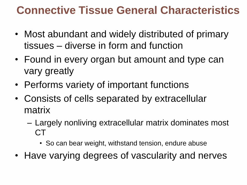
Connective Tissue General Characteristics
• Most abundant and widely distributed of primary
tissues – diverse in form and function
• Found in every organ but amount and type can
vary greatly
• Performs variety of important functions
• Consists of cells separated by extracellular
matrix
– Largely nonliving extracellular matrix dominates most
CT
• So can bear weight, withstand tension, endure abuse
• Have varying degrees of vascularity and nerves

Functions of Connective Tissue
• Enclose organs as a capsule and separate
organs into layers
• Connect tissues to one another
• Support and movement, but not contraction
• Storage
• Cushion and insulate
• Transport
• Protect

• Connective tissue proper - binding and support, insulating,
storing reserve fuel
• Cartilage – supporting, protecting, connecting

• Bone - supporting and protecting
• Blood - transporting substances, regulation

Structural Elements of Connective Tissue:
Ground Substance
• Unstructured gels that fills space between cells– Medium through which solutes diffuse between blood
capillaries and cells
• Components– Interstitial fluid
– Cell adhesion proteins – sticky nectins
– Hydrophilic Proteoglycans• Protein core and
• Glycosaminoglycans– large polysaccharides
» Example: chrondroitin sulfate and hyaluronic acid
• Trap water in varying amounts, affecting viscosity of ground substance

Structural Elements of Connective Tissue: Fibers
• Collagen
– Cross-linked fibrils of tropocollagen protein
– Strongest and most abundant type, provides flexible,
high-tensile strength
• Elastic fibers
– Networks of long, thin, elastin fibers that allow for stretch
and recoil
– Stretch limited by collagen fibers present in tissue
• Reticular
– Short, fine, highly branched collagenous fibers (different
chemistry and form than collagen fibers)
– Branch, forming spongy networks that offer more "give"

Structural Elements of Connective Tissue:
Cells
• "Blast" cells
– Immature form; mitotically active; secrete ground substance and fibers;
‘reawaken’ when needed
– Fibroblasts in connective tissue proper
– Chondroblasts in cartilage
– Osteoblasts in bone
– Hemocytoblasts ‘blood makers’ in bone marrow – always active
• "Cyte" cells
– Mature form; maintain matrix; detect and respond to new stresses
– Chondrocytes in cartilage
– Osteocytes in bone
• “Clast” cells – unique origin, destroy bone matrix in normal
processes

Other Cell Types in Connective Tissues
• Fat cells - adipocytes– Store nutrients, insulate, pad
• White blood cells– Several types whose names will be learned later
– Tissue response to injury
• Mast cells– Initiate local inflammatory response upon detection of
foreigners
– Secrete substances to control inflammation
• Macrophages– Phagocytic cells that "eat" dead cells, microorganisms,
abnormal cells; function in immune system

Extracellular matrix
Ground substanceFibers
• Collagen fiber
• Elastic fiber
• Reticular fiber
Capillary
Neutrophil
Mast cell
Fat cell
Lymphocyte
Fibroblast
Macrophage
Cell types
Figure 4.7 Areolar
connective tissue:
A prototype (model)
connective tissue.
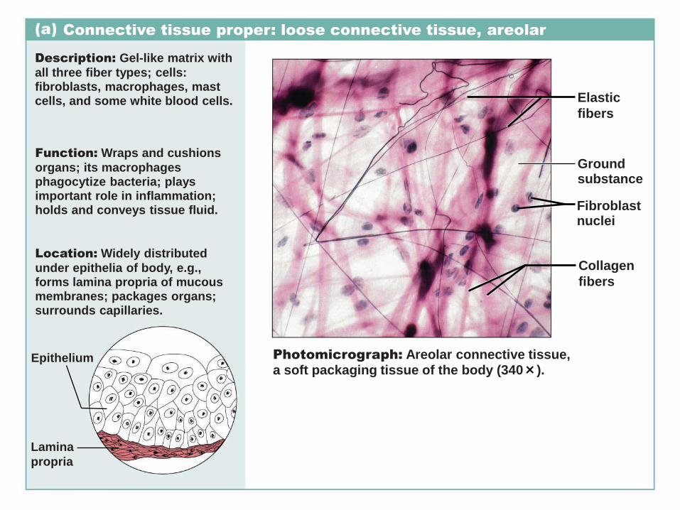
Connective tissue proper: loose connective tissue, areolar
Description: Gel-like matrix with
all three fiber types; cells:fibroblasts, macrophages, mastcells, and some white blood cells. Elastic
fibers
Function: Wraps and cushions
organs; its macrophagesphagocytize bacteria; playsimportant role in inflammation;holds and conveys tissue fluid.
Groundsubstance
Fibroblastnuclei
Collagen
fibers
Location: Widely distributed
under epithelia of body, e.g.,forms lamina propria of mucousmembranes; packages organs;surrounds capillaries.
Epithelium Photomicrograph: Areolar connective tissue,a soft packaging tissue of the body (340×).
Lamina
propria

Connective tissue proper: loose connective tissue, adipose
Description: Matrix as inareolar, but very sparse; closelypacked adipocytes, or fat cells,have nucleus pushed to theside by large fat droplet.
Function: Provides reservefood fuel; insulates againstheat loss; supports andprotects organs.
Location: Under skin insubcutaneous tissue; aroundkidneys and eyeballs; withinabdomen; in breasts.
Adiposetissue
Photomicrograph: Adipose tissue from thesubcutaneous layer under the skin (350×).
Nucleus ofadipose(fat) cell
Fat droplet
Mammary
glands

Connective tissue proper: loose connective tissue, reticular
Description: Loose networkof reticular fibers in a gel-likeground substance; reticularcells lie on the network.
Function: Fibers form a softinternal skeleton (stroma) thatsupports other cell typesincluding white blood cells,mast cells, and macrophages.
Whiteblood cell(lymphocyte)
Reticularfibers
Location: Lymphoid organs(lymph nodes, bone marrow,and spleen).
Photomicrograph: Dark-staining networkof reticular connective tissue fibers formingthe internal skeleton of the spleen (350×).
Spleen

CT Proper Function Location
Loose and loose fibrous CT
Areolar Wraps and cushions organs; its
macrophages phagocytize bacteria;
plays important role in inflammation;
holds and conveys tissue fluid
Widely distributed under
epithelia of body, e.g., forms
lamina propria of mucous
membranes; packages
organs; surrounds capillaries
Adipose Provides reserve food fuel; insulates
against heat loss; supports and
protects organs
Under skin in subcutaneous
tissue; around kidneys and
eyeballs; within abdomen; in
breasts
Reticular Fibers form a soft internal skeleton
(stroma) that supports other cell types
including white blood cells, mast cells,
and macrophages
Lymphoid organs (lymph
nodes, bone marrow, and
spleen)
Connective Tissue Proper: All connective tissues except bone, cartilage and blood
Note: Most CT have been studied in the laboratory so micrographs are not provided
here. There are many micrographs located in lab and lecture texts and via internet
image searches

Connective tissue proper: dense connective tissue, dense regular
Description: Primarily parallelcollagen fibers; a few elasticfibers; major cell type is thefibroblast.
Function: Attaches muscles tobones or to muscles; attachesbones to bones; withstandsgreat tensile stress whenpulling force is applied in onedirection.
Location: Tendons, mostligaments, aponeuroses.
Collagenfibers
Nuclei offibroblasts
Shoulderjoint
Photomicrograph: Dense regular connectivetissue from a tendon (430×).
Ligament
Tendon

Connective tissue proper: dense connective tissue, dense irregular
Description: Primarilyirregularly arranged collagenfibers; some elastic fibers;fibroblast is the major cell type.
Nuclei of
fibroblasts
Function: Withstands tensionexerted in many directions;provides structural strength.
Location: Fibrous capsules oforgans and of joints; dermis ofthe skin; submucosa ofdigestive tract.
Shoulder
joint
Photomicrograph: Dense irregular connectivetissue from the fibrous capsule of a joint (430×).
Fibrousjointcapsule
Collagen
fibers

Connective tissue proper: dense connective tissue, elastic
Description: Dense regularconnective tissue containing ahigh proportion of elastic fibers.
Function: Allows tissue torecoil after stretching; maintainspulsatile flow of blood througharteries; aids passive recoil oflungs following inspiration.
Elastic
fibers
Location: Walls of largearteries; within certain ligamentsassociated with the vertebralcolumn; within the walls of thebronchial tubes.
Photomicrograph: Elastic connective tissuein the wall of the aorta (250×).
Aorta
Heart

CT Proper Function Location
Dense fibrous CT
Dense regular Attaches muscles to bones or to
muscles; attaches bones to bones;
withstands great tensile stress
when pulling force is applied in one
direction
Tendons, most ligaments,
aponeuroses
Dense
irregular
Withstands tension exerted in many
directions; provides structural
strength
Fibrous capsules of organs
and of joints; dermis of the
skin; submucosa of digestive
tract
Elastic Allows tissue to recoil after
stretching; maintains pulsatile flow
of blood through arteries; aids
passive recoil of lungs following
inspiration
Walls of large arteries; within
certain ligaments associated
with the vertebral column;
within the walls of the
bronchial tubes

Cartilage: hyaline
Description: Amorphous butfirm matrix; collagen fibers forman imperceptible network;chondroblasts produce thematrix and when mature(chondrocytes) lie in lacunae.
Function: Supports andreinforces; serves as resilientcushion; resists compressivestress.
MatrixLocation: Forms most of theembryonic skeleton; covers theends of long bones in jointcavities; forms costal cartilagesof the ribs; cartilages of thenose, trachea, and larynx.
Photomicrograph: Hyaline cartilage froma costal cartilage of a rib (470×).
Chondrocyte
in lacuna
Costal
cartilages

Cartilage: fibrocartilage
Description: Matrix similar tobut less firm than that in hyalinecartilage; thick collagen fiberspredominate.
Function: Tensile strengthallows it to absorb compressiveshock.
Chondrocytes
in lacunae
Collagen
fiber
Location: Intervertebral discs;pubic symphysis; discs of kneejoint.
Intervertebral
discs
Photomicrograph: Fibrocartilage of an
intervertebral disc (125×). Special stainingproduced the blue color seen.

Cartilage: elastic
Description: Similar to hyalinecartilage, but more elastic fibersin matrix.
Function: Maintains the shape ofa structure while allowing greatflexibility.
Location: Supports the externalear (pinna); epiglottis.
Photomicrograph: Elastic cartilage fromthe human ear pinna; forms the flexibleskeleton of the ear (800x).
Chondrocytein lacuna
Matrix

Others: bone (osseous tissue)
Description: Hard, calcifiedmatrix containing manycollagen fibers; osteocyteslie in lacunae. Very wellvascularized.
Function: Supports andprotects (by enclosing);provides levers for the musclesto act on; stores calcium andother minerals and fat; marrowinside bones is the site forblood cell formation(hematopoiesis).
Location: Bones
Central
canal
Lacunae
Lamella
Photomicrograph: Cross-sectional view ofbone (125×).

Connective tissue: blood
Description: Red and whiteblood cells in a fluid matrix(plasma). Red blood
cells(erythrocytes)
White blood
cells:• Lymphocyte• Neutrophil
Function: Transportrespiratory gases, nutrients,wastes, and other substances.
Location: Contained withinblood vessels.
Plasma
Photomicrograph: Smear of human blood (1670×);shows two white blood cells surrounded by redblood cells.

CT Type Function Location
Hyaline
cartilage
Supports and reinforces; serves as
resilient cushion; resists
compressive stress
Lacks nerves, blood vessels
Most abundant type
Forms most of the embryonic
skeleton; covers the ends of long
bones in joint cavities; forms costal
cartilages of the ribs; cartilages of
the nose, trachea, and larynx
Elastic
cartilage
Maintains the shape of a structure
while allowing great flexibility
Supports the external ear (pinna);
epiglottis
Fibro-
cartilage
Matrix similar to but less firm than
that in hyaline cartilage; thick
collagen fibers predominate
Intervertebral discs; pubic
symphysis; discs of knee joint
Bone Supports and protects; provides
levers for the muscles to act on;
stores calcium and other minerals
and fat; marrow inside bones is the
site for blood cell formation
doh
Blood Transport respiratory gases,
nutrients, wastes, and other
substances
Contained within blood vessels
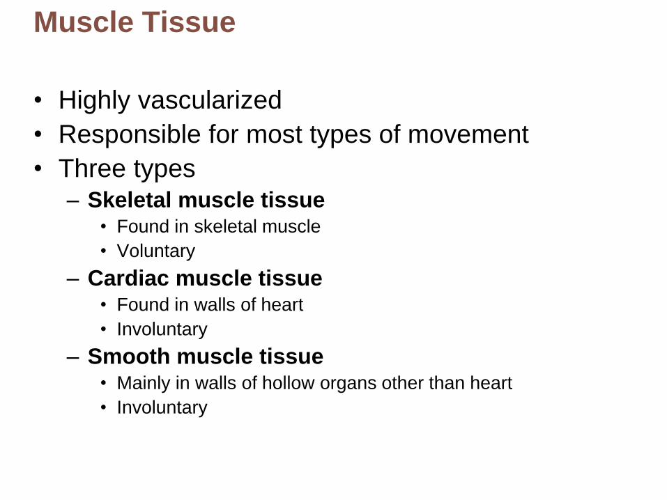
Muscle Tissue
• Highly vascularized
• Responsible for most types of movement
• Three types– Skeletal muscle tissue
• Found in skeletal muscle
• Voluntary
– Cardiac muscle tissue• Found in walls of heart
• Involuntary
– Smooth muscle tissue• Mainly in walls of hollow organs other than heart
• Involuntary
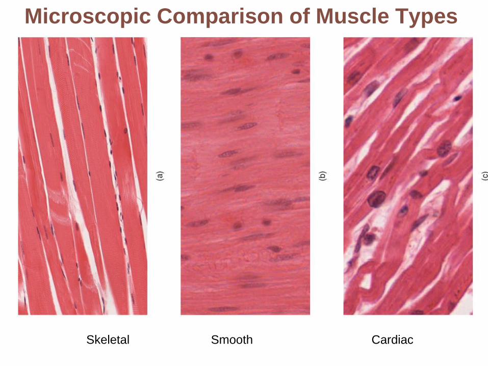
Microscopic Comparison of Muscle Types
Skeletal Smooth Cardiac

Nervous Tissue
• Main component of nervous system
– Brain, spinal cord, nerves
– Regulates and controls body functions
• Neurons
– Specialized nerve cells that generate and conduct
nerve impulses
• Neuroglia
– Supporting cells that support, insulate, and protect
neurons

Nervous tissue
Description: Neurons arebranching cells; cell processesthat may be quite long extend fromthe nucleus-containing cell body;also contributing to nervous tissueare nonexcitable supporting cells.
Function: Neurons transmitelectrical signals from sensoryreceptors and to effectors (musclesand glands) which control theiractivity; supporting cells supportand protect neurons.
Location: Brain, spinal
cord, and nerves.
Photomicrograph: Neurons (350x).
Neuronprocesses
Nuclei ofsupportingcells
Cell bodyof a neuron
Neuron processes Cell body
Axon Dendrites

Tissue Repair
• Necessary when barriers are penetrated and tissues are damaged– Temporary tissues form to close the breach
– Cells divide and migrate to replace temporary tissue
– Dead cells, debris, foreign invaders must be removed
• Occurs in two major ways– Regeneration
• Same kind of tissue replaces destroyed tissue
• Original function restored
– Fibrosis or Replacement• Connective tissue scar replaces destroyed tissue
• Original function lost
• More likely in severe injury

Slide 1
Scab
Blood clot inincised wound
Epidermis
Vein
Inflammatorychemicals
Migrating whiteblood cell
ArteryInflammation sets the stage:
• Severed blood vessels bleed.
• Inflammatory chemicals are released.
• Local blood vessels become more permeable, allowing white blood cells,
fluid, clotting proteins, and other plasma proteins to seep into the injured area.
• Clotting occurs; surface dries and forms a scab.
1

Regenerating epithelium
Area of granulationtissue ingrowth
Macrophage
Budding capillary
Fibroblast
Organization restores the blood supply:
• The clot is replaced by granulation tissue, which restores the vascular
supply.
• Fibroblasts produce collagen fibers that bridge the gap.
• Macrophages phagocytize dead and dying cells and other debris.
• Surface epithelial cells multiply and migrate over the granulation tissue.
2

Regeneratedepithelium
Fibrosed area
Regeneration and fibrosis effect permanent repair:
• The fibrosed area matures and contracts; the epithelium thickens.
• A fully regenerated epithelium with an underlying area of scar tissue
results.
3

Regenerative Capacity in Different Tissues
A. Regenerate extremely well
– Epithelial tissues, bone, areolar connective tissue,
dense irregular connective tissue, blood-forming
tissue
B. Moderate regenerating capacity
– Smooth muscle and dense regular connective tissue
C. Virtually no functional regenerative capacity
– Cardiac muscle and nervous tissue of brain and
spinal cord
– New research shows cell division does occur
• Efforts underway to coax them to regenerate better


![Histology Slides - mediconotes.commediconotes.com/freenotes/basic/histology_laboratory_slides.pdf[Histology] Histology Slides MedicoNotes provides real laboratory Histological slides](https://img.pdfslide.net/doc/110x75/5ae110e87f8b9a5a668e6aa3/histology-slides-histology-histology-slides-mediconotes-provides-real-laboratory.jpg)