Embed Size (px)
Citation preview

Slide 1
ICD-O CODINGThird Edition
PART IAn introduction
to theICD-O-3 Coding Manual
This is part 1 of a 2-part slide presentation. Use the navigation buttons below this panel to advance the slides. Be sure that you have printed out the Part I exercises worksheet provided in the course. You will need this as you go through the presentation.

Slide 2
Objectives
Introduction to the ICD-O-3 ICD-O-3 Coding Rules Rules for Topography Rules for Morphology Grade/Differentiation
International
Classification of
Diseases for
Oncology
Third EditionIC
D-O
In this lesson, we are going to talk about: •The Rules in ICD-O-3 •How to use the different sections of ICD-O-3 •How to code the primary site, histology, behavior and grade of neoplasms Take out your ICD-O-3 and have it ready. You should follow along and take the time to look things up when indicated. Also, you may want to have your FORDS within reach. There are a few areas that will include information from the FORDS.
Slide 3
Purpose of Coding
Standardize collection
Compare dataRegionalNationalInternational
Dual Coding SystemTopographyMorphology
Why do we convert the information from the medical record into codes? Coding allows us to compare the data more easily with reports and analysis. The other alternative being text, which isn’t very practical. It is much easier to group similar histologies and primary sites by a code number than by written text. Any numbering system could have been used for this. But, the reason we use the ICD-O-3 is to standardize cancer data collection and to allow what you coded to be compared to what someone else coded across town, in another state, and around the world. The “International” Classification of Diseases for Oncology is used around the world, not just in the U.S. This allows data to be combined into larger databases containing information collected by many reporting sources around the world. ICD-O-3 is considered a dual classification system, meaning that this one book provides us the coding systems for assigning both the topography and morphology codes of the tumor.

Slide 4
ICD-O-3 Organization
1. Instructions for use…………..…………..…..pages 1-42
2. Topography - Numerical List……….…….pages 43-65
Behavior and Grade……………………….…pages 66-67
3. Morphology - Numerical List……………...pages 69-104
4. Alphabetic Index………………….………...….pages 105-218
5. Appendices………………………………….…...pages 219-240
Page 8page
First, let’s orient you to the ICD-O-3 manual. There are 5 main sections of the ICD-O-3. Turn to each of these sections in your ICD-
O-3 manual and review them as we talk about them. The page numbers are listed on the slide so that you can follow along.
Section 1 contains some preliminary instructions for how to use the manual. It explains
the format of ICD-O-3 and defines the other four main sections. We will spend most of our time during this presentation talking about these instructions.

Slide 5
Section 2: Topography – Numerical List
Primary Site
“C” Code C __ __ . __
Where is it?
9, 43-65
Section 2 begins on page 45 and lists all of the topography codes in order by the topography code number. When we say “Topography”, what we are referring to is the “C” Code or the primary site. The topography tells us where in the body the tumor originated
. Originated is a key word here. We ALWAYS want to code the site where the tumor started growing.
Slide 6
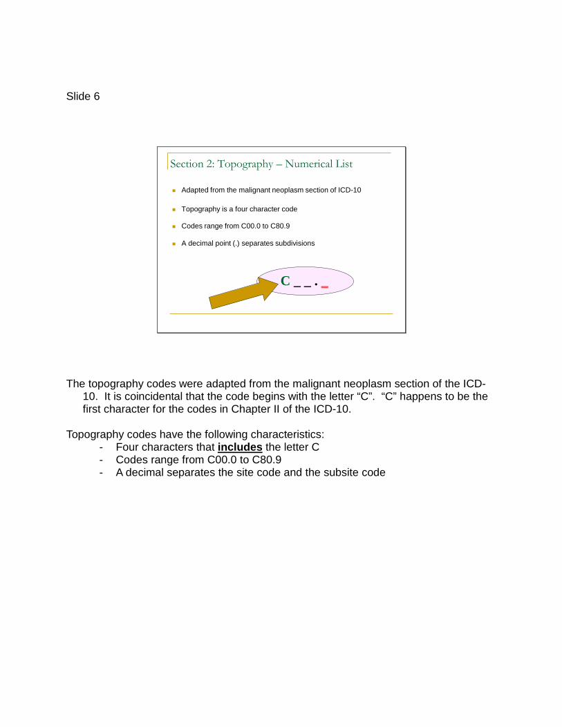
Section 2: Topography – Numerical List
Adapted from the malignant neoplasm section of ICD-10
Topography is a four character code
Codes range from C00.0 to C80.9
A decimal point (.) separates subdivisions
C _ _ . _
The topography codes were adapted from the malignant neoplasm section of the ICD-10. It is coincidental that the code begins with the letter “C”. “C” happens to be the first character for the codes in Chapter II of the ICD-10.
Topography codes have the following characteristics:
- Four characters that includes- Codes range from C00.0 to C80.9
the letter C
- A decimal separates the site code and the subsite code

Slide 7
Structure of the Topography Code
Breast, Upper Inner Quadrant
C 5 0 . 2
C
Site
Subsite
This is the structure of the topography code. It is a four-character code that includes the C. C is always part of the topography code. Then, you have a 2 digit site code (in green) and a 1 digit subsite code (in yellow) that is separated by a decimal point.
Find C50.2 in Section 2, the topography numerical index in your ICD-O-3. In this example, C50.2 tells us that this cancer originated in the upper inner quadrant of
the breast. 50 is the site code for breast. 2 is the subsite code for upper inner quadrant.
(You should have found C50.2 on page 58.)
Slide 8
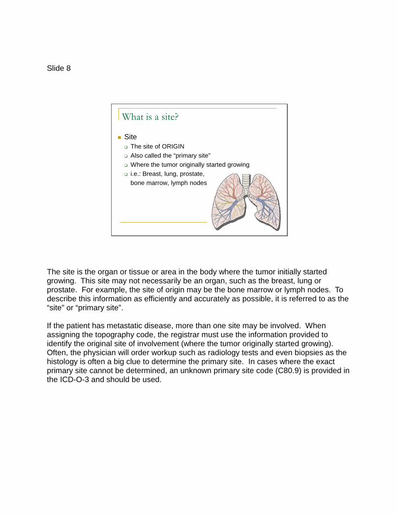
What is a site?
Site The site of ORIGIN Also called the “primary site” Where the tumor originally started growing i.e.: Breast, lung, prostate,
bone marrow, lymph nodes
The site is the organ or tissue or area in the body where the tumor initially started growing. This site may not necessarily be an organ, such as the breast, lung or prostate. For example, the site of origin may be the bone marrow or lymph nodes. To describe this information as efficiently and accurately as possible, it is referred to as the “site” or “primary site”. If the patient has metastatic disease, more than one site may be involved. When assigning the topography code, the registrar must use the information provided to identify the original site of involvement (where the tumor originally started growing). Often, the physician will order workup such as radiology tests and even biopsies as the histology is often a big clue to determine the primary site. In cases where the exact primary site cannot be determined, an unknown primary site code (C80.9) is provided in the ICD-O-3 and should be used.

Slide 9
Your turn…
Exercise 1: What is the Site?
An important part of coding site and histology correctly is being able to recognize the terminology in the medical record before assigning a code to that term.
Complete Exercise 1 that was provided in the instructor’s notes.
When finished, check your answers.

Slide 10
What is a subsite?
Subsite Further divisions of the site of origin i.e.: Breast, upper inner quadrant;
lung, upper lobe
Not all sites have applicablesubsites
i.e.: Prostate, kidney
Some sites can be divided into more specific areas within the organ. For example, the breast can be divided into quadrants: upper inner, lower inner, upper outer, and lower outer. The lung can be divided into lobes: upper lobe, middle lobe, and lower lobe. The colon can be divided into segments: ascending, transverse, descending, and sigmoid. Coding the subsite allows us to describe the exact location of the primary tumor more specifically. In some primary sites, multiple tumors in more than one subsite
could mean that the patient has more than one primary, rather than a metastasis. For other primary sites, this may only be one primary cancer. Also, the staging criteria and surgery codes may differ for different subsites within an organ. Recording the subsite when it is known is important in capturing the most complete information.
For some sites, dividing the organ into more specific areas is not necessary. Therefore, the ICD-O-3 will not provide applicable subsites codes for that site. A good example of this is the prostate. The prostate does have left and right lobes, but they were not given specific subsites codes in the ICD-O-3. There is only one topography code for the prostate (C61.9) regardless of where the tumor originated within the prostate.

Slide 11
Your turn…
Exercise 2: What is the Subsite?
Complete Exercise 2 that was provided in the instructor’s notes.
When finished, check your answers.

Slide 12
Tips for Assigning the Topography Code
Assign the primary site where the tumor originated
Do not automatically code the site the specimen was taken from
Assign C80.9 if the primary site cannot be determined Exhaust all resources before assigning an unknown Update if the exact site is determined even if months
or years later
The site in which the tumor started is always assigned as the primary site. Read all documentation carefully and thoroughly to pinpoint the primary site as accurately as possible. In some situations, a biopsy of a site other than the primary site may be done. This is due to several reasons such as that area is easier to biopsy than the primary site when they are just trying to obtain a tissue diagnosis. Do not automatically code the biopsy site - unless it is the same as the primary site, of course. If the site cannot be determined, then assign the site code of C80.9. As with all other unknowns coded in the abstract, you should exhaust all resources including contacting the managing physician if necessary. Sometimes the disease is so diffuse, it is difficult to determine where the cancer originated. Or, the patient may refuse further workup. In this case, we have no other option but to code to an unknown site. If at a later time, the exact site is determined, the site code should be updated and a correction sent to the central registry.

Slide 13
Determining the Organ/Tissue of Origin
NCI Public DomainR
One of the most important pieces of information for a cancer registrar in assigning the site and histology code is to understand the disease process for cancer. Understanding how cancer cells form and grow and spread will help you understand why you are instructed to assign one code over another. Much of this was introduced in the beginning of this course.
Before we can even think about what codes to assign, the first thing (after we have determined that the
case is reportable) is to determine the primary site. The primary site drives everything else in the abstract. The staging system that you use is based on the primary site. The surgery codes that you use are based on primary site. Note: In some instances, like with lymphoma and leukemia, histology is a driving factor as well.
When you read through a chart, you have to determine a couple of factors. • What organs or tissues are involved? • Is there more than one tumor to consider? • Is this where the tumor originated or is this a metastatic tumor? If there is, for example, a tumor in the colon and the lung, you have to consider if the tumor started in the
colon and metastasize to the lung. Or, did one tumor start in the colon and a completely different tumor start in the lung. When reading the diagnostic reports, you have to make this clear distinction. In cases where the distinction is not clear, we have the multiple primary rules as our guideline.
In theory, this sounds pretty easy. But, the wording in the charts can be quite confusing. The physicians
may even be having a difficult time deciding what the primary site is. You may have to read the documentation several times to try to figure out what message the physician is conveying. You may have to wait for a later admission to see if more information has become available, or even contact the managing physician. Because so much depends on this information, it is important that we take the time and use all of our resources to code this as accurately as possible.
Once we know the organ or tissue where the tumor began, then we can become more concerned about
which specific site code to assign to describe that information. This is where the ICD-O-3 comes in.

Slide 14
Section 3: Morphology - Numerical List
Cell Type
“M” Codes M __ __ __ __ / __ __
What is it?
9, 69-104
Between sections 2 and 3 is a list of the valid codes for behavior and grade. Behavior and grade are part of the complete morphology code. There are a few coding instructions for assigning these two values in the ICD-O-3, but you will find more detailed coding instructions in the FORDS. Behavior and grade will be discussed in more detail later. Section 3 is similar to section 2. Instead of the topography codes, section 3 lists the morphology codes in order by the morphology code number. The morphology code describes the cell type of the tumor and its biologic activity, in other words, the characteristics of the tumor. Often the terms “Histology” and “Morphology” are used interchangeably. When you hear either term, the type of cancer or cell type is what the person is referring to. Morphology just goes a little farther, adding behavior and grade to the cell type.

Slide 15
Section 3: Morphology - Numerical List
Displays the structure of the coded morphology nomenclature Primary point of reference Morphology is a five digit code Codes range from M-8000/0 – M-9989/3 Additional 6th digit for grade
M- _ _ _ _ / _ _
The morphology code has the following characteristics: • Five digit code ranging from 8000 to 9989. • The 5 digits consist of 4 digits for the cell type + 1 digit for the behavior code (the
number after the slash). • The sixth digit is considered part of the morphology code and is the grade or
differentiation of the tumor. Note: The word “nomenclature” is used in the ICD-O-3 and is a general term that refers
to a system of names or codes used in a particular discipline, such as anatomy – or in this instance – morphology.
Section 2 and 3 are called the numerical lists – because the codes are listed in
numerical order. The general instructions in the ICD-O-3 state that the numerical lists are the primary point of reference. What does that mean?
The numerical lists are most often used when you already know the code and you need
to know what the terminology is for that code. For example, if you ran a report from the cancer registry database of all of the histologies for the colon cancer, it is best to group the histologies by the morphology code. The best and easiest place to find the term for that code is in the numerical list. Here, the terminology from the medical record has already been coded and recorded in the database. Therefore, the numerical listing is considered the primary reference point for looking up a code to see what the terminology is for that code. Why? Because it is listed by the code in numerical order and easy to reference.
The numerical lists are also used to double check what was looked up in the alphabetic
index to make sure the most complete and accurate code is being selected. An example of this will be provided later in the presentation.

Slide 16
Structure of the Morphology Code
Well Differentiated Adenocarcinoma
M-8140 / 3 1
Cell TypeBehavior
Differentiation (Grade)
This is the structure of the complete morphology code. The first four digits indicate the specific histology term or cell type. The fifth digit, after the slash is a behavior code, which indicates whether a tumor is malignant, benign, in situ, or uncertain whether malignant or benign. A separate one-digit code for histologic grading or differentiation is provided. For hematopoietic cases, this element of the code is used to identify the immunophenotype (T-, B-, Null-, and NK-cell origin). Find 8140/3 in the numerical index in your ICD-O-3. In this example, 8140/31 tells us that this is a malignant well differentiated adenocarcinoma. 8140 (in green) is the cell type code for adenocarcinoma. /3 (in red) is the behavior code indicating that this is malignant or invasive cancer. And the 1 (in yellow) is the differentiation code for well differentiated. (You should have found 8140/3 on page 72.)
Slide 17
Your turn…
Exercise 3: What is the histologic term?
Complete Exercise 3 that was provided in the instructor’s notes
When finished, check your answers
Slide 18
Structure of a Complete ICD-O-3 Code
Diagnostic term:Poorly differentiated squamous cell carcinoma,
upper lobe of lung
C34.1 M-8070/33
Topography Morphology
10 Digits
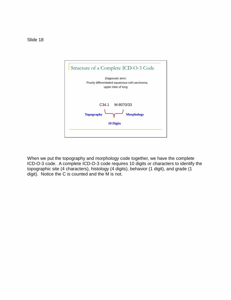
When we put the topography and morphology code together, we have the complete ICD-O-3 code. A complete ICD-O-3 code requires 10 digits or characters to identify the topographic site (4 characters), histology (4 digits), behavior (1 digit), and grade (1 digit). Notice the C is counted and the M is not.

Slide 19
Format of the Numerical Lists
Terms are listed only once Term are listed in numerical order by the code number
Preferred term is listed in bold
8012/3 Large cell carcinoma, NOS
8013/3 Large cell neuroendocrine carcionoma
8014/3 Large cell carcinoma with thabdoid phenotype
To help organize terms in the ICD-O-3, the terms were listed using a specific format. The format used in the numerical lists is different than the format used in the alphabetic list. For example, in the numerical lists, a particular code may have more than one term that can be described by that code. These multiple terms are listed in a specific way that will give them some meaning to us as users of these codes. Turn to page 45 in your ICD-O-3 manual. This is the beginning of the topography numerical list. In the numerical lists, each code is only listed once. And, they are listed in numerical order by the code number. Topography codes range from C00.0 to C80.9. C00.0 is the lowest number in the range so it will be listed first. C80.9 is the highest so it will be listed last (page 65). C00.0 is only listed once. C00.1 is only listed once. And so on. Now, let’s look at the morphology list on page 69. The range of morphology codes is 8000 to 9989. Again, each morphology code is listed in numerical order and only listed once. Notice the order when a code has more than one behavior code listed. For example, look at 8010 (carcinoma, NOS). Benign is listed first (8010/0), then in situ (8010/2), then invasive (8010/3). In both numerical lists, each code has 1 bold term listed beside it. The term in bold is the preferred term. Each and every code in both the topography and morphology numerical lists will have one boldfaced term listed beside it. For some codes, the boldfaced term is the only term listed for that code. There are several examples on page 69. 8012/3, 8013/3, 8014/3 for example.

Slide 20
Format of the Numerical Lists
Synonyms Are indented Term that means the same as the boldface term
Equivalent terms Not a synonym of the boldface term Not indented Not sufficiently different to have its own code
9085/3 Mixed germ cell tumorMixed teratoma and seminoma
8033/3 Pseudosarcomatous carcinomaSarcomatoid carcinoma
Other codes will have more than one term listed beside it. The terms listed under the bold term may be indented or it may not be.
If the term is
indented under the preferred term (in bold), it is considered a synonym of that boldface term. Synonym means another way of saying the same thing. Look at code 8033/3 in your ICD-O-3. Sarcomatoid carcinoma is a synonym of pseudosarcomatous carcinoma. So, these two terms mean exactly the same.
Terms not
indented are considered equivalent terms. These equivalent terms are not synonyms of the preferred term. For coding purposes in the ICD-O-3, an equivalent term is not quite exactly the same thing as what is being described by the term in bold. However, the equivalent term shares some component of the preferred term, but not all, therefore it is not “exactly” the same. For the topography, the equivalent term may be a subdivision of the preferred term. In morphology, the two terms may involve the same type of cell. Because the difference between the two are not significantly different, they were not given their own code, but instead were listed under the related term. To summarize, it is not exactly the same, but is not different enough to have its own code.
Look at code 9085/3 on page 90. Mixed germ cell tumor would describe all morphologies coded to 9085/3 and is the preferred term. Mixed teratoma and seminoma is an equivalent term. It is not exactly the same as mixed germ cell tumor but it is not significantly different enough to have been given its own code. Both of these terms should be assigned the code of 9085/3.
Whether the term is in bold or is indented is usually not huge factor when assigning the
histology code. What is important is that you understand that all of these terms are involving the same cell type and why the coding book was formatted in this way.

Slide 21
Your turn…
Exercise 4: Practice Using the Numerical Indices in the ICD-O-3.
Complete Exercise 4 that was provided in the instructor’s notes
When finished, check your answers
Slide 22
Section 4: Alphabetic Index
Topography and Morphology are listed together
Terms are listed alphabetically Terms may be listed more than once
Example: Squamous Cell Carcinoma can be found under: “S” for Squamous Cell or “C” for Carcinoma
Exception: Lymphoma and Leukemia
10, 105
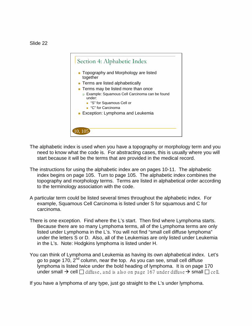
The alphabetic index is used when you have a topography or morphology term and you need to know what the code is. For abstracting cases, this is usually where you will start because it will be the terms that are provided in the medical record.
The instructions for using the alphabetic index are on pages 10-11. The alphabetic
index begins on page 105. Turn to page 105. The alphabetic index combines the topography and morphology terms. Terms are listed in alphabetical order according to the terminology association with the code.
A particular term could be listed several times throughout the alphabetic index. For
example, Squamous Cell Carcinoma is listed under S for squamous and C for carcinoma.
There is one exception. Find where the L’s start. Then find where Lymphoma starts.
Because there are so many Lymphoma terms, all of the Lymphoma terms are only listed under Lymphoma in the L’s. You will not find “small cell diffuse lymphoma” under the letters S or D. Also, all of the Leukemias are only listed under Leukemia in the L’s. Note: Hodgkins lymphoma is listed under H.
You can think of Lymphoma and Leukemia as having its own alphabetical index. Let’s
go to page 170, 2nd column, near the top. As you can see, small cell diffuse lymphoma is listed twice under the bold heading of lymphoma. It is on page 170 under small cell diffus e , a nd is a ls o on pa ge 167 unde r diffus e small ce ll.
If you have a lymphoma of any type, just go straight to the L’s under lymphoma.

Slide 23
Format of the Alphabetic Index
Boldface header Words that have 3 or more terms that include that
word Modifying terms are indented under the bolded term NOS is always listed first Modifying term is more specific than the NOS term
Topographic (C codes) and Morphologic (M codes) terms are not mixed under a single heading
Spaces indicate: A change from topographic to morphologic terms The end of a group
Terms in the alphabetic index are listed using a specific format as well. Turn back to page 105. Notice that not all codes have a term that is in bold beside it. In the alphabetic index, a bold term means something different than a bold term in the numerical index. In the alphabetic index, any word that has three or more terms that include that word have a boldface heading. The terms that include that word are indented under the bold header. Look at Abdomen under “A”. The term Abdomen has 7 terms that contain the word abdomen. Since that is 3 or more, abdomen is given a boldface header. The terms listed under the bolded term are indented and are considered modifying terms because they describe the bolded term more specifically. This formatting system is basically a way of grouping terms together, instead of having to repeat the word “abdomen” seven times. When there is a list of terms grouped under a boldface header, often the first term listed will be “NOS”. If NOS is appropriate and applies, NOS will always be listed first under the heading and not in alphabetical order under N. NOS is a non-specific description to say that the bolded term could not be defined more specifically. We know it was the abdomen, but there is not enough information to say it was the skin or the muscle. When there is a choice between a more specific modifying term and the NOS term, the more specific term should be coded.
Under the group of 7 “abdomen” terms, you will see a space, then 8822/1 abdominal desmoid and 8822/1 abdominal fibromatosis. Although “abdominal” is part of the term for both of the 8822 morphology terms, there are only 2 of them (not 3), so it is not given a bold header. Also, topography and morphology terms are not mixed under a single bold heading. Under these two codes, there is another space, then a bold heading for Abdominal. Listed under it are 4 topography terms that contain the word abdominal. Because there are more than 3, abdominal is given a bold header. Even though abdominal is also part of the codes for 8822, they are not combined under the abdominal heading containing the topography terms. Morphology terms are listed together and topography terms are listed together. Topography and morphology terms are not mixed under a single bold heading. A space (or blank line) indicates either a change from topographic to morphologic terms or designates the end of a group.

Slide 24
Your turn…
Exercise 5: Practice Using the Alphabetical Index in the ICD-O-3.
Complete Exercise 5 that was provided in the instructor’s notes
When finished, check your answers

Slide 25
Tumor-like Lesions and Conditions
Can be confused with neoplasms end in “oma” pre-malignant conditions
Appear as M------- (see SNOMED)
NOT Reportable
No code at all…..then it is not reportable.
Tumor-like Lesions and Conditions can also be found listed in the alphabetic Index. Often they will end in “oma”, but not always. These are not considered to be neoplasms, but rather pre-malignant conditions, and therefore do not have an ICD-O-3 morphology code. Instead, they are listed with an ‘M’ and 7 dashes followed by “see SNOMED” in parentheses. SNOMED is the Systematized Nomenclature of Medicine. There are different editions of SNOMED currently in use, so the specific code is not listed. On this same page (105), you can see a few examples at the bottom of each column. The important thing to remember is if you have a diagnosis, and you look up the term and it has an M and dashes, then it is not reportable and does not need to be abstracted. This also brings up another point. If the term in the medical record cannot be found anywhere in the ICD-O-3 (after checking all combinations of the term and perhaps even checking with a mentor for assistance), then it is not reportable. All reportable histologies will be listed in the ICD-O-3 with a code. Be careful that you have looked up every possible combination, especially with tricky terms that may not sound reportable but are, before determining that the case is not reportable.

Slide 26
“NOS”…and How to Use It
Just as with all of the other data items, you want to code the site code as specifically as possible and the morphology code as specifically as possible. As was briefly mentioned in the earlier slide, some terms have an ‘NOS’ associated with it. NOS is the acronym for Not Otherwise Specified. NOS is similar to NEC in ICD-9. NOS is a term used to describe the site or histology in a general sense. Often, this means that the term could be described in more detail but there wasn’t enough information available to assign a more specific code.

Slide 27
“NOS” – Where is it?
Usually not on pathology or other reports
Usually only listed under those terms that appear with additional modifying words
Listed first under the bold header
Indicates that other, more specific, modifiers of the term may be listed elsewhere
120, 164
Carcinoma
NOS will usually not be found on any pathology or other reports in the medical record. It is a term related to cancer registry coding applications. Also, not all terms have a NOS term listed with it. Only those terms that have more specific modifying words with a more specific code have a NOS. There are times when we don’t have enough information to code more specifically. The NOS term is a clue to us that there may be a more specific code to choose from and we should look further before choosing the NOS term. Find carcinoma under the C’s. Carcinoma is a general term (an NOS term). The first term indented under carcinoma is NOS. Squamous cell and basal cell are more specific types of carcinoma and are also listed under carcinoma. The words ‘basal’ and ‘squamous’ are modifying words for the term ‘carcinoma’. If the histology is known to be squamous cell carcinoma, this should be coded over carcinoma, NOS as it is more specific. Look at page 164, 1st column, at the term “LOBE”. Lobe doesn’t have an NOS code listed under it even though we know there are more specific choices (or modifying terms). You can’t assign the site code in your abstract as Lobe, NOS. Because various organs in the body contain lobes, such as the brain and the lung, we aren’t even given an option to be so vague. If you ran a report and it said LOBE NOS, this doesn’t even put us in the vicinity of any one particular area. If you know it was in one lobe of the lung, but you don’t know which one, you code to Lung NOS (p165) not to Lobe NOS. This is one reason there may not be an NOS option.
Slide 28
“NOS” - When to use it? Term is not modified
”adenocarcinoma” Code to 8140/3 Adenocarcinoma, NOS
Term has an adjective that does not appear elsewhere ”atypical adenocarcinoma” Code to 8140/3 Adenocarcinoma, NOS
Term is used in the general sense ”endocrine gland” Code to C75.9 Endocrine gland, NOS
Topography subsite code for NOS = .9106
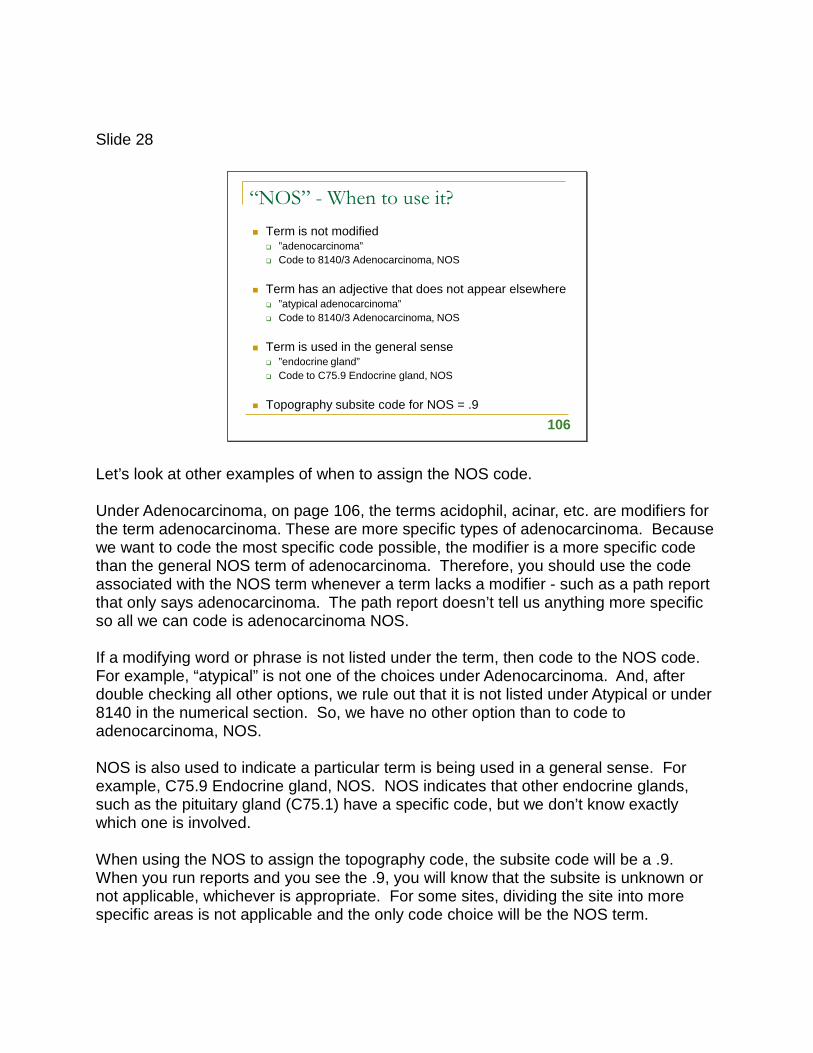
Let’s look at other examples of when to assign the NOS code. Under Adenocarcinoma, on page 106, the terms acidophil, acinar, etc. are modifiers for the term adenocarcinoma. These are more specific types of adenocarcinoma. Because we want to code the most specific code possible, the modifier is a more specific code than the general NOS term of adenocarcinoma. Therefore, you should use the code associated with the NOS term whenever a term lacks a modifier - such as a path report that only says adenocarcinoma. The path report doesn’t tell us anything more specific so all we can code is adenocarcinoma NOS. If a modifying word or phrase is not listed under the term, then code to the NOS code. For example, “atypical” is not one of the choices under Adenocarcinoma. And, after double checking all other options, we rule out that it is not listed under Atypical or under 8140 in the numerical section. So, we have no other option than to code to adenocarcinoma, NOS. NOS is also used to indicate a particular term is being used in a general sense. For example, C75.9 Endocrine gland, NOS. NOS indicates that other endocrine glands, such as the pituitary gland (C75.1) have a specific code, but we don’t know exactly which one is involved. When using the NOS to assign the topography code, the subsite code will be a .9. When you run reports and you see the .9, you will know that the subsite is unknown or not applicable, whichever is appropriate. For some sites, dividing the site into more specific areas is not applicable and the only code choice will be the NOS term.

Slide 29
Alphabetic Index Numerical
EndocrineC75.9 gland, NOSC75.8 glands, multipleC25.4 pancreas
M-8360/1 Endocrine adenomas, multiple
.
.
.
C75 OTHER ENDOCRINEGLANDS AND RELATEDSTRUCTURES
C75.0 Parathyroid glandC75.1 Pituitary glandC75.2 Craniopharyngeal ductC75.3 Pineal glandC75.4 Carotid bodyC75.5 Aortic body and other…C75.8 Overlapping lesion…C75.9 Endocrine gland, NOS
This is a very important step for making sure that you are choosing the best code. This step isn’t specified anywhere in the general instructions but is a basic fundamental of coding. When you are given a diagnosis, all you have is the terminology, so you have to look it up in the Alphabetic Index first to get the code. But, you should not stop there. You should always go back and look the code up on the Numerical List as well. For example, the Discharge Summary says “endocrine gland”. First look in the Alphabetic Index, under E. Page 137 shows endocrine gland with a site code of C75.9 and that’s all. Because we now know the site code for the endocrine gland, we can go back to the numerical section, to C75 in Topography Numerical List (page 63). You will notice that this can be coded much more specifically than “endocrine gland, nos.” This is a clue that it is possible to assign this to a more specific code and we should research this further. When you discover that the ICD-O-3 provides more specific choices, go back to the medical record and see if you can find reference for a specific structure before coding to the NOS term. If not, then C75.9 is by all means the most appropriate code.

Slide 30
Alphabetic Index NumericalCarcinoma, continued
Squamous cellM-8070/3 NOSM-8070/6 NOS, metastaticM-8075/3 acantholyticM-8075/3 adenoidM-8083/3 basaloidM-8084/3 clear cell type M-8070/2 in situ, NOS
.
.
.
805-808 SQUAMOUS CELL NEOPLASMS
8070/3 Squamous cell carcinoma NOSEpidermoid carcinoma, NOSSquamous carcinomaSquamous cell epithelioma
Squamous cell carcinoma, epidermoid type
Here is another example where cross referencing between the two sections can also help solve coding dilemmas. Look at page 70-71. Notice the main group headings: •805-808 Squamous Cell neoplasms •809-811 Basal Cell neoplasms •812-813 Transitional Cell papillomas and carcinomas All of the codes in the 805-808 range are considered squamous cell neoplasms. If you have a complex diagnosis and are not sure if you found the most specific code in the alphabetic index, try coming back to the numerical list for those codes to see if you can narrow it down a little further. Seeing them all grouped together may help you decide the most appropriate code. For example, let’s say we have a squamous cell carcinoma, epidermoid type. Squamous cell carcinoma, NOS has a code of 8070. The NOS is telling us that there may be more a specific code. Our term stated “epidermoid type” so we want to see if there is a more specific code for the epidermoid type. If you look under squamous cell in the alphabetic listing (pg 124 under “carcinoma” or pg 203 under “squamous”), you will see “epidermoid” is not listed under the squamous cell heading.

Before we rule out that there is not a more specific code, we should go back and check the numerical index as well. Find squamous cell in the numerical index. The alphabetic index told us the code was 8070 so let’s look at the 805-808 category on pg 70. Find 8070/3 for the squamous cell carcinoma. You will see there are 3 synonymous terms listed under squamous cell carcinoma in the numerical index. One of them is epidermoid. Therefore, we now know that SCC and SCC epidermoid type have the same ICD-O-3 code of 8070. Even though we did not find a more specific code, what we did do is determine that indeed 8070 was the most specific code and there was not another code that would have been even more specific. Now, with this diagnosis, what else could we have done? Looked under “epidermoid” in the Alphabetic Index. In the alphabetic index, go back to the E’s under carcinoma, pg 121, or under the E’s on pg 137. You will find Epidermoid is listed and the code is 8070. The take home message here is don’t go to one version of the wording and say, well I don’t see anything that matches my diagnosis, so this is the best code I can use. Look it up under all of the different words and terms. Cross reference these sections until you can rule out all other code options.

Slide 32
Section 5: Appendices
1. New ICD-O, Third Edition, Codes2. New Morphology Terms and Synonyms3. Terms That Changed Morphology Code4. Terms That Changed From Tumor-Like Lesions to
Neoplasms5. Terms Deleted From ICD-O, Third Edition6. Terms That Changed Behavior Code
219
Section 5 contains the Appendices. These are important for DATA ANALYSIS and comparing OLD and NEW data. The appendices list the changes between the ICD-O-2 and ICD-O-3. Some of the appendices include information that affect reportability beginning in 2001. Registrars should not use these appendices to assign the morphology code to current cases.
Slide 33
International
Classification of
Diseases for
Oncology
Third Edition
ICD
-O
International
Classification of
Diseases for
Oncology
Second Edition
ICD
-O
Prior to01/01/2001
On or After01/01/2001
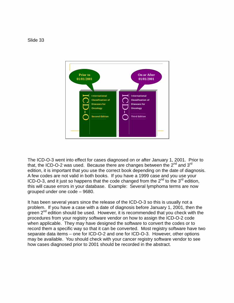
The ICD-O-3 went into effect for cases diagnosed on or after January 1, 2001. Prior to that, the ICD-O-2 was used. Because there are changes between the 2nd and 3rd edition, it is important that you use the correct book depending on the date of diagnosis. A few codes are not valid in both books. If you have a 1999 case and you use your ICD-O-3, and it just so happens that the code changed from the 2nd to the 3rd edition, this will cause errors in your database. Example: Several lymphoma terms are now grouped under one code – 9680. It has been several years since the release of the ICD-O-3 so this is usually not a problem. If you have a case with a date of diagnosis before January 1, 2001, then the green 2nd edition should be used. However, it is recommended that you check with the procedures from your registry software vendor on how to assign the ICD-O-2 code when applicable. They may have designed the software to convert the codes or to record them a specific way so that it can be converted. Most registry software have two separate data items – one for ICD-O-2 and one for ICD-O-3. However, other options may be available. You should check with your cancer registry software vendor to see how cases diagnosed prior to 2001 should be recorded in the abstract.

Slide 34
IARC Rules Multiple Primary Neoplasms Do not use rules on pages 35 and 37, and Table 25 In the U.S., registries follow the SEER multiple
primary rules
Basis of Diagnosis Do not use rules on page 38 and Table 26 In the U.S., use Diagnostic Confirmation codes FORDS manual
Because this is the INTERNATIONAL Classification of Diseases for Oncology, there is some information that pertains only to international registries and not to registries in the U.S. The multiple primary rules on pages 35 and 37 are for international registries. Registries in the U.S. follow the SEER MP/H Rules. There are some differences. For example, SEER counts each segment of the colon as a separate site where IARC does not. Also, do not use the rules for the Basis of Diagnosis on page 38 and in Table 26. In the U.S. we use the data item “diagnostic confirmation” to specify if the cancer was confirmed histologically, cytologically, on radiology, etc. You may want to write “IARC Rules – DO NOT USE” at the top of each of these pages to remind you while you are abstracting.
Slide 35
What if more than one term is stated?
As the ICD-O-3 is used all over the world, it was never meant to provide us specific information on how to determine groups or families of histologies or how to choose one code over another if more than one term is used to describe the tumor. Our other coding manuals provide additional information to help fill that gap. For example, you will find more instructions on how to code grade in the FORDS. If there is more than one histology term stated, we should use the SEER Multiple Primary and Histology (MP/H) coding rules to guide us in making a decision. This will be discussed in a later lesson. When coding, you have to pull all of the information from all of our available resources together.

Slide 36
Summary
1. Determine where the tumor originated2. Look up every term in the diagnosis3. Look terms up in the alphabetic index4. Cross reference the numeric index5. Use NOS as clue to investigate for
more specific information6. Code the most specific term allowable
This concludes the information provided in the general instructions. Important points to remember: •Look up every term in the diagnosis, look them up in both the alphabetic and the numerical indices. •Use NOS as a clue to look for more specific information. •Code the most specific term allowable. •Make notes of the codes that go along with the terms you have found. You will need them in the MP/H manual!

Slide 37
ICD-O CODINGThird Edition
This concludes Part I
You have reached the end of Part 1 of the ICD-O-3 Coding slide presentation. Close this window to return to your course, or use the navigation buttons below to view the presentation again.