Embed Size (px)
Citation preview

Page 1/25
Multiscale imaging of the rat brain using anintegrated diceCT and histology work�owPaul M. Gignac
Oklahoma State University Center for Health SciencesHaley D. O’Brien
Oklahoma State University Center for Health SciencesJimena Sanchez
Universidad VeracruzanaDolores Vazquez Sanroman ( [email protected] )
Oklahoma State University Center for Health Sciences
Research Article
Keywords: immuno�uorescence, �uorescence, cresyl violet, iodine, eosinophilic, isoelectric
Posted Date: February 19th, 2021
DOI: https://doi.org/10.21203/rs.3.rs-158407/v1
License: This work is licensed under a Creative Commons Attribution 4.0 International License. Read Full License
Page 2/25
AbstractAdvancements in tissue visualization techniques have spurred signi�cant gains in the biomedicalsciences by enabling researchers to integrate their datasets across anatomical scales. Of particularimport are techniques that enable the interpolation of multiple hierarchical scales in samples taken fromthe same individuals. This study demonstrates that two-dimensional histology techniques can beemployed on neural tissues following three-dimensional diffusible iodine-based contrast-enhancedcomputed tomography (diceCT) without causing tissue degradation. This represents the �rst step towarda multiscale pipeline for brain visualization. We studied brains from adolescent male Sprague-Dawleyrats, comparing experimental (diceCT-stained then de-stained) to control (without diceCT) brains toevaluate neural tissues for immunolabeling integrity, compare somata sizes, and distinguish neuronsfrom glial cells within the telencephalon and diencephalon. We hypothesized that if experimental andcontrol samples do not differ signi�cantly in quantitative metrics, brain tissues are robust to the chemical,temperature, and radiation environments required for these multiple, successive imaging protocols.Visualizations for experimental brains were �rst captured via micro-computed tomography scanning ofisolated, iodine-infused specimens. Samples were then cleared of iodine, serially sectioned, and preparedagain using immuno�uorescent, �uorescent, and cresyl violet labeling, followed by imaging with confocaland light microscopy, respectively. Our results show that many neural targets are resilient to diceCTimaging and compatible with downstream histological staining as part of a low-cost, multiscale brainimaging pipeline.
1. IntroductionScienti�c gains in understanding biological organization and complexity are closely associated withtechnological and methodological gains in bio-imaging. Visualization tools widely used in biomedicaland clinical imaging, such as light-sheet microscopy (Voie et al. 1993), magnetic resonance imaging(MRI: Jackson and Langham 1968; Lauterbur 1974; Damadian et al. 1976; Henshaw et al. 1977; µMRI:Smith et al. 1996; Dodd et al. 1999), white matter tractography (diffusion tensor image analysis; Basser etal. 1994; Jellison et al. 2004), X-ray micro-computed tomography (µCT; Houns�eld 1977; Feldkamp et al.1989; Rüegsegger et al. 1996), and positron emission tomography (Ter-Pogossian et al. 1975; Phelps etal. 1976; Muehllehner et al. 1976; Hawkins et al. 1992; Jones and Townsend 2017), are continuouslyre�ned to allow investigators to improve the visualization of gross anatomical, microscopic, and cellular-level structure and function of biological tissues. These methodological enhancements have spurredsigni�cant advancement within the biomedical sciences by enabling researchers to integrate datasetsacross anatomical scales––from the low-resolution capture of gross structures to the �ne-resolutioncapture of subcellular cytoskeletal elements. Synthetic approaches that combine multiple imagingtechniques have the potential to maximize single-specimen data extraction that spans hierarchical,logarithmic scales of tissue organization.
Here, we evaluate the e�cacy of using diffusible iodine-based contrast-enhanced computed tomography(diceCT; Gignac et al. 2016) as a low-cost foundation for multiscale anatomical imaging integration by

Page 3/25
combining high-resolution cellular and gross anatomical brain-visualization techniques. Although same-brain, multiscale visualization has been conducted previously by combining MRI with histology (e.g., Dinget al. 2016), the relatively coarse resolution of most readily-available MRI equipment limits the spatialaccuracy of placing microscopic-scale information from histology into macro- and mesoscopicanatomical contexts. µMRI can achieve more satisfactory spatial resolutions as high as 25 microns,potentially overcoming this limitation; however, µMRI scans remain cost-prohibitive, time-consuming, andare hampered by specimen size limitations (de Crespigny et al. 2008). Nonetheless, µMRI remains apopular imaging modality because it is non-disruptive to biological macromolecules, including proteins(Smith et al. 1996). We propose the use of diceCT (Gignac et al. 2016), which is commonly employed inorganismal biology research, as a novel, inexpensive, and high-resolution ex vivo alternative to µMRI.DiceCT is an X-ray µCT-imaging technique that utilizes iodine (in the form of Lugol's iodine [I2KI] oralcoholic iodine [I2E]) as a contrast agent to permit three-dimensional (3D) visualization of tissue-levelneuroanatomy at resolutions superior to MRI-based modalities (de Crespigny et al. 2008; Anderson andMaga 2015; Hughes et al. 2016b; Gignac and Kley 2018). Iodine absorbs X-rays and binds preferentiallyto sugars and fatty tissues, making it excellent for nervous system visualization due to its cleardifferentiation of myelinated and non-myelinated structures at micron-scale resolution using µCT (Gignacand Kley 2018).
The application of µCT for studying how the brain is organized has lagged behind its use as a diagnostictool in biomedical studies (Sengle et al., 2013; Hopkins et al., 2015; Karreman et al. 2016; Morales et al.2016; Shami et al. 2017). However, µCT-scanner technologies and software optimizations are improvingrapidly, allowing increasingly faster scans at micro-and nano-scale resolutions (e.g., Pelc 2015). The swiftevolution of µCT imaging is now being leveraged to document a broad array of tissue types at variousspatial scales, thereby providing a substrate for multi-scale visualization as, or perhaps more, viable thanµMRI. For example, diceCT methods achieve simultaneous visualization of hard and soft tissues, such asthe skull with its brain and cranial nerves intact (Gignac and Kley, 2018). They also include reversibleprocessing steps, allowing multiscale, fully-registered imaging of the same tissues with more traditionalhistological approaches. Multi-tool and correlative imaging, such as that widely used in pathology for thestructural examination of human tissues (McDonald and Hayes 1969; Morales et al. 2016), enablesmultiscale co-registration datasets from the same sample. Multiscale imaging via diceCT, therefore, holdsthe potential to maximize the amount of neuroanatomical information retrieved from one individual,which is otherwise practically unattainable using single-scale and destructive approaches (Caplan et al.2011; Burnett et al. 2014).
Recently, diceCT samples have been used successfully in subsequent thin-section histological protocols.These studies have followed a variety of methodologies, including harnessing the iodine already presentin the sample from initial contrast-enhancement (Jeffery et al. 2011) or following a dehydration rinse inethanol with re-staining using either Giemsa (Herdina et al. 2015), thionin (Hughes et al. 2016a),hematoxylin and eosin (Chen et al. 2018), Marius yellow, crystal red, methyl blue, and Luxol fast bluesolutions (Heimel et al. 2019). Histological results secondary to diceCT appear to have produced clear

Page 4/25
identi�cation of two-dimensional (2D) muscular, epithelial, connective, and peripheral nervous tissues.Collectively, these studies illustrate the potential for multiscale diceCT-to-histology tissue-imagingpipelines. However, no studies have formally tested post-diceCT histology against non-diceCT controls,nor have any reported successful immunocytochemical and immuno�uorescence tissue stainingfollowing diceCT.
Brain tissue is considered to be incredibly delicate, with neural proteins highly susceptible to degradationunder non-idealized chemical and/or thermal conditions (Gwóźdź et al. 1970; Bowler and Tirri 1974;Gwóźdź et al. 1978; Millan et al. 1979; Gold et al. 1985) when compared to somatic tissues (Burger andFuhrman 1964). Destructive ex vivo conditions may include formalin �xation, weeks in a salt-rich andaqueous contrast solution (e.g., I2KI), repeated staining with refreshed solutions, storage at roomtemperature, exposure to high levels of X-rays, and de-staining with alternating baths of sodiumthiosulfate (Na2S2O3) and deionized water or 70% ethanol—each of which represents a step in diceCTpreparation, imaging, and de-staining protocols (Gignac et al. 2016). The long-term resiliency of nervoustissues to such conditions has not been quanti�ed. To directly address the susceptibility of neurons andglial cells to degradation during diceCT preparation and imaging, we compare diceCT treatment andcontrol neural tissue samples by: 1) evaluating the speci�city and integrity of �uorescence,immuno�uorescence, and cresyl violet staining methods; 2) analyzing preserved morphologies; and 3)quantifying neural cells in four regions of interest within the telencephalon and diencephalon. Wehypothesize that, given the proper conditions, brain tissue can remain robust to the chemical,temperature, and radiation environments required for multiple imaging protocols, and we quantitativelyevaluated this hypothesis by comparing diceCT-histology preparations and histology-only controls. Tohelp guide future studies, we also document instances in our evaluation of these protocols whereindeviating from certain steps in the process could preclude successful histological staining. Overall, wedemonstrate that diceCT is non-destructive of neural tissues, and our results support the use of diceCTimaging in lieu of µMRI as part of a multiscale brain imaging pipeline.
2. Methods
2.1 Brain samplingSix adolescent (120–150 g body weight) male Sprague-Dawley rats (Charles River Laboratories, Franklin,MA, USA) were single-housed on a 12/12-hour light/dark cycle with ad libitum access to food and water.All rats were deeply anesthetized with an Avertin/xylazine cocktail and perfused transcardially with coldsaline solution (0.9% NaCl) followed by cold 4% paraformaldehyde. After perfusion, cervical vertebraewere removed, followed by postcranial musculature and surrounding connective tissue. Dorsal portionsof the parietal and temporal skull bones were detached, and the entire occipital bone was excised. Theremaining tissues connecting the skull and brain were severed, and all cranial nerves cut at the roots.Once extracted, brains were placed in 4% paraformaldehyde solution and refrigerated (4º C) for 24 hours,followed by a 0.1M Phosphate Buffer Solution (PBS-30%) with refrigeration (4º C) for 48–96 hours, at

Page 5/25
which full point immersion was observed. Three brain samples were prepared for diceCT imaging,followed by cresyl violet, �uorescence, and immuno�uorescence staining. Three additional brain sampleswere prepared as histological controls.
2.2 DiceCT protocols
2.2.1 StainingFollowing 24 hours of �xation and up to 96 hours of sucrose immersion, experimental rat brains (n = 3)were submerged into a 3% weight-by-volume (w/v) solution of Lugol's iodine (I2KI) for 21 days (at roomtemperature), following protocols outlined in Gignac et al. (2016) and Gignac and Kley (2018). In anaqueous solution, iodine (I2) and potassium iodide (KI) become Triiodide (iodine trimers; I3–) and
potassium ions (K+), and iodine trimers bind differentially to the carbohydrates and lipids present inneural tissues. All three samples were fully submerged in an iodine solution, placed on an electric rocker,and protected from light for stain infusion duration. Upon completion, specimens were washed in adeionized water bath for two hours to remove unbound, excess staining agent from external surfaces,which further aids in the differentiation of neural tissues during X-ray imaging (Gignac and Kley 2018).Specimens were then sealed in 50 ml polypropylene centrifuge tubes with polyethylene foam and �lledwith deionized water for CT scanning. Foam and centrifuge tubes were chosen as packing material andcontainers, respectively, for their relatively low density compared to iodine-stained tissues. Samples werefully submerged in water to prevent desiccation during scanning.
2.2.2 µCT scanningAll specimens were µCT scanned on a GE phoenix v|tome|x s240 high-resolution micro-focus CT system(General Electric, Fair�eld, CT, USA) at the American Museum of Natural History Microscopy and ImagingFacility (New York, NY, USA). Scan parameters were optimized on a specimen-by-specimen basis, guidedby the scout X-ray image and accompanying histogram. All scan parameters follow therecommendations of Gignac and Kley (2018). Due to the similarity of each specimen's size, shape, tissueconstituency, and staining, all scans were performed within a narrow range of 110–120 kilovolts, 110–120 micro-Amperage, 333-millisecond exposure timing, and using six× multi-frame averaging on atungsten target with a 0.1 mm copper �lter. All scans were completed in 107 minutes at 20–25micrometer (µm) resolutions (isometric voxels) to demonstrate equivalency to the highest µMRIresolutions currently available. After scanning, µCT data were reconstructed on an HP z800 workstation(Hewlett-Packard, Palo Alto, CA, USA) equipped with VG Studio Max (Volume Graphics GmbH, Heidelberg,Germany) and exported as TIFF stacks. Stacks were imported into AvizoLite 2020 (Thermo FisherScienti�c Inc., Waltham, MA) for 3D model building based on contrast differences between white andgray matter structures, as described in Gignac and Kley (2018).
2.2.3 Chemical de-stainingFollowing X-ray imaging, we used a chemical de-staining process to remove iodine from brain tissues.Brains were sectioned transversely into anterior and posterior halves (parallel to the planned plane of

Page 6/25
histological section) to increase the surface area available for the de-staining agent's diffusion. Ourteam's preliminary efforts identi�ed this bisection of the brains into anterior-posterior halves as a vitalstep to ensure complete removal of the triiodide contrast agent. Without this step, subsequenthistological sectioning resulted in shearing, which left the tissues unsuitable for further histologicalprocessing. All specimens were chemically de-stained by submersion in a 1% w/v solution of sodiumthiosulfate on an electric rocker for seven days, followed by three days of a deionized water bath toremove excess de-staining agent (Gignac et al. 2016). In solution, sodium thiosulfate reacts withTriiodide, reducing it to iodide (I–) and chemically de-staining soft tissues (Trevorrow and Fashena 1935;Shakhashiri 1983; Kondo et al. 2001). This protocol was undertaken twice in total. Repetition ensuredthat all iodine was chemically unbound from and physically �ushed out of samples before histologicalsectioning and re-staining. We recommend visual inspection of tissue slides at the beginning ofhistological preparation to con�rm that samples are de-stained and free of any residual brown to orangecoloration.
2.3 Histology preparationHistological control specimens (n = 3) were treated following protocols previously published (Vazquez-Sanroman et al., 2017). De-stained (i.e., post-diceCT) and control brains were transferred to a PBS-30%sucrose solution for one week. In preparation for cryo-sectioning, samples were embedded usingHistoprep frozen tissue embedding medium (Thermo Fisher Scienti�c Inc.) and immersed in liquidnitrogen for 20 seconds. Consecutive coronal sections, 15 µm in thickness, were obtained using acryostat on a − 6°C setup (Ag Protect Leica CM 1860; Leica Camera AG, Wetzlar, Germany) and stored inan antifreeze solution (30% ethylene glycol, 25% glycerol, 30% sucrose in PBS) at − 20°C. Histoprep andliquid nitrogen immersion were critical for optimizing tissue sectioning protocol. Besides, sections thickerthan 15 µm tended to disintegrate upon handling, which left them unreliable for staining.
Areas from the telencephalon and diencephalon were pre-selected for histological analyses based onexpected clear anatomical boundaries and identi�able neuronal or glial cell populations. We probed avariety of brain regions to select cell-type-speci�c markers that distinguish neurons and glial cells. Wealso tested for differences between control and post-diceCT preparations to determine if diceCT interfereswith immuno�uorescence, �uorescence, and standard histological stains commonly used to identifyneural phenotypes. We used �ve stains: three immuno�uorescent labels (anti-parvalbumin [PRV], anti-glial �brillary antigen protein [GFAP], and anti-DARPP-32), one �uorescent label for DNA (4’,6-diamidino-2-phenylindole [DAPI]), as well as a cresyl violet stain for RNA. These were applied as relevant to the medialprefrontal cortex (PRV, DAPI, cresyl violet), mammillary nucleus (GFAP, DAPI), the dentate gyrus of thehippocampus (PRV, DAPI, cresyl violet), and the substantia nigra pars reticulata (anti-DARPP-32, DAPI).These targeted antibodies allowed us to characterize phenotypes for cell membrane proteins,intermediate �laments, nucleic acids, and dopamine-cAMP-regulated phosphoprotein. See Table 1 for asummary of histological preparations.
2.3.1 Fluorescent staining

Page 7/25
In order to qualitatively and quantitatively examine overall cellular morphologies and to ensure theviability of tissues for antibody reactivity, tissue sections containing portions of the medial prefrontalcortex (mPFC), mammillary nucleus (MM), the dentate gyrus of the hippocampus (DG), and substantianigra pars reticulata (SNr) from all experimental and control specimens were prepared forimmuno�uorescent staining. Free-�oating sections were rinsed seven times for 15 minutes each at roomtemperature with Triton X-100:1× Tris-PBS (Tris-HCl 10 mM, sodium phosphate buffer 10 mM, 0.9% NaCl,pH 7.4) and incubated at 4º C with the following primary antibodies: (1) mouse monoclonal anti-parvalbumin (PRV) antibody (no. 235, Swant Inc., Marly, Switzerland), diluted 1:1000; (2) mousemonoclonal anti-glial �brillary antigen protein (GFAP) (no. 3670, Cell Signaling Technology, Danvers, MA,USA), diluted 1:500; or (3) goat polyclonal anti-DARPP-32 (no. sc-271111, Santa Cruz Biotechnology,Dallas, TX, USA), diluted 1:200. Incubations were at 4° C for 24 hours in PBS 0.1M Triton X-100containing 3% donkey serum (no. sc-2044, Santa Cruz Biotechnology). After rinsing, tissue was protectedfrom light during a two-hour incubation at room temperature with one of the following secondaryantibodies with conjugated �uorochromes: (1) Alexa Fluor 488 donkey anti-mouse (no. A32766 ThermoFisher Scienti�c) diluted 1:500; or (2) Alexa Fluor 488 donkey anti-goat (no. 705-545-147 Jackson Labs,Bar Harbor, ME, USA), diluted 1:500; followed by 4′,6-diamidino-2-phenylindole (DAPI) staining for oneminute (no. sc-3598, Santa Cruz Biotechnology). Once the �uorescence reaction occurred, sections weremounted using Mowiol 4–88 reagent (475904-100GM, MilliporeSigma, Burlington, MA, USA).
2.3.2 Cresyl violet stainingTo test the viability of tissues and cellular morphologies for non-�uorescent labeling, cresyl violet stainingwas performed using the mPFC and DG sections. The number of cells was quantitively analyzed fromDG-containing sections. Free-�oating slices [inferred Bregma coordinates: DG: (− 5.3 mm); mPFC: (3.20mm); Paxinos and Watson, 2003] were rinsed and then mounted on gelatin-coated slides. Sections werestained for eight minutes in 0.1% cresyl violet (no. 190-M, Sigma-Aldrich Corp., St. Louis, MO, USA),followed by two rinses of distilled water for 30 seconds each. Sections were subsequently dehydrated inascending ethanol concentrations (50–100%; one minute each) and cleared in two xylene changes.Slides were then covered with the DPX mounting medium (no. 06522; Sigma-Aldrich Corp.) and imaged at×4, ×10, ×20, and ×100 magni�cations.
2.4 Densitometry measurements and cellular metricsAll �uorescent-labeled sections for each region of interest (mPFC, MM, DG, SNr) were examined usingconfocal microscopy (Nikon C1 Digital Eclipse Modular Confocal Microscope System; Nikon Corp., Tokyo,Japan). Confocal images were taken in single XY planes at a resolution of 1024 × 1024 and 100 Hzspeed. Laser intensity, gain, and offset were maintained constant for each image acquisition.Quantitative evaluations were made using ImageJ software (National Institutes of Health, Bethesda, MD,USA). For the densitometry analysis (GFAP �uorescence), images were transformed from RGB into binary(black and white) to de�ne objects and backgrounds. ImageJ plugins for noise reduction and backgroundsubtraction were applied across all images for each condition. GABAergic neurons visualized in the post-diceCT and control tissues were con�rmed by the positive staining of PRV. Immunoreactivity was

Page 8/25
abundant in somata, axons, terminals, and cells with the general appearance of those reported by earlierinvestigations, including perfused-�xed brains (Alonso et al., 1990). Parvalbumin-immunoreactive somatadisplayed varying morphologies and sizes. All immunoreactivity analyses were determined by measuringdensitometry in 40,000 µm2 regions of interest. Results were expressed as region-speci�c mean valuesand standard errors of the mean (SEM). Densitometry measurements were statistically compared usingone-way ANOVAs. Quantitative evaluations of cellular morphologies were made using ImageJ software.Region-speci�c cell sizes and nuclei counts between control and post-diceCT groups were expressed asmean values with SEM and compared using one-way ANOVAs. Student's t-tests were employed tocompare somata diameters between tissue treatments. In all statistical tests, experimental samples werecompared to controls with the signi�cance set to 𝛼 < 0.05. All statistical comparisons were conductedusing the STATISTICA 7 software package (Statsoft Inc., Tulsa, OK, USA).
2.4.1 Medial prefrontal cortex: Parvalbumin, DAPI, andcresyl violet stainingFor mPFC-containing tissue sections, we quanti�ed PRV and DAPI reactivity and compared treatmentsusing a one-way ANOVA. Parvalbumin immunoreactive neurons in the mPFC have an identi�ablestructure and are well recognized as GABAergic interneurons. Therefore, they represent an excellent targetphenotype to study changes in cellular morphologies. We considered PRV-GABAergic neurons to be thosecells that expressed PRV in the soma and axonal projections (Alonso et al., 1990). Additionally, weestimated the number of nuclei positively stained by DAPI and assessed somatic diameter, also for one-way ANOVA. Cresyl violet was performed and compared qualitatively between post-diceCT and non-diceCT samples and with cresyl violet-stained sections from the DG.
2.4.2 Mammillary nucleus: DAPI and glial �brillary acidicproteinWe stained MM sections with DAPI and also labeled the sections using a GFAP antibody. We quanti�edthe GFAP-immunoreactivity of astrocytes and compared their numbers by treatment using a one-wayANOVA and compared cell-sizes using a student's t-test.
2.4.3 Dentate gyrus: Cresyl violet and parvalbumin stainingFor DG-containing tissue sections, cresyl violet was performed and qualitatively compared between post-diceCT and non-diceCT samples from the DG and the mPFC. Nuclei counts were also drawn from cresylviolet-stained samples and compared using one-way ANOVA. Somata sizes for PRV-positive neuronsbetween treatments were analyzed using a student's t-test.
2.4.4 Substantia nigra: DARPP-32For SNr sections, we quanti�ed DARPP-32 immunoreactivity for one-way ANOVA to determine whetherdiceCT conditions affect intracellular messengers' quality and speci�city coupled to speci�c receptors for
Page 9/25
the phosphoprotein DARPP-32, which is typically high for the SNr (Hemmings and Greengard 1986). Also,we contrasted cell sizes for DARPP-32-immunoreactive neurons in the SNr using a student's t-test.
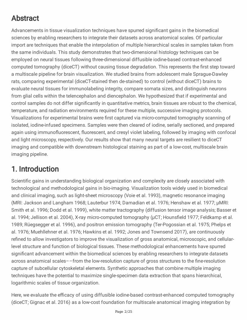
2.5 Digital reconstructionEach diceCT brain was digitally rendered into 3D, using Avizo software running on a Mac Pro computer(Apple Inc.; Cupertino, CA, USA). The slice tool was used to move through the 3D volume in planarsections to match 2D histology images with 3D diceCT anatomy. Digital images of the histology sectionswere virtually placed into anatomical position with their complementary diceCT volume by matchinghomologous neuroanatomical features. Anatomical relationships were veri�ed against rat-brain atlases(Paxinos and Watson 2003; Swanson 2018).
3. Results
3.1 DiceCT imagingDiceCT revealed that soft-tissue traits, even delicate myelinated structures (Gignac and Kley, 2018), couldbe effectively imaged and visualized (Fig. 1). Clear visualization of tissue-level structures relied on severalsteps to ensure imaging noise was minimized, including a post-staining wash in deionized water,moderate X-ray exposure timing during CT scanning, and several-fold multi-frame image averaging incaptured X-ray image processing.
3.2 Histological phenotypes
3.2.1 Medial prefrontal cortex: Parvalbumin, DAPI, andcresyl violet stainingParvalbumin immunoreactivity in mPFC neurons was within previously reported ranges (e.g., Vazquez-Sanroman et al., 2017). Cells showed a morphologically heterogeneous subpopulation of non-pyramidalinterneurons with PRV-positive axons and pericellular basket-like morphologies that enveloped theperikarya of nearby cell bodies (Fig. 2A, B). DiceCT staining did not alter the number of PRV-positiveneurons in comparison to control brain tissue (one-way ANOVA; F1,10 = 8.36, p = 0.89) (Fig. 2E,F). As anadditional histological control, DAPI staining was used to determine the number of nuclei and assessgross cell morphology (Fig. 2A, B). The diceCT pipeline did not modify the appearance of cells stainedwith DAPI, nor surface immunostaining intensity (one-way ANOVA; F1,10 = 5.26, p = 0.96). Somatadiameters in PRV-DAPI-positive cells were similar between post-diceCT and control brain tissues (t = 0.44(F1,14), p = 0.24) (Fig. 2G). Bright�eld light microscopy of cresyl violet-treated post-diceCT sections wassu�cient to reveal cytoarchitectural landmarks (Figs. 1D, 2C). Post-diceCT cresyl violet staining quality inthe mPFC was comparable to that of the post-diceCT cresyl violet-stained DG treatments (see below).

Page 10/25
3.2.2 Mammillary nucleus: DAPI and glial �brillary acidicproteinQualitatively, diceCT preparation, imaging, and de-staining did not modify the appearance of cells stainedwith DAPI. The distribution of GFAP immunoreactive elements in controls was consistent with previousreports (Rahati et al., 2016; Vazquez-Sanroman et al., 2017). Intensely stained, evenly distributedastrocytes with full dendritic body labeling characterized both post-diceCT and control tissues. However,surface densitometry of cell body and astrocyte projections revealed that diceCT-treated brain tissuedecreased GFAP levels compared to controls (one-way ANOVA; F1,8 = 6.33, p < 0.01) (Fig. 3A–C).
3.2.3 Dentate gyrus: cresyl violet and parvalbumin stainingBright�eld light microscopy of cresyl violet-treated experimental sections revealed su�ciently robuststaining to delimit speci�c brain regions and other cytoarchitectural landmarks in the DG. This resultindicates both the successful removal of iodine and the cresyl violet dye's in�ltration into the cell bodiesof the treatment tissues (Fig. 4A). Cresyl violet-stained cells from post-diceCT treatments were notstructurally different as compared to those from control brains. Likewise, cresyl violet stainingsubsequent to diceCT staining, imaging, and de-staining procedures did not alter cell nuclei counts (one-way ANOVA; F1,10 = 18.22, p = 0.66). Experimental tissues, therefore, showed preserved cell morphology,neuronal density, and tissue integrity at high-power magni�cation (Fig. 4A). In addition, the appearance ofPRV immunoreactivity was visible in post-diceCT and control brain tissues (Fig. 4C), and neuronalsomata diameters for experimental treatments did not differ from those of the controls (t = 1.24 (F1,10), p = 0.92) (Fig. 4C,D).
3.2.4 Substantia nigra: DARPP-32We found consistent DARPP-32 expression in post-diceCT and control brains (Fig. 5B). We observedstrong staining in cell bodies and projections of dopaminergic neurons within the SNr (Fig. 5B). DARPP-32 co-localization with DAPI demonstrated nuclear staining in both tissue treatments. We found nosigni�cant difference in somata diameters between experimental and control brain tissues (t = 0.84(F1,14), p = 0.41) (Fig. 5C)
4. DiscussionX-ray imaging of non-mineralized soft tissues is a signi�cant challenge for neuroanatomical studies,causing the brain to be intrinsically di�cult to document through such means. Contrast agents, likeiodine, potentially address this issue by enabling the visualization of soft neuroanatomical features athigh resolutions (Wong et al. 2013; Lautenschlager et al. 2014; Gignac et al. 2016; Parlanti et al., 2017;Naumann and Olsson 2018). However, neural tissues' resilience to multi-faceted imaging pipelines hasnot previously been quanti�ed nor tested explicitly. We developed a µCT-based imaging work�ow that

Page 11/25
validates neural proteins' durability, neuro�laments, neurotransmitters, and nucleic acids after iodinestaining. Our results indicated that a broad array of neural phenotypes were preserved following theiodine stain, imaging, and de-stain process.
Further, we superimposed neuroanatomical datasets to visualize structural diversity for the sameindividual, successfully demonstrating that brain tissues' suitability for a multimodal visualizationpipeline is an important animal model (Fig. 1). DiceCT, therefore, can be used as a readily available, low-cost alternative to µMRI for combined cellular and gross-level imaging modalities in organismal andbiomedical neuroscience applications. Below we discuss the implications of our �ndings, factors thatmay in�uence work�ow products, and the potential that multiscale, same-sample approaches hold foradvancing neuroanatomical research.
4.1 Effects of diceCT on histologyOur approach allowed us to survey tissue integrity, which was maintained throughout the work�ow'sdiceCT and histology steps. DiceCT samples undergo formalin �xation, thermal shifts betweenrefrigerated (4º C), room (25º C), and CT-scanner cabinet (> 25º C) temperatures, weeks in iodine stainwithout refrigeration, exposure to high-powered X-rays, and de-staining using alternating baths of sodiumthiosulfate and deionized water (Gignac et al. 2016). Exposure to and changes between theseenvironments hold the potential to stress and damage brain tissue beyond its capacity to maintainreactivity to histological stains. Instead, we found that numerous cellular targets remained robust toadditional histological stains after implementing diceCT protocols. Tissue sectioning and targetability forimmunohistochemistry with �uorescence, �uorescent labeling, and standard acetate-based cytologicalstaining were maintained after diceCT treatment. This was evidenced by visual and quantitativecon�rmation of su�cient histological labeling following de-staining with sodium thiosulfate. Anti-parvalbumin targets calcium-binding proteins and GABAergic neurons, anti-DARPP-32, which targets D1dopamine receptors, DAPI, which targets nucleic acids; and cresyl violet, which targets RNA, all showedhigh histological �delity in experimental and control tissues. The histological staining agents' bindingwithout color pollution from residual iodine illustrates that iodine was adequately removed from braintissues by the de-staining protocol. The reversibility of diceCT staining is an essential potentialdifferentiator when considering multimodal contrast-enhanced imaging methods (see the survey ofcontrast agents in Gignac et al. 2016) and a key factor that enables the integrated imaging pipelinepresented here. We found it highly valuable to bisect the brains for de-staining, parallel to the plane forhistological sectioning, because this provided an additional surface area for sodium thiosulfate anddeionized water to diffuse, improving the e�ciency of de-staining. By anticipating the plane of thehistological section, we minimized unnecessary physical damage to the samples. We recommend similarapproaches by other workers undertaking diceCT-histology imaging projects.
Our results show a straightforward pattern of histological robusticity of rodent brain tissues and cellularstructures after diceCT. However, we also found that GFAP intensity deviated from the staining mentionedabove outcomes. Anti-GFAP densitometry comparisons quanti�ed differences in stain expression during

Page 12/25
astrocyte staining, and astrocyte intensities illustrated that experimental brains showed signi�cantlyreduced targetability of GFAP (Table 1). Glial �brillary antigen protein expression is modulated in part bylipopolysaccharides (Kang et al. 2019), which are upregulated during astrocyte proliferation. The a�nityof Triiodide for lipids and carbohydrates may contribute to a triiodide-lipopolysaccharide binding thatdepolymerizes GFAP. Consuming GFAP by this process would reduce available targets for anti-GFAP, alsoreducing immunostaining intensity. Notably, this phenomenon is likely more pronounced duringadolescence, when astrocytogenesis is high (Leal et al. 1997; Akdemir et al. 2020), which characterizesour sample. Thus, the GFAP intensity reduction documented here following diceCT staining may be lesspronounced for adult brains. Further evaluation of post-diceCT histology that targets positively chargedmacromolecules is undoubtedly warranted. Regardless of the mechanism, reduced anti-GFAP valuesremained su�cient, even in our adolescent sample, for use as a "phenotyping" marker to distinguish cellsas belonging to glial versus neuronal populations (Table 1; Fig. 3).
The combination of unaffected anti-parvalbumin, anti-DARPP-32, DAPI, and cresyl violet labelingalongside reduced anti-GFAP labeling leads to a broad and preliminary guideline: cellular targets that areeosinophilic appear more likely to be robust to the diceCT staining and de-staining protocols. Eosinophilicneural proteins, dopamine receptors, and nucleic acids, including those we targeted histologically, havenet negative charges (i.e., low isoelectric points; Haiech et al. 1979; Hemmings et al. 1984; Lipfert et al.2014; O'Dowd et al. 2014). This suggests that they repel the negatively charged Triiodide (I3-) moleculesused in diceCT staining. Therefore, Triiodide may fail to bind these substrates, likely contributing to theirpreservation for multimodal staining pipelines using diceCT. This suggests that histology, especiallyimmunohistochemistry, following diceCT should target substrates that are basic, have low isoelectricpoints, or are eosinophilic to produce the best quality results.
We found comparable somata diameter measurements between control and experimental tissues,identifying no signi�cant differences in neuronal size in the mPFC, DG, and SNr. This �nding indicatesthat soft-tissue distortions, such as volumetric shrinkage, may occur due to iodine staining (Vickerton etal. 2013; Gignac et al., 2016) not appear to impact neuron size, at least after de-staining and histologicalpreparation. Likewise, comparable amounts of mPFC-labeled cells between control and experimentalbrains indicate that the addition of diceCT to brain-histology protocols does not impact cell densities.This suggests that any osmotic water loss due to the staining agent's high-salt content during the diceCTphase of the pipeline can be reconstituted during the de-staining process by employing deionized waterbaths. Thus, the diceCT imaging pipeline we articulate here does not demonstrably alter brain tissuesamples' cellular con�guration. Taken together, we anticipate that combining diceCT with histologicalanalyses holds enormous promise as a powerful tool for documenting neuroanatomy across spatialscales and in 3D.
4.2 Considerations for the preservation of nervous tissuesNervous tissues must be rapidly �xed with an appropriate concentration and duration of formaldehyde toconduct histological preparations of the brain (Fox et al. 1985; Hughes et al. 2016b). Consequently, not all

Page 13/25
approaches to preserving samples will be compatible with our pipeline, and brain preservation methodsshould be referenced before imaging attempts. For example, although suitable diceCT images of thebrain have been achieved from ethanol-�xed specimens (e.g., Prötzel et al. 2018), formaldehyde �xationof nervous tissues is considered to be optimal for many steps of histology after diceCT. These includetissue sectioning, immunohistochemical staining, and other cell-based stains (Berod et al. 1981). Toensure that the most neuroanatomical data can be gleaned from a single sample, researchers shouldconsider what type of �xative and method of specimen preservation is required to enable continued useof the tissue during downstream investigations. For example, earlier histology efforts by AMK using post-diceCT rat brain samples did not exhibit reactivity when subjected to a wide variety of stainingtechniques, including immunohistochemistry and cytological-based stains. These prior results wereprimarily attributed to excessive sample storage post-de-staining in formaldehyde. Therefore, we stronglyrecommend avoiding over-�xation in order to achieve the best results. Finally, our study's potentiallimitation is that brain tissues can be distorted from shrinkage due to �xation and over-straining (Gignacand Kley 2018). There are physical methods and mathematical approaches to address these issues(Weisbecker 2012; Taylor et al. 2020); however, some may be incompatible with the multimodal pipelinedescribed here.
5. ConclusionsTo our knowledge, we are the �rst to demonstrate that strong cytoplasmic labeling of neurons with cresylviolet staining and immuno�uorescence after diceCT brain imaging is preserved. The multiscaleexamination of precious biological samples enables an advantageous alternative to single-modalitypipelines. Multiscale, same-sample approaches provide greater detail for anatomical regions than ispossible by using one instrument or method alone. Beyond the conglomerate of staining techniquesdescribed here, we also suggest exploring additional essential cellular markers (e.g., tyrosine hydroxylase,choline acetyltransferase) and transcription factors (e.g., c-Fos) following diceCT, may provide fruitfuladditions to integrated brain-imaging pipelines. We further recommend µCT scanning specimens beforeany contrast enhancement to capture skeletal morphology, such as the braincase or whole skull. Thesemore traditional µCT datasets can be combined with whole-brain diceCT and region-of-interest histologyto study structural anatomy across orders of magnitude in spatial scale that achieves ample coverage ofexternal morphology and internal anatomy—resulting in a cell-to-skull hierarchy of data. Whetherharnessed for model organisms (Cole 1944; Preuss 2000; Keifer and Summers 2016; Khan et al. 2018),translational research (Bonnier et al. 2014; Vazquez-Sanroman et al. 2017; Kim et al. 2019), orcomparative neuroanatomical studies (Yartsev 2017), pipelines such as these hold great promise forunlocking data that can be unintentionally trapped by single-method preparations, especially those thatdo not adequately preserve deep internal soft tissues, such as the brain. To unlock general principles ofnervous system organization across the tree of life (Butler and Hodos 2005; Manger et al. 2008; Carlson2012; Striedter et al. 2014; Brenowitz and Zakon 2015; Russell et al. 2017), highly integrative approaches,as we have demonstrated here, are essential (Schwenk et al. 2009; Dowling et al. 2010; Vogelstein et al.2014).

Page 14/25
DeclarationsAcknowledgments
We owe special thanks to all UTEP Systems Neuroscience Laboratory members, especially Daniel F.Hughes and Arshad M. Khan, for their valuable comments and feedback. We thank M. Hill and the AMNHMicroscopy and Imaging Facility for assistance.
Funding
PMG was supported by NSF (1450850, 1457180, & 1754659) and the American Association for Anatomy.DVS, HDO, and PMG were supported by the OSU-CHS department of Anatomy and Cell Biology. DVS andHDO were also supported by the OSU-CHS O�ce of the Vice President for Research. JS was supported bya pre-doctoral fellowship from the National Mexican Council of Education (CONACyt)
Compliance with ethical standards
Research involving human participants and animals
The Oklahoma State University Institutional Animal Care and Use Committee approved all surgical andtesting procedures and conformed to NIH guidelines.
Con�ict of interest
The authors declare no con�icts of interest.
ReferencesAkdemir ES, Huang A Y-S, Deneen B (2020) Astrocytogenesis: where, when, and how [version 1; peerreview: 2 approved] F1000Research 2020, 9(F1000 Faculty Rev):233.https://doi.org/10.12688/f1000research.22405.1
Alonso JR, Covenas R, Lara J, Aijon J (1990) Distribution of parvalbumin immunoreactivity in the ratseptal area. Brain Res Bull 24:41–48. https://doi.org/10.1016/0361-9230(90)90287-A
Anderson R, Maga AM (2015) A novel procedure for rapid imaging of adult mouse brains with microCTusing iodine-based contrast. PLoS ONE 10:e0142974. https://doi.org/10.1371/journal.pone.0142974
Basser PJ, Mattiello J, LeBihan D (1994) Estimation of the effective self-diffusion tensor form the NMRspin echo. J Mag Res B 103:247–224. https://doi.org/10.1006/jmrb.1994.1037
Berod A, Hartman BK, Pujol JF (1981) Importance of �xation in immunohistochemistry: Use offormaldehyde solutions at variable pH for the localization of tyrosine hydroxylase. J Histoch & Cytoch29:844–850. https://doi.org/10.1177%2F29.7.6167611

Page 15/25
Bonnier G, Roche A, Romascano D et al (2014) Advanced MRI unravels the nature of tissue alterations inearly multiple sclerosis. Ann Clin Transl Neurol 1(6):423–432. https://doi.org/10.1002/acn3.68
Bowler K, Tirri R (1974) The temperature characteristics of synaptic membrane ATPases from immatureand adult rat brain. J Neurochem 23:611–613. https://doi.org/10.1111/j.1471-4159.1974.tb06068.x
Brenowitz EA, Zakon HH (2015) Emerging from the bottleneck: Bene�ts of the comparative approach tomodern neuroscience. Trends Neurosci 38:273–278. https://doi.org/10.1016/j.tins.2015.02.008
Burger FJ, Fuhrman FA (1964) Evidence of injury by heat in mammalian tissues. Am J Physiol 206:1057–1064. https://doi.org/10.1152/ajplegacy.1964.206.5.1057
Burnett TL, McDonald SA, Gholinia A et al (2014) Correlative tomography. Scienti�c Reports 4:4711.https://doi.org/10.1038/srep04711
Butler A B, Hodos W (2005) Comparative Vertebrate Neuroanatomy: Evolution and Adaptation. JohnWiley & Sons. [ISBN: 978-0-471-73384-3]
Caplan J, Niethammer M, Taylor II RM, Czymmek KJ (2011) The power of correlative microscopy:Multimodal, multi-scale, multi-dimensional. Curr Opin Struc Biol 21:686–693.https://doi.org/10.1016/j.sbi.2011.06.010
Carlson BA (2012) Diversity matters: Diversity Matters: The importance of comparative studies and thepotential for synergy between neuroscience and evolutionary biology. Arch Neurol 69: 987–993.https://doi.org/10.1001/archneurol.2012.77
Chen Y, Lin G, Chen Y, Fok A, Slack JM (2012) Micro‐computed tomography for visualizing limb skeletalregeneration in young Xenopus frogs. Anat Rec 295:1562–1565. https://doi.org/10.1002/ar.22496
Cole FJ (1944) A History of Comparative Anatomy from Aristotle to the Eighteenth Century. MacMillan &Co. [ISBN: 9780486602240]
Damadian R, Minkoff L, Goldsmith M, Stanford M, Koutcher J (1976) Field focusing nuclear magneticresonance (FONAR): visualization of a tumor in a live animal. Science 194:1430–1432.https://doi.org/10.1126/science.1006309
de Crespigny A, Bou-Reslan H., Nishimura MC, Phillips H, Carano RA, D'Arceuil HE (2008) 3D micro-CTimaging of the postmortem brain. J Neurosci Meth 171:207–213.https://doi.org/10.1016/j.jneumeth.2008.03.006
Ding SL, Royall JJ, Sunkin SM et al (2016) Comprehensive cellular‐resolution atlas of the adult humanbrain. J Comp Neurol 524:3127–3481. https://doi.org/10.1002/cne.24080

Page 16/25
Dodd SJ, Williams M, Suhan JP, Williams DS, Koretsky AP, Ho C (1999) Detection of single mammaliancells by high-resolution magnetic resonance imaging. Biophys J 76:103-109.https://doi.org/10.1016/S0006-3495(99)77182-1
Dowling AP, Bauchan GR, Ochoa R, Beard JJ (2010) Scanning electron microscopy vouchers andgenomic data from an individual specimen: Maximizing the utility of delicate and rare specimens.Acarologia 50:479–485. https://doi.org/10.1051/acarologia/20101983
Feldkamp LA, Goldstein SA, Par�tt AM, Jesion G, Kleerekoper M (1989) The direct examination of three-dimensional bone architecture in vitro by computed tomography. J Bone Min Res 4:3–11.https://doi.org/10.1002/jbmr.5650040103
Fox CH, Johnson FB, Whiting J, Roller PP (1985) Formaldehyde �xation. J Histochem Cytochem 33: 845–853. https://doi.org/10.1177/33.8.3894502
Gignac PM, Kley NJ (2018) The utility of diceCT imaging for high-throughput comparativeneuroanatomical studies. Brain Behav Evol 91:180–190. https://doi.org/10.1159/000485476
Gignac PM, Kley NJ, Clarke JA (2016) Diffusible iodine-based contrast-enhanced computed tomography(diceCT): An emerging tool for rapid, high-resolution, 3-D imaging of metazoan soft tissues. J Anat228:889–909. https://doi.org/10.1111/joa.12449
Gold S, Cahani M, Sohmer H, Horowitz M, Shahar A (1985) Effects of body temperature elevation onauditory nerve-brain-stem evoked responses and EEGs in rats. Electroencephalogr Clin Neurophysiol 60,146–153. https://doi.org/10.1016/0013-4694(85)90021-5
Gwóźdź B, Dyduch A, Grzybek H, Panz B (1978). Structural changes in brain mitochondria of micesubjected to hyperthermia. Exp Path Bd 15:124–126. https://doi.org/10.1016/S0014-4908(78)80077-5
Gwóźdź B, Krauze M, Dyduch A (1970) Investigations on oxidative phosphorylation in the brain tissue ofanimals subjected to high temperature. Acta Physiol Pol 21: 239–245. [PMID: 5420887]
Haiech J, Derancourt J, Pechère J-F, Demaille JG (1979) Magnesium and calcium binding toparvalbumins: evidence for differences between parvalbumins and an explanation of their relaxingfunction. Biochemistry 19:2752–2758. https://doi.org/10.1021/bi00580a010
Hawkins RA, Hoh C, Glaspy J et al (1992) The role of positron emission tomography in oncology andother whole-body applications. Sem Nucl Med 22:268–284. https://doi.org/10.1016/S0001-2998(05)80121-7
Heimel P, Swiadek NV, Slezak P, Kerbl M, Schneider C, Nürnberger S, Redl H, Teuschle AH Hercher D (2019)Iodine-Enhanced Micro-CT Imaging of Soft Tissue on the Example of Peripheral Nerve Regeneration.Contrast Media Mol Imaging 2019:7483745:1–15. https://doi.org/10.1155/2019/7483745

Page 17/25
Hemmings Jr HC, Greengard P (1986) DARPP-32, a dopamine-regulated phosphoprotein. Prog Brain Res69:149–159. https://doi.org/0.1016/S0079-6123(08)61056-0
Hemmings Jr HC, Nairn AC, Aswad DW, Greengard P (1984) DARPP-32, a dopamine- and adenosine 3':5'-monophosphate-regulated phosphoprotein enriched in dopamine-innervated brain regions. II. Puri�cationand characterization of the phosphoprotein from bovine caudate nucleus. J Neuros 4:99–110.https://doi.org/10.1523/JNEUROSCI.04-01-00099.1984
Herdina AN, Plenk H, Benda P (2015) Correlative 3D-imaging of Pipistrellus penis micromorphology:Validating quantitative microCT images with undecalci�ed serial ground section histomorphology. J.Morph 276:695–706. https://doi.org/10.1002/jmor.20372
Hinshaw WS, Bottomley PA, Holland GN (1977) Radiographic thin-section image of the human wrist bynuclear magnetic resonance. Nature 270:722–723. https://doi.org/10.1038/270722a0
Hopkins TM, Heilman AM, Liggett JA, LaSance K, Little KJ, Hom DB, Minteer DM, Marra KG, Pixley SK(2015) Combining micro-computed tomography with histology to analyze biomedical implants forperipheral nerve repair. J Neurosci Meth 255:122–130. https://doi.org/10.1016/j.jneumeth.2015.08.016
Houns�eld GN (1977) The E.M.I. Scanner. Proc Roy Soc Lond B 195:281–289.https://doi.org/10.1098/rspb.1977.0008
Hughes DF, Gignac PM, Greenbaum E, Khan AM (2016a) Documenting brain diversity in �eld-caughtlizards, from skull to cell: Initial development of a processing pipeline for top down, multi-scale structuralanalyses of a single brain by integrating specialized microcomputed tomography (diceCT), Nissl-basedcytoarchitectonics, and immunohistochemistry. Program No. 778.02. 2016 Neuroscience MeetingPlanner.
Hughes DF, Walker EM, Gignac PM et al (2016b) Rescuing perishable neuroanatomical information froma threatened biodiversity hotspot: Remote �eld methods for brain tissue preservation validated bycytoarchitectonic analysis, immunohistochemistry, and x-ray microcomputed tomography. PLoS ONE11:e0155824. https://doi.org/10.1371/journal.pone.0155824
Jackson JA, Langham WH (1968) Whole-Body NMR Spectrometer. Rev Sci Instrum 39:510–5.13.https://doi.org/10.1063/1.1683420
Jeffery NS, Stephenson RS, Gallagher JA, Jarvis JC, Cox PG (2011) Micro-computed tomography withiodine staining resolves the arrangement of muscle �bres. J Biomech 44:189–192.https://doi.org/10.1016/j.jbiomech.2010.08.027
Jellison BJ, Field AS, Medow J, et al (2004) Diffusion tensor imaging of cerebral white matter: a pictorialreview of physics, �ber tract anatomy, and tumor imaging patterns. Am J Neuroradiol 25:356–369.[PMID: 15037456]

Page 18/25
Jones T, Townsend D (2017) History and future technical innovation in positron emission tomography. JMed Imaging 4:011013. https://doi.org/10.1117/1.JMI.4.1.011013
Kang J-B, Park D-J, Shah M-A, Kim M-O, Koh P-O (2019) Lipopolysaccharide induces neuroglia activationand NF-kB activation in cerebral cortex of adult mice. Lab Anim Res 35:19.https://doi.org/10.1186/s42826-019-0018-9
Karreman MA, Mercier L, Schieber NL et al (2016) Fast and precise targeting of single tumor cells in vivoby multimodal correlative microscopy. J Cell Sci 129:444–456. https://doi.org/10.1242/jcs.181842
Keifer J, Summers CH (2016) Putting the "biology" back into "neurobiology": The strength of diversity inanimal model systems for neuroscience research. Front Sys Neurosci 10:69.https://doi.org/10.3389/fnsys.2016.00069
Khan AM, Grant AH, Martinez A et al (2018) Mapping molecular datasets back to the brain regions theyare extracted from: Remembering the native countries of hypothalamic expatriates and refugees. AdvNeurobiol 21:101–193. https://doi.org/10.1007/978-3-319-94593-4_6
Kim C-B, Park S-J, Jeong J-C (2019) Construction of 3D-rendering imaging of an ischemic rat brain modelusing the planar FMMD technique. Sci Rep 9:19050. https://doi.org/10.1038/s41598-019-55585-x
Kondo H, Fukuda H, Ono H et al (2001) Sodium thiosulfate solution spray for relief of irritation caused byLugol's stain in chromoendoscopy. Gastrointest Endosc 53:199–202.https://doi.org/10.1067/mge.2001.110730
Leal RB, Gonçalves CA, Rodnight R (1997) Calcium-dependent phosphorylation of glial �brillary acidprotein (GFAP) in the rat hippocampus: a comparison of the kinase/phosphatase balance in immatureand mature slices using tryptic phosphopeptide mapping. Dev Brain Res 105:1–10.https://doi.org/10.1016/s0165-3806(97)00113-2
Lautenschlager S, Bright JA, Ray�eld EJ (2014) Digital dissection-using contrast-enhanced computedtomography scanning to elucidate hard-and soft-tissue anatomy in the Common Buzzard Buteo buteo. JAnat 224:412–431. https://doi.org/10.1111/joa.12153
Lauterbur PC (1974) Magnetic resonance zeugmatography. Pure Appl Chem 40:119–157.https://doi.org/10.1351/pac197440010149
Lipfert J, Doniach S, Das R, Herschlag D (2014) Understanding nucleic acid-ion interactions. Annu RevBiochem 83:813–841. https://doi.org/10.1146/annurev-biochem-060409-092720
Manger P, Cort J, Ebrahim N, Goodman A, Henning J, Karolia M, Rodrigues SL, Strkalj G (2008) Is 21stcentury neuroscience too focused on the rat/mouse model of brain function and dysfunction? FrontNeuroanat 2:5. https://doi.org/10.3389/neuro.05.005.2008

Page 19/25
McDonald LW, Hayes TL (1969) Correlation of scanning electron microscope and light microscopeimages of individual cells in human blood and blood clots. Exp Mol Pathol 10:186–198.
Millan N, Murdock LL, Bleier R, Siegel FL (1979) Effects of the acute hyperthermia on polyribosomes, invivo protein synthesis and ornithine decarboxylase activity in the neonatal rat brain. J Neurochem32:311–317.
Mikula, S., Trotts, I., Stone, J., and Jones, E.G. 2007. Internet-Enabled High-Resolution Brain Mapping andVirtual Microscopy. NeuroImage. 35(1):9-15
Morales AG, Stempinski ES, Xiao X, Patel A, Panna A, Olivier KN, McShane PJ, Robinson C, George AJ,Donahue DR, Chen P (2016) Micro‐CT scouting for transmission electron microscopy of human tissuespecimens. J of Microsc 263:113–117.
Muehllehner G, Buchin MP, Dudek JH (1976) Performance parameters of a positron imaging camera. IEEET Nucl Sci 23:528–537. https://doi.org/10.1109/TNS.1976.4328300
Naumann A, Olsson L (2018) Three‐dimensional reconstruction of the cranial and anterior spinal nervesin early tadpoles of Xenopus laevis (Pipidae, Anura). J Comp Neurol 526:836–857.
O'Dowd BF, Ji X, Nguyen T, George S R (2012) Two amino acids in each of D1 and D2 dopamine receptorcytoplasmic regions are involved in D1-D2 heteromer formation. Biochem Biophys Res Commun417(1):23–28. https://doi.org/10.1016/j.bbrc.2011.11.027
Parlanti P, Cappello V, Brun F, Tromba G, Rigolio R, Tonazzini I, Cecchini M, Piazza V, Gemmi M (2017) Sizeand specimen-dependent strategy for x-ray micro-CT and TEM correlative analysis of nervous systemsamples. Sci Rep 7:2858.
Paxinos G, Watson C (2003) The Rat Brain in Stereotaxic Coordinates, 7th edition. Elsevier. [ISBN:9780123919496]
Pelc NJ (2014). Recent and future directions in CT imaging. Ann Biomed Eng 42(2):260–268.https://doi.org/10.1007/s10439-014-0974-z
Phelps ME, Hoffman EJ, Mullani NA, Higgins CS, Pogossian MMT (1976) Design considerations for apositron emission transaxial tomograph (PETT III). IEEE Trans Nuc Sci 23:516–522.https://doi.org/10.1109/TNS.1976.4328298
Preuss TM (2000) Taking the measure of diversity: Comparative alternatives to the model-animalparadigm in cortical neuroscience. Brain, Behavior and Evolution 55:287–299.
Prötzel D, Vences M, Hawlitschek O, Scherz MD, Ratsoavina FM, Glaw F (2018) Endangered beauties:Micro-CT cranial osteology, molecular genetics and external morphology reveal three new species of

Page 20/25
chameleons in the Calumma boettgeri complex (Squamata: Chamaeleonidae). Zool J Linn Soc 184:471–498.
Rahati M, Nozari M, Eslami H, Shabani M, Basiri M (2016) Effects of enriched environment on alterationsin the prefrontal cortex GFAP- and S100B-immunopositive astrocytes and behavioral de�cits in MK-801-treated rats. Neuroscience 326:105–116. https://doi.org/10.1016/j.neuroscience.2016.03.065
Rüegsegger P, Koller B, Müller R (1996) A microtomographic system for the nondestructive evaluation ofbone architecture. Calci�ed Tissue Int 58:24–29. https://doi.org/10.1007/bf02509542
Russell JJ, Theriot JA, Sood P, Marshall WF, Landweber LF, Fritz-Laylin L, Polka JK, Oliferenko S, GerbichT, Gladfelter A, Umen, J (2017) Non-model model organisms. BMC Biol 15:55.
Schwenk K, Padilla DK, Bakken GS, Full RJ (2009) Grand challenges in organismal biology. Integr CompBiol 49:7–14. https://doi.org/10.1093/icb/icp034.
Sengle G, Tufa SF, Sakai LY, Zulliger MA, Keene DR (2013) A correlative method for imaging identicalregions of samples by micro-CT, light microscopy, and electron microscopy: Imaging adipose tissue in amodel system. J Histochem Cytochem 61:263–271.
Shakhashiri B (1983) Chemical Demonstrations: A Handbook for Teachers of Chemistry, Vol. 3. Madison:University of Wisconsin Press.
Shami GJ, Cheng D, Braet F (2017) Combined multidimensional microscopy as a histopathology imagingtool. J Cell Physiol 232:249–256.
Smith BR, Linney E, Huff DS, Johnson GA (1996) Magnetic resonance microscopy of embryos. ComputMed Imag Grap. 20:483–490. https://doi.org/10.1016/S0895-6111(96)00046-8
Striedter GF et al (2014) NSF workshop report: Discovering general principles of nervous systemorganization by comparing brain maps across species. J Comp Neurol 522:1445–1453.
Swanson, LW (2018) Brain maps 4.0—Structure of the rat brain: An open access atlas with global nervoussystem nomenclature ontology and �atmaps. J Comp Neurol 526:935–943.
Taylor MS, O'Brien HD, Gignac PM (2020). Shrinkage after swimming in iodine? Evaluating the use ofhydrogel stabilization for reinforcing nervous tissues before iodine diffusion. Society for Integrative andComparative Biology Abstracts 122-2.
Ter-Pogossian MM, Phelps ME, Hoffman EJ, Mullani NA (1975) A Positron-emission transaxialtomography for nuclear imaging (PETT). Radiology 114:89–98. https://doi.org/10.1148/114.1.89
Trevorrow V, Fashena G (1935) The determination of iodine in biological material. J Biol Chem 110:29–38.

Page 21/25
Vazquez‐Sanroman DB, Monje RD, Bardo MT (2017) Nicotine self‐administration remodels perineuronalnets in ventral tegmental area and orbitofrontal cortex in adult male rats. Addict Biol 22:1743–1755.
Vickerton P, Jarvis J, Jeffery N (2013) Concentration-dependent specimen shrinkage in iodine-enhancedmicroCT. J Anat 223:185–193. http://doi.org/10.1111/joa.12068
Voie AH, Burns DH, Spelman FA (1993) Orthogonal-plane �uorescence optical sectioning: Three-dimensional imaging of macroscope biological specimens. J Microsc 170:229–236.https://doi.org/10.1111/j.1365-2818.1993.tb03346.x
Vogelstein JT, Park Y, Ohyama T, Kerr RA, Truman JW, Priebe CE, Zlatic M (2014) Discovery of brainwideneural-behavioral maps via multiscale unsupervised structure learning. Science 344:386–392.
Weisbecker V (2012) Distortion in formalin-�xed brains: Using geometric morphometrics to quantify theworst-case scenario in mice. Brain Struct Funct 217:677–685.
Wong MD, Spring S, Henkelman RM (2013) Structural stabilization of tissue for embryo phenotypingusing micro-CT with iodine staining. PLoS ONE 8:e84321.
Yartsev MM (2017) The emperor's new wardrobe: Rebalancing diversity of animal models in neuroscienceresearch. Science 358:466–469.
TableTable 1. Significance values for ANOVA and t-tests of control and post-diceCT samples stained using anti-Parvalbumin, anti-GFAP,
DAPI, anti-DARPP-32, and cresyl violet with target neural phenotype indicated. Significance (𝛼 < 0.05; indicated by asterisk) was
identified for anti-GFAP densitometry measurements only. Somata diameters, cell counts, and other fluorescent intensity
comparisons were not significantly different.
Histological Prep Neural Phenotype Significance
anti-Parvalbumin GABAergic interneurons mPFC: p = 0.89 ; DG: p = 0.92
anti-GFAP Glial cell intermediate filament proteins MM: p < 0.01*
DAPI Nucleic acids mPFC: p = 0.96
anti-DARPP-32 Phosphoprotein in D1 receptor neurons SNr: p = 0.41
cresyl violet RNA DG: p = 0.66
Figures

Page 22/25
Figure 1
2D images of an adolescent rat brain imaged using diceCT in (A) parasagittal, (B, E) coronal, and (C)transverse views alongside a (D) 3D projection that has been digitally dissected to show the mPFC andDG. In (D), 2D cresyl violet histological images from the mPFC and DG overlay their positions in the 3Dmodel to illustrate the integration of multiscale bio-imaging approaches. Neuroanatomical comparison(E) of diceCT (left) and cresyl violet histology (right) with regions of interest labeled. Cresyl violet imagedownloaded from BrainMaps.org (Mikula et al., 2007). Abbreviations: BST, bed nuclei of stria terminalis[Johnston]; fx, column of the fornix; GPe, globus pallidus; LHA, lateral hypothalamic area; LPO, lateralpreoptic area; MPN, medial preoptic nucleus; PVH, paraventricular nucleus hypothalamus; PVT,paraventricular nucleus thalamus; SI, substantia innominate [Reil, Reichert]; sm, stria medullaris; and V3h,third ventricle. Nomenclature following Swanson, 2018

Page 23/25
Figure 2
(A, B) Immunostained and (C) cresyl violet stained mPFC (scale bar is 20 μm). PRVimmunoreactivity(green), DAPI labeling (blue), and their co-visualization (merge) in mPFC from (A) post-diceCT and (B) control subjects; (D) sampled mPFC areas (red) [Paxinos and Watson, 2003]. (E) Mean(±SEM) number of PRV immunoreactive cells for post-diceCT and control mPFC per mm2. (F) Mean

Page 24/25
(±SEM) number of DAPI-positive cells for post-diceCT and control mPFC per mm2. (G) Mean (±SEM) celldiameter of DAPI-positive cells for post-diceCT and control mPFC.
Figure 3
GFAP-immunoreactive (green) and DAPI-positive (blue) cells in (A) post-diceCT and (B) and control MM.Scale bar is 20 μm. (C) Mean (±SEM) attenuation units (a.u.) of GFAP-positive cells for post-diceCT andcontrol MM; (D) sampled MM area (red) [Paxinos and Watson, 2003].

Page 25/25
Figure 4
(A) cresyl violet-stained DG, insets mark regions magni�ed (4–100x; scale bars: 600, 250, 60, 25 μm) insuccessive panels from left to right. (B) Sampled DG area (red) [Paxinos and Watson, 2003]. (C) PRVimmunoreactivity (green) and DAPI-positive (blue) cells in post-diceCT and control DG. (D) Mean (±SEM)cell diameter of DAPI-positive cells for post-diceCT and control DG.
Figure 5
(A) Sampled area a SNr (red) [Paxinos and Watson, 2003]; (B) DARPP-32-immunoreactive (green) andDAPI-positive (blue) cells in post-diceCT and control SN. (C) Mean (±SEM) cell diameter of DAPI-positivecells for post-diceCT and control SNr.