Embed Size (px)
Citation preview
at SciVerse ScienceDirect
International Biodeterioration & Biodegradation xxx (2012) 1e13
Contents lists available
International Biodeterioration & Biodegradation
journal homepage: www.elsevier .com/locate/ ibiod
Microscopic, chemical, and molecular-biological investigation of the decayedmedieval stained window glasses of two Catalonian churches
Guadalupe Piñar a,*, Maite Garcia-Valles b, Domingo Gimeno-Torrente b, Jose Luis Fernandez-Turiel c,Jörg Ettenauer a, Katja Sterflinger a
a Institute of Applied Microbiology, Department of Biotechnology, Vienna Institute of Bio Technology (VIBT), University of Natural Resources and Life Sciences, Muthgasse 11,A-1190 Vienna, Austriab Facultat de Geologia, Universitat de Barcelona (UB), c/ Martí i Franquès s/n, 08028 Barcelona, Spainc Institut de Ciències de la Terra Jaume Almera, CSIC, c/ Solé i Sabarís s/n, 08028 Barcelona, Spain
a r t i c l e i n f o
Article history:Received 28 November 2011Received in revised form21 February 2012Accepted 22 February 2012Available online xxx
Keywords:Stained glassesBiodeteriorationBio-pittingPatinasMineral precipitationMicrobial communitiesMolecular methods
* Corresponding author. Tel.: þ43 1 47654 6943; faE-mail address: [email protected] (G. Pi
0964-8305/$ e see front matter � 2012 Elsevier Ltd.doi:10.1016/j.ibiod.2012.02.008
Please cite this article in press as: Piñar, G., ewindow glasses of two Catalonian churches
a b s t r a c t
We investigated the decayed historical church window glasses of two Catalonian churches, both underMediterranean climate. Glass surfaces were studied by scanning electron microscopy (SEM), energydispersive spectrometry (EDS), and X-ray diffraction (XRD). Their chemical composition was determinedby wavelength-dispersive spectrometry (WDS) microprobe analysis. The biodiversity was investigated bymolecular methods: DNA extraction from glass, amplification by PCR targeting the16S rRNA and ITSregions, and fingerprint analyses by denaturing gradient gel electrophoresis (DGGE). Clone librariescontaining either PCR fragments of the bacterial 16S rDNA or the fungal ITS regions were screened byDGGE. Clone inserts were sequenced and compared with the EMBL database. Similarity values rangedfrom 89 to 100% to known bacteria and fungi. Biological activity in both sites was evidenced in the formof orange patinas, bio-pitting, and mineral precipitation. Analyses revealed complex bacterial commu-nities consisting of members of the phyla Proteobacteria, Bacteroidetes, Firmicutes, and Actinobacteria.Fungi showed less diversity than bacteria, and species of the genera Cladosporium and Phoma weredominant. The detected Actinobacteria and fungi may be responsible for the observed bio-pittingphenomenon. Moreover, some of the detected bacteria are known for their mineral precipitationcapabilities. Sequence results also showed similarities with bacteria commonly found on deterioratedstone monuments, supporting the idea that medieval stained glass biodeterioration in the Mediterraneanarea shows a pattern comparable to that on stone.
� 2012 Elsevier Ltd. All rights reserved.
1. Introduction
Medieval stained glass windows are part of our cultural heri-tage, but due to their permanent exposure to environmentalconditions they have been damaged over centuries. To protect andconserve this valuable material, it is necessary to understand thelong-term environmental corrosion processes on glass. Stainedglass is made up of several components: network formers, stabi-lizers, modifiers, and coloring elements (Römich, 1999). The mainnetwork formers used in medieval stained glass were silica andphosphorus. In addition, several metals, such as Cu, Co, and Mn,were used to color the glass (Bamford, 1977; Newton and Davison,1989). Nevertheless, if we consider the main chemical elements,historic glass can be classified into two types: K-rich and Na-rich
x: þ43 1 47654 6675.ñar).
All rights reserved.
t al., Microscopic, chemical, an, International Biodeteriorati
glass (Newton and Fuchs, 1988; Brill, 1999). Most Central Euro-pean medieval stained glass produced between the 12th and 15thcenturies was K-rich in composition. However, the coeval EuropeanMediterranean glass shows the continuation of a Roman-like Na-rich glassmaking tradition. A common feature of medieval stainedglass is the presence of corrosion, patina development, and mineralcrust growth over the glass, which has caused serious damage tomany Central European stained glass windows. For this reason,studies on glass decay have become important in several countries.Most of these studies consider that glass corrosion and decay arerelated mainly to a physicochemical process (Newton and Davison,1989; Schreiner, 1991). Nevertheless, since the beginning of the20th century there has been evidence of biological induction instained glass decay (Krumbein et al., 1995). Other important factorsthat enhance corrosion are environmental pollution (i.e., CO2 andSOx in urban areas, where most stained glass windows are located)and, in the case of biodeterioration, the presence of organic carbonon the glass.
dmolecular-biological investigation of the decayed medieval stainedon & Biodegradation (2012), doi:10.1016/j.ibiod.2012.02.008
G. Piñar et al. / International Biodeterioration & Biodegradation xxx (2012) 1e132
The result of glass decay is a sharp decrease of the flux andnetwork modifiers on the surface and contiguous mass of glass(leaching). This leads to the genesis of a gel surface (or planarvolume) of the glass depleted in practically all glass componentsexcept network formers (Newton and Davison, 1989; Sterpenichand Libourel, 2001; Garcia-Valles et al., 2003). The leachedelements may combine with other (i.e., atmospheric) componentsto form complex salts. The most soluble salts are removed bymoisture and rain, but the others remain on the glass surface asmineral products forming patinas and crusts, as sulfates (gypsumand syngenite), calcite, Ca-oxalates, etc. In KeCa-rich medievalglasses, when the surface pH might reach a level greater than 9,advanced corrosion of the outer level of silica-rich glass occurs(Garcia-Valles et al., 2003).
As mentioned above, nowadays biological corrosion of glass isa well-known phenomenon. Glass biodeterioration is the result ofmetabolic activities of complex microbial communities composedmainly of fungi (Nagamuttu, 1967; Kaiser et al., 1996; Schabereiter-Gurtner et al., 2001b), bacteria (Rölleke et al., 1999; Marvasi et al.,2009), and lichens (Mellor, 1924). The organic residues present onhistorical glass, as dead microbial material, metabolites of auto-trophic bacteria, animal faeces, and dust deposits, promote themicrobial growth. The role of microorganisms in glass decayincludes both chemical and mechanical destruction of glass. Themycelia of filamentous fungi and Actinobacteria initiate botha mechanical destruction and the creation of a leaching environ-ment by the adsorption of water, enhancing the chemicaldestruction of glasses. Furthermore, the production of metabolicproducts, as organic and inorganic acids, can lead to pH changes,redox-reactions leaching and chelation of special glass compo-nents. In summary, microbial growth on glass surfaces producesseveral types of damage, such as bio-pitting corrosion, cracks, andpatina formation (Krumbein et al., 1991; Drewello and Weissmann,1997).
Investigations of microbial colonization of historical glass haveso far been based on culture-dependent methods (Krumbein et al.,1991; Drewello and Weissmann, 1997), with only the exceptionsusing culture-independent techniques (Rölleke et al., 1999;Schabereiter-Gurtner et al., 2001b; Carmona et al., 2006). Indetermining the appropriate measures to take against microbialgrowth on historical glass, it is important to get an overview of theinhabiting microbial populations. However, the first problem foundwhenworking with samples taken from cultural assets is the smallquantity of sample material available, which is in most cases notsufficient for reliable cultivation assays. Furthermore, microbes areusually members of complex microbial communities and dependon special nutrients; therefore only a minority of them can becultivated under conventional laboratory conditions. By contrast,cultivation-independent methods enable the detection of slowlygrowing, fastidious, or uncultivable microorganisms, allowinga more complete picture of the inhabiting microbial communitiesthan do traditional cultivation techniques (Schabereiter-Gurtneret al., 2001a).
The objective of this study was the chemical and biologicalcharacterization of the stained window glasses showing signs ofbiodeterioration of two Catalonian churches. To this end, glasssurfaces were investigated using scanning electron microscopy(SEM), energy dispersive spectrometry (EDS), and X-ray diffraction(XRD). In addition, the chemical glass compositionwas investigatedusing wavelength-dispersive spectrometry (WDS) microprobeanalysis.
The biodiversity of themicro-biota associatedwith the observeddecay was investigated by the following molecular methods: DNAextraction from glass samples, amplification by PCR targetingthe16S rRNA and ITS regions, and DNA fingerprint analyses by
Please cite this article in press as: Piñar, G., et al., Microscopic, chemical, anwindow glasses of two Catalonian churches, International Biodeteriorati
denaturing gradient gel electrophoresis (DGGE). In parallel clonelibraries containing either PCR fragments of the 16S rDNA or the ITSregions were screened by DGGE and selected clone inserts weresequenced and compared with the EMBL database.
2. Materials and methods
2.1. Sampling
Glass samples were obtained from different restoration andconservation projects in two Mediterranean coastal cities ofnortheastern Spain, Tarragona and Barcelona. This area hasa Mediterranean climate with rainfalls mainly concentrated inspring and fall (around 580 mm yr�1) and is characterized bymoderate weather, warm summers (21e30 �C), and mild sunnywinters (6e14 �C). Both buildings are located ca. 150 m from themarine shoreline.
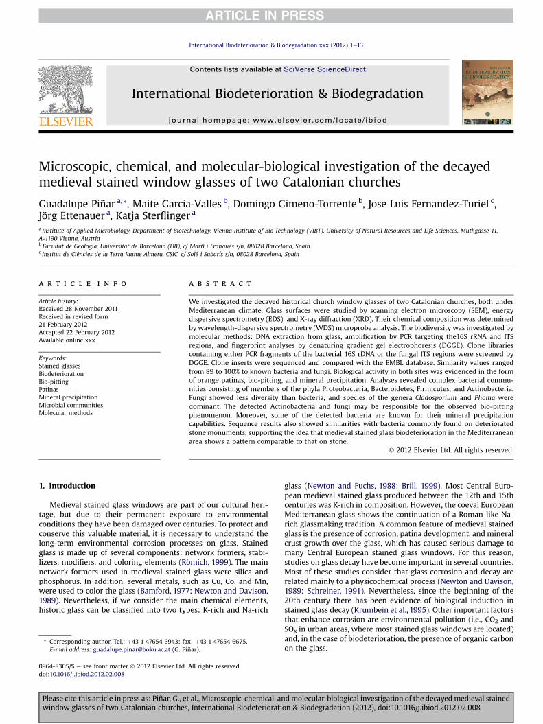
The rosette glasses of the transept from the Cathedral of Tarra-gona (Fig. 1) date back to the beginning of the 14th century. Theyshow evidence of damage and repair after theWar of Independence(1808e1812) as well. All stained glass windows show originalblack-fired draws (grisailles), but our present study is only con-cerned with the composition and corrosion of the main pieces ofglass.
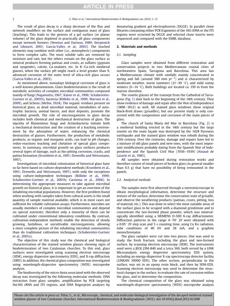
The church of Santa Maria del Mar in Barcelona (Fig. 2) isa medieval building erected in the 14th century but the largerosette on the main façade was destroyed by the 1428 Pyreneesearthquake and the stained glass window was rebuilt during the15th century. Over the centuries, damage and repairs have led toa mixture of old glass panels and new ones, with the most impor-tant modifications probably dating from the Spanish War of Inde-pendence and the Spanish Civil War (1936e1939) (Ainaud deLasarte et al., 1985).
All samples were obtained during restoration works andtherefore consist of small pieces of broken glass (in general smallerthan 0.5 g) that have no possibility of being remounted in thepanels.
2.2. Analytical methods
The samples were first observed through a stereomicroscope toobtain morphological information, determine the structure andtexture of the surface, determine the conservation state of grisaille,and observe the weathering products (patinas, crusts, pitting, lossof material, etc.). This was done to select the most suitable areas ofthe surface glass to be scraped with a diamond grindstone and toconcentrate the neo-formed phase powder, which was mineral-ogically identified using a SIEMENS D-500 X-ray diffractometer.Diffraction patterns in the range 4e70� 2q were obtained witha 0.05� 2q step scan and 5 s counting time, using Cu Ka radiation,tube conditions of 40 kV and 28 mA, and a graphitemonochromator.
The glass samples were cut into two pieces. One was used tostudy the fresh fracture, including the glass and neo-formedsurface, by scanning electron microscopy (SEM). The instrumentsused were a JEOL J3M-840 and a Leica 360, both served by a LINKMicroanalysis energy dispersive spectrometry EDS system,including an energy-dispersive X-ray spectroscopy detector facility(LINKAN 10000 EDS). The other section, perpendicular to thesurface, was set in an epoxy resin block, and then by SEM-EDS.Scanning electron microscopy was used to determine the struc-tural changes in the surface, to evaluate the rate of corrosionwithinthe glass, and to determine the composition.
The chemical composition of the glass was obtained usingwavelength-dispersive spectrometry (WDS) microprobe analysis
dmolecular-biological investigation of the decayedmedieval stainedon & Biodegradation (2012), doi:10.1016/j.ibiod.2012.02.008
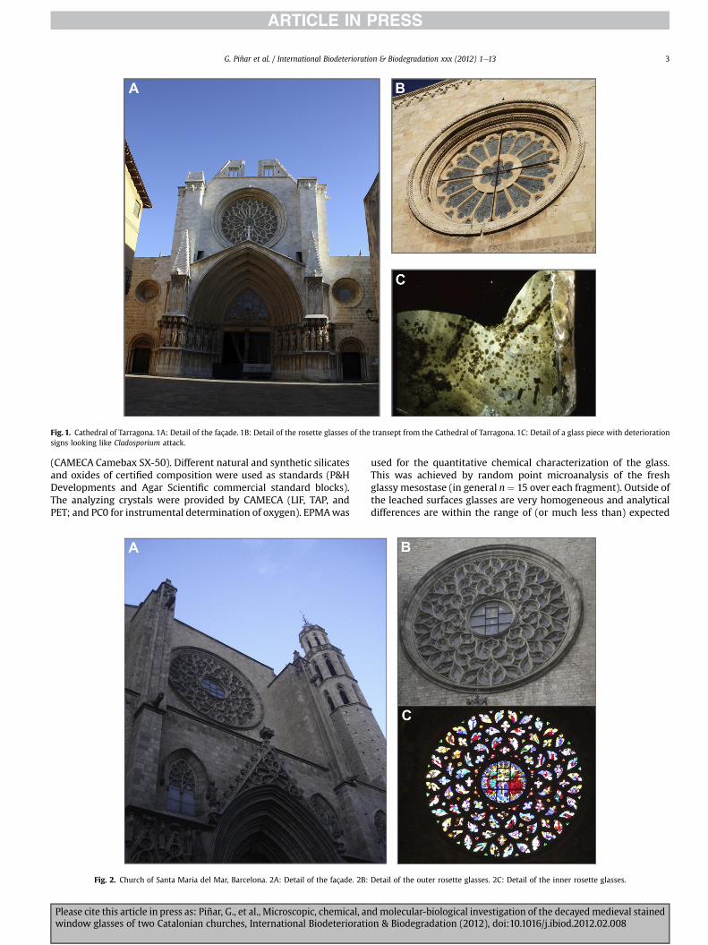
Fig. 1. Cathedral of Tarragona. 1A: Detail of the façade. 1B: Detail of the rosette glasses of the transept from the Cathedral of Tarragona. 1C: Detail of a glass piece with deteriorationsigns looking like Cladosporium attack.
G. Piñar et al. / International Biodeterioration & Biodegradation xxx (2012) 1e13 3
(CAMECA Camebax SX-50). Different natural and synthetic silicatesand oxides of certified composition were used as standards (P&HDevelopments and Agar Scientific commercial standard blocks).The analyzing crystals were provided by CAMECA (LIF, TAP, andPET; and PC0 for instrumental determination of oxygen). EPMAwas
Fig. 2. Church of Santa Maria del Mar, Barcelona. 2A: Detail of the façade. 2B:
Please cite this article in press as: Piñar, G., et al., Microscopic, chemical, anwindow glasses of two Catalonian churches, International Biodeteriorati
used for the quantitative chemical characterization of the glass.This was achieved by random point microanalysis of the freshglassy mesostase (in general n¼ 15 over each fragment). Outside ofthe leached surfaces glasses are very homogeneous and analyticaldifferences are within the range of (or much less than) expected
Detail of the outer rosette glasses. 2C: Detail of the inner rosette glasses.
d molecular-biological investigation of the decayed medieval stainedon & Biodegradation (2012), doi:10.1016/j.ibiod.2012.02.008
Fig. 3. Patina, leaching surface, and bio-pitting observed on glass samples. 3A: Macroscopic pictures corresponding to two Tarragona glass samples. Left image shows silica-leachedsurface directly in contact with glass and before patina (orange at bottom and more dark at top) developed in sample T-5. Right image shows detail of patina and bio-pitting
G. Piñar et al. / International Biodeterioration & Biodegradation xxx (2012) 1e134
Please cite this article in press as: Piñar, G., et al., Microscopic, chemical, and molecular-biological investigation of the decayedmedieval stainedwindow glasses of two Catalonian churches, International Biodeterioration & Biodegradation (2012), doi:10.1016/j.ibiod.2012.02.008
G. Piñar et al. / International Biodeterioration & Biodegradation xxx (2012) 1e13 5
instrumental error. Zoned glasses (i.e., red glasses consisting ofa deep red plate sandwiched between white glass) were alsostudied by acquisition of a profile of points orthogonal to the planaroptical anisotropies. The analytical accuracy and precision was alsocontrolled by means of internal standards (Brill, 1999).
2.3. Molecular analysis
2.3.1. DNA extraction from glass samplesExtraction of DNA was performed directly from the glass
samples using the method previously described by Sert andSterflinger (2010) with the following modifications: Prior to lysis,a piece of glass (0.2e0.25 g) was triturated with a mortar and theobtained powder, together with 500 ml lysing buffer, was added tothe tubes of the lysing matrix E (MP Biomedicals). The mixture wasshaken in a cell disrupter (Thermo Savant FastPrep, FP120, Hol-brook, USA) at full speed for 40 s, and incubated for 1 h at 65 �C.Afterward, the mixture was shaken again at full speed for 40 s andthen centrifuged for 10 min at 10,000 g. The supernatant wastransferred to a new Eppendorf tube and an approximately equalvolume of chloroform/isoamyl alcohol was added, mixed thor-oughly, and centrifuged for 5 min in a microcentrifuge. This stepwas repeated using phenol/chloroform/isoamyl alcohol. Thesupernatant was transferred to a new Eppendorf tube and furtherpurified using the QIAamp Viral RNA mini kit (Qiagen, Hilden,Germany) following the instructions of the manufacturer. The finalelution step was repeated twice with 100 ml of 80 �C preheatedddH2O (Sigma Aldrich, St. Louis, USA). The purified DNA was useddirectly for PCR amplification.
2.3.2. PCR amplification of extracted DNAFor the analysis of fungal sequences, fragments of about 700 bp
in size corresponding to the ITS1, the ITS2 region, and the adjacent5.8S rRNA gene, were amplified with the primer pair ITS1 and ITS4(White et al., 1990). For DGGE analysis, a nested PCRwas performedwith the PCR product of the first round as template DNA using theprimers ITS1GC with a 37-base GC clamp attached to the 50 end(Muyzer et al., 1993) and ITS2. All reactions were carried out asdescribed in Michaelsen et al. (2006).
For the identification of bacterial 16S rRNA sequences, DNA wasamplified with the primer pair 341f/907r (Muyzer et al., 1993;Teske et al., 1996). For DGGE analysis, 200-bp fragments of the 16SrDNA were amplified with a nested PCR using the eubacterialspecific primer 341f-GC with a 40-bp GC clamp added to its 50 end(Muyzer et al., 1993) and the universal consensus primer 518r(Neefs et al., 1990). PCR conditions were as described bySchabereiter-Gurtner et al. (2001a). All PCR products were analyzedby electrophoresis in a 2% (w/v) agarose gel.
2.3.3. Denaturing gradient gel electrophoresis (DGGE)The DGGEwas performed as previously described (Muyzer et al.,
1993) using a D-Code system (BioRad) in �0.5 TAE (20 mM Tris,10 mM acetate, 0.5 mM Na2 EDTA; pH7.8 with 8% (w/v) acryl-amide). Gels were run at a constant temperature of 60 �C witha voltage of 200 V during 3.5 h and 6 h, respectively, for bacterialand fungal fingerprints. The linear chemical gradient of
developed in red glass plaqué in sample T-6. 3B: SEM analyses on sample T-6 show the degrered plaqué glass (sample T-35). Glass and patina have been affected by biological activity obssurface corresponding to sample STM-16 from Santa Maria del Mar. The right image shows cOccasionally there is an alignment perpendicular to this main direction. The left image, obsepit penetration in the green glass zone. This penetration was short, due to the toxicity ofpictures of K-glass from Tarragona (T-5) showing advanced glass decay caused by bio-pittincolour in this figure legend, the reader is referred to the web version of this article.)
Please cite this article in press as: Piñar, G., et al., Microscopic, chemical, anwindow glasses of two Catalonian churches, International Biodeteriorati
denaturants used in this study [100% denaturing solution contains7 M urea and 40% (v/v) formamide] are indicated in the figures.
After completion of electrophoresis, gels were stained ina 1 mg ml�1 ethidium bromide solution (stock: 10 mg ml�1) for20 min and afterward visualized by a UVP documentation system(BioRad Transilluminator, Universal Hood; Mitsubishi P93D-printer).
2.3.4. Creation of clone libraries and sequence analysisTo obtain a detailed phylogenetic identification of the microbial
community members, clone libraries containing either ITS fungalregions (fungal community) or 16S rRNA fragments (bacterialcommunity) were carried out. For fungal clone libraries, the DNAtemplate was amplified using the primers ITS1/ITS4 as mentionedabove. For bacterial clone libraries, the primer pair 341f/907r wasused asmentioned above. The PCR products were purified using theQIAquick PCR Purification Kit Protocol (Qiagen, Hilden, Germany)and resuspended in ddH2O water.
Purified PCR products were ligated into the pGEM-T easy Vectorsystem (Promega, Vienna, Austria) following the instructions of themanufacturer. The ligation products were transformed into One-shot TOP10 cells (Invitrogen). These cells allow the identificationof recombinants (white colonies) on an indicator LB medium con-taining ampicillin (100 mg ml�1), streptomycin (25 mg ml�1), and X-Gal (5-bromo-4-chloro-3-indolyl-ß-1-galactopyranoside; 0.1 mM)(Sambrook et al., 1989).
Clones were screened in a DGGE gel and sequenced as describedby Schabereiter-Gurtner et al. (2001a). Comparative sequenceanalysis was performed by comparing pair-wise insert sequenceswith those available in the public online database NCBI using theBLAST search program (Altschul et al., 1997). The resultingsequences of the bacterial and fungal clones have been deposited atGenBank: Genetic sequence database at the National Center forBiotechnical Information (NCIB) (GenBank ID: BA123456)(Tables 3and 4).
3. Results
3.1. Patinas, leached surface, and glass characterization
Stereomicroscope observations showed different situations oneach glass sample. All glass samples, except T-28 (Cathedral ofTarragona) and STM-13 (church of Santa Maria del Mar), werecovered by discontinuous orange and/or beige patina (Figs. 1C and3A and C). In addition, glasses presented traces of biological activityas bio-pitting (Fig. 3A and B), isolate micro-cracks and someinterconnected micro-cracks developed in glass, in patina, in oldpaints, or in grisaille (Fig. 3C).
Some samples showed a leached surface (Figs. 1C and 3A left)and also evidence of a gel with a silica composition, as determinedby SEM. The more external part of the patina showed to be orangeand the bottom beige (Fig. 3A, left). Occasionally, the patina wasformed in glass surfaces previously attacked by microorganisms(Fig. 3A), producing a more or less penetrating pitting into the glass(Fig. 3B). This destroyed surface can also be filled (constructiveactivity) with authigenic minerals (patina).
e of penetration of this activity into the glass. 3C: Orange and beige patina developed inerved as small pits. The SEM picture shows the pit evolution to inner part. 3D: Decayedircles isolated or one after the other forming a row arranged in a preferential direction.rved with microscopy in the thin section perpendicular to the glass surface, shows thethe saturated copper, chromophore element found in this glass (3.71% CuO). 3e: SEMg; note the curved-branched irregular pitting. (For interpretation of the references to
d molecular-biological investigation of the decayed medieval stainedon & Biodegradation (2012), doi:10.1016/j.ibiod.2012.02.008
G. Piñar et al. / International Biodeterioration & Biodegradation xxx (2012) 1e136
SEM observations of the glass corroborated the morphology andevolution of pits (Fig. 3B and D), and the advanced glass decay.Fig. 3D shows initial development of curved-branched irregularpitting not directly related to cracking surface. Each pit appears asan incipient nodule-like glass decay form overprinted on thecurved branch of pit progression. These branches of pits tend tocoalesce and provide a deeply penetrated upper film of the glass,sometimes associated with an external surface patina. We couldalso observe that in plaqué glass the bio-pitting stopped when themicroorganisms arrived at the colored layers, which contain heavymetals e e.g., copper e that are noxious to microorganisms(Fig. 3C). The diameter of the pit holes decreases from the external(50 mm) to the internal part (20 mm, Fig. 3C). This activity was alsoobserved in the K-rich glass STM-16 (Fig. 3D). Fig. 3E showsa macroscopic photograph with a bio-pitting arrangement made inthe glass surface, and SEM shows the detail of the pit hole sectionrelated to organic activity.
The mineralogical composition of patinas, determined by XRDand corroborated by SEM-EDAX analyses, revealed the presence ofgypsum (CaSO4$2H2O), bixbyite (Mn2O3), syngenite [K2Ca(SO4)2$H2O], thenardite [(a-Na2SO4)], calcite (CaCO3), and quartz(SiO2), as well as the presence of clay minerals and Ca-oxalate(weddellite, CaC2O4$2H2O, and whewellite, CaC2O4$H2O), deter-mined in orange patina.
The chemical composition of the investigated samples obtainedfrom the Cathedral of Tarragona and the church of Santa Maria delMar are showed in Tables 1and 2, respectively. In all samples thevitrifying compound was SiO2. Depending on the flushingcompound type used, K2O or Na2O, we distinguish two maincompositional groups of glasses: Na-rich and K-rich (or KeCa-rich).The Na-rich glasses typically show greater silica content (>68%)than the K-rich or KeCa-rich glasses (around 50%). The othernetwork-forming, Al2O3, has a uniformvaluew2% independently ofthe K- or Na-rich character. The K-group glasses always containanother oxide, P2O5 (3e4%), which is absent in the Na-groupglasses. Three (T-6, T-31, and T-35) out of six samples from theCathedral of Tarragona corresponded to red plaqué glass, formed byred layer between 160 and 200 mm thick and colorless 2.5 mm. Thecolor exhibited can be due to the presence of CuO (comparing theresults in Table 1). Two of the five samples from Santa Maria delMar were plaqué as well, STM-16 green and STM-19 blue. The firstone was double plaqué in cross section, consisting of a 60-mm-thicklayer that was colorless, one layer green to 720 mm, anothercolorless to 720 mm, other green and colorless also to ca. 720 mm.The main chemical compounds are the same, but the green color isdue to enriched Cu, w3.7% CuO; when this value is >1% the colorobtained is green. The blue plaqué was formed by blue glass ofa 150-mm-thick layer and other colorless to 2.8 mm. Comparing theoxide constituents among the blue and colorless layers, differences
Table 1Chemical composition of the glass samples obtained from the Cathedral of Tarragona.
Sample T-3 T-5 T-6
Color Blue Browneyellow Red ColorlessSiO2 48.78 49.09 48.69 47.71K2O 16.08 18.75 18.10 19.04Na2O 0.35 0.30 0.43 0.39P2O5 3.83 3.97 3.74 4.40CaO 19.02 17.66 17.84 19.03MgO 4.98 4.82 4.99 5.10Al2O3 2.33 2.20 2.36 2.42MnO 1.24 1.18 0.98 1.12FeO 1.07 0.59 0.44 0.47CuO 0.26 0.18 0.95 0.12PbO 0.04 0.00 0.06 0.04TiO2 0.17 0.15 0.05 0.15
Please cite this article in press as: Piñar, G., et al., Microscopic, chemical, anwindow glasses of two Catalonian churches, International Biodeteriorati
were observed in Mn and Fe, showing higher values in the bluezones.
3.2. Molecular analysis
For molecular analyses, pieces of glass (0.2e0.25 g) were usedfor direct DNA extraction. The DNA extracts were amplified by PCRwith primers targeting the 16S rRNA gene of bacteria, as well as theITS regions of fungi. The bacterial and fungal amplified fragmentswere further analyzed by DGGE analysis, revealing fingerprints forboth bacterial and fungal communities.
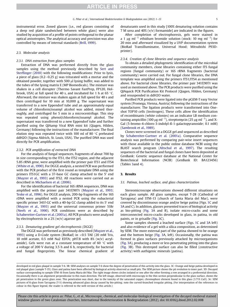
Fig. 4 shows the DGGE profiles derived from the bacterial andfungal communities colonizing the samples obtained from theCathedral of Tarragona and the church of Santa Maria del Mar inBarcelona. In general, bacterial DGGE profiles showed a highercomplexity in the community structure, with many dominantbands as well as some other faint bands (Fig. 4A). The DGGE profilesderived from the fungal communities showed fewer bands, indi-cating a lower biodiversity of fungi on the glass samples (Fig. 4B).
To obtain a phylogenetic identification of the individualmembers of the bacterial and fungal communities inhabiting theglasses of the two investigated locations, two K-rich glass samples(one from each location) were selected for the creation of clonelibraries containing either the bacterial 16S rRNA gene or the fungalITS regions and the 5.8S rRNA gene. The resulting bacterial andfungal clones were further screened by DGGE and clones wereselected for sequencing. The obtained sequences were comparedwith 16S rRNA gene sequences and ITS regions of known bacteriaand fungi, respectively, listed in the EMBL database. Figs. 5and 6show the DGGE profiles derived from bacterial (A) and fungal (B)clones selected for sequencing obtained from samples T-5 (Cathe-dral of Tarragona) and STM-16 (church of Santa Maria del Mar),respectively. Tables 3and 4 show the phylogenetic affiliations of thebacterial and fungal clones, respectively, obtained from bothsamples.
3.3. Microflora associated with the glass material
3.3.1. BacteriaTable 3 shows the phylogenetic affiliations of all bacterial clones
obtained in this study. A total of 19 and eight bacterial sequenceswere obtained from samples T-5 and STM-16, respectively.
From sample T-5 (Table 3A), nine sequences (47.3% of thebacterial sequences obtained from this sample) showed high scoresimilarities, ranging from 90 to 100%, with members of the Pro-teobacteria (Alpha-, Beta-, and Gamma-proteobacteria) beingrelated to species of the genera Methylobacterium (K59), Bradyrhi-zobium (K91), Ralstonia (K15), Delftia (K67), Variovorax (K93), Aci-netobacter (K79), and Thermomonas (K90), as well as to two
T-28 T-31 T-35
Purple Red Colorless Red Colorless67.61 49.27 48.97 46.68 43.424.54 19.09 19.10 19.59 21.41
15.83 0.36 0.35 0.46 0.580.66 4.39 4.25 3.88 4.307.42 17.78 17.37 19.28 20.780.25 4.75 4.62 5.05 4.742.04 2.30 2.27 2.59 2.480.96 1.01 1.00 1.20 1.340.50 0.53 0.54 0.53 0.580.03 0.32 0.04 0.44 0.070.02 0.01 0.01 0.09 0.110.11 0.17 0.16 0.18 0.18
d molecular-biological investigation of the decayedmedieval stainedon & Biodegradation (2012), doi:10.1016/j.ibiod.2012.02.008
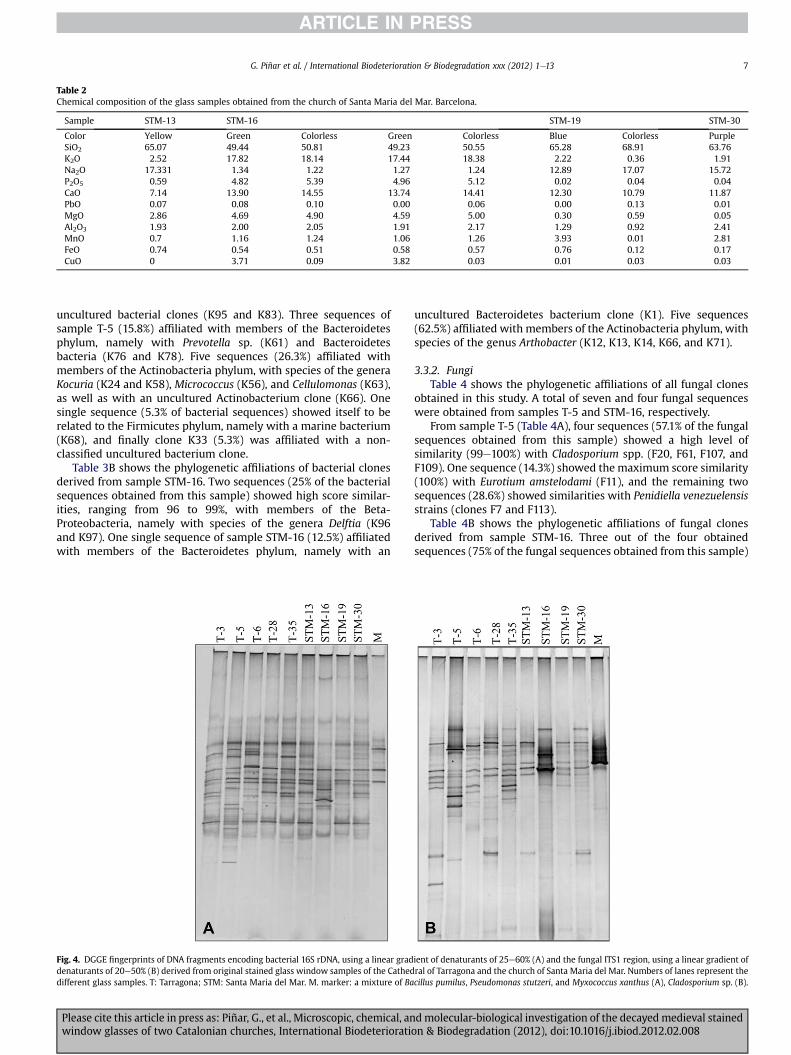
Table 2Chemical composition of the glass samples obtained from the church of Santa Maria del Mar. Barcelona.
Sample STM-13 STM-16 STM-19 STM-30
Color Yellow Green Colorless Green Colorless Blue Colorless PurpleSiO2 65.07 49.44 50.81 49.23 50.55 65.28 68.91 63.76K2O 2.52 17.82 18.14 17.44 18.38 2.22 0.36 1.91Na2O 17.331 1.34 1.22 1.27 1.24 12.89 17.07 15.72P2O5 0.59 4.82 5.39 4.96 5.12 0.02 0.04 0.04CaO 7.14 13.90 14.55 13.74 14.41 12.30 10.79 11.87PbO 0.07 0.08 0.10 0.00 0.06 0.00 0.13 0.01MgO 2.86 4.69 4.90 4.59 5.00 0.30 0.59 0.05Al2O3 1.93 2.00 2.05 1.91 2.17 1.29 0.92 2.41MnO 0.7 1.16 1.24 1.06 1.26 3.93 0.01 2.81FeO 0.74 0.54 0.51 0.58 0.57 0.76 0.12 0.17CuO 0 3.71 0.09 3.82 0.03 0.01 0.03 0.03
G. Piñar et al. / International Biodeterioration & Biodegradation xxx (2012) 1e13 7
uncultured bacterial clones (K95 and K83). Three sequences ofsample T-5 (15.8%) affiliated with members of the Bacteroidetesphylum, namely with Prevotella sp. (K61) and Bacteroidetesbacteria (K76 and K78). Five sequences (26.3%) affiliated withmembers of the Actinobacteria phylum, with species of the generaKocuria (K24 and K58), Micrococcus (K56), and Cellulomonas (K63),as well as with an uncultured Actinobacterium clone (K66). Onesingle sequence (5.3% of bacterial sequences) showed itself to berelated to the Firmicutes phylum, namely with a marine bacterium(K68), and finally clone K33 (5.3%) was affiliated with a non-classified uncultured bacterium clone.
Table 3B shows the phylogenetic affiliations of bacterial clonesderived from sample STM-16. Two sequences (25% of the bacterialsequences obtained from this sample) showed high score similar-ities, ranging from 96 to 99%, with members of the Beta-Proteobacteria, namely with species of the genera Delftia (K96and K97). One single sequence of sample STM-16 (12.5%) affiliatedwith members of the Bacteroidetes phylum, namely with an
Fig. 4. DGGE fingerprints of DNA fragments encoding bacterial 16S rDNA, using a linear graddenaturants of 20e50% (B) derived from original stained glass window samples of the Catheddifferent glass samples. T: Tarragona; STM: Santa Maria del Mar. M. marker: a mixture of Ba
Please cite this article in press as: Piñar, G., et al., Microscopic, chemical, anwindow glasses of two Catalonian churches, International Biodeteriorati
uncultured Bacteroidetes bacterium clone (K1). Five sequences(62.5%) affiliated with members of the Actinobacteria phylum, withspecies of the genus Arthobacter (K12, K13, K14, K66, and K71).
3.3.2. FungiTable 4 shows the phylogenetic affiliations of all fungal clones
obtained in this study. A total of seven and four fungal sequenceswere obtained from samples T-5 and STM-16, respectively.
From sample T-5 (Table 4A), four sequences (57.1% of the fungalsequences obtained from this sample) showed a high level ofsimilarity (99e100%) with Cladosporium spp. (F20, F61, F107, andF109). One sequence (14.3%) showed the maximum score similarity(100%) with Eurotium amstelodami (F11), and the remaining twosequences (28.6%) showed similarities with Penidiella venezuelensisstrains (clones F7 and F113).
Table 4B shows the phylogenetic affiliations of fungal clonesderived from sample STM-16. Three out of the four obtainedsequences (75% of the fungal sequences obtained from this sample)
ient of denaturants of 25e60% (A) and the fungal ITS1 region, using a linear gradient ofral of Tarragona and the church of Santa Maria del Mar. Numbers of lanes represent thecillus pumilus, Pseudomonas stutzeri, and Myxococcus xanthus (A), Cladosporium sp. (B).
d molecular-biological investigation of the decayed medieval stainedon & Biodegradation (2012), doi:10.1016/j.ibiod.2012.02.008
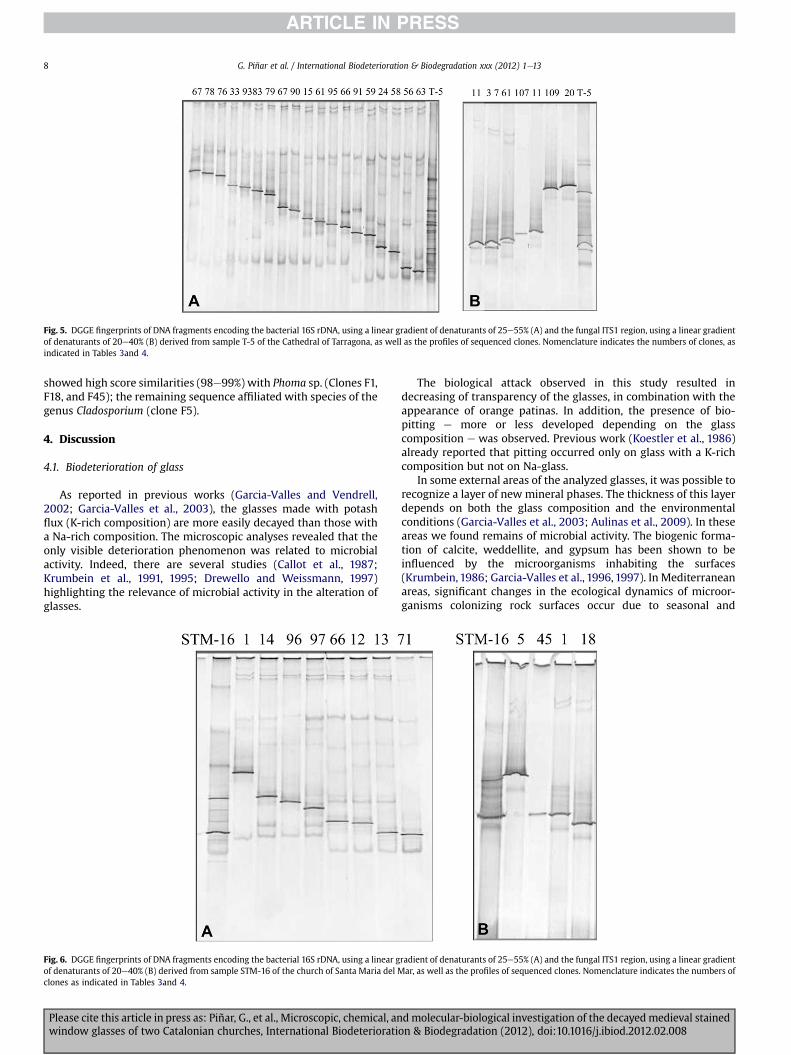
Fig. 5. DGGE fingerprints of DNA fragments encoding the bacterial 16S rDNA, using a linear gradient of denaturants of 25e55% (A) and the fungal ITS1 region, using a linear gradientof denaturants of 20e40% (B) derived from sample T-5 of the Cathedral of Tarragona, as well as the profiles of sequenced clones. Nomenclature indicates the numbers of clones, asindicated in Tables 3and 4.
G. Piñar et al. / International Biodeterioration & Biodegradation xxx (2012) 1e138
showed high score similarities (98e99%) with Phoma sp. (Clones F1,F18, and F45); the remaining sequence affiliated with species of thegenus Cladosporium (clone F5).
4. Discussion
4.1. Biodeterioration of glass
As reported in previous works (Garcia-Valles and Vendrell,2002; Garcia-Valles et al., 2003), the glasses made with potashflux (K-rich composition) are more easily decayed than those witha Na-rich composition. The microscopic analyses revealed that theonly visible deterioration phenomenon was related to microbialactivity. Indeed, there are several studies (Callot et al., 1987;Krumbein et al., 1991, 1995; Drewello and Weissmann, 1997)highlighting the relevance of microbial activity in the alteration ofglasses.
Fig. 6. DGGE fingerprints of DNA fragments encoding the bacterial 16S rDNA, using a linear gof denaturants of 20e40% (B) derived from sample STM-16 of the church of Santa Maria del Mclones as indicated in Tables 3and 4.
Please cite this article in press as: Piñar, G., et al., Microscopic, chemical, anwindow glasses of two Catalonian churches, International Biodeteriorati
The biological attack observed in this study resulted indecreasing of transparency of the glasses, in combination with theappearance of orange patinas. In addition, the presence of bio-pitting e more or less developed depending on the glasscomposition e was observed. Previous work (Koestler et al., 1986)already reported that pitting occurred only on glass with a K-richcomposition but not on Na-glass.
In some external areas of the analyzed glasses, it was possible torecognize a layer of new mineral phases. The thickness of this layerdepends on both the glass composition and the environmentalconditions (Garcia-Valles et al., 2003; Aulinas et al., 2009). In theseareas we found remains of microbial activity. The biogenic forma-tion of calcite, weddellite, and gypsum has been shown to beinfluenced by the microorganisms inhabiting the surfaces(Krumbein, 1986; Garcia-Valles et al., 1996, 1997). In Mediterraneanareas, significant changes in the ecological dynamics of microor-ganisms colonizing rock surfaces occur due to seasonal and
radient of denaturants of 25e55% (A) and the fungal ITS1 region, using a linear gradientar, as well as the profiles of sequenced clones. Nomenclature indicates the numbers of
d molecular-biological investigation of the decayedmedieval stainedon & Biodegradation (2012), doi:10.1016/j.ibiod.2012.02.008
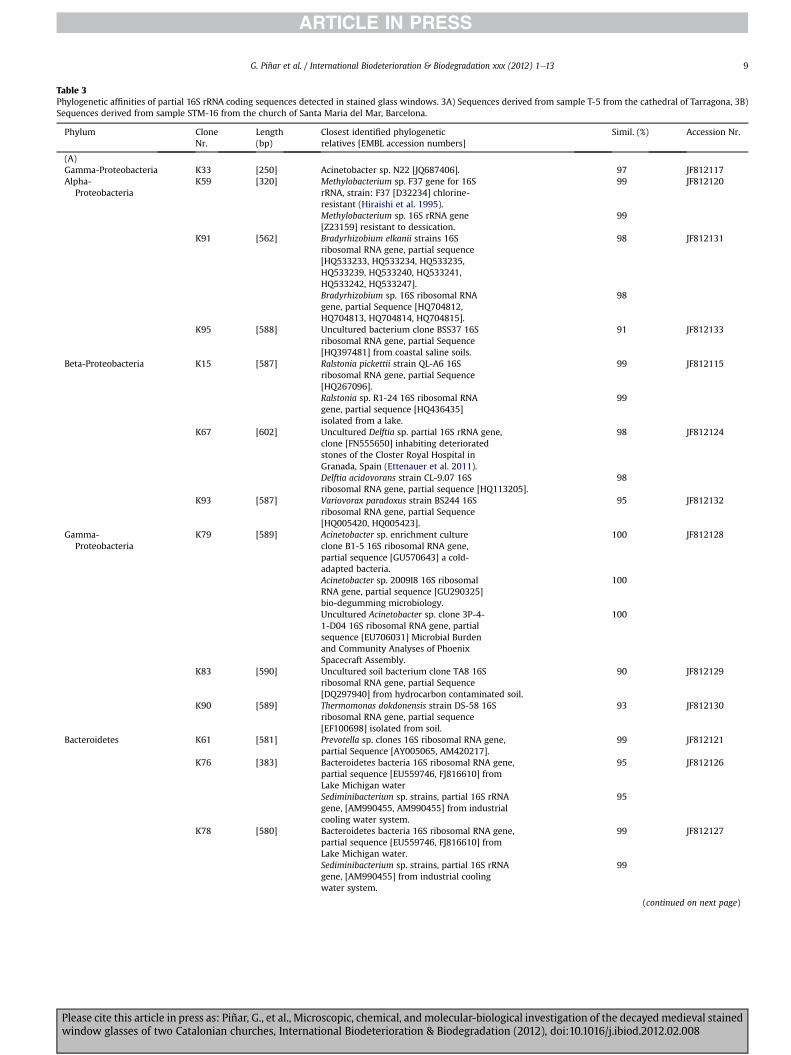
Table 3Phylogenetic affinities of partial 16S rRNA coding sequences detected in stained glass windows. 3A) Sequences derived from sample T-5 from the cathedral of Tarragona, 3B)Sequences derived from sample STM-16 from the church of Santa Maria del Mar, Barcelona.
Phylum CloneNr.
Length(bp)
Closest identified phylogeneticrelatives [EMBL accession numbers]
Simil. (%) Accession Nr.
(A)Gamma-Proteobacteria K33 [250] Acinetobacter sp. N22 [JQ687406]. 97 JF812117Alpha-
ProteobacteriaK59 [320] Methylobacterium sp. F37 gene for 16S
rRNA, strain: F37 [D32234] chlorine-resistant (Hiraishi et al. 1995).
99 JF812120
Methylobacterium sp. 16S rRNA gene[Z23159] resistant to dessication.
99
K91 [562] Bradyrhizobium elkanii strains 16Sribosomal RNA gene, partial sequence[HQ533233, HQ533234, HQ533235,HQ533239, HQ533240, HQ533241,HQ533242, HQ533247].
98 JF812131
Bradyrhizobium sp. 16S ribosomal RNAgene, partial Sequence [HQ704812,HQ704813, HQ704814, HQ704815].
98
K95 [588] Uncultured bacterium clone BSS37 16Sribosomal RNA gene, partial Sequence[HQ397481] from coastal saline soils.
91 JF812133
Beta-Proteobacteria K15 [587] Ralstonia pickettii strain QL-A6 16Sribosomal RNA gene, partial Sequence[HQ267096].
99 JF812115
Ralstonia sp. R1-24 16S ribosomal RNAgene, partial sequence [HQ436435]isolated from a lake.
99
K67 [602] Uncultured Delftia sp. partial 16S rRNA gene,clone [FN555650] inhabiting deterioratedstones of the Closter Royal Hospital inGranada, Spain (Ettenauer et al. 2011).
98 JF812124
Delftia acidovorans strain CL-9.07 16Sribosomal RNA gene, partial sequence [HQ113205].
98
K93 [587] Variovorax paradoxus strain BS244 16Sribosomal RNA gene, partial Sequence[HQ005420, HQ005423].
95 JF812132
Gamma-Proteobacteria
K79 [589] Acinetobacter sp. enrichment cultureclone B1-5 16S ribosomal RNA gene,partial sequence [GU570643] a cold-adapted bacteria.
100 JF812128
Acinetobacter sp. 2009I8 16S ribosomalRNA gene, partial sequence [GU290325]bio-degumming microbiology.
100
Uncultured Acinetobacter sp. clone 3P-4-1-D04 16S ribosomal RNA gene, partialsequence [EU706031] Microbial Burdenand Community Analyses of PhoenixSpacecraft Assembly.
100
K83 [590] Uncultured soil bacterium clone TA8 16Sribosomal RNA gene, partial Sequence[DQ297940] from hydrocarbon contaminated soil.
90 JF812129
K90 [589] Thermomonas dokdonensis strain DS-58 16Sribosomal RNA gene, partial sequence[EF100698] isolated from soil.
93 JF812130
Bacteroidetes K61 [581] Prevotella sp. clones 16S ribosomal RNA gene,partial Sequence [AY005065, AM420217].
99 JF812121
K76 [383] Bacteroidetes bacteria 16S ribosomal RNA gene,partial sequence [EU559746, FJ816610] fromLake Michigan water
95 JF812126
Sediminibacterium sp. strains, partial 16S rRNAgene, [AM990455, AM990455] from industrialcooling water system.
95
K78 [580] Bacteroidetes bacteria 16S ribosomal RNA gene,partial sequence [EU559746, FJ816610] fromLake Michigan water.
99 JF812127
Sediminibacterium sp. strains, partial 16S rRNAgene, [AM990455] from industrial coolingwater system.
99
(continued on next page)
G. Piñar et al. / International Biodeterioration & Biodegradation xxx (2012) 1e13 9
Please cite this article in press as: Piñar, G., et al., Microscopic, chemical, and molecular-biological investigation of the decayed medieval stainedwindow glasses of two Catalonian churches, International Biodeterioration & Biodegradation (2012), doi:10.1016/j.ibiod.2012.02.008
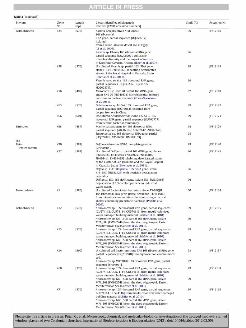
Table 3 (continued )
Phylum CloneNr.
Length(bp)
Closest identified phylogeneticrelatives [EMBL accession numbers]
Simil. (%) Accession Nr.
Actinobacteria K24 [570] Kocuria aegyptia strain YIM 7000316S ribosomalRNA gene, partial sequence [DQ059617]isolatedfrom a saline, alkaline desert soil in Egypt(Li et al. 2006).
96 JF812116
Kocuria sp. HI-A4a 16S ribosomal RNA gene,partial sequence [DQ205297], culturablemicrobial diversity and the impact of tourismin Kartchner Caverns, Arizona (Ikner et al. 2007).
96
K58 [570] Uncultured Kocuria sp. partial 16S rRNA gene,clone F-K34 [FN555669] inhabiting deterioratedstones of the Royal Hospital in Granada, Spain(Ettenauer et al. 2011).
99 JF812119
Kocuria rosea strains 16S ribosomal RNA gene,partial Sequences [HQ830206, HQ538755,HQ202874].
99
K56 [409] Micrococcus sp. RNE 20 partial 16S rRNA gene,strain RNE 20 [FR749831] Microbiological inducedcorrosion in nuclear materials (Forte-Giacoboneet al. 2011).
97 JF812118
K63 [570] Cellulomonas sp. Mn5-4 16S ribosomal RNA gene,partial sequence [HQ730135] isolated fromcopper iron ore in China.
99 JF812122
K66 [601] Uncultured Actinobacterium clone JBS_5Y17 16Sribosomal RNA gene, partial sequence [EU702777]from benthic bacterial community.
89 JF812123
Fimicutes K68 [487] Marine bacteria gene for 16S ribosomal RNA,partial sequence [AB607166; AB607142; AB607143].
98 JF812125
Enterococcus sp. 16S ribosomal RNA gene, partial[HQ677826, AB596997, HM584103].
98
(B)Beta-
ProteobacteriaK96 [587] Delftia acidovorans SPH-1, complete genome
[CP000884].99 JF812140
K97 [587] Uncultured Delftia sp. partial 16S rRNA gene, clones[FN435923, FN435934, FN435975, FN434405,FN434411, FN434425] inhabiting deteriorated stonesof the Closter of San Jeronimo and the Royal Hospitalin Granada, Spain (Ettenauer et al. 2011).
96 JF812141
Delftia sp. R-41380 partial 16S rRNA gene, strainR-41380. [FR682925] with pesticide degradationcapability.
96
Delftia sp. EK3 16S rRNA gene, isolate EK3. [AJ237966]degradation of 1,3-dichloropropene in industrialwaste water.
96
Bacteroidetes K1 [580] Uncultured Bacteroidetes bacterium clone A3-01QJH16S ribosomal RNA gene, partial sequence [EU434886]from microbial communities colonizing a single naturalshelter containing prehistoric paintings (Portillo et al.2009).
100 JF812134
Actinobacteria K12 [570] Arthrobacter sp. 16S ribosomal RNA gene, partial sequences[GU574112, GU574114, GU574116] from mould-colonizedwater damaged building material (Schäfer et al. 2010).
99 JF812135
Arthrobacter sp. M71_S08 partial 16S rRNA gene, isolateM71_S08 [FM992748] from the deep oligotrophic EasternMediterranean Sea (Gärtner et al. 2011).
99
K13 [570] Arthrobacter sp. 16S ribosomal RNA gene, partial sequences[GU574112, GU574114, GU574116] from mould-colonizedwater damaged building material (Schäfer et al. 2010).
99 JF812136
Arthrobacter sp. M71_S08 partial 16S rRNA gene, isolateM71_S08 [FM992748] from the deep oligotrophic EasternMediterranean Sea (Gärtner et al. 2011).
99
K14 [590] Uncultured soil bacterium clone TA8 16S ribosomal RNA gene,partial Sequence [DQ297940] from hydrocarbon contaminatedsoil.
93 JF812137
Arthrobacter sp. WPCB182 16S ribosomal RNA gene, partialsequence [FJ006921].
92
K66 [570] Arthrobacter sp. 16S ribosomal RNA gene, partial sequences[GU574112, GU574114, GU574116] from mould-colonizedwater damaged building material (Schäfer et al. 2010).
99 JF812138
Arthrobacter sp. M71_S08 partial 16S rRNA gene, isolateM71_S08 [FM992748] from the deep oligotrophic EasternMediterranean Sea (Gärtner et al. 2011).
99
K71 [570] Arthrobacter sp. 16S ribosomal RNA gene, partial sequencesGU574114, GU574116] from mould-colonized water damagedbuilding material (Schäfer et al. 2010).
99 JF812139
Arthrobacter sp. M71_S08 partial 16S rRNA gene, isolateM71_S08 [FM992748] from the deep oligotrophic EasternMediterranean Sea (Gärtner et al. 2011).
99
Please cite this article in press as: Piñar, G., et al., Microscopic, chemical, and molecular-biological investigation of the decayedmedieval stainedwindow glasses of two Catalonian churches, International Biodeterioration & Biodegradation (2012), doi:10.1016/j.ibiod.2012.02.008
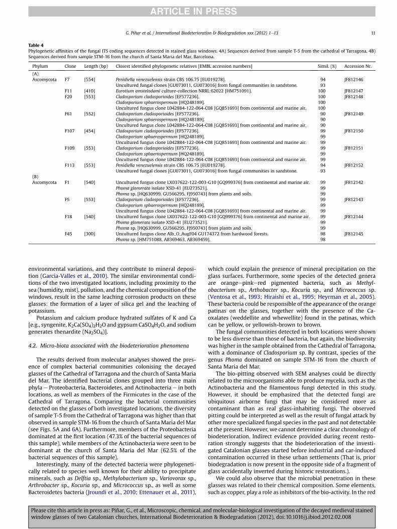
Table 4Phylogenetic affinities of the fungal ITS coding sequences detected in stained glass windows. 4A) Sequences derived from sample T-5 from the cathedral of Tarragona, 4B)Sequences derived from sample STM-16 from the church of Santa Maria del Mar, Barcelona.
Phylum Clone Length (bp) Closest identified phylogenetic relatives [EMBL accession numbers] Simil. (%) Accession Nr.
(A)Ascomycota F7 [554] Penidiella venezuelensis strain CBS 106.75 [EU019278]. 94 JF812146
Uncultured fungal clones [GU073011, GU073016] from fungal communities in sandstone. 93F11 [410] Eurotium amstelodami culture-collection NRRL:62022 [HM751091]. 100 JF812147F20 [553] Cladosporium cladosporioides [EF577236]. 100 JF812148
Cladosporium sphaerospermum [HQ248189]. 100Uncultured fungus clone L042884-122-064-C08 [GQ851693] from continental and marine air. 100
F61 [552] Cladosporium cladosporioides [EF577236]. 90 JF812149Cladosporium sphaerospermum [HQ248189]. 90Uncultured fungus clone L042884-122-064-C08 [GQ851693] from continental and marine air. 90
F107 [454] Cladosporium cladosporioides [EF577236]. 99 JF812150Cladosporium sphaerospermum [HQ248189]. 99Uncultured fungus clone L042884-122-064-C08 [GQ851693] from continental and marine air. 99
F109 [553] Cladosporium cladosporioides [EF577236]. 99 JF812151Cladosporium sphaerospermum [HQ248189]. 99Uncultured fungus clone L042884-122-064-C08 [GQ851693] from continental and marine air. 99
F113 [553] Penidiella venezuelensis strain CBS 106.75 [EU019278]. 94 JF812152Uncultured fungal clones [GU073011, GU073016] from fungal communities in sandstone. 93
(B)Ascomycota F1 [540] Uncultured fungus clone LX037622-122-003-G10 [GQ999376] from continental and marine air. 99 JF812142
Phoma glomerata isolate XSD-41 [EU273521]. 99Phoma sp. [HQ630999, GU566295, FJ950743] from plants and soils. 99
F5 [553] Cladosporium cladosporioides [EF577236]. 99 JF812143Cladosporium sphaerospermum [HQ248189]. 99Uncultured fungus clone L042884-122-064-C08 [GQ851693] from continental and marine air. 99
F18 [540] Uncultured fungus clone LX037622-122-003-G10 [GQ999376] from continental and marine air. 99 JF812144Phoma glomerata isolate XSD-41 [EU273521]. 99Phoma sp. [HQ630999, GU566295, FJ950743] from plants and soils. 99
F45 [300] Uncultured fungus clone Alb_O_AugF04 GU174372 from hardwood forests. 98 JF812145Phoma sp. [HM751088, AB369463, AB369459]. 98
G. Piñar et al. / International Biodeterioration & Biodegradation xxx (2012) 1e13 11
environmental variations, and they contribute to mineral deposi-tion (Garcia-Valles et al., 2010). The similar environmental condi-tions of the two investigated locations, including proximity to thesea (humidity, mist), pollution, and the chemical composition of thewindows, result in the same leaching corrosion products on theseglasses: the formation of a layer of silica gel and the leaching ofpotassium.
Potassium and calcium produce hydrated sulfates of K and Ca[e.g., syngenite, K2Ca(SO4)2H2O and gypsumCaSO4H2O, and sodiumgenerates thenardite (Na2SO4)].
4.2. Micro-biota associated with the biodeterioration phenomena
The results derived from molecular analyses showed the pres-ence of complex bacterial communities colonising the decayedglasses of the Cathedral of Tarragona and the church of Santa Mariadel Mar. The identified bacterial clones grouped into three mainphyla e Proteobacteria, Bacteroidetes, and Actinobacteria e in bothlocations, as well as members of the Firmicutes in the case of theCathedral of Tarragona. Comparing the bacterial communitiesdetected on the glasses of both investigated locations, the diversityof sample T-5 from the Cathedral of Tarragona was higher than thatobserved in sample STM-16 from the church of Santa Maria del Mar(see Figs. 5A and 6A). Furthermore, members of the Proteobacteriadominated at the first location (47.3% of the bacterial sequences ofthis sample), while members of the Actinobacteria were seen to bedominant at the church of Santa Maria del Mar (62.5% of thebacterial sequences of this sample).
Interestingly, many of the detected bacteria were phylogeneti-cally related to species well known for their ability to precipitateminerals, such as Delftia sp., Methylobacterium sp., Variovorax sp.,Arthrobacter sp., Kocuria sp., and Micrococcus sp., as well as someBacteroidetes bacteria (Jroundi et al., 2010; Ettenauer et al., 2011),
Please cite this article in press as: Piñar, G., et al., Microscopic, chemical, anwindow glasses of two Catalonian churches, International Biodeteriorati
which could explain the presence of mineral precipitation on theglass surfaces. Furthermore, some species of the detected generaare orangeepinkered pigmented bacteria, such as Methyl-obacterium sp., Arthobacter sp., Kocuria sp., and Micrococcus sp.(Ventosa et al., 1993; Hiraishi et al., 1995; Heyrman et al., 2005).These bacteria could be responsible of the appearance of the orangepatinas on the glasses, together with the presence of the Ca-oxalates (weddellite and whewellite) found in the patinas, whichcan be yellow, or yellowish-brown to brown.
The fungal communities detected in both locations were shownto be less diverse than those of bacteria, but again, the biodiversitywas higher in the sample obtained from the Cathedral of Tarragona,with a dominance of Cladosporium sp. By contrast, species of thegenus Phoma dominated on sample STM-16 from the church ofSanta Maria del Mar.
The bio-pitting observed with SEM analyses could be directlyrelated to the microorganisms able to produce mycelia, such as theActinobacteria and the filamentous fungi detected in this study.However, it should be emphasized that the detected fungi areubiquitous airborne fungi that may be considered more ascontaminant than as real glass-inhabiting fungi. The observedpitting could be interpreted as well as the result of fungal attack byother more specialized fungal species in the past and not detectableat the present. However, we cannot determine a clear chronology ofbiodeterioration. Indirect evidence provided during recent resto-ration strongly suggests that the biodeterioration of the investi-gated Catalonian glasses started before industrial and car-inducedcontamination occurred in these urban settlements (That is, priorbiodegradation is now present in the opposite side of a fragment ofglass accidentally inverted during historic restorations.).
We could also observe that the microbial penetration in theseglasses was related to their chemical composition. Some elements,such as copper, play a role as inhibitors of the bio-activity. In the red
dmolecular-biological investigation of the decayed medieval stainedon & Biodegradation (2012), doi:10.1016/j.ibiod.2012.02.008
G. Piñar et al. / International Biodeterioration & Biodegradation xxx (2012) 1e1312
flashed glasses (Fig. 3E), the microbial development was stoppedwhen the organisms reached the Cu-rich colored layer and, thus,they stopped their destructive activity. In those samples showingancient bio-activity, we observed a non-uniform spatial distribu-tion of pitting on the surface of the glasses. In some cases, the bio-pitting formed a pattern of channels like those shown in Fig. 3E.
Last but not least, our sequence results show high similaritieswith bacteria already found on deteriorated stone monuments(Jroundi et al., 2010; Ettenauer et al., 2011), building materials(Schäfer et al., 2010), caves (Ikner et al., 2007), and prehistoricpaintings (Portillo et al., 2009), and associated with the corrosion ofmaterials (Forte-Giacobone et al., 2011). Many of the relatedmicroorganismswere previously isolated from stones and artworksin the Mediterranean area (Portillo et al., 2009; Jroundi et al., 2010;Ettenauer et al., 2011) which supports the idea that medievalstained glass biodeterioration in the Mediterranean area showspatterns comparable to those developed on the stone (Garcia-Valleset al., 2003, 2010). This is logical if we think that, in historicbuildings, both the rock and the glass have been exposed to thesame atmospheric conditions, which makes for similar mecha-nisms of decay on both materials. Furthermore, taking intoconsideration that Tarragona and Barcelona are cities located at theMediterranean seaside, it is not surprising to find sequences relatedto Methylobacterium sp. able to tolerate chloride (Hiraishi et al.,1995), Kocuria aegyptia isolated from saline environments (Liet al., 2006), oligotrophic species of the genus Arthrobacter iso-lated from the Mediterranean sea (Gärtner et al., 2011), as well asfungal species detected in marine air (see Table 4).
Comparing our data with previous studies focusing on the bio-logical deterioration of historical glass, we find a lack of studies thatcombine different methodologies, with very few exceptions(Carmona et al., 2006). To date, published studies have focused onthe microscopic and chemical (Garcia-Valles and Vendrell, 2002,2003) or on the microbial characterization of historical glasssamples by culture-dependent techniques (Marvasi et al., 2009).Only a few published works have reported on the molecular char-acterization of historical glass samples (Rölleke et al., 1999;Schabereiter-Gurtner et al., 2001b; Carmona et al., 2006). In thefirst two molecular studies mentioned above, the authors investi-gated glass samples derived from the historical window of thechapel in Stockkämpen, Germany. Rölleke et al. (1999) analyzed thebacterial community of this window via DGGE and 16S rDNAsequence analysis. They identified Flexibacter sp., as well asmembers of the ammonia-oxidizing genus Nitrosospira and severalmembers of the Actinobacteria, such as Arthrobacter, Frankia, andGeodermatophilus. Schabereiter-Gurtner et al. (2001b) analyzed thefungal communities inhabiting the same window by 18S rDNA-based DGGE fingerprinting, creation of clone libraries, andsequencing, and compared the detected community with the oneobtained from other glass windows located at the church of St.Michael and Johann Baptist in Brakel, Germany. Even thoughauthors found fungal communities consisting of five to eightdifferent fungi on these two glass windows, the molecular strategythey followed was fastidious and very time-consuming, as it wasnecessary to use at least three different fungal primer combinationsto screen 18S rDNA clone libraries. Finally, Carmona et al. (2006)investigated the microbial communities (bacteria and fungi) asso-ciated with the historical stained glasses of the Cartuja de Mira-floresMonastery (Spain). The bacterial communities were analyzedby 16S rDNA-based DGGE fingerprinting, although no attempt atidentification was made. Fungi were identified by cloning andsequencing of the 18S rDNA.
Nowadays it is well known that there is a lack of taxonomicresolution between fungal 18S rRNA genes to closely related taxaand, therefore, this region is not so accurate for an accurate
Please cite this article in press as: Piñar, G., et al., Microscopic, chemical, anwindow glasses of two Catalonian churches, International Biodeteriorati
phylogenetic identification of fungi (Anderson and Cairney, 2004).In recent years, ITS regions have become the selected geneticmarker for studies on fungal communities in environmentalsamples (White et al., 1990). For this reason, in previous studies wedeveloped a protocol to obtain DGGE fingerprints derived fromfungal ITS regions of DNA directly extracted from different mate-rials of cultural heritage (Michaelsen et al., 2006). To date, we havesuccessfully applied this molecular strategy to analyze fungalcommunities colonizing paper (Michaelsen et al., 2006, 2009,2010), stone monuments (Piñar et al., 2009), parchment (Piñaret al., 2011), oil paintings on canvas (Lopez-Miras, personalcommunication), human remains (Piñar, unpublished data) and, inthe present study, glass samples. This strategy has proven easierand more reliable than the one using DGGE fingerprints derivedfrom 18S rDNA fragments.
In addition, with the exception of some fewworks (Rölleke et al.,1999; Carmona et al., 2006; Marvasi et al., 2009) bacteria werethought not to be dominant colonizers of glass surfaces and not toplay a relevant role in the observed deterioration phenomena. Theresults obtained in the present study clearly show that bacterialcommunities were more diverse than those of fungi and that thedetected bacteria are phylogenetically related to well-knownbacteria possessing metabolic activities able to produce theobserved biodeterioration of the investigated glasses. This facthighlights the relevance of bacteria, together with fungi, in thebiodeterioration of historical glass.
Acknowledgment
This study was financed by the Austrian Science Fund (FWF)project “Hertha-Firnberg T137.”G. Piñar is currently financed by theAustrian Science Fund (FWF) project “Elise-Richter V194-B20.” Theanalytical work was conducted at the Serveis Científico-Tècnics ofthe Universitat de Barcelona; we acknowledge all the technical andscientific staff involved in the experiments for their assistanceduring data acquisition. P. Valldepérez (restorer) and J. Figuerola(architect) are acknowledged for the access to facilities providedduring the rosette restoration of the Cathedral of Tarragona. P.Valldepérez is also acknowledged for the access to facilitiesprovided during the rosette restoration of Santa Maria del Mar,Barcelona.
References
Ainaud de Lasarte, J., Vila-Grau, J., Escudero-Ribot, M.A., 1985. Els vitralls medievalsde l’església de Santa Maria del Mar de Barcelona. Corpus Vitrearum Medi Aevi.Institut d’Estudis Catalans, Barcelona, p. 404.
Altschul, S.F., Madden, T.L., Schäffer, A.A., Zhang, J., Zhang, Z., Miller, W., Lipman, J.D.,1997. Gapped BLAST and PSI-BLAST: a new generation of protein databasesearch programs. Nucleic Acids Research 25, 3389e3402.
Anderson, I.C., Cairney, J.W.G., 2004. Diversity and ecology of soil fungal commu-nities: increased understanding through the application of molecular tech-niques. Environmental Microbiology 6, 769e779.
Aulinas, M., Garcia-Vallès, M., Gimeno, D., Fernandez-Turiel, J.L., Ruggieri, F.,Pugès, M., 2009. Weathering patinas on the medieval (S. XIV) stained glasswindows of the Pedralbes Monastery (Barcelona, Spain). Environmental Scienceand Pollution Research 16, 443e452.
Bamford, C.R., 1977. Colour Generation and Control in Glass. Glass Science andTechology 2. Elsevier, pp. 224.
Brill, R.H., 1999. Chemical analysis of early glasses. The Corning Museum of Glass 2,335.
Callot, G., Maurette, M., Pottier, L., Dubois, A., 1987. Biogenic etching of micro-fractures in amorphous and crystalline silicates. Nature 328, 147e149.
Carmona, N., Laiz, L., Gonzalez, J.M., Garcia-Heras, M., Villegas, M.A., Saiz-Jimenez, C., 2006. Biodeterioration of historic stained glasses from the Cartujade Miraflores (Spain). International Biodeterioration and Biodegradation 58,155e161.
Drewello, R., Weissmann, R., 1997. Microbially influenced corrosion of glass. AppliedMicrobiology and Biotechnology 47, 337e346.
Ettenauer, J., Piñar, G., Sterflinger, K., Gonzalez-Muñoz, M.T., Jroundi, F., 2011.Molecular monitoring of the microbial dynamics occurring on historical
dmolecular-biological investigation of the decayedmedieval stainedon & Biodegradation (2012), doi:10.1016/j.ibiod.2012.02.008
G. Piñar et al. / International Biodeterioration & Biodegradation xxx (2012) 1e13 13
limestone buildings during and after the in situ application of differentbio-consolidation treatments. Science of the Total Environment 409,5337e5352.
Forte-Giacobone, A.F., Rodriguez, S.A., Burkart, A.L., Pizarro, R.A., 2011. Microbio-logical induced corrosion of AA 6061 nuclear alloy in highly diluted media byBacillus cereus RE 10. International Biodeterioration and Biodegradation 65,1161e1168.
Garcia-Valles, M., Vendrell, M., 2002. The glasses of the transept’s rosette of thecathedral of Tarragona: characterization, classification and decay. BoletínSociedad Española de Cerámica y Vidrio 41, 217e224.
Garcia-Valles, M., Blázquez, F., Vendrell, M., 1996. Studies of patinas and decaymechanisms leading to the restoration of Santa Maria de Montblanc. Studies inConservation 41, 1e8.
Garcia-Valles, M., Vendrell, M., Krumbein, W., Urzì, C., 1997. Coloured mineralcoatings on monument surfaces as result of biomineralization: the caseof the Tarragona cathedral (Catalonia). Applied Geochemistry 12,255e266.
Garcia-Valles, M., Gimeno-Torrente, D., Martínez-Manent, S., Fernandez-Turiel, J.L.,2003. Medieval stained glass in a Mediterranean climate: typology, weatheringand glass decay, and associated bio-mineralization processes and products.American Mineralogist 88, 1996e2006.
Garcia-Valles, M., Aulinas, M., Lopez-Melcion, J.B., Moya Garra, A., 2010. Patinasdeveloped in environmental burial conditions: the Neolithic steles of Reguersde Seró (Lleida, Spain). Environmental Science and Pollution Research 17,1287e1299.
Gärtner, A., Blümel, M., Wiese, J., Imhoff, J.F., 2011. Isolation and characterisation ofbacteria from the Eastern Mediterranean deep sea. Antonie Van Leeuwenhoek100, 421e435.
Heyrman, J., Verbeeren, J., Schumann, P., Swings, J., De Vos, P., 2005. Six novelArthrobacter species isolated from deteriorated mural paintings. InternationalJournal of Systematic and Evolutionary Microbiology 55, 1457e1464.
Hiraishi, A., Furuhata, K., Matsumoto, A., Koike, K.A., Fukuyama, M., Tabuchi, K.,1995. Phenotypic and genetic diversity of chlorine-resistant Methylobacteriumstrains isolated from various environments. Applied and EnvironmentalMicrobiology 61, 2099e2107.
Ikner, L.A., Toomey, R.S., Nolan, G., Neilson, J.W., Pryor, W.M., Maier, R.M., 2007.Culturable microbial diversity and the impact of tourism in Kartchner caverns,Arizona. Microbial Ecology 53, 30e42.
Jroundi, F., Fernández-Vivas, A., Rodriguez-Navarro, C., Bedmar, E.J., Gonzalez-Muñoz, M.T., 2010. Bioconservation of deteriorated monumental calcarenitestone and identification of bacteria with carbonatogenic activity. MicrobialEcology 60, 39e54.
Kaiser, J.P., Trumpler, S., Raschle, P., 1996. Fungal growth on medieval glass paint-ings. In: Heitz, E., Flemming, H.-C., Sand, W. (Eds.), Microbially InfluencedCorrosion of Materials. Springer, Berlin, pp. 353e357.
Koestler, R.J., Santoro, E.D., Ransick, L., Brill, R.H., Lynn, M., 1986. Preliminary SEMstudy of microbiologically induced deterioration of high alkali-low lime glass.In: O’Rear, C.E., Llewellyn, G.C. (Eds.), Biodeterioration Research 1. Plenum, NewYork, pp. 295e307.
Krumbein, W.E., 1986. Biotransfer of Minerals by Microbes and microbial mats. In:Riding (Ed.), Biomineralization in Lower Plants and Animals. Clarendon Press,Oxford UK, pp. 55e72.
Krumbein, W.E., Urzi, C., Gehrmann, C., 1991. Biocorrosion and biodeterioration ofantique and medieval glass. Geomicrobiology Journal 9, 139e165.
Krumbein, W.E., Gorbushina, A.A., Palinska, K., Sterflinger, K., 1995. The paradoxonof glass. stability and decay; biological deterioration and transformation;conservation and restoration. In: Lefèvre, R.-A., Pallot-Frossard, I. (Eds.), Actesdu Cours Intensif Européen. Ravello, 28-30/04/95, pp. 108e124.
Li, W.J., Zhang, Y.Q., Schumann, P., Chen, H.H., Hozzein, W.N., Tian, X.P., Xu, L.H.,Jian, C.L., 2006. Kocuria aegyptia sp. nov., a novel actinobacterium isolated froma saline, alkaline desert soil in Egypt. International Journal of Systematic andEvolutionary Microbiology 56, 733e737.
Marvasi, M., Vedovato, E., Balsamo, C., Macherelli, A., Dei, L., Mastromei, G.,Perito, B., 2009. Bacterial community analysis on the medieval stained glasswindow “Natività” in the Florence Cathedral. Journal of Cultural Heritage 10,124e133.
Mellor, E., 1924. The decay of window glass from the point of view of the lichenousgrowth. Journal of the Society of Glass Technology 8, 182e186.
Please cite this article in press as: Piñar, G., et al., Microscopic, chemical, anwindow glasses of two Catalonian churches, International Biodeteriorati
Michaelsen, A., Pinzari, F., Ripka, K., Lubitz, K., Piñar, G., 2006. Application ofmolecular techniques for the identification of fungal communities colonisingpaper material. International Biodeterioration and Biodegradation 58, 133e141.
Michaelsen, A., Piñar, G., Montanari, M., Pinzari, F., 2009. Biodeterioration andrestoration of a 16th-century book using a combination of conventional andmolecular techniques: a case study. International Biodeterioration andBiodegradation 63, 161e168.
Michaelsen, A., Piñar, G., Pinzari, F., 2010. Molecular and microscopical investigationof the microflora inhabiting a deteriorated Italian manuscript dated from thethirteenth century. Microbial Ecology 60, 69e80.
Muyzer, G., De Waal, E.C., Uitterlinden, A.G., 1993. Profiling of complex microbialpopulations by denaturing gradient gel electrophoresis analysis of polymerasechain reaction-amplified genes coding for 16S rRNA. Applied and Environ-mental Microbiology 59, 695e700.
Nagamuttu, S., 1967. Molds on optical glass and control measures. InternationalBiodeterioration Bulletin 3, 25e27.
Neefs, J.M., Van de Peer, Y., Hendriks, L., De Wachter, R., 1990. Compilation of smallribosomal subunit RNA sequences. Nucleic Acids Research 18, 2237e2317.
Newton, R.G., Davison, S., 1989. Conservation of Glass. Butterworth-Heineman,Oxford, U.K, pp. 318.
Newton, R.G., Fuchs, 1988. Chemical compositions and weathering of some medi-eval glasses from York Minster. Part 1. Glass Technology 29, 43e48.
Piñar, G., Ripka, K., Weber, J., Sterflinger, K., 2009. The micro-biota of a sub-surfacemonument: the medieval chapel of St. Virgil (Vienna, Austria). InternationalBiodeterioration and Biodegradation 63, 851e859.
Piñar, G., Pinzari, F., Sterflinger, K., 2011. Modern technologies as basis for thepreservation of parchment. In: López Montes, A.M., Collado Montero, F., MedinaFlórez, V., Espejo Arias, T., García Bueno, A. (Eds.), Proceedings of the 18thInternational Meeting on Heritage Conservation, Granada, Spain, pp. 250e253.ISBN 978-84-338-5339-4, Granada.
Portillo, M.C., Alloza, R., Gonzalez, J.M., 2009. Three different phototrophic microbialcommunities colonizing a single natural shelter containing prehistoric paint-ings. Science of the Total Environment 407, 4876e4881.
Rölleke, S., Gurtner, C., Drewello, U., Drewello, R., Lubitz, W., Weissmann, R., 1999.Analysis of bacterial communities on historical glass by denaturing gradient gelelectrophoresis of PCR-amplified gene fragments coding for 16S rRNA. Journalof Microbiological Methods 36, 107e114.
Römich, H., 1999. Historic glass and its interaction with the environment. In:Tennet, N.H. (Ed.), The Conservation of Glass and Ceramics. James and James(Science Publishers) Ltd, London, pp. 5e14.
Sambrook, J., Fritsch, E.F., Maniatis, T., 1989. Molecular Cloning: A LaboratoryManual, second ed. Cold Spring Harbor Laboratory, Cold Spring Harbor.
Schabereiter-Gurtner, C., Piñar, G., Lubitz, W., Rolleke, S., 2001a. An advancedmolecular strategy to identify bacterial communities on art objects. Journal ofMicrobiological Methods 45, 77e87.
Schabereiter-Gurtner, C., Piñar, G., Lubitz, W., Rolleke, S., 2001b. Analysis of fungalcommunities on historical church window glass by denaturing gradient gelelectrophoresis and phylogenetic 18S rDNA sequence analysis. Journal ofMicrobiological Methods 47, 345e354.
Schäfer, J., Jäckel, U., Kämpfer, P., 2010. Analysis of actinobacteria from mould-colonized water damaged building material. Systematic and Applied Microbi-ology 33, 260e268.
Schreiner, M., 1991. Glass of the past: the degradation and deterioration of medievalglass artifacts. Mikrochimica Acta 11, 255e264.
Sert, H.B., Sterflinger, K., 2010. A new Coniosporium species from historical marblemonuments. Mycological Progress 9, 353e359.
Sterpenich, J., Libourel, G., 2001. Using stained glass windows to understand thedurability of toxic waste matrices. Chemical Geology 174, 181e193.
Teske, A., Wawer, C., Muyzer, G., Ramsing, N.B., 1996. Distribution of sulphate-reducing bacteria in a stratified fjord (Mariager Fjord, Denmark) as evaluatedby most-probable-number counts and DGGE of PCR-amplified ribosomal DNAfragments. Applied and Environmental Microbiology 62, 1405e1415.
Ventosa, A., Marquez, M.C., Kocur, M., Tindall, B.J., 1993. Comparative study ofMicrococcus sp. strains CCM 168 and CCM 1405 and members of the genusSalinicoccus. International Journal of Systematic Bacteriology 43, 245e248.
White, T.J., Bruns, T., Lee, S., Taylor, J., 1990. Amplification and Direct Sequencing ofFungal Ribosomal RNA Genes for Phylogenetics. PCR Protocols: A Guide toMethods and Applications. Academic Press, New York, pp. 315e322.
dmolecular-biological investigation of the decayed medieval stainedon & Biodegradation (2012), doi:10.1016/j.ibiod.2012.02.008