Embed Size (px)
Citation preview
Low-oxygen response is triggered by an ATP-dependent shift in oleoyl-CoA in ArabidopsisRomy R. Schmidta,1, Martin Fuldab, Melanie V. Paulc, Max Andersa, Frederic Pluma, Daniel A. Weitsa, Monika Kosmaczd,Tony R. Larsone, Ian A. Grahame, Gerrit T. S. Beemsterf, Francesco Licausig,h, Peter Geigenbergerc, Jos H. Schippersa,and Joost T. van Dongena,1
aInstitute of Biology I, Rheinisch-Westfälische Technische Hochschule Aachen University, 52074 Aachen, Germany; bAlbrecht von Haller Institute of PlantSciences, Goettingen University, 37077 Goettingen, Germany; cDepartment Biology I, Ludwig Maximilian University of Munich, 82152 Planegg-Martinsried,Germany; dMax Planck Institute of Molecular Plant Physiology, 14476 Potsdam, Germany; eDepartment of Biology, University of York, Heslington, YO105DD York, United Kingdom; fIntegrated Molecular Plant Physiology Research Group, University of Antwerp, G.U.613, 2020 Antwerpen, Belgium; gPlantLab,Institute of Life Sciences, Scuola Superiore Sant’Anna, 56017 Pisa, Italy; and hDipartimento di Biologia,Università di Pisa, 56126 Pisa, Italy
Edited by Julia Bailey-Serres, University of California, Riverside, CA, and approved November 5, 2018 (received for review June 10, 2018)
Plant response to environmental stimuli involves integration ofmultiple signals. Upon low-oxygen stress, plants initiate a set ofadaptive responses to circumvent an energy crisis. Here, we revealhow these stress responses are induced by combining (i) energy-dependent changes in the composition of the acyl-CoA pool and(ii) the cellular oxygen concentration. A hypoxia-induced declineof cellular ATP levels reduces LONG-CHAIN ACYL-COA SYNTHETASEactivity, which leads to a shift in the composition of the acyl-CoApool. Subsequently, we show that different acyl-CoAs induceunique molecular responses. Altogether, our data disclose a rolefor acyl-CoAs acting in a cellular signaling pathway in plants. Uponhypoxia, high oleoyl-CoA levels provide the initial trigger to releasethe transcription factor RAP2.12 from its interaction partner ACYL-COABINDING PROTEIN at the plasma membrane. Subsequently, accordingto the N-end rule for proteasomal degradation, oxygen concentration-dependent stabilization of the subgroup VII ETHYLENE-RESPONSEFACTOR transcription factor RAP2.12 determines the level ofhypoxia-specific gene expression. This research unveils a specificmechanism activating low-oxygen stress responses only when a de-crease in the oxygen concentration coincides with a drop in energy.
low-oxygen stress | integrative signaling | acyl-CoA | ERFVII | ACBP
Flooding contributes almost 60% to the worldwide cost anddamage to crops provoked by natural disasters (1). Due to
heavy precipitation and concomitant waterlogging or floodingevents in large areas of the world, climate change will causeplants to be even more frequently exposed to oxygen-limitingconditions (hypoxia) in the near future (2).In plants, subgroup VII ETHYLENE-RESPONSE FACTOR
(ERFVII) transcription factors act as key regulators of hypoxicgene expression (3–6). During nonstress conditions, the ERFVIIprotein RELATED TO APETALA 2.12 (RAP2.12) is seques-tered to the plasma membrane via direct interaction with ACYL-CoA BINDING PROTEIN (ACBP) (3, 7–9). Upon hypoxia,RAP2.12 is released from the plasma membrane and subsequentlyaccumulates in the nucleus (3, 7, 9). Further, the stability ofERFVII proteins is tightly controlled in an oxygen-dependentmanner employing the Cys branch of the N-end rule (3, 4).That is, ERFVII protein degradation is prevented under hypoxicconditions when N end rule-assisted degradation is impaired dueto oxygen limitation (10). Although the homeostatic regulation ofadaptive responses to low-oxygen stress in plants is well in-vestigated (3, 4, 11), the identity of the initial trigger to releaseRAP2.12 from its membrane docking protein ACBP remainsunknown and the existence of multiple signal queues that areintegrated into low-oxygen specific responses is likely (12).ACBPs represent an evolutionarily conserved protein family
found in Escherichia coli, yeast, animals, and plants (13, 14) andparticipate in the regulation of unbound acyl-CoA levels by seques-tration and transportation of acyl-CoAs (15, 16). The interactionbetween members of a protein family capable of reversibly binding
acyl-CoAs with the ERFVII proteins RAP2.12 (3, 7) and RAP2.3 (8,9) provided a first indication that acyl-CoAs can be involved in therelease of ERFVII transcription factor protein during hypoxia. Weelaborated this mode of action with experiments on RAP2.12 as arepresentative member of ERFVII transcription factors.Acyl-CoAs are intermediates in both lipid catabolism and
anabolism. In the catabolic pathway, fatty acids are activated inthe cytosol by ACYL-CoA SYNTHETASES before their trans-port into mitochondria or peroxisomes where β-oxidation occurs.In plants, lipid anabolism occurs through two pathways: de novofatty acid synthesis takes place in plastids and the generated fattyacids can be incorporated into complex lipids within the plastidby the so-called prokaryotic pathway. Alternatively, the fatty acidmay be exported from the plastid to the cytosol to becomesubstrate for the eukaryotic lipid biosynthesis pathway in theendoplasmic reticulum (ER). Transport of fatty acids from theplastid, through the cytosol into the ER is mainly mediated viapalmitoyl-CoA (C16:0-CoA) and oleoyl-CoA (C18:1-CoA) thatare produced from palmitic and oleic acid by the enzyme LONG-CHAIN ACYL-CoA SYNTHETASES (LACS) at the outerplastid membrane in root and shoot tissues (17–20). In addition
Significance
To control adaptive responses to the ever-changing environ-ment that plants are continuously exposed to, plant cells mustintegrate a multitude of information to make optimal deci-sions. Here, we reveal how plants can link information aboutthe cellular energy status with the actual oxygen concentrationof the cell to trigger a response reaction to low-oxygen stress.We reveal that oleoyl-CoA has a moonlighting function in anenergy (ATP)-dependent signal transduction pathway in plants,and we provide a model that explains how diminishing oxygenavailability can initiate adaptive responses when it coincideswith a decreased energy status of the cell.
Author contributions: R.R.S., M.F., T.R.L., I.A.G., F.L., P.G., J.H.S., and J.T.v.D. designedresearch; R.R.S. coordinated the experiments; R.R.S., M.F., M.V.P., M.A., F.P., D.A.W.,M.K., T.R.L., G.T.S.B., F.L., and J.H.S. performed research; R.R.S., M.F., M.V.P., T.R.L.,I.A.G., G.T.S.B., P.G., J.H.S., and J.T.v.D. analyzed data; and R.R.S., J.H.S., and J.T.v.D. wrotethe paper.
The authors declare no conflict of interest.
This article is a PNAS Direct Submission.
This open access article is distributed under Creative Commons Attribution-NonCommercial-NoDerivatives License 4.0 (CC BY-NC-ND).
Data deposition: The data reported in this paper have been deposited in the Gene Ex-pression Omnibus (GEO) database, www.ncbi.nlm.nih.gov/geo (accession no. GSE97186).1To whom correspondence may be addressed. Email: [email protected] [email protected].
This article contains supporting information online at www.pnas.org/lookup/suppl/doi:10.1073/pnas.1809429115/-/DCSupplemental.
Published online December 3, 2018.
www.pnas.org/cgi/doi/10.1073/pnas.1809429115 PNAS | vol. 115 | no. 51 | E12101–E12110
PLANTBIOLO
GY
Dow
nloa
ded
by g
uest
on
Janu
ary
29, 2
022
to their involvement in lipid metabolism, acyl-CoAs are alsoknown to modulate the activity of numerous enzymes, ionchannels, and transcription factors in animals and microorgan-isms (15). Examples of acyl-CoAs directly affecting transcriptionfactor activity by functioning as ligands have been reported forhumans (HNF-4α) (21) and E. coli (FadR) (22). For plants, di-rect involvement of acyl-CoAs controlling transcription factoractivity was not demonstrated yet, although an indirect regula-tory role has been suggested (23).Hypoxia has detrimental effects on the plant’s cellular ho-
meostasis, in the first place, because oxidative phosphorylation inmitochondria is reduced, which ultimately leads to a decrease ofthe cellular energy charge. This results in ATP-consuming met-abolic processes being attenuated, including fatty acid synthesisand processing (24). Consequently, the export of newly synthe-sized fatty acids from the plastid into the cytosol is affected, sincethe activation to acyl-CoAs is ATP dependent (18, 25). There-fore, an energy-related impact of hypoxia on acyl-CoA levels inthe cytosol is expected and investigated here.In this study, we reveal a combinatory signaling network by
which the reduced energy level under low-oxygen stress is in-tegrated into the ERFVII-dependent hypoxic signaling cascade.We show that dynamic responses of C18:1-CoA and C16:0-CoAlevels to hypoxia constitute an early molecular trigger, leading todissociation of the ACBP:RAP2.12 complex thereby activatingthe molecular low-oxygen response cascade. We describe an in-tegrative signaling mechanism in which adaptive gene expressionupon low-oxygen stress results from the specific combination of(i) low-energy triggered release of the transcription factorRAP2.12 from ACBP as mediated by an acyl-CoA signal; and (ii)low-oxygen dependent stabilization of the RAP2.12 proteinaccording to the N-end rule of protein degradation.
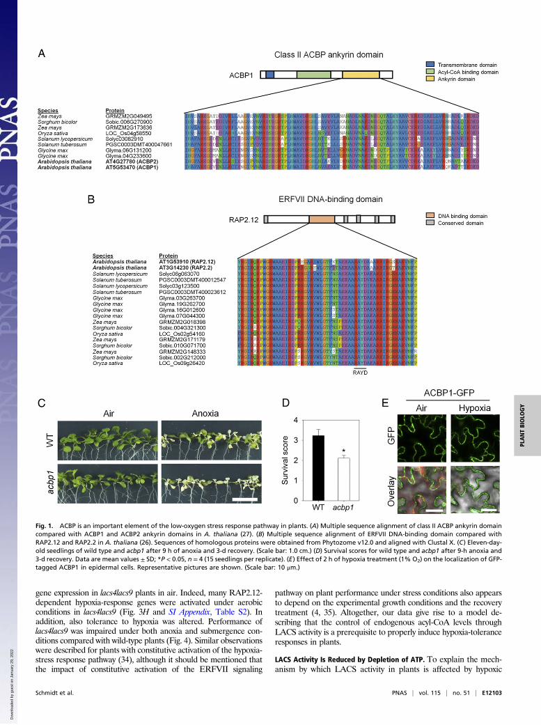
ResultsC18:1-CoA Promotes Dissociation of the ACBP:RAP2.12 Complex inVitro and in Vivo. In Arabidopsis, the ERFVII transcription fac-tor RAP2.12 is constitutively expressed but sequestered at theplasma membrane by binding to ACBP1 during normoxic con-ditions, while it accumulates in the nucleus under oxygen con-centrations below 10% (vol/vol) (3, 7). The interaction domainsof ACBP1 and RAP2.12 were previously identified in Arabi-dopsis (3, 26, 27) and appear to be well conserved among plantspecies (Fig. 1 A and B), indicating that complex formation ofboth proteins is a general feature in plants. Importantly, changingthe expression of ACBP1 affects tolerance to low oxygen (28) (Fig.1 C and D) similar to what was shown previously for RAP2.12 (3).During hypoxia GFP-tagged ACBP1 remains at the plasmamembrane (Fig. 1E), while GFP-tagged RAP2.12 was shown toaccumulate in the nucleus upon hypoxia (3, 7). This indicates thatRAP2.12 dissociates from ACBP1 before its relocation to thenucleus. Consequently, the release of RAP2.12 from ACBP1 isconsidered as a trigger that activates adaptive gene expression inresponse to hypoxia.To investigate if acyl-CoAs interfere with the interaction be-
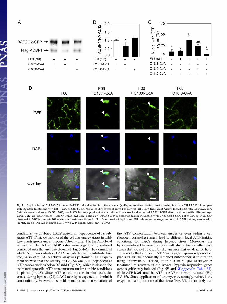
tween ACBP1 and RAP2.12, we performed an in vitro affinityassay (SI Appendix, Fig. S1) in the presence of either oleoyl-CoA(C18:1-CoA), which is a preferred substrate for ACBP1, orpalmitoyl-CoA (C16:0-CoA) that is not a strongly interactingagent (29, 30). In vitro exposure of an ACBP1:RAP2.12 proteincomplex to C18:1-CoA, but not C16:0-CoA, significantly de-creased the binding affinity between the two proteins as indicatedby the reduced ratio of Flag-tagged ACBP1 and CFP-taggedRAP2.12 protein (Fig. 2 A and B). Apparently, interaction be-tween ACBP1 and C18:1-CoA reduces the binding capacity ofACBP1 for the transcription factor RAP2.12. To confirm thatC18:1-CoA–induced dissociation of RAP2.12 from ACBP1 alsooccurs in vivo, we exposed detached leaves of plants expressing35S:RAP2.12-GFP to various acyl-CoAs under aerobic conditions.
Application of C18:1-CoA, but not C18:0-CoA or C16:0-CoA,significantly induced nuclear accumulation of RAP2.12-GFP, in-dicating that RAP2.12 was released from ACBP1 in vivo after theapplication of C18:1-CoA, but not of C16:0-CoA (Fig. 2 C and D).Translocation of RAP2.12 to the nucleus was previously describedto occur during hypoxic conditions (3, 7). Therefore, we tested ifapplication of acyl-CoA to leaves might have induced hypoxia dueto increased beta-oxidation or mitochondrial respiration. How-ever, no increase of the oxygen consumption rate by leaf tissueafter treatment with acyl-CoAs was observed, indicating that ourexperimental treatment did not affect the oxygen concentration ofthe tissue in this experiment (SI Appendix, Fig. S2). We concluded,remobilization of the transcription factor RAP2.12 from theplasma membrane into the nucleus can be triggered in vivo byincreasing the level of C18:1-CoA.
Acyl-CoAs Provoke Distinct Transcript Responses. To determine ifspecific transcriptional responses are provoked by the applica-tion of different acyl-CoAs, RNA-Seq transcriptome analysis wasperformed on wild-type seedlings exposed to either 1 mM C18:1-CoA, C18:0-CoA, or C16:0-CoA. Uptake of these externallyapplied acyl-CoAs is mediated by ABCD transporters that firstcleave the CoA group from the acyl chain, allowing the resultingfatty acid to cross lipid membranes. Once inside the cell, CoA isimmediately reattached, which traps the acyl-CoA in the cellularcompartment in which it has been imported (31). This analysisrevealed that each of these acyl-CoAs modulates distinct sets ofgenes (Fig. 3A and Dataset S1). The high specificity of inducedchanges in gene expression underlines the eligibility of acyl-CoAsas signaling molecules in plants. To investigate the biologicalfunction of the differentially regulated genes, a Gene Ontology(GO) enrichment analysis (32) was carried out. While applica-tion of C18:0-CoA or C16:0-CoA mainly affected the expressionof genes related to reproductive development and hormonesignaling, C18:1-CoA mainly modulated the expression of genesassociated with hypoxia and low-oxygen responses (Fig. 3B andDataset S2). This result was confirmed by qPCR-assisted ex-pression profiling executed on wild-type plants incubated withC18:1-CoA. Indeed, RAP2.12-regulated hypoxia-response geneswere induced by C18:1-CoA treatment, while C18:0-CoA andC16:0-CoA had only minor effects on the expression of thesegenes (Fig. 3C and SI Appendix, Table S1). This observation isreadily explained by our earlier observation that RAP2.12 reloc-alizes to the nucleus upon C18:1-CoA application (Fig. 2 C andD). Therefore, it is concluded that C18:1-CoA provides a specificcellular signal that is substantially involved in the control of geneexpression by releasing RAP2.12 from ACBP1.
Increase of C18:1-CoA to C16:0-CoA Ratio Induces Hypoxic GeneExpression in Vivo. In a physiological context, C18:1-CoA–mediateddissociation of RAP2.12 from ACBP1 only makes sense when theendogenous acyl-CoA pool responds to hypoxia. Indeed, HPLC-assisted quantification of acyl-CoAs revealed a shift to elevatedC18:1-CoA (Fig. 3D) and C20:0-CoA (SI Appendix, Fig. S3) levelswith less C16:0-CoA (Fig. 3E) after 3 h of hypoxia, while no sig-nificant changes of the total pool of acyl-CoAs included in ouranalyses were observed (SI Appendix, Fig. S3). These dynamic re-sponses of specific acyl-CoAs to changing environmental conditionsas exemplified here for hypoxia are in line with the suggestion thatacyl-CoAs in plants can play a role in stress signaling.A similar shift of the acyl-CoA pool as observed during low-
oxygen conditions was observed in transgenic plants in which twoLACS genes, LACS4 and -9, were knocked out (33) (Fig. 3 F andGand SI Appendix, Fig. S4). Under aerobic conditions, lacs4lacs9double knockout plants have elevated C18:1-CoA and reducedC16:0-CoA levels. To provide further proof that endogenouschanges of the C18:1-CoA or C16:0-CoA level can have an effect onlow-oxygen responses of plants, we tested the induction of hypoxic
E12102 | www.pnas.org/cgi/doi/10.1073/pnas.1809429115 Schmidt et al.
Dow
nloa
ded
by g
uest
on
Janu
ary
29, 2
022
gene expression in lacs4lacs9 plants in air. Indeed, many RAP2.12-dependent hypoxia-response genes were activated under aerobicconditions in lacs4lacs9 (Fig. 3H and SI Appendix, Table S2). Inaddition, also tolerance to hypoxia was altered. Performance oflacs4lacs9 was impaired under both anoxia and submergence con-ditions compared with wild-type plants (Fig. 4). Similar observationswere described for plants with constitutive activation of the hypoxia-stress response pathway (34), although it should be mentioned thatthe impact of constitutive activation of the ERFVII signaling
pathway on plant performance under stress conditions also appearsto depend on the experimental growth conditions and the recoverytreatment (4, 35). Altogether, our data give rise to a model de-scribing that the control of endogenous acyl-CoA levels throughLACS activity is a prerequisite to properly induce hypoxia-toleranceresponses in plants.
LACS Activity Is Reduced by Depletion of ATP. To explain the mech-anism by which LACS activity in plants is affected by hypoxic
Fig. 1. ACBP is an important element of the low-oxygen stress response pathway in plants. (A) Multiple sequence alignment of class II ACBP ankyrin domaincompared with ACBP1 and ACBP2 ankyrin domains in A. thaliana (27). (B) Multiple sequence alignment of ERFVII DNA-binding domain compared withRAP2.12 and RAP2.2 in A. thaliana (26). Sequences of homologous proteins were obtained from Phytozome v12.0 and aligned with Clustal X. (C) Eleven-day-old seedlings of wild type and acbp1 after 9 h of anoxia and 3-d recovery. (Scale bar: 1.0 cm.) (D) Survival scores for wild type and acbp1 after 9-h anoxia and3-d recovery. Data are mean values ± SD; *P < 0.05, n = 4 (15 seedlings per replicate). (E) Effect of 2 h of hypoxia treatment (1% O2) on the localization of GFP-tagged ACBP1 in epidermal cells. Representative pictures are shown. (Scale bar: 10 μm.)
Schmidt et al. PNAS | vol. 115 | no. 51 | E12103
PLANTBIOLO
GY
Dow
nloa
ded
by g
uest
on
Janu
ary
29, 2
022
conditions, we analyzed LACS activity in dependence of its sub-strate ATP. First, we monitored the cellular energy status in wild-type plants grown under hypoxia. Already after 2 h, the ATP levelas well as the ATP-to-ADP ratio were significantly reducedcompared with the air-treated control (Fig. 5 A–C). To examine atwhich ATP concentration LACS activity becomes substrate lim-ited, an in vitro LACS activity assay was performed. This experi-ment showed that the activity of LACS4 was ATP dependent atATP concentrations below 0.8 mM (Fig. 5D), which is close to theestimated cytosolic ATP concentration under aerobic conditionsin planta (36–38). Since ATP concentrations in plant cells de-crease during hypoxia (24), LACS activity is expected to diminishconcomitantly. However, it should be mentioned that variations of
the ATP concentration between tissues or even within a cell(between organelles) might lead to different local ATP-limitingconditions for LACS during hypoxic stress. Moreover, thehypoxia-induced low-energy status will also influence other pro-cesses that are not covered by the analyses that we describe here.To verify that a drop in ATP can trigger hypoxia responses of
plants in air, we chemically inhibited mitochondrial respirationusing antimycin-A. Indeed, after 3 h of 50 μM antimycin-Atreatment of rosettes in air, several hypoxia-responsive geneswere significantly induced (Fig. 5E and SI Appendix, Table S3),while ATP levels and the ATP-to-ADP ratio were reduced (Fig.5 F–H). Since application of antimycin-A strongly reduced theoxygen consumption rate of the tissue (Fig. 5I), it is unlikely that
Fig. 2. Application of C18:1-CoA induces RAP2.12 relocalization into the nucleus. (A) Representative Western blot showing in vitro ACBP1:RAP2.12 complexstability after treatment with C18:1-CoA or C16:0-CoA. Pluronic F68 treatment served as control. (B) Quantification of ACBP1-to-RAP2.12-ratio as shown in A.Data are mean values ± SD *P < 0.05, n = 8. (C) Percentage of epidermal cells with nuclear localization of RAP2.12-GFP after treatment with different acyl-CoAs. Data are mean values ± SD; *P < 0.05. (D) Localization of RAP2.12-GFP in detached leaves incubated with 0.1% C18:1-CoA, C18:0-CoA or C16:0-CoAdissolved in 0.01% pluronic F68 under normoxic conditions for 3 h. Treatment with pluronic F68 only served as negative control. DAPI staining was used toidentify nuclei. Arrows indicate nuclei with GFP signal. (Scale bar: 10 μm.)
E12104 | www.pnas.org/cgi/doi/10.1073/pnas.1809429115 Schmidt et al.
Dow
nloa
ded
by g
uest
on
Janu
ary
29, 2
022

the induction of hypoxia-responsive genes is the consequence oflow-oxygen concentrations here. Also an antimycin-A–inducedROS burst appeared unlikely to be responsible for the gene in-duction, since the induced genes responded similarly when inaddition to antimycin-A also 1 mM of the hydrogen peroxidescavenger dimethylthiourea (DMTU) (39, 40) was supplied (Fig.5E). Although it cannot be concluded that reduced ATP levelsare exclusively responsible for triggering hypoxia responses inplants without performing dose–response analyses of individualand combined compounds, the data provide evidence that the
cellular energy status is involved in the regulation of hypoxicgene expression.
DiscussionIn this study, we demonstrate that acyl-CoAs provoke distincttranscriptional responses in plants, suggesting that they are involvedin different signaling pathways. Specifically, binding of C18:1-CoAto ACBP triggers dissociation of the ACBP:RAP2.12 complex uponan hypoxia-induced energy crisis, resulting in mobilization of thetranscription factor RAP2.12 into the nucleus. Consequently,RAP2.12-mediated gene expression is induced. Therewith we reveal
Fig. 3. Changing oleoyl-CoA levels induces low-oxygen responsive gene expression. (A) Number of significantly differentially expressed genes in leaves after1.5-h treatment with different acyl-CoAs as determined by RNA-Seq (FDR-adjusted P value <0.05). (B) Number of GO classes in which differentially expressedgenes are significantly overrepresented under acyl-CoA treatments as shown in A. (C) qPCR analysis of differential expression of hypoxia-responsive genesafter acyl-CoA treatment in air (reference: F68 only). Data are presented as mean values *P < 0.05, n = 5. (D) C18:1-CoA levels increase upon hypoxia in wildtype. Data are mean values ± SD; *P < 0.05, n = 3. (E) C16:0-CoA levels decrease upon hypoxia in wild type. Data are mean values ± SD; *P < 0.05, n = 3. (F)C18:1-CoA levels are increased in lacs4lacs9 double mutants grown in air. Data are mean values ± SD; *P < 0.05, n = 3. (G) C16:0-CoA levels are lowered inlacs4lacs9 double mutants. Data are mean values ± SD; *P < 0.05, n = 3. (H) Expression data for hypoxia-responsive genes comparing wild type and lacs4-1 lacs9-2in air or hypoxia (2 h 1% O2; mean values, *P < 0.05, n = 4).
Schmidt et al. PNAS | vol. 115 | no. 51 | E12105
PLANTBIOLO
GY
Dow
nloa
ded
by g
uest
on
Janu
ary
29, 2
022

the trigger of the ERFVII-mediated signaling cascade to activatecellular hypoxia-tolerance responses in plants (Fig. 6).When the oxygen availability to cells diminishes, mitochondria
produce less ATP due to a reduced activity of oxidative phos-phorylation (41). Indeed, our plants showed a rapid decrease oftheir energy status upon hypoxia. Already within 2 h, the ATP-to-ADP ratio dropped significantly and remained decreasingthroughout the rest of the experiment (Fig. 5C). Consequently,the activity of ATP-mediated reactions within the cell is expectedto reduce too (24). Here, we show that ATP concentration-dependent LACS activity reaches its maximum at an ATP con-centration of 1 mM (Fig. 5D) which resembles the concentrationin a nonstressed plant cell (36–38). This means that any decreaseof the cellular energy status is translated into a reduction ofLACS activity. The enzyme LACS is among others located in theplastidial outer envelope where it is provided with C16:0 andC18:1 fatty acids from the stroma by thioesterases that are lo-cated in the inner envelope (42, 43). Using CoA and ATP ascosubstrates, LACS activates fatty acids and releases them asC16:0-CoA and C18:1-CoA into the cytosol (18). When the levelof ATP drops and LACS activity decreases, the export rate offatty acids will be reduced. As the elongation and rapid desatu-ration reactions in the plastidial stroma from C16:0 to C18:1commence, the ratio C18:1 compared with C16:0 that is providedto LACS from the stroma is likely to increase through time underthese conditions. As a consequence, the ratio of C18:1-CoA to
C16:0-CoA that is released by LACS into the cytosol will in-crease and would readily explain why we observe an increasedlevel of C18:1-CoA compared with C16:0-CoA in plants thatwere exposed to low oxygen (Fig. 3 D and E) as well as in lac-s4lacs9 double knockout lines in air (Fig. 3 F and G).Acyl-CoA fatty acid esters bind to ACBP proteins. Here, we
show that specifically the interaction between C18:1-CoA withACBP1 results in release of RAP2.12 from the ACBP1:RAP2.12 complex while C16:0-CoA does not affect the in-teraction between RAP2.12 and ACBP1 (Fig. 2). A shift of theratio between C18:1-CoA and C16:0-CoA as described above,will therefore lead to the release of RAP2.12 from ACBP1. In-deed, application of C18:1-CoA, but not C16:0-CoA, increasedthe number of nuclei in which GFP-tagged RAP2.12 accumu-lated (Fig. 2 C and D). Consequently, also up-regulation ofhypoxia-responsive marker genes was observed (Fig. 3 A–C).Similar to this external application of acyl-CoAs, also an en-
dogenous shift of the C18:1-CoA to C16:0-CoA ratio as observedin lacs4lacs9 mutant lines resulted in the up-regulation ofhypoxia-responsive genes already during aerobic conditions (Fig.3H). Altogether, these data describe how the ultimate trigger forrelease of RAP2.12 from ACBP1 is constituted by an energycrisis-provoked response of the acyl-CoA pool under hypoxia(Fig. 6).Changes of the cellular energy status happen all of the time as
most biotic and abiotic stress conditions affect energy metabolism
Fig. 4. Decreased tolerance of lacs4 lacs9 knockout lines to anoxia and submergence. (A) Eleven-day-old seedlings of wild type and lacs4-1 lacs9-2 after 9 h ofanoxia and 3-d recovery. (Scale bar: 1.0 cm.) (B) Survival scores for wild type and lacs4-1 lacs9-2 after 9-h anoxia and 3-d recovery. Data are mean values ± SD;*P < 0.05, n = 4 (15 seedlings per replicate). (C) Phenotype of wild type and lacs4 lacs9 mutant grown in air (control), or after 3- or 4-d submergence-inducedhypoxic treatment. (Scale bar: 2 cm.) Photographs were taken 4 d after the submergence treatment. (D) Absolute dry weight of wild-type and lacs4-1 lacs9-2plants grown in air. Data represent mean ± SD (three replicate experiments with every 12 plants per genotype). Asterisk indicates significant differences afterone-way ANOVA (P < 0.05). (E) Absolute fresh weight of wild-type and lacs4-1 lacs9-2 plants grown in air. Data represent mean ± SD (three replicate ex-periments with every 12 plants per genotype). Asterisk indicates significant differences after one-way ANOVA (P < 0.05). (F) Relative fresh weight of wild-typeand lacs4-1 lacs9-2 plants grown in air, or after 3 or 4 d of submergence followed by 4 d of recovery. Data represent mean ± SD (three replicate experimentswith every 12 plants per genotype). Asterisk indicates significant differences after one-way ANOVA (P < 0.05). (G) Percentage of plants that survived the 3 or4 d of flooding-induced hypoxia, respectively (mean values ± SD, three replicate experiments with every 12 plants per genotype). *P < 0.05 according to one-way ANOVA.
E12106 | www.pnas.org/cgi/doi/10.1073/pnas.1809429115 Schmidt et al.
Dow
nloa
ded
by g
uest
on
Janu
ary
29, 2
022

in one way or another (44). It would be most detrimental for plantfitness, when each fluctuation of the ATP level immediately led tothe activation of hypoxic gene expression (34). Therefore, thelifetime of ERFVII proteins depends on the actual cellular oxygenconcentration via the Cys branch of the N-end rule for proteaso-mal protein degradation (3, 4, 11). Only when an energy crisisis provoked by low-oxygen conditions, the stabilization ofRAP2.12 enables the protein to accumulate in the nucleus in asufficient amount to activate hypoxic gene expression. However,when RAP2.12 is released from ACBP1 due to an energy deficitthat is not related to low-oxygen stress, the protein will be rapidly
degraded due to proteasomal activity. Therefore, we propose thatthe ACBP1:RAP2.12 complex forms the initial hub capable ofintegrating signal inputs related to the cellular energy charge withoxygen concentration-dependent determination of the lifetime ofRAP2.12 protein (Fig. 6). Subsequently, RAP2.12 protein that isnewly synthesized after the onset of hypoxia does still undergo N-end rule-assisted stabilization but may not be linked directly to theenergy status of the cell.Constitutive activation of the molecular stress response to low
oxygen in the lacs4lacs9 mutant background led to reduced tol-erance to low oxygen as well as to flooding stress (Fig. 4). This
Fig. 5. Decreasing the cellular ATP level constitutes limiting conditions for LACS activity and induces the expression of low-oxygen responsive genes. (A) ATPlevels under hypoxia (mean ± SD, *P < 0.05, n = 5). (B) Concentration of ADP in wild-type seedlings grown under long-day conditions and exposed to hypoxia.Data shown are given in nanomoles per gram fresh weight and represent the mean ± SD of independent replicates (n = 5). (C) ATP-to-ADP-ratio underhypoxia (mean ± SD, *P < 0.05, n = 5). (D) In vitro LACS activity depends on ATP concentration (mean ± SD, *P < 0.05, n = 5). The gray area marks the ATP-concentration range usually determined in plant cells. (E) Differential expression of hypoxia-responsive genes after 3 h of 1 mM DMTU and/or 50 μMantimycin-A (AA) treatment under aerobic conditions (reference: mock-treated control). Data are presented as mean ± SD, *P < 0.05, n = 5. (F) ATP levels after3 h of 50 μM AA treatment (mean ± SD, *P < 0.05, n = 5). (G) Concentration of ADP in wild-type seedlings exposed to 3 h of 50 μM AA treatment. Datarepresent mean ± SD (n = 5). Asterisk indicates significant differences after one-way ANOVA (P < 0.05). (H) ATP-to-ADP-ratio after 3 h of 50 μM AA treatment(mean ± SD, *P < 0.05, n = 5). (I) Oxygen consumption rate in wild-type leaves upon 3 h of 50 μM AA treatment. Data represent mean ± SD (n = 7). Asteriskindicates significant difference after Student’s t test (P < 0.05).
Schmidt et al. PNAS | vol. 115 | no. 51 | E12107
PLANTBIOLO
GY
Dow
nloa
ded
by g
uest
on
Janu
ary
29, 2
022

observation is congruent with earlier observations that consti-tutive activation of the hypoxia-stress response in plants viaoverexpression of a stable version of RAP2.12 protein reducedtolerance to hypoxia (34), although other studies indicate thatthe latter phenotype is likely conditional to growth conditionsand recovery treatment too (4, 34). This underlines the impor-tance of a timely control of stress responses that are optimallyadjusted to the actual environmental conditions. The integrationof (i) energy-dependent changes in C18:1-CoA levels as cellulartrigger signal with (ii) the homeostatic control of the lifetime ofRAP2.12 in an oxygen concentration-dependent manner pro-vides a highly specific control mechanism to initiate hypoxic re-sponses. The mechanism guarantees that a full low-oxygenresponse is activated only when hypoxia is detrimental for theplant’s energy status.The ATP dependence of oleoyl-CoA synthesis by LACS in
combination with the impact of oleoyl-CoA on the interactionbetween RAP2.12 and ACBP exposes mitochondrial activity asan early trigger for hypoxia signaling. Consistently, manipulatingmitochondrial ATP synthesis using inhibitors of specific re-spiratory complexes like antimycin-A induced RAP2.12-controlledhypoxic gene expression even under aerobic conditions (45) (Fig.5E). It is striking that the induction of hypoxic gene expression inair by acyl-CoAs (Fig. 3 C and H) or antimycin-A (Fig. 5E) waslower compared with the induction of these genes by hypoxicconditions (Fig. 3H). This observation stresses the impact of ad-ditional oxygen-dependent regulatory mechanisms, such as the N-end rule-mediated reduction of RAP2.12 lifetime in air. In thiscontext, it is worth mentioning that the control of low-oxygenstress responses is not only linked to the oxygen and energy sta-tus of the cell, but it is also known to be influenced by othercellular factors such as nitric oxide (46, 47), hydrogen peroxide(48), calcium (49, 50), and potassium (51). In the near future, itwill be intriguing to expand the mechanistic explanation of howthe energy and oxygen status of a cell is integrated upon low-oxygen stress with these additional signaling components (12).ACBPs are found in all kingdoms of life, while ERFVIIs are
highly conserved among higher plants. Moreover, the interactiondomains of both protein families are highly conserved in plants(Fig. 1). Therefore, the ACBP:ERFVII signaling hub as pre-sented here may represent a universal mechanism in plants toinitiate hypoxia-induced stress responses via the integration ofmultiple cellular signals. Moreover, the specific interaction ofACBPs with various acyl-CoAs on the one hand (Fig. 2) and thedistinct cellular responses provoked by individual acyl-CoAs onthe other hand (Fig. 3 A and B) suggest that many additional
possibilities may exist of how acyl-CoAs can modulate cellularsignaling pathways in plants.
Materials and MethodsPlant Materials. Arabidopsis thaliana ecotype Col-0 was used as wild type forall analyses. The 35S:RAP2.12-GFP line and lacs4-1 lacs9-2 and lacs4-2 lacs9-7double knockout lines were described previously (3, 33). The acbp1 knockoutline (SAIL_683_C03) was obtained from the Nottingham Arabidopsis StockCenter (SI Appendix, Fig. S5).
Growth Conditions and Analysis of Oxygen Deprivation Response. For testinganoxia tolerance of seedlings, seeds were sown on half-strength MS mediumcontaining 0.5% (wt/vol) sucrose, stratified for 48 h at 4 °C, and germinatedat 21 °C day/18 °C night with a photoperiod of 16 h light (150 μmol·m−2·s−1)and 8 h dark. At day 11, seedlings were exposed to full anoxia, by placing theculture plates in an environment containing 100% nitrogen, for 9 h in the darkto avoid photosynthetic oxygen production. After 3 d of recovery, the survivalscore was determined as previously described (4). For submergence assays,seeds were sown in moist soil, stratified at 4 °C in the dark for 48 h, andgerminated at 21 °C day/18 °C night with a photoperiod of 16 h light and 8 hdarkness. The 4-wk-old plants were submerged in water in 40-cm high plasticcontainers and kept in the dark to avoid photosynthetic oxygen production.Leaves stayed 10 cm under the water surface. After 3 or 4 d, water was re-moved from the boxes and plants were placed back under photoperiodicconditions (16 h/8 h, light/dark). Submergence tolerance was assayed after 4 dof recovery.
For acyl-CoA pool measurements, plants were grown on horizontal agarplates containing 0.8% agar in 1/2 MS medium (pH 5.7) with 15 mM sucrosefor 2 wk under long-day conditions (16 h in 150 μmol photons·m−2·s−1 at 21 °Cand 8 h in 0 μmol photons·m−2·s−1 at 19 °C). After 2.5 h in light, they weresubjected to hypoxia in the dark by exposition to a stream of air containing1% (vol/vol) oxygen, supplemented with nitrogen and 350 ppm carbon di-oxide. Plants were harvested by freezing in liquid nitrogen after 2 h, 3 h, and4 h of hypoxia. For normoxic control, untreated plants were harvested si-multaneously with the start of hypoxic treatment.
For expression analysis after acyl-CoA treatment, wild-type seeds weresown in 24-well plates containing half-strength liquid MS medium, stratifiedfor 48 h at 4 °C, and germinated at 21 °C day/18 °C night with a photoperiodof 16 h light (150 μmol·m−2·s−1) and 8 h dark. At day 14, seedlings weretreated with different acyl-CoAs at a final concentration of 0.1% in 0.01%pluronic F68. Pluronic is a nonfatty acid-based detergent which means thatthe detergent properties of pluronic in solubilizing fatty acyls are not con-founded by fatty acyls derived from the detergent itself (52).
Cloning of Constructs. Coding sequences (CDSs) were amplified from a cDNAtemplate using Phusion High Fidelity DNA polymerase (Thermo Fisher Sci-entific). The ACBP1 CDS was cloned into pENTR-D and recombined intopK7FWG2 (51) to fuse it with GFP. For in vitro expression, the CDS ofACBP1 was fused N terminally with a Flag tag by PCR and cloned into pF3AWG BYDV (Promega), while the CDS of RAP2.12 was fused C terminally to
Fig. 6. Triggering low-oxygen responses in plants integrates the cellular energy and oxygen status via modulation of oleoyl-CoA levels. Oxygen limitationreduces cellular ATP levels, which results in increased C18:1-CoA levels. Dissociation of ERFVII protein (as shown here for RAP2.12) bound to ACBP1 at theplasma membrane is promoted by C18:1-CoA. Free ERFVII protein is stable under low-oxygen conditions and relocalizes into the nucleus to activate hypoxicresponses.
E12108 | www.pnas.org/cgi/doi/10.1073/pnas.1809429115 Schmidt et al.
Dow
nloa
ded
by g
uest
on
Janu
ary
29, 2
022
CFP by PCR and cloned into pF3A WG BYDV (Promega). A complete list of allprimers used is provided in SI Appendix, Table S4.
Plant Transformation. Transgenic ACBP1-GFP plants were generated by trans-forming wild-type plants with the vector pK7FWG2 (53) containing the ACBP1CDS fused in frame to GFP at its C terminus. T0 seeds were screened forkanamycin resistance and the presence of GFP signals by confocal microscopy.
qRT-PCR. RNA extraction, digestion of genomic DNA, cDNA synthesis, andqRT-PCR analysis were performed as described previously (54). For all experi-ments, four to five independent biological replicates were used, as indicated inthe figure and table legends. For normalization, UBIQUITIN10 expression wasused according to ref. 3. Primers for hypoxia core genes, LACS genes, andUBI10 are given in SI Appendix, Table S4.
RNA-Seq Analysis. Illumina HiSeq sequencing was performed according tostandardized protocols as described in detail in SI Appendix. Transcriptomeanalysis was performed by means of CLC Genomics Workbench v.6 using theA. thaliana reference sequence (Tair10). Expression values were normalizedusing quantile normalization and pairwise statistical analyses comparing thetreatments performed using false discovery rate (FDR)-corrected P valuesbased on Baggerly’s test (55).
Confocal Imaging. For protein localization studies, GFP signals were imagedand analyzed with a Leica DM6000 TCS SP8 confocal microscope (LeicaMicrosystems). Nuclear staining was performed by using DAPI (molecularprobes) according to the manufacturer’s instructions. For quantification ofnuclear translocation of RAP2.12 after acyl-CoA treatment, 20 DAPI-stainednuclei per plant (five plants in total) were analyzed per treatment. Leaves of5-wk-old soil-grown 35S:RAP2.12-GFP plants were incubated for 3 h withdifferent acyl-CoAs at a final concentration of 0.1% in 0.01% pluronic F68 ina 24-well plate under continuous shaking in the light. The experiments wererepeated three times.
In Vitro Binding Assay. Themethod to determine whether acyl-CoAs affect theinteraction between ACBP1 and RAP2.12 is explained in detail in SI Appendix,Supplementary Information Text and Fig. S1. In brief, both proteins weresynthesized using wheat germ extract (54). The full CDS of RAP2.12 wasfused C terminally with the CDS of CFP, and the CDS of ACBP1 was N-terminally fused with a FLAG tag. RAP2.12-CFP protein was bound to GFP–Trap-A beads (Chromotek) and incubated with ACBP1 protein overnight.Subsequently, beads were resuspended in wash buffer containing different
acyl-CoAs at a final concentration of 0.1% in 0.01% pluronic F68, or as amock control of only 0.01% F68. After one night of incubation, the bufferwas replaced and the composition of the protein complex retained to thebeads was analyzed on a Western blot.
Analysis of Acyl-CoA Esters. Acyl-CoAs were extracted, derivatized, and an-alyzed using HPLC as described earlier (56). Detailed information about themethod is provided in SI Appendix.
LACS4 in Vitro Enzyme Assay. The in vitro LACS enzyme assay was carried outas described previously (17) using protein that was heterologously expressedin Escherichia coli (57). Details of the method are explained in SI Appendix.
ATP and ADP Quantification. ATP and ADP were extracted with 16% tri-chloroacetic acid (33) and analyzed after derivatization by HPLC as describedpreviously (58). Details of the procedure are described in SI Appendix.
Analysis of Oxygen Consumption Rates. For the determination of oxygenconsumption rates, 4-wk-old plants grown on soil under short-day conditions(8 h in 160 μmol photons m−2·s−1 at 20 °C and 16 h in 0 μmol photons m−2·s−1
at 16 °C) were used. Starting 3 h before measurement, the plants weresprayed every hour either with 50 mM Mes buffer (pH 6.5) containing 50 μMantimycin-A (59) or with 50 mM Mes buffer for control. This treatment wasdone in the last hour of dark phase and 2 h into the light phase, and thenmeasurement of oxygen consumption rates at normoxic conditions wasperformed as described before (34) using the respective spraying solution. Ina similar setup, the effect of different acyl-CoAs on the oxygen consumptionrate was tested by incubating leaf disks in acyl-CoAs at a final concentrationof 0.1% in 0.01% pluronic F68.
Statistical Analysis. Statistical evaluation of significant variation betweentreatments or genotypes was done by performing Student’s t test or one-wayANOVA where appropriate.
Data Availability. The RNA-Seq gene expression data are available in NCBI’sGene Expression Omnibus (GEO) through GEO Series accession no. GSE97186.
ACKNOWLEDGMENTS. We thank Sandro Parlanti and Frauke Augstein forvaluable support. This work was supported by grants (to J.T.v.D.) (DO 1298/2-2)and (to P.G.) (GE 878/7-2) from the German Science Foundation (DFG). M.F.was supported by the DFG (Grant DFG FU 430/5-1).
1. Food and Agricultural Organization of the United Nations (FAO) (2015) The impact of
disasters on agriculture and food security. (Food and Agricultural Organization of the
United Nations, Rome) Report I5128E/1/11.15.2. Hirabayashi Y, et al. (2013) Global flood risk under climate change. Nat Clim Chang 3:
816–821.3. Licausi F, et al. (2011) Oxygen sensing in plants is mediated by an N-end rule pathway
for protein destabilization. Nature 479:419–422.4. Gibbs DJ, et al. (2011) Homeostatic response to hypoxia is regulated by the N-end rule
pathway in plants. Nature 479:415–418.5. Bui LT, Giuntoli B, Kosmacz M, Parlanti S, Licausi F (2015) Constitutively expressed ERF-
VII transcription factors redundantly activate the core anaerobic response in Arabi-
dopsis thaliana. Plant Sci 236:37–43.6. Gasch P, et al. (2016) Redundant ERF-VII transcription factors bind to an evolutionarily
conserved cis-Motif to regulate hypoxia-responsive gene expression in Arabidopsis.
Plant Cell 28:160–180.7. Kosmacz M, et al. (2015) The stability and nuclear localization of the transcription
factor RAP2.12 are dynamically regulated by oxygen concentration. Plant Cell Environ
38:1094–1103.8. Li HY, Chye ML (2004) Arabidopsis Acyl-CoA-binding protein ACBP2 interacts with an
ethylene-responsive element-binding protein, AtEBP, via its ankyrin repeats. Plant
Mol Biol 54:233–243.9. Abbas M, et al. (2015) Oxygen sensing coordinates photomorphogenesis to facilitate
seedling survival. Curr Biol 25:1483–1488.10. van Dongen JT, Licausi F (2015) Oxygen sensing and signaling. Annu Rev Plant Biol 66:
345–367.11. Weits DA, et al. (2014) Plant cysteine oxidases control the oxygen-dependent branch
of the N-end-rule pathway. Nat Commun 5:3425.12. Schmidt RR, Weits DA, Feulner CFJ, van Dongen JT (2018) Oxygen sensing and in-
tegrative stress signaling in plants. Plant Physiol 176:1131–1142.13. Grevengoed TJ, Klett EL, Coleman RA (2014) Acyl-CoA metabolism and partitioning.
Annu Rev Nutr 34:1–30.14. MengW, Su YC, Saunders RM, Chye ML (2011) The rice acyl-CoA-binding protein gene
family: Phylogeny, expression and functional analysis. New Phytol 189:1170–1184.
15. Neess D, Bek S, Engelsby H, Gallego SF, Færgeman NJ (2015) Long-chain acyl-CoA
esters in metabolism and signaling: Role of acyl-CoA binding proteins. Prog Lipid
Res 59:1–25.16. Lung SC, Chye ML (2016) Deciphering the roles of acyl-CoA-binding proteins in plant
cells. Protoplasma 253:1177–1195.17. Shockey JM, Fulda MS, Browse JA (2002) Arabidopsis contains nine long-chain acyl-
coenzyme a synthetase genes that participate in fatty acid and glycerolipid metab-
olism. Plant Physiol 129:1710–1722.18. Koo AJ, Ohlrogge JB, Pollard M (2004) On the export of fatty acids from the chlo-
roplast. J Biol Chem 279:16101–16110.19. Li N, Xu C, Li-Beisson Y, Philippar K (2016) Fatty acid and lipid transport in plant cells.
Trends Plant Sci 21:145–158.20. Troncoso-Ponce MA, Nikovics K, Marchive C, Lepiniec L, Baud S (2016) New insights on
the organization and regulation of the fatty acid biosynthetic network in the model
higher plant Arabidopsis thaliana. Biochimie 120:3–8.21. Hertz R, Magenheim J, Berman I, Bar-Tana J (1998) Fatty acyl-CoA thioesters are li-
gands of hepatic nuclear factor-4alpha. Nature 392:512–516.22. DiRusso CC, Heimert TL, Metzger AK (1992) Characterization of FadR, a global tran-
scriptional regulator of fatty acid metabolism in Escherichia coli. Interaction with the
fadB promoter is prevented by long chain fatty acyl coenzyme A. J Biol Chem 267:
8685–8691.23. Lung SC, et al. (2018) Arabidopsis ACYL-COA-BINDING PROTEIN1 interacts with STE-
ROL C4-METHYL OXIDASE1-2 to modulate gene expression of homeodomain-leucine
zipper IV transcription factors. New Phytol 218:183–200.24. Geigenberger P (2003) Response of plant metabolism to too little oxygen. Curr Opin
Plant Biol 6:247–256.25. Grattan RP, Roger SC (1981) Acyl-CoA may be a neglected product in studies of fatty
acid synthesis by isolated chloroplasts. FEBS Lett 135:182–186.26. Okamuro JK, Caster B, Villarroel R, Van Montagu M, Jofuku KD (1997) The
AP2 domain of APETALA2 defines a large new family of DNA binding proteins in
Arabidopsis. Proc Natl Acad Sci USA 94:7076–7081.27. Du ZY, Chye ML (2013) Interactions between Arabidopsis acyl-CoA-binding proteins
and their protein partners. Planta 238:239–245.
Schmidt et al. PNAS | vol. 115 | no. 51 | E12109
PLANTBIOLO
GY
Dow
nloa
ded
by g
uest
on
Janu
ary
29, 2
022

28. Xie LJ, et al. (2015) Arabidopsis acyl-CoA-binding protein ACBP3 participates in plant
response to hypoxia by modulating very-long-chain fatty acid metabolism. Plant J 81:
53–67.29. Chye ML, Li HY, Yung MH (2000) Single amino acid substitutions at the acyl-CoA-
binding domain interrupt 14[C]palmitoyl-CoA binding of ACBP2, an Arabidopsis acyl-
CoA-binding protein with ankyrin repeats. Plant Mol Biol 44:711–721.30. Du ZY, Arias T, Meng W, Chye ML (2016) Plant acyl-CoA-binding proteins: An
emerging family involved in plant development and stress responses. Prog Lipid Res
63:165–181.31. Theodoulou FL, Carrier DJ, Schaedler TA, Baldwin SA, Baker A (2016) How to move an
amphipathic molecule across a lipid bilayer: Different mechanisms for different ABC
transporters? Biochem Soc Trans 44:774–782.32. Proost S, et al. (2009) PLAZA: A comparative genomics resource to study gene and
genome evolution in plants. Plant Cell 21:3718–3731.33. Jessen D, Roth C, Wiermer M, Fulda M (2015) Two activities of long-chain acyl-
coenzyme A synthetase are involved in lipid trafficking between the endoplasmic
reticulum and the plastid in Arabidopsis. Plant Physiol 167:351–366.34. Paul MV, et al. (2016) Oxygen sensing via the ethylene response transcription factor
RAP2.12 affects plant metabolism and performance under both normoxia and hyp-
oxia. Plant Physiol 172:141–153.35. Riber W, et al. (2015) The greening after extended darkness1 is an N-end rule path-
way mutant with high tolerance to submergence and starvation. Plant Physiol 167:
1616–1629.36. Gout E, Rébeillé F, Douce R, Bligny R (2014) Interplay of Mg2+, ADP, and ATP in the
cytosol and mitochondria: Unravelling the role of Mg2+ in cell respiration. Proc Natl
Acad Sci USA 111:E4560–E4567.37. Soccio M, Laus MN, Trono D, Pastore D (2013) A new simple fluorimetric method to
assay cytosolic ATP content: Application to durum wheat seedlings to assess modu-
lation of mitochondrial potassium channel and uncoupling protein activity under
hyperosmotic stress. Biologia 68:421–432.38. van Dongen JT, Schurr U, Pfister M, Geigenberger P (2003) Phloem metabolism and
function have to cope with low internal oxygen. Plant Physiol 131:1529–1543.39. Dröse S, Brandt U (2008) The mechanism of mitochondrial superoxide production by
the cytochrome bc1 complex. J Biol Chem 283:21649–21654.40. Guo H, et al. (2016) Plastid-nucleus communication involves calcium-modulated
MAPK signalling. Nat Commun 7:12173.41. Gupta KJ, Zabalza A, van Dongen JT (2009) Regulation of respiration when the ox-
ygen availability changes. Physiol Plant 137:383–391.42. Dörmann P, Voelker TA, Ohlrogge JB (1995) Cloning and expression in Escherichia coli
of a novel thioesterase from Arabidopsis thaliana specific for long-chain acyl-acyl
carrier proteins. Arch Biochem Biophys 316:612–618.
43. Jones A, Davies HM, Voelker TA (1995) Palmitoyl-acyl carrier protein (ACP) thio-esterase and the evolutionary origin of plant acyl-ACP thioesterases. Plant Cell 7:359–371.
44. Baena-González E, Sheen J (2008) Convergent energy and stress signaling. TrendsPlant Sci 13:474–482.
45. Wagner S, Van Aken O, Elsässer M, Schwarzländer M (2018) Mitochondrial energysignaling and its role in the low-oxygen stress response of plants. Plant Physiol 176:1156–1170.
46. Gibbs DJ, et al. (2014) Nitric oxide sensing in plants is mediated by proteolytic controlof group VII ERF transcription factors. Mol Cell 53:369–379.
47. Vicente J, et al. (2017) The Cys-Arg/N-end rule pathway is a general sensor of abioticstress in flowering plants. Curr Biol 27:3183–3190.e4.
48. Pucciariello C, Parlanti S, Banti V, Novi G, Perata P (2012) Reactive oxygen species-driven transcription in Arabidopsis under oxygen deprivation. Plant Physiol 159:184–196.
49. Sedbrook JC, Kronebusch PJ, Borisy GG, Trewavas AJ, Masson PH (1996) TransgenicAEQUORIN reveals organ-specific cytosolic Ca2+ responses to anoxia and Arabidopsisthaliana seedlings. Plant Physiol 111:243–257.
50. Bose J, Pottosin II, Shabala SS, Palmgren MG, Shabala S (2011) Calcium efflux systemsin stress signaling and adaptation in plants. Front Plant Sci 2:85.
51. Wang F, Chen ZH, Shabala S (2017) Hypoxia sensing in plants: On a quest for ionchannels as putative oxygen sensors. Plant Cell Physiol 58:1126–1142.
52. Poirier Y, Erard N, Petétot JM (2001) Synthesis of polyhydroxyalkanoate in the peroxisomeof Saccharomyces cerevisiae by using intermediates of fatty acid beta-oxidation. ApplEnviron Microbiol 67:5254–5260.
53. Karimi M, De Meyer B, Hilson P (2005) Modular cloning in plant cells. Trends Plant Sci10:103–105.
54. Schmidt R, et al. (2013) Salt-responsive ERF1 regulates reactive oxygen species-dependent signaling during the initial response to salt stress in rice. Plant Cell 25:2115–2131.
55. Baggerly KA, Deng L, Morris JS, Aldaz CM (2003) Differential expression in SAGE:Accounting for normal between-library variation. Bioinformatics 19:1477–1483.
56. Larson TR, Graham IA (2001) Technical advance: A novel technique for the sensitivequantification of acyl CoA esters from plant tissues. Plant J 25:115–125.
57. Overath P, Pauli G, Schairer HU (1969) Fatty acid degradation in Escherichia coli. Aninducible acyl-CoA synthetase, the mapping of old-mutations, and the isolation ofregulatory mutants. Eur J Biochem 7:559–574.
58. Zhang L, et al. (2008) Overriding the co-limiting import of carbon and energy intotuber amyloplasts increases the starch content and yield of transgenic potato plants.Plant Biotechnol J 6:453–464.
59. De Clercq I, et al. (2013) Themembrane-boundNAC transcription factor ANAC013 functions inmitochondrial retrograde regulation of the oxidative stress response in Arabidopsis. Plant Cell25:3472–3490.
E12110 | www.pnas.org/cgi/doi/10.1073/pnas.1809429115 Schmidt et al.
Dow
nloa
ded
by g
uest
on
Janu
ary
29, 2
022