Embed Size (px)
Citation preview
Magnetic resonance imaging demonstrates compartmental muscle mechanismsof human vertical fusional vergence
Joseph L. Demer1,2 and Robert A. Clark1
1Department of Ophthalmology, David Geffen Medical School, University of California, Los Angeles, California;and 2Department of Neurology, David Geffen Medical School, University of California, Los Angeles, California
Submitted 5 November 2014; accepted in final form 9 January 2015
Demer JL, Clark RA. Magnetic resonance imaging demonstratescompartmental muscle mechanisms of human vertical fusional ver-gence. J Neurophysiol 113: 2150–2163, 2015. First published January14, 2015; doi:10.1152/jn.00871.2014.—Vertical fusional vergence(VFV) normally compensates for slight vertical heterophorias. Weemployed magnetic resonance imaging to clarify extraocular musclecontributions to VFV induced by monocular two-prism diopter(1.15°) base-up prism in 14 normal adults. Fusion during prismviewing requires monocular infraduction. Scans were repeated with-out prism, and with prism shifted contralaterally. Contractility indi-cated by morphometric indexes was separately analyzed in medial andlateral vertical rectus and superior oblique (SO) putative compart-ments, and superior and inferior horizontal rectus extraocular muscleputative compartments, but in the whole inferior oblique (IO). Imagesconfirmed appropriate VFV that was implemented by the inferiorrectus (IR) medial compartment contracting ipsilateral and relaxingcontralateral to prism. There was no significant contractility in the IRlateral compartment. The superior but not inferior lateral rectus (LR)compartment contracted significantly in the prism viewing eye, butnot contralateral to prism. The IO contracted ipsilateral but notcontralateral to the prism. In the infraducting eye, the SO medialcompartment relaxed significantly, while the lateral compartment wasunchanged; contralateral to prism, the SO lateral compartment con-tracted, while the medial compartment was unchanged. There was nocontractility in the superior or medial rectus muscles in either eye.There was no globe retraction. We conclude that the vertical compo-nent of VFV is primarily implemented by IR medial compartmentcontraction. Since appropriate vertical rotation is not directly imple-mented, or is opposed, by associated differential LR and SO compart-mental activity, and IO contraction, these actions probably implementa torsional component of VFV.
eye movement; extraocular muscle; magnetic resonance imaging;vertical vergence
VERTICAL BINOCULAR MISALIGNMENT occurs under both normaland pathological conditions and induces diplopia, unless com-pensated by motor or sensory processes. Geometric consider-ations produce physiological vertical image disparity duringnear viewing in tertiary gazes (Schor et al. 1994). Somevertical image disparity can be fused into a single percept bysensory and motor mechanisms. Sensory fusion, which de-pends upon the vertical extent of Panum’s fusional area (Schorand Tyler 1981), is typically limited to only about 0.25°amplitude (Hara et al. 1998). Most fusion of vertical disparityis therefore attributed to a dysconjugate motor mechanism,vertical fusional vergence (VFV).
Maximal amplitudes of VFV are typically much smaller thanhorizontal fusional vergence amplitudes (Sharma and Abdul-Rahim 1992), being typically 1–1.5° (Mottier and Mets 1990;von Noorden 1990). The amplitude of normal VFV is alsogreater at near than at distance (Bharadwaj et al. 2007; Hara etal. 1998), although viewing distance does not influence thesensory component of vertical fusion (Hara et al. 1998).
The extraocular muscle (EOM) mechanisms implementingVFV have been historically, yet controversially deduced fromobserved eye movements, as well as conventional notions ofEOM anatomy and actions. Based upon video observations ofcyclotorsion and globe translation during VFV of up to 1° incentral gaze, Enright (1992) supposed that VFV may be im-plemented exclusively by the superior oblique (SO) muscles,acting against fixed tone in the inferior oblique (IO) muscles.Enright’s view was supported by an afterimage study of VFVin response to a three- or four-prism diopter (PD; roughly 1.5or 2°) prism that was suggestive of an oblique EOM contribu-tion (Cheeseman and Guyton 1999). However, Enright’s pos-tulated SO mechanism was disputed by observations that therelative contributions of the two eyes were independent of thehorizontal gaze angle, and therefore inconsistent with expectedvariation associated with SO actions (Van Rijn and Collewijn1994). The foregoing discrepancy may have been due to smallnumbers of subjects in each study, and to what has sinceemerged to be idiosyncratic properties of VFV. Irsch et al. usedscleral magnetic search coils to demonstrate cycloversion (sim-ilarly-directed torsion in the two eyes) during VFV, and arguedthat Enright was correct in attributing VFV to the obliqueEOMs (Irsch et al. 2013). The VFV of standing subjects inresponse to a two PD base-down prism was found to be greaterwhen the nondominant eye viewed through the prism thanwhen the dominant eye viewed through the prism; both VFVresponses exceeded the geometric demand of the prism(Matheron et al. 2008). In eight subjects viewing a virtualreality display, neither cyclovergence nor cycloversion wascorrelated with VFV (Hara et al. 1998). Therefore, even thedirection of torsion associated with VFV has been consideredindividually idiosyncratic (Steffen et al. 2002).
Additional studies of prism-induced VFV reported the ori-entation of Listing’s plane (LP), but again have been inconsis-tent. Mikhael et al. (1995) reported that monocular verticalprisms of 0.75–3.5° rotated the vertical primary position ofeither eye in proportion to the amount of VFV, but variablyrotated horizontal primary position. Straumann and Muller(1994) found that fusion with 0.75° vertical prism inducednasal rotation of LP without change in vertical primary posi-tion. Steffen et al. (2002) found no consistent changes in
Address for reprint requests and other correspondence: J. L. Demer, SteinEye Institute, 100 Stein Plaza, UCLA, Los Angeles, CA 90095-7002 (e-mail:[email protected]).
J Neurophysiol 113: 2150–2163, 2015.First published January 14, 2015; doi:10.1152/jn.00871.2014.
2150 0022-3077/15 Copyright © 2015 the American Physiological Society www.jn.org
by 10.220.32.247 on October 10, 2016
http://jn.physiology.org/D
ownloaded from
horizontal primary position induced by 3.9–6.2° VFV, but asignificant change in relative binocular orientation of the pri-mary positions.
Analysis of the relationship between torsional and verticaleye movements has been used to infer EOM mechanisms ofVFV in phoria-adapted normal subjects (Irsch et al. 2013) andpathological vertical heterophorias attributed to SO weakness(Mudgil et al. 2002). In 10 subjects with SO weakness, thekinematic behavior was highly diverse (Mudgil et al. 2002).The ratio of cyclovergence to VFV ranged from 0.2 to 1.5, andthree different EOM actuators were inferred in different indi-viduals: 1) predominantly the oblique EOMs; 2) predominantlythe vertical rectus EOMs; and 3) the paretic SO in the hyper-tropic eye plus the contralateral superior rectus (SR) (Cheese-man and Guyton 1999). This study offered the dubious postu-late that a weak or paralyzed SO implements VFV in someindividuals.
Compartmentalization has recently emerged as a novel fea-ture of EOM function that might alter the kinematic assump-tions foundational to prior studies of VFV. Because humanrectus EOM tendons are wide (Apt 1980; Apt and Call 1980;Demer 2009), their intrinsic parallel fibers individually insert atsubstantially differing scleral sites that have correspondinglydifferent oculorotary actions. The motor nerve to each hori-zontal rectus EOM divides into superior and inferior branchessupplying non-overlapping distributions, forming segregatedneuromuscular compartments maintained throughout their en-tire lengths (da Silva Costa et al. 2011; Peng et al. 2010). Theseneuromuscular compartments contain roughly parallel EOMfibers having only sparse lateral interconnections (Demer et al.2010; Lim et al. 2007) and thus minimal transverse mechanicalcoupling. Consequently, only about 5% of passive tensileloading of one transverse half of an isolated rectus EOMtendon is reflected in force observed in the opposite half-tendon (Shin et al. 2012). Calcium-induced depolarization inone bovine rectus compartment generates contractile forcemostly confined to that compartment (Shin et al. 2014). Theinferior rectus (IR) muscle is innervated diffusely by oneintramuscular motor nerve branch, but its lateral third is inner-vated additionally but selectively by a separate nerve branch,providing the possibility of some differential compartmentalfunction (da Silva Costa et al. 2011). While the intramuscularmotor nerve arborization of the SR does not exhibit topo-graphic segregation compatible with selective compartmentalcontrol, torsional and vertical actions could be implemented bydifferential compartmental contraction in the lateral rectus(LR) and medial rectus (MR) where motor innervation ishighly segregated (da Silva Costa et al. 2011). For example,selective compartmental contraction of the inferior compart-ments of the LR (LRi), MR (MRi), or both might infraduct theeye, as required for VFV.
The potential for differential compartmental control of theSO has recently been recognized based upon three-dimensionalreconstructions of the intramuscular trochlear nerve in mon-keys and humans (Le et al. 2014b). The trochlear nervebifurcates external to the SO into medial and lateral branchesthat innervate non-overlapping compartments of EOM fibersdemarcated by a border slightly oblique to the long axis of theEOM cross section. Tracing of EOM and tendon fibers in thehuman SO indicates that medial compartment fibers insert nearthe globe equator and thus have predominantly torsional action
in central gaze, while the lateral compartment fibers insertposteriorly and have predominantly vertical action in centralgaze (Le et al. 2014b). Taken together, these findings implythat the torsional and vertical actions of the SO need not belinked, but might be controlled independently if inputs to theirmotor pools differ. If so, differential actions of the SO com-partments could explain inconsistencies in prior studies ofVFV. Moreover, the motor nerves to the IO muscle of rabbit,cow, monkey, and human also bifurcate prior to entry into theEOM belly, and each of the divisions innervates non-overlap-ping groups of EOM fibers (Le et al. 2014a). Thus differentialcompartmental control appears anatomically possible for bothhorizontal and both oblique EOMs; a total of 11 distinctoculorotary EOM compartments could contribute to eye move-ments.
It is clear that the three externally observable degrees ofrotational freedom of each eye, horizontal, vertical, and tor-sional, are highly overdetermined by the number of EOMcompartments available to effect ocular rotation. The contri-butions of particular EOMs or EOM compartments thereforecannot be ascertained from observations of eye movementbehavior alone. It is necessary to directly observe the contrac-tile behavior of EOMs and their compartments. Several directmeasures of muscle behavior can indicate “contractility”(Clausen 2003). Magnetic resonance imaging (MRI) allowsdirect observation of function in individual EOMs. Unlikeforce measurement, MRI indicates contractility by morpholog-ical changes associated with EOM shortening. Miller (1989)introduced the approach for rectus EOMs by analyzing changesin cross-sectional area distribution, and that method has beenextended by others (Tian et al. 2000). Analysis of EOM crosssections by MRI shows atrophy and reduced thickening in thefields of activation of denervated rectus EOMs (Demer 2003;Demer and Miller 1999), and the normal and paretic SO (Clarkand Demer 2011; Clark et al. 1998; Demer and Miller 1995;Jiang and Demer 2008; Kono and Demer 2003; Kono et al.2009) and IO (Demer et al. 2003b; Ela-Dalman et al. 2008;Kono and Demer 2003). After trochlear neurectomy in mon-key, MRI and histology demonstrate SO atrophy comparable toMRI measurements in living humans with SO palsy (Demer etal. 2010).
Although isometric studies of function record developedtension at fixed muscle length (Lennerstrand et al. 2006),muscle contraction in a literal sense can be studied by mea-suring isotonic length changes during constant loading (Wilkie1956). The concept of contractility includes shortening andthickening, features conveniently approached by imaging.Moreover, there is evidence that EOM volume increases duringcontraction (Yoo et al. 2014). For horizontal rectus EOMs,there are high correlations with conjugate horizontal ductionangle for MRI measures of both maximum cross-sectional areaand posterior partial volume (PPV), so that both measures canrepresent contractility (Clark and Demer 2012c). Differentialcompartmental contractility in human EOMs is demonstrableby MRI. In humans, the superior MR (MRs) compartmentexhibits greater contractility that the MRi compartment (Demerand Clark 2014). Differential horizontal rectus contraction hasbeen demonstrated in the LRi compartment during ocularcounterrolling (Clark and Demer 2012a), in the MRs compart-ment during vertical duction (Demer and Clark 2013), and
2151MRI OF EXTRAOCULAR MUSCLES IN VERTICAL FUSIONAL VERGENCE
J Neurophysiol • doi:10.1152/jn.00871.2014 • www.jn.org
by 10.220.32.247 on October 10, 2016
http://jn.physiology.org/D
ownloaded from
threefold greater in MRs during conjugate adduction than inhorizontal convergence (Demer and Clark 2014).
During horizontal convergence to a target aligned to oneeye, MRI has demonstrated extorsion of the rectus pulley array,absence of retraction of the aligned eye inconsistent with MRand LR cocontraction (Demer et al. 2003a), and corelaxation ofall compartments of the aligned MR and LR (Demer and Clark2014). Based upon the transverse widths of the horizontalrectus EOM tendons, it has been estimated that differentialcompartmental activity in the horizontal rectus EOMs alonecould suffice to implement the amplitude of normal VFV (daSilva Costa et al. 2011). This study aimed to employ MRI toevaluate the contributions of individual EOM actuators, includ-ing the horizontal rectus and SO compartments, to human VFVinduced by monocular prism viewing. Such mechanistic ob-servations are essential to understanding the central neuralcontrol strategies for control of vergence.
MATERIALS AND METHODS
Subjects. Fourteen healthy adult, paid volunteers of mean age 21.6 � 2.1yr (SD, range 19–27 yr) were recruited by advertisement and gavewritten, informed consent according to a protocol conforming to theDeclaration of Helsinki and approved by the Institutional ReviewBoard at the University of California, Los Angeles. Subjects under-
went examinations by an ophthalmologist-author verifying normalcorrected visual acuity, normal ocular versions, orthotropia at nearand distance, 40 arcsec contour stereopsis by crossed polarizationtesting at near (Titmus), and ability to fuse at least two PD (1.14°)vertical prism before either eye.
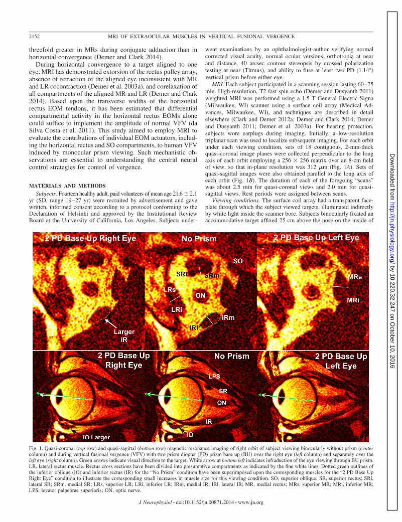
MRI. Each subject participated in a scanning session lasting 60–75min. High-resolution, T2 fast spin echo (Demer and Dusyanth 2011)weighted MRI was performed using a 1.5 T General Electric Signa(Milwaukee, WI) scanner using a surface coil array (Medical Ad-vances, Milwaukee, WI), and techniques are described in detailelsewhere (Clark and Demer 2012a; Demer and Clark 2014; Demerand Dusyanth 2011; Demer et al. 2003a). For hearing protection,subjects wore earplugs during imaging. Initially, a low-resolutiontriplanar scan was used to localize subsequent imaging. For each orbitunder each viewing condition, sets of 18 contiguous, 2-mm-thickquasi-coronal image planes were collected perpendicular to the longaxis of each orbit employing a 256 � 256 matrix over an 8-cm fieldof view, so that in-plane resolution was 312 �m (Fig. 1A). Sets ofquasi-sagittal images were also obtained parallel to the long axis ofeach orbit (Fig. 1B). The duration of each of the foregoing “scans”was about 2.5 min for quasi-coronal views and 2.0 min for quasi-sagittal views. Rest periods were assigned between scans.
Viewing conditions. The surface coil array had a transparent face-plate through which the subject viewed targets, illuminated indirectlyby white light inside the scanner bore. Subjects binocularly fixated anaccommodative target affixed 25 cm above the nose on the inside of
Fig. 1. Quasi-coronal (top row) and quasi-sagittal (bottom row) magnetic resonance imaging of right orbit of subject viewing binocularly without prism (centercolumn) and during vertical fusional vergence (VFV) with two prism diopter (PD) prism base up (BU) over the right eye (left column) and separately over theleft eye (right column). Green arrows indicate visual direction to the target. White arrow at bottom left indicates infraduction of the eye viewing through BU prism.LR, lateral rectus muscle. Rectus cross sections have been divided into presumptive compartments as indicated by the fine white lines. Dotted green outlines ofthe inferior oblique (IO) and inferior rectus (IR) for the “No Prism” condition have been superimposed upon the corresponding muscles for the “2 PD Base UpRight Eye” condition to illustrate the corresponding small increases in muscle size for this viewing condition. SO, superior oblique; SR, superior rectus; SRl,lateral SR; SRm, medial SR; LRs, superior LR; LRi, inferior LR; IRm, medial IR; IRl, lateral IR; MR, medial rectus; MRs, superior MR; MRi, inferior MR;LPS, levator palpebrae superioris; ON, optic nerve.
2152 MRI OF EXTRAOCULAR MUSCLES IN VERTICAL FUSIONAL VERGENCE
J Neurophysiol • doi:10.1152/jn.00871.2014 • www.jn.org
by 10.220.32.247 on October 10, 2016
http://jn.physiology.org/D
ownloaded from
the scanner bore, consisting of a 5 � 5 mm, black on white cross of1-mm stroke width surrounded by five finely ruled, concentric squaresof progressively larger dimensions to a maximum of 20 � 20 mm.
Most normal subjects have sufficient VFV reserve to maintainfusion, despite a two PD vertical disparity during distance viewing,and slightly more during near viewing (von Noorden 1990). Thiscondition requires 1.14° monocular infraduction by the viewing eye.Prior to MRI, each subject was evaluated for the ability to maintainVFV for the prism viewing condition. Initial subjects were also testedwith three PD base up, but since many could not maintain VFV andreported vertical diplopia with the stronger prism, prism power waslimited to two PD. Alternatively, the prism base could have been setdown to evoke a monocular supraduction, or one PD split base up inone eye with one PD base down in the contraprism eye to evokeantisymmetric vertical ductions, but these alternatives were not cho-sen to permit simplification of analysis. Consequently, for scansinvolving VFV, a two PD acrylic prism was affixed base up to thetransparent faceplate of the surface coil mask over one eye, and thesubject’s ability to binocularly fuse the target in the scanner withoutdiplopia was verified subjectively. Quasi-sagittal and quasi-coronalimages of both orbits were each obtained three times during the samescanning session: first, during viewing without prism; second, duringright eye viewing through prism; and third, during left eye viewingthrough prism. Before each scan, subjects were verbally coached tofuse the target binocularly, and their success was confirmed after eachscan.
Analysis. Digital MRI images were quantified using ImageJ64 andcustom analysis programs written in MatLab (MathWorks 2011,Boston, MA). Potential subjective bias was minimized by quasi-automated analysis subsequent to structure outlining in ImageJ64, andautomated analysis of subsequent steps in MatLab. Moreover, theauthors did not have strong a priori expectations of quantitative resultsfor individual EOMs, and, as will be seen below, the results wereoften surprising. Anteroposterior globe position was determined inquasi-sagittal, midglobe images from the difference between horizon-tal coordinates of the anterior border of the inferior orbital rim and theglobe centroid as manually outlined. Contractility of the IO wasinferred from changes in its manually outlined cross section inquasi-sagittal images at the midpoint of the IR (Demer et al. 2003b).In quasi-coronal images, each rectus EOM and the SO was manuallyoutlined and cropped to include only its belly (Clark and Demer2012b, 2012c), avoiding inclusion of adjacent nonmuscular structures.Subsequent analytic steps were automated. For each rectus EOM, theangle of a linear best-fit through the maximum transverse dimensionwas computed (Clark and Demer 2012a), and the image rotated toalign that best-fit line to vertical for horizontal EOMs, and horizontalfor vertical EOMs. Superior and inferior horizontal rectus compart-mental areas were calculated as cross-sectional areas above and belowthe perpendicular bisector of that best-fit line, omitting a band 20% ofthe length of the best fit line to account for anatomical variation in thecompartmental border; corresponding medial and lateral vertical rec-tus compartmental areas were calculated relative to that perpendicularbisector (Clark and Demer 2012a), but again omitting the central 20%to account for anatomical variation in the border. For the SO, abootstrap analysis was performed as described in the RESULTS sectionto determine the angular orientation of a line, oblique to the long axisof the SO cross section that optimally discriminated compartmentalfunction, again omitting the central 20% of the cross section toaccount for possible curvature or other irregularities in the compart-mental boundary.
Longitudinal cross-sectional area distributions of rectus EOMs andthe SO were plotted to evaluate data trends. For this purpose, imageplane sets were referenced to the globe-optic nerve junction, permit-ting averaging over multiple subjects. However, because intersubjectvariation in overall EOM size can obscure the effects of small changesin contractility in each subject, a normalized analysis was thenperformed for statistical purposes.
Cross sections in four contiguous image planes �4, �5, �6, and�7 (8 to 14 mm posterior to the globe-optic nerve junction) weresummed and multiplied by the 2-mm slice thickness to form PPVs.Change in PPV in horizontal rectus EOMs is a robust correlate ofduction angle, accounting for �85% of variance in duction of groupsof subjects, and �97% of variance within individuals (Clark andDemer 2012c). We have also demonstrated that, in 13 normal sub-jects, change in PPV of vertical rectus EOMs is also highly correlatedwith vertical duction angle in normal subjects (unpublished data),accounting for �84% of variance in duction in groups of subjects, and�90% of variance within individuals (unpublished data). The changein PPV of the normal SO also correlates well with vertical duction,accounting for �50% of variance in duction in groups of subjects, and�70% of variance within individuals (unpublished data).
As conventionally done in MRI studies of EOM function (Demerand Clark 2014), contractility was defined as the percentage changefrom that measured without prism viewing. Because of the highlycurved path of the IO, the only image plane analyzed for the IO wasthat closest to the center of the IR muscle; whole-muscle cross-sectional area changes in this plane are considered to best reflect IOcontractility (Demer and Clark 2005; Demer et al. 2003a, 2003b).
While every EOM contains an oculorotary global layer (GL) thatinserts upon the sclera, and an orbital layer (OL) that inserts uponconnective tissues, the borders between these layers are seldomdiscriminable by MRI. The automated parsing of EOMs into com-partments therefore included both GL and OL contributions. Cross-sectional data were analyzed using ANOVA; two-way comparisons ofmeans were made using the two-tailed Student’s t-test, both byGraphPad Prism (GraphPad Software, La Jolla, CA).
Although data were analyzed separately for right eyes alone andleft eyes alone, trends were similar for both. Right and left eyes were,therefore, pooled for analysis reported here.
RESULTS
Angular eye positions. Differences in horizontal and verticaleye positions were determined from the measured globe radius,and changes in positions of the globe-optic nerve junctionrelative to the centroid of the bony orbit. As described else-where (Demer et al. 2003a), these values can be determined atsubpixel resolution, providing a precise measure of rotationalocular orientation. Horizontal and vertical eye positions werecomputed as prism viewing minus contraprism positions, in-cluding each eye in each role for 28 total observations. Eyesduring prism viewing averaged 0.07 � 0.26° (standard error ofthe mean, SEM) abduction relative to that during contraprismviewing, a difference not significantly different from zero.Eyes during prism viewing averaged 0.85 � 0.30° infraductionrelative to during contraprism viewing, significantly differentfrom zero (P � 0.01), but not significantly different from theideal VFV of 1.15° (P � 0.1). This means that, on average,subjects performed the motor task accurately, achieving thedesired amount of VFV without confounding horizontalvergence.
Globe position. Anteroposterior globe position was analyzedin quasi-sagittal images to seek evidence of EOM cocontrac-tion or corelaxation during VFV. The mean horizontal globecentroid was located 5.98 � 0.57 (SE) mm posterior to theanterior border of the inferior orbital rim during viewingwithout prism, and during VFV with two PD monocular baseup, prism was 6.15 � 0.56 mm posterior in the orbit contralat-eral to prism, and 6.07 � 0.59 mm posterior in the orbitipsilateral to prism. While ANOVA demonstrated highly sig-nificant variation among individual orbits (P � 0.0001), there
2153MRI OF EXTRAOCULAR MUSCLES IN VERTICAL FUSIONAL VERGENCE
J Neurophysiol • doi:10.1152/jn.00871.2014 • www.jn.org
by 10.220.32.247 on October 10, 2016
http://jn.physiology.org/D
ownloaded from
was no significant effect of prism viewing condition (P �0.346), nor a significant effect by paired t-testing of the changein anteroposterior globe position within individual orbits (P �0.5). This means that VFV was not associated with globeretraction or proptosis exceeding measurement variability.
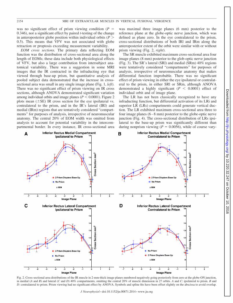
EOM cross sections. The primary data reflecting EOMfunction was the distribution of cross-sectional area along thelength of EOMs; these data include both physiological effectsof VFV, but also a large contribution from intersubject ana-tomical variability. There was a suggestion in some MRIimages that the IR contracted in the infraducting eye thatviewed through base-up prism, but quantitative analysis ofpooled subject data demonstrated that the increase in cross-sectional area was small in any single image plane (Fig. 1, left).There was no significant effect of prism viewing on IR crosssections, although ANOVA demonstrated significant variationamong individual orbits and image planes (P � 0.0001). Figure 2plots mean (�SE) IR cross section for the eye ipsilateral vs.contralateral to the prism, and in the IR’s lateral (IRl) andmedial (IRm) regions that are tentatively considered “compart-ments” for purposes of analysis, irrespective of neuromuscularanatomy. The central 20% of EOM width was omitted fromanalysis to account for potential variability in the intercom-partmental border. In every instance, IR cross-sectional area
was maximal three image planes (6 mm) posterior to thereference plane at the globe-optic nerve junction, which wasdefined as plane zero. In the eye contralateral to the prism,cross-sectional distributions of both IRl and IRm along theanteroposterior extent of the orbit were similar with or withoutprism viewing (Fig. 2, right).
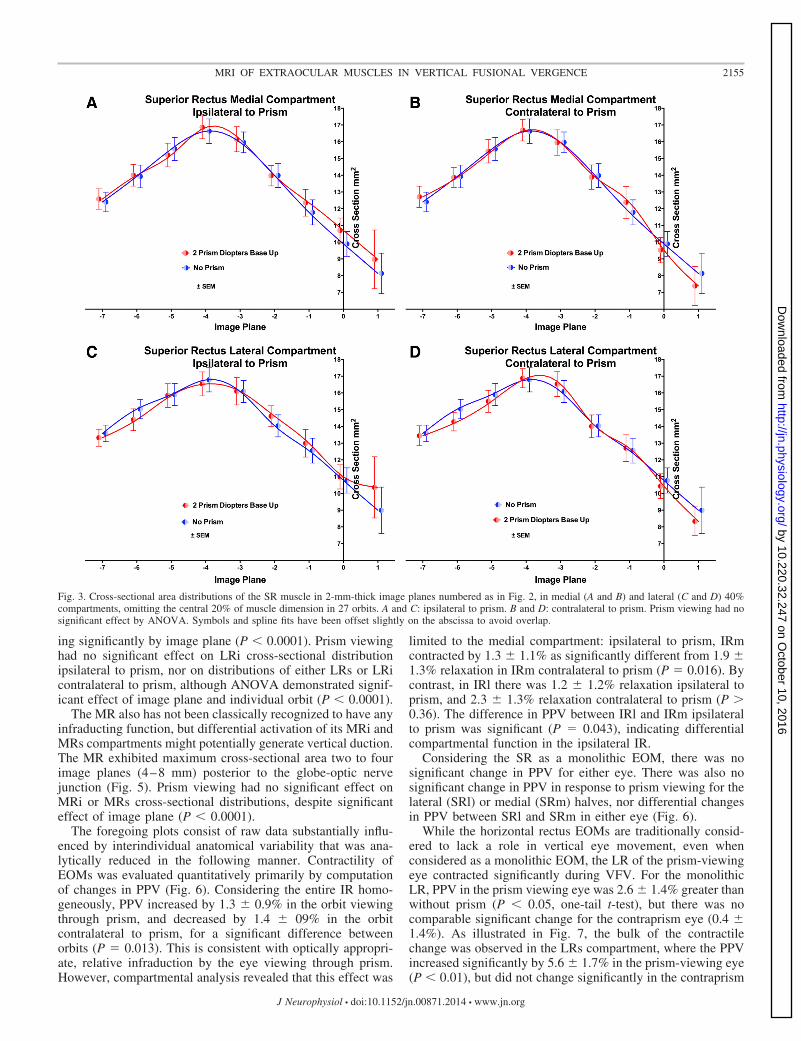
The SR muscle exhibited maximum cross-sectional area fourimage planes (8 mm) posterior to the glob-optic nerve junction(Fig. 3). The SR’s lateral (SRl) and medial (SRm) 40% regionswere tentatively considered “compartments” for purposes ofanalysis, irrespective of neuromuscular anatomy that makesdifferential function improbable. There was no significanteffect of prism viewing in either the eye ipsilateral or contralat-eral to the prism, in either SRl or SRm, although ANOVAdemonstrated a highly significant (P � 0.0001) effect ofindividual orbit and of image plane.
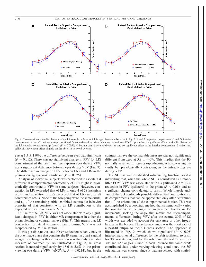
The LR has not been classically recognized to have anyinfraducting function, but differential activation of its LRi andsuperior LR (LRs) compartments could generate vertical duc-tion. The LR exhibited maximum cross-sectional area three tofour image planes (6–8 mm) posterior to the globe-optic nervejunction (Fig. 4). The cross-sectional distribution of LRs ipsi-lateral to the base-up prism was significantly different thanduring nonprism viewing (P � 0.0056), while of course vary-
Fig. 2. Cross-sectional area distributions of the IR muscle in 2-mm-thick image planes numbered negatively going posteriorly from zero at the globe-ON junction,in medial (A and B) and lateral (C and D) 40% compartments, omitting the central 20% of muscle dimension in 27 orbits. A and C: ipsilateral to prism. B andD: contralateral to prism. Prism viewing had no significant effect by ANOVA. Symbols and spline fits have been offset slightly on the abscissa to avoid overlap.
2154 MRI OF EXTRAOCULAR MUSCLES IN VERTICAL FUSIONAL VERGENCE
J Neurophysiol • doi:10.1152/jn.00871.2014 • www.jn.org
by 10.220.32.247 on October 10, 2016
http://jn.physiology.org/D
ownloaded from
ing significantly by image plane (P � 0.0001). Prism viewinghad no significant effect on LRi cross-sectional distributionipsilateral to prism, nor on distributions of either LRs or LRicontralateral to prism, although ANOVA demonstrated signif-icant effect of image plane and individual orbit (P � 0.0001).
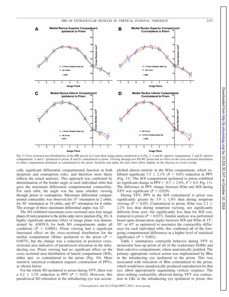
The MR also has not been classically recognized to have anyinfraducting function, but differential activation of its MRi andMRs compartments might potentially generate vertical duction.The MR exhibited maximum cross-sectional area two to fourimage planes (4–8 mm) posterior to the globe-optic nervejunction (Fig. 5). Prism viewing had no significant effect onMRi or MRs cross-sectional distributions, despite significanteffect of image plane (P � 0.0001).
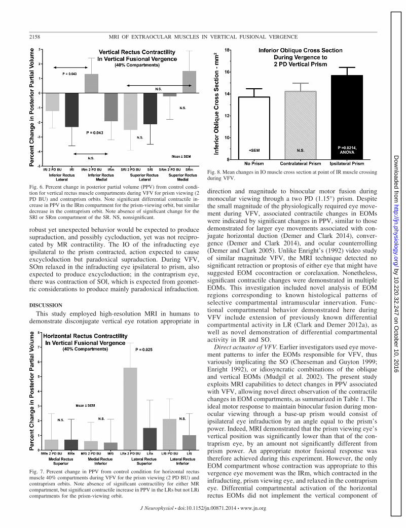
The foregoing plots consist of raw data substantially influ-enced by interindividual anatomical variability that was ana-lytically reduced in the following manner. Contractility ofEOMs was evaluated quantitatively primarily by computationof changes in PPV (Fig. 6). Considering the entire IR homo-geneously, PPV increased by 1.3 � 0.9% in the orbit viewingthrough prism, and decreased by 1.4 � 09% in the orbitcontralateral to prism, for a significant difference betweenorbits (P � 0.013). This is consistent with optically appropri-ate, relative infraduction by the eye viewing through prism.However, compartmental analysis revealed that this effect was
limited to the medial compartment: ipsilateral to prism, IRmcontracted by 1.3 � 1.1% as significantly different from 1.9 �1.3% relaxation in IRm contralateral to prism (P � 0.016). Bycontrast, in IRl there was 1.2 � 1.2% relaxation ipsilateral toprism, and 2.3 � 1.3% relaxation contralateral to prism (P �0.36). The difference in PPV between IRl and IRm ipsilateralto prism was significant (P � 0.043), indicating differentialcompartmental function in the ipsilateral IR.
Considering the SR as a monolithic EOM, there was nosignificant change in PPV for either eye. There was also nosignificant change in PPV in response to prism viewing for thelateral (SRl) or medial (SRm) halves, nor differential changesin PPV between SRl and SRm in either eye (Fig. 6).
While the horizontal rectus EOMs are traditionally consid-ered to lack a role in vertical eye movement, even whenconsidered as a monolithic EOM, the LR of the prism-viewingeye contracted significantly during VFV. For the monolithicLR, PPV in the prism viewing eye was 2.6 � 1.4% greater thanwithout prism (P � 0.05, one-tail t-test), but there was nocomparable significant change for the contraprism eye (0.4 �1.4%). As illustrated in Fig. 7, the bulk of the contractilechange was observed in the LRs compartment, where the PPVincreased significantly by 5.6 � 1.7% in the prism-viewing eye(P � 0.01), but did not change significantly in the contraprism
Fig. 3. Cross-sectional area distributions of the SR muscle in 2-mm-thick image planes numbered as in Fig. 2, in medial (A and B) and lateral (C and D) 40%compartments, omitting the central 20% of muscle dimension in 27 orbits. A and C: ipsilateral to prism. B and D: contralateral to prism. Prism viewing had nosignificant effect by ANOVA. Symbols and spline fits have been offset slightly on the abscissa to avoid overlap.
2155MRI OF EXTRAOCULAR MUSCLES IN VERTICAL FUSIONAL VERGENCE
J Neurophysiol • doi:10.1152/jn.00871.2014 • www.jn.org
by 10.220.32.247 on October 10, 2016
http://jn.physiology.org/D
ownloaded from
eye at 1.5 � 1.9%; the difference between eyes was significant(P � 0.012). There was no significant change in PPV for LRicompartment of the prism and contraprism eyes during VFV,nor a significant difference between eyes during VFV (Fig. 7).The difference in change in PPV between LRs and LRi in theprism-viewing eye was significant (P � 0.025).
Analysis of individual subjects was performed to ascertain ifdifferential compartmental contractility of LRi might idiosyn-cratically contribute to VFV in some subjects. However, con-traction in LRi exceeded that of LRs in only 4 of 28 ipsiprismorbits, and relaxation in LRi exceeded that of LRs in 8 of 28contraprism orbits. None of the foregoing were the same orbits,and all of the remaining orbits exhibited contractile behavioropposite of that consistent with an LR contribution to theexpected vertical direction of VFV.
Unlike for the LR, VFV was not associated with any signif-icant changes in PPV in either MR compartment in either theprism viewing or contraprism eye (Fig. 7). This means that LRcontraction ipsilateral to base-up prism during VFV was notreciprocated by MR relaxation.
It was possible to evaluate IO cross section reliably only inthe one image plane that contains the IR center in quasi-sagittalimages, so change in this cross section was the only availablemeasure of contractility. As illustrated in Fig. 8, IO crosssection increased significantly by 18.6 � 5.6% in the prism-viewing eye during VFV (ANOVA, P � 0.0214), but in the
contraprism eye the comparable measure was not significantlydifferent from zero at 5.8 � 4.0%. This implies that the IO,normally assumed to have a supraducting action, was signifi-cantly but paradoxically contracting in the infraducting eyeduring VFV.
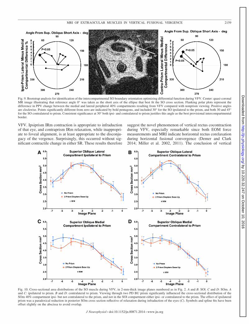
The SO has well-established infraducting function, so it isinteresting that, when the whole SO is considered as a mono-lithic EOM, VFV was associated with a significant 4.2 � 1.2%reduction in PPV ipsilateral to the prism (P � 0.01), and nosignificant change contralateral to prism. Whole muscle anal-ysis of the SO confounds possible differential contributions inits compartments that can be appreciated only after determina-tion of the orientation of the compartmental border. This wasaccomplished by a bootstrap method that systematically variedthe orientation of the angle of an assumed border in 15°increments, seeking the angle that maximized intercompart-mental differences during VFV after the central 20% of SOwidth was excluded to account for curvature or other irregu-larities in the border. The reference angle was the short axis ofa best-fit ellipse to the SO cross section. The approach isillustrated in Fig. 9, which shows significant (P � 0.05)intercompartmental differences for the orbit ipsilateral to prismfor 30° orientation, and for the orbit contralateral to prism for30° and 45° angles. Since in each instance the same orbitscontributed data under varying viewing conditions, the 30°orientation was chosen, since it was associated with statisti-
Fig. 4. Cross-sectional area distributions of the LR muscle in 2-mm-thick image planes numbered as in Fig. 2. A and B: superior compartment. C and D: inferiorcompartment. A and C: ipsilateral to prism. B and D: contralateral to prism. Viewing through two PD BU prism had a significant effect on the distribution ofthe LR superior compartment ipsilateral (P � 0.0056; A) but not contralateral to the prism, and no significant effect in the inferior compartment. Symbols andspline fits have been offset slightly on the abscissa to avoid overlap.
2156 MRI OF EXTRAOCULAR MUSCLES IN VERTICAL FUSIONAL VERGENCE
J Neurophysiol • doi:10.1152/jn.00871.2014 • www.jn.org
by 10.220.32.247 on October 10, 2016
http://jn.physiology.org/D
ownloaded from
cally significant differential compartmental function in bothipsiprism and contraprism roles, and therefore most likelyreflects the actual anatomy. This approach was confirmed bydetermination of the border angle in each individual orbit thatgave the maximum differential compartmental contractility.For each orbit, the angle was the same whether viewingthrough prism or contraprism. Maximum differential compart-mental contractility was observed for 15° orientation in 2 orbits,for 30° orientation in 19 orbits, and 45° orientation for 6 orbits.The average of these maximum differential angles was 32°.
The SO exhibited maximum cross-sectional area four imageplanes (8 mm) posterior to the globe-optic nerve junction (Fig. 10). Ahighly significant anatomic effect of image plane was demon-strated by ANOVA for both SO compartments under allconditions (P � 0.0001). Prism viewing had a significantfunctional effect on the cross-sectional distribution for themedial compartment (SOm) ipsilateral to the prism (P �0.0075), but the change was a reduction in posterior cross-sectional area indicative of paradoxical relaxation in the infra-ducting eye. Prism viewing had no significant effect on thecross-sectional area distribution of lateral compartment (SOl),either ipsi- or contralateral to the prism (Fig. 10). Moresensitive statistical evaluation requires construction of PPVs,as shown below.
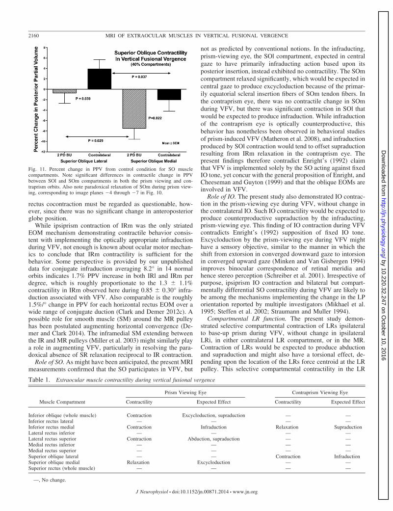
For the whole SO ipsilateral to prism during VFV, there wasa 4.2 � 1.2% reduction in PPV (P � 0.02). However, thisparadoxical SO relaxation in the infraducting eye was accom-
plished almost entirely in the SOm compartment, which ex-hibited significant 7.5 � 2.1% (P � 0.01) reduction in PPV(Fig. 11). The SOl compartment ipsilateral to prism exhibitedno significant change in PPV (�0.7 � 2.0%, P � 0.5, Fig. 11).The difference in PPV change between SOm and SOl duringVFV was significant (P � 0.029).
During VFV, PPV in the SOl contralateral to prism wassignificantly greater by 3.9 � 1.8% than during nonprismviewing (P � 0.05). Contralateral to prism, SOm was 2.2 �2.2% less than during nonprism viewing, not significantlydifferent from zero, but significantly less than for SOl con-tralateral to prism (P � 0.037). Similar analysis was performedbased upon demarcation angles between SOl and SOm of 15°,30°, or 45° as optimized to maximize the contractility differ-ence for each individual orbit; this confirmed all of the fore-going compartmental differences at a higher level of statisticalsignificance (P � 0.001).
Table 1 summarizes contractile behavior during VFV tomonocular base-up prism of all of the oculorotary EOMs andtheir selective compartments, where anatomically justified. Theprimary appropriate vertical action was implemented by IRmin the infraducting eye ipsilateral to the prism. This wasassociated with relaxation of IRm contralateral to the prism,which would have paradoxically produced supraduction by thateye, albeit appropriately augmenting vertical vergence. Themost striking contractility observed during VFV was contrac-tion in LRs in the infraducting eye ipsilateral to prism; this
Fig. 5. Cross-sectional area distributions of the MR muscle in 2-mm-thick image planes numbered as in Fig. 2. A and B: superior compartment. C and D: inferiorcompartment. A and C: ipsilateral to prism. B and D: contralateral to prism. Viewing through two PD BU prism had no effect on the cross-sectional distributionof either compartment ipsilateral or contralateral to the prism. Symbols and spline fits have been offset slightly on the abscissa to avoid overlap.
2157MRI OF EXTRAOCULAR MUSCLES IN VERTICAL FUSIONAL VERGENCE
J Neurophysiol • doi:10.1152/jn.00871.2014 • www.jn.org
by 10.220.32.247 on October 10, 2016
http://jn.physiology.org/D
ownloaded from
robust yet unexpected behavior would be expected to producesupraduction, and possibly cycloduction, yet was not recipro-cated by MR contractility. The IO of the infraducting eyeipsilateral to the prism contracted, action expected to causeexcycloduction but paradoxical supraduction. During VFV,SOm relaxed in the infraducting eye ipsilateral to prism, alsoexpected to produce excycloduction; in the contraprism eye,there was contraction of SOl, which is expected from geomet-ric considerations to produce mainly paradoxical infraduction.
DISCUSSION
This study employed high-resolution MRI in humans todemonstrate disconjugate vertical eye rotation appropriate in
direction and magnitude to binocular motor fusion duringmonocular viewing through a two PD (1.15°) prism. Despitethe small magnitude of the physiologically required eye move-ment during VFV, associated contractile changes in EOMswere indicated by significant changes in PPV, similar to thosedemonstrated for larger eye movements associated with con-jugate horizontal duction (Demer and Clark 2014), conver-gence (Demer and Clark 2014), and ocular counterrolling(Demer and Clark 2005). Unlike Enright’s (1992) video studyof similar magnitude VFV, the MRI technique detected nosignificant retraction or proptosis of either eye that might havesuggested EOM cocontraction or corelaxation. Nonetheless,significant contractile changes were demonstrated in multipleEOMs. This investigation included novel analysis of EOMregions corresponding to known histological patterns ofselective compartmental intramuscular innervation. Func-tional compartmental behavior demonstrated here duringVFV include extension of previously known differentialcompartmental activity in LR (Clark and Demer 2012a), aswell as novel demonstration of differential compartmentalactivity in IR and SO.
Direct actuator of VFV. Earlier investigators used eye move-ment patterns to infer the EOMs responsible for VFV, thusvariously implicating the SO (Cheeseman and Guyton 1999;Enright 1992), or idiosyncratic combinations of the obliqueand vertical EOMs (Mudgil et al. 2002). The present studyexploits MRI capabilities to detect changes in PPV associatedwith VFV, allowing novel direct observation of the contractilechanges in EOM compartments, as summarized in Table 1. Theideal motor response to maintain binocular fusion during mon-ocular viewing through a base-up prism would consist ofipsilateral eye infraduction by an angle equal to the prism’spower. Indeed, MRI demonstrated that the prism viewing eye’svertical position was significantly lower than that of the con-traprism eye, by an amount not significantly different fromprism power. An appropriate motor fusional response wastherefore achieved during this experiment. However, the onlyEOM compartment whose contraction was appropriate to thisvergence eye movement was the IRm, which contracted in theinfraducting, prism viewing eye, and relaxed in the contraprismeye. Differential compartmental activation of the horizontalrectus EOMs did not implement the vertical component of
Fig. 6. Percent change in posterior partial volume (PPV) from control condi-tion for vertical rectus muscle compartments during VFV for prism viewing (2PD BU) and contraprism orbits. Note significant differential contractile in-crease in PPV in the IRm compartment for the prism-viewing orbit, but similardecrease in the contraprism orbit. Note absence of significant change for theSRl or SRm compartment of the SR. NS, nonsignificant.
Fig. 7. Percent change in PPV from control condition for horizontal rectusmuscle 40% compartments during VFV for the prism viewing (2 PD BU) andcontraprism orbits. Note absence of significant contractility for either MRcompartment, but significant contractile increase in PPV in the LRs but not LRicompartments for the prism-viewing orbit.
Fig. 8. Mean changes in IO muscle cross section at point of IR muscle crossingduring VFV.
2158 MRI OF EXTRAOCULAR MUSCLES IN VERTICAL FUSIONAL VERGENCE
J Neurophysiol • doi:10.1152/jn.00871.2014 • www.jn.org
by 10.220.32.247 on October 10, 2016
http://jn.physiology.org/D
ownloaded from
VFV. Ipsiprism IRm contraction is appropriate to infraductionof that eye, and contraprism IRm relaxation, while inappropri-ate to foveal alignment, is at least appropriate to the disconju-gacy of the vergence. Surprisingly, this occurred without sig-nificant contractile change in either SR. These results therefore
suggest the novel phenomenon of vertical rectus cocontractionduring VFV, especially remarkable since both EOM forcemeasurements and MRI indicate horizontal rectus corelaxationduring horizontal fusional convergence (Demer and Clark2014; Miller et al. 2002, 2011). The conclusion of vertical
Fig. 9. Bootstrap analysis for identification of the intercompartmental SO boundary orientation optimizing differential function during VFV. Center: quasi-coronalMR image illustrating that reference angle 0° was taken as the short axis of the ellipse that best fit the SO cross section. Flanking polar plots represent thedifference in PPV change between the medial and lateral peripheral 40% compartments resulting from VFV compared with nonprism viewing. Positive anglesare clockwise. Points significantly different from zero are indicated by bold pentagons, and included 30° for the SO ipsilateral to the prism, and both 30 and 45°for the SO contralateral to prism. Consistent significance at 30° both ipsi- and contralateral to prism justifies this angle as the best provisional intercompartmentalborder.
Fig. 10. Cross-sectional area distributions of the SO muscle during VFV, in 2-mm-thick image planes numbered as in Fig. 2. A and B: SOl. C and D: SOm. Aand C: ipsilateral to prism. B and D: contralateral to prism. Viewing through two PD BU prism significantly influenced the cross-sectional distribution of theSOm 40% compartment ipsi- but not contralateral to the prism, and not in the SOl compartment either ipsi- or contralateral to the prism. The effect of ipsilateralprism was a paradoxical reduction in posterior SOm cross section reflective of relaxation during infraduction of the eyes (C). Symbols and spline fits have beenoffset slightly on the abscissa to avoid overlap.
2159MRI OF EXTRAOCULAR MUSCLES IN VERTICAL FUSIONAL VERGENCE
J Neurophysiol • doi:10.1152/jn.00871.2014 • www.jn.org
by 10.220.32.247 on October 10, 2016
http://jn.physiology.org/D
ownloaded from
rectus cocontraction must be regarded as questionable, how-ever, since there was no significant change in anteroposteriorglobe position.
While ipsiprism contraction of IRm was the only striatedEOM mechanism demonstrating contractile behavior consis-tent with implementing the optically appropriate infraductionduring VFV, not enough is known about ocular motor mechan-ics to conclude that IRm contractility is sufficient for thebehavior. Some perspective is provided by our unpublisheddata for conjugate infraduction averaging 8.2° in 14 normalorbits indicates 1.7% PPV increase in both IRl and IRm perdegree, which is roughly proportionate to the 1.3 � 1.1%contractility in IRm observed here during 0.85 � 0.30° infra-duction associated with VFV. Also comparable is the roughly1.5%/° change in PPV for each horizontal rectus EOM over awide range of conjugate duction (Clark and Demer 2012c). Apossible role for smooth muscle (SM) around the MR pulleyhas been postulated augmenting horizontal convergence (De-mer and Clark 2014). The inframedial SM extending betweenthe IR and MR pulleys (Miller et al. 2003) might similarly playa role in augmenting VFV, particularly in resolving the para-doxical absence of SR relaxation reciprocal to IR contraction.
Role of SO. As might have been anticipated, the present MRImeasurements confirmed that the SO participates in VFV, but
not as predicted by conventional notions. In the infraducting,prism-viewing eye, the SOl compartment, expected in centralgaze to have primarily infraducting action based upon itsposterior insertion, instead exhibited no contractility. The SOmcompartment relaxed significantly, which would be expected incentral gaze to produce excycloduction because of the primar-ily equatorial scleral insertion fibers of SOm tendon fibers. Inthe contraprism eye, there was no contractile change in SOmduring VFV, but there was significant contraction in SOl thatwould be expected to produce infraduction. While infraductionof the contraprism eye is optically counterproductive, thisbehavior has nonetheless been observed in behavioral studiesof prism-induced VFV (Matheron et al. 2008), and infraductionproduced by SOl contraction would tend to offset supraductionresulting from IRm relaxation in the contraprism eye. Thepresent findings therefore contradict Enright’s (1992) claimthat VFV is implemented solely by the SO acting against fixedIO tone, yet concur with the general proposition of Enright, andCheeseman and Guyton (1999) and that the oblique EOMs areinvolved in VFV.
Role of IO. The present study also demonstrated IO contrac-tion in the prism-viewing eye during VFV, without change inthe contralateral IO. Such IO contractility would be expected toproduce counterproductive supraduction by the infraducting,prism-viewing eye. This finding of IO contraction during VFVcontradicts Enright’s (1992) supposition of fixed IO tone.Excycloduction by the prism-viewing eye during VFV mighthave a sensory objective, similar to the manner in which theshift from extorsion in converged downward gaze to intorsionin converged upward gaze (Minken and Van Gisbergen 1994)improves binocular correspondence of retinal meridia andhence stereo perception (Schreiber et al. 2001). Irrespective ofpurpose, ipsiprism IO contraction and bilateral but compart-mentally differential SO contractility during VFV are likely tobe among the mechanisms implementing the change in the LPorientation reported by multiple investigators (Mikhael et al.1995; Steffen et al. 2002; Straumann and Muller 1994).
Compartmental LR function. The present study demon-strated selective compartmental contraction of LRs ipsilateralto base-up prism during VFV, without change in ipsilateralLRi, in either contralateral LR compartment, or in the MR.Contraction of LRs would be expected to produce abductionand supraduction and might also have a torsional effect, de-pending upon the location of the LRs force centroid at the LRpulley. This selective compartmental contractility in the LR
Fig. 11. Percent change in PPV from control condition for SO musclecompartments. Note significant differences in contractile change in PPVbetween SOl and SOm compartments in both the prism viewing and con-traprism orbits. Also note paradoxical relaxation of SOm during prism view-ing, corresponding to image planes �4 through �7 in Fig. 10.
Table 1. Extraocular muscle contractility during vertical fusional vergence
Prism Viewing Eye Contraprism Viewing Eye
Muscle Compartment Contractility Expected Effect Contractility Expected Effect
Inferior oblique (whole muscle) Contraction Excycloduction, supraduction — —Inferior rectus lateral — — — —Inferior rectus medial Contraction Infraduction Relaxation SupraductionLateral rectus inferior — — — —Lateral rectus superior Contraction Abduction, supraduction — —Medial rectus inferior — — — —Medial rectus superior — — — —Superior oblique lateral — — Contraction InfraductionSuperior oblique medial Relaxation Excycloduction — —Superior rectus (whole muscle) — — — —
—, No change.
2160 MRI OF EXTRAOCULAR MUSCLES IN VERTICAL FUSIONAL VERGENCE
J Neurophysiol • doi:10.1152/jn.00871.2014 • www.jn.org
by 10.220.32.247 on October 10, 2016
http://jn.physiology.org/D
ownloaded from
extends the finding of LR differential compartmental contrac-tility first reported for ocular counterrolling, in which selectivecontractility was demonstrated in LRi (Clark and Demer2012a). No contractile change in LRi was observed in thepresent study during VFV, nor in either MRi or MRs. Absenceof contribution of the MR to VFV stands in contrast to MRIevidence for significantly less MRs, but not MRi, contractilityduring horizontal convergence than during adduction (Demerand Clark 2014). Selective compartmental control of LR ispossible because the abducens nerve bifurcates into inferiorand superior divisions prior to entry into the LR belly (da SilvaCosta et al. 2011; Peng et al. 2010) and then arborizes withinnon-overlapping compartments of fibers that have only weaktransverse mechanical coupling among generally parallel mus-cle (Shin et al. 2012, 2014) and tendon fibers (Shin et al. 2013).There is preliminary evidence that topographically distinctabducens motoneuron pools may selectively innervate the twoLR compartments (Demer et al. 2013), consistent with evi-dence that neuoropathic LR atrophy in human LR abducensparesis is often limited to LRs (Clark and Demer 2014). Thefrequent occurrence of small-angle vertical strabismus in thesetting of human abducens palsy (Pihlblad and Demer 2014)also supports a differential compartmental role for the LRs inVFV.
Paradoxical horizontal rectus behavior. Robust contractilityof LRs of the prism-viewing eye during VFV was not recip-rocated by contractile change in either MR compartment, norreflected in horizontal eye movement. During static VFV, forcebalance between MR and LR would always be expected, unlessother EOMs or passive elastic forces contribute. The presentfinding of absence of reciprocal behavior in the horizontalrectus agonist-antagonist pair is reminiscent of the recent,perplexing finding in humans that LR contractility did notsignificantly differ between convergence and conjugate adduc-tion, yet contractility in MRs during convergence was onlyabout one-third of that in conjugate adduction (Demer andClark 2014). However, during VFV, contraction in LRs may berequired to offset the secondary adducting effect of contractionin IRm during infraduction. Another possible contributor toforce balance is SM, which is abundant in a band over 1 mmthick from the IR to MR pulleys (Demer et al. 1997; Kono etal. 2002b; Miller et al. 2003). Most of this SM is composed ofbundles arranged in the anteroposterior direction (Kono et al.2002b) that could load or unload the IR pulley suspension, andso indirectly alter IR tension. While the SM in the pulleysuspensions receives autonomic innervation (Demer et al.1997) whose presumed slow dynamic properties could becompatible with VFV, nothing is presently known about thisSM’s function.
Compartmental SO function. The present finding duringVFV of robust differential compartmental SO contractility isthe first demonstration of this phenomenon and may be under-stood in light of recent anatomical findings. Dissection ofhuman and bovine orbits demonstrates that the structure of theSO and its tendon does not differ significantly from the rectusEOMs (Le et al. 2014a, 2014b). The SO tendon readily unrollsinto a configuration of parallel muscle fibers in continuity withcorresponding parallel tendon fibers that can be traced to itsbroad scleral insertion (Le et al. 2014a; Le et al. 2014b).External to the SO belly, the trochlear nerve bifurcates intomedial and lateral divisions arborizing within non-overlapping
medial and lateral compartments of muscle fibers that in turncorrespond with predominantly equatorial and retroequatorialscleral insertions, respectively (Le et al. 2014a, 2014b). Froma starting position in central gaze as employed here, geometrydictates that the equatorial insertion of the SOm fibers mustgenerate predominantly cycloduction, while the retroequatorialinsertion of SOl fibers must generate predominantly infraduc-tion. Thus the observed relaxation of SOm in the prism-viewing eye during VFV contributes to the excycloductionimplemented by the IO and IR. The observed SOl contractionin the non-prism-viewing eye during VFV would contribute toa counterproductive infraduction of the eye that was alreadyaligned on the target without prism viewing.
Compartmental vertical rectus function. Anatomical studiesof intramuscular SR innervation suggested absence of com-partmentally segregated peripheral innervation, although a se-lective lateral motor nerve division was demonstrated in the IRsupplying partially overlapping innervation of diffuse, whole-muscle innervation provided by the main division (da SilvaCosta et al. 2011). Since this study detected no significant SRcontractility during VFV, the data provide no further insightinto possible SR compartmentalization. However, the IR con-traction observed during VFV was present only in IRm, con-stituting the first demonstration of differential compartmentalfunction in this EOM. Given the pattern of overlapping intra-muscular innervation in IR, this behavior might be explainedby profound reduction in selective partial innervation to IRl inthe eye infraducting during prism viewing, while the sameregion of EOM fibers remains partly innervated by moderatelyincreased firing by the nerve branch that diffusely projects tothe entire IR. No data are available concerning possible selec-tive innervation to specific fiber types in either IR compart-ment. While the present MRI method was not designed todetect compartmental function in the IO, there is anatomicalevidence for compartmental innervation of that EOM as well(Le et al. 2014a).
Because experimental time constraints did not permit com-parative study of conjugate infraduction similar to the 1.15°angle of VFV, is it impossible to know if differential compart-mental function in the IR is unique to vergence as opposed toconjugate vertical gaze. We have obtained unpublished datausing identical MRI and analytic technique in normal humansubjects during conjugate infraduction, showing no significantdifference between IRl and IRm contractility for conjugateinfraduction averaging 8° (13 orbits of 8 subjects), or formaximal infraduction averaging 21° (25 orbits of 13 subjects).Systematic studies of small-angle conjugate infraduction, con-jugate supraduction, and VFV in response to base-down prismare warranted in further investigations.
Analytic considerations for compartmentalization. The fore-going compartmental analysis should be considered capable ofdemonstrating differential compartmental function in EOMs,but incapable of excluding it for several reasons. The presentanalysis parsed rectus EOMs into two compartments havingequal transverse dimensions corresponding to the average lo-cation of anatomical demarcations occasionally observable byMRI in the LR (Demer and Clark 2014), and to averageproportions consistently demonstrated by histological recon-struction in a small number of serially sectioned orbits for LR(da Silva Costa et al. 2011; Peng et al. 2010) and MR (da SilvaCosta et al. 2011). The present analysis parsed the SO into
2161MRI OF EXTRAOCULAR MUSCLES IN VERTICAL FUSIONAL VERGENCE
J Neurophysiol • doi:10.1152/jn.00871.2014 • www.jn.org
by 10.220.32.247 on October 10, 2016
http://jn.physiology.org/D
ownloaded from
equal medial and lateral regions, again based upon histologicalobservations consistently made in a small number of seriallysectioned orbits (Le et al. 2014b). While these assumptionsseem reasonable for average relative compartmental propor-tions for the horizontal rectus EOMs and the SO, the relativeproportions are expected to vary among individual subjects.This implies that there was probably some misattribution offunction in one EOM compartment to that of the fellowcompartment, which would have the statistical effect of dimin-ishing differences between compartments, and increasing vari-ance, so that small but real differential compartmental effectscould appear statistically insignificant. Moreover, for the SO,the mediolateral intercompartmental border might be variablyinclined from the vertical as a result of curvature of theadjacent superomedial orbital wall, along which the SO bellycourses. An incorrect assumption about the anatomical orien-tation of the intercompartmental SO border would also reduceobserved differences in compartmental behavior and mightincrease variance. In every case, the analytic assumptions herewould all tend to diminish the chances of detecting differentialcompartmental behavior, but never create an illusion of it.Moreover, the significant differential compartmental behaviorthat was detected here in LR and SO during VFV probablyunderestimates the actual magnitude of differential effect andmight have missed more subtle differential behavior altogether.
GL and OL. The analytic method employed here also poolsthe GL and OL in each EOM compartment, although the latterdo not directly generate ocular rotation. The GL and OL wouldprobably have contributed equally to each assumed compart-mental parsing. The two layers probably contract simultane-ously during static fixations (Kono et al. 2002a), and noanatomical evidence has yet emerged of separate motor nervetrunks to these two layers. Elucidation of physiological rela-tionships among the transverse EOM compartments, and theOL/GL, will require higher resolution methods of observingEOM function that have yet to be developed and should ideallybe correlated with companion studies of motoneuron behavior.
ACKNOWLEDGMENTS
Nicolasa de Salles provided technical assistance.
GRANTS
This study was supported by National Eye Institute Grants EY-08313 andcore grant EY-00331. J. L. Demer was supported by an unrestricted awardfrom Research to Prevent Blindness and holds the Leonard Apt Chair ofPediatric Ophthalmology.
DISCLOSURES
No conflicts of interest, financial or otherwise, are declared by the author(s).
AUTHOR CONTRIBUTIONS
Author contributions: J.L.D. conception and design of research; J.L.D.performed experiments; J.L.D. and R.A.C. analyzed data; J.L.D. and R.A.C.interpreted results of experiments; J.L.D. prepared figures; J.L.D. draftedmanuscript; J.L.D. and R.A.C. edited and revised manuscript; J.L.D. approvedfinal version of manuscript.
REFERENCES
Apt L. An anatomical reevaluation of rectus muscle insertions. Trans AmOphthalmol Soc 78: 365–375, 1980.
Apt L, Call NB. An anatomical reevaluation of rectus muscle insertions.Ophthalmic Surg 12: 108–112, 1980.
Bharadwaj SR, Hoenig MP, Sivaramakrishnan VC, Karthikeyan B, Si-monian D, Mau K, Rastani S, Schor CM. Variation of binocular-verticalfusion amplitude with convergence. Invest Ophthalmol Vis Sci 48: 1592–1600, 2007.
Cheeseman EW, Guyton DL. Vertical fusional vergence: the key to disso-ciated vertical deviation. Arch Ophthalmol 117: 1188–1191, 1999.
Clark RA, Demer JL. Differential lateral rectus compartmental contractionduring ocular counter-rolling. Invest Ophthalmol Vis Sci 53: 2887–2896,2012a.
Clark RA, Demer JL. Enhanced vertical rectus contractility by magneticresonance imaging in superior oblique palsy. Arch Ophthalmol 129: 904–908, 2011.
Clark RA, Demer JL. Functional magnetic resonance imaging (MRI) withselective zonal analysis suggests that differential compartmental medialrectus (MR) contractility augments vertical duction. Program No. 371.01.In: 2012 Neuroscience Meeting Planner. New Orleans, LA: Society forNeuroscience, 2012b.
Clark RA, Demer JL. Functional morphometry of horizontal rectus extraoc-ular muscles during ocular duction. Invest Ophthalmol Vis Sci 53: 7375–7379, 2012c.
Clark RA, Demer JL. Lateral rectus superior compartment palsy. Am JOphthalmol 15: 479–487, 2014.
Clark RA, Miller JM, Demer JL. Displacement of the medial rectus pulleyin superior oblique palsy. Invest Ophthalmol Vis Sci 39: 207–212, 1998.
Clausen T. Na�-K� pump regulation and skeletal muscle contractility.Physiol Rev 83: 1269–1324, 2003.
da Silva Costa RM, Kung J, Poukens V, Yoo L, Tychsen L, and Demer JL.Intramuscular innervation of primate extraocular muscles: unique compart-mentalization in horizontal recti. Invest Ophthalmol Vis Sci 52: 2830–2836,2011.
Demer JL. A 12-year, prospective study of extraocular muscle imaging incomplex strabismus. J AAPOS 6: 337–347, 2003.
Demer JL. Extraocular muscles. In: Duane’s Clinical Ophthalmology, editedby Tasman W and Jaeger EA. Hagerstown, MD: Lipincott, 2009, p. 1–30.
Demer JL, Clark RA. Differential compartmental function of medial rectusmuscle during converged and conjugate ocular adduction. J Neurophysiol112: 845–855, 2014.
Demer JL, Clark RA. Magnetic resonance imaging (MRI) demonstratesdifferential compartmental contractility of medial rectus muscle duringvertical duction. In: ARVO 2013 Annual Meeting Abstracts. Rockville: MD,ARVO, 2013, p. 1302.
Demer JL, Clark RA. Magnetic resonance imaging of human extraocularmuscles during static ocular counter-rolling. J Neurophysiol 94: 3292–3302,2005.
Demer JL, Dusyanth A. T2 fast spin echo magnetic resonance imaging ofextraocular muscles. J AAPOS 15: 17–23, 2011.
Demer JL, Kono R, Wright W. Magnetic resonance imaging of humanextraocular muscles in convergence. J Neurophysiol 89: 2072–2085, 2003a.
Demer JL, Miller JM. Magnetic resonance imaging of the functional anatomyof the superior oblique muscle. Invest Ophthalmol Vis Sci 36: 906–913,1995.
Demer JL, Miller JM. Orbital imaging in strabismus surgery. In: ClinicalStrabismus Management: Principles and Techniques, edited by RosenbaumAL and Santiago AP. Philadelphia, PA: Saunders, 1999, p. 84–98.
Demer JL, Mittelman-Smith M, Micevyh P, Poukens V, Foeller P, Tych-sen LT. Do topographically distinct abducens motor neuron pools innervatethe superior and inferior compartments of the lateral rectus muscle? ProgramNo. 363.09. In: 2013 Neuroscience Meeting Planner. San Diego, CA:Society for Neuroscience, 2013.
Demer JL, Oh SY, Clark RA, Poukens V. Evidence for a pulley of theinferior oblique muscle. Invest Ophthalmol Vis Sci 44: 3856–3865, 2003b.
Demer JL, Poukens V, Miller JM, Micevych P. Innervation of extraocularpulley smooth muscle in monkeys and humans. Invest Ophthalmol Vis Sci38: 1774–1785, 1997.
Demer JL, Poukens V, Ying H, Shan X, Tian J, Zee DS. Effects ofintracranial trochlear neurectomy on the structure of the primate superioroblique muscle. Invest Ophthalmol Vis Sci 51: 3485–3493, 2010.
Ela-Dalman N, Velez FG, Demer JL, Rosenbaum AL. High resolutionmagnetic resonance imaging demonstrates reduced inferior oblique musclesize in isolated inferior oblique palsy. J AAPOS 12: 602–607, 2008.
Enright JT. Unexpected role of the oblique muscles in the human verticalfusional reflex. J Physiol 451: 279–293, 1992.
2162 MRI OF EXTRAOCULAR MUSCLES IN VERTICAL FUSIONAL VERGENCE
J Neurophysiol • doi:10.1152/jn.00871.2014 • www.jn.org
by 10.220.32.247 on October 10, 2016
http://jn.physiology.org/D
ownloaded from
Hara N, Steffen H, Roberts DC, Zee DS. Effects of horizontal vergence onthe motor and sensory components of vertical fusion. Invest Ophthalmol VisSci 39: 2268–2276, 1998.
Irsch K, Guyton DL, Ramey NA, Adyanthaya RS, Ying HS. Verticalvergence adaptation produces objective vertical deviation that changes withhead tilt. Invest Ophthalmol Vis Sci 54: 3108–3114, 2013.
Jiang L, Demer JL. Magnetic resonance imaging of the functional anatomy ofthe inferior rectus muscle in superior oblique muscle palsy. Ophthalmology115: 2079–2086, 2008.
Kono R, Clark RA, Demer JL. Active pulleys: magnetic resonance imagingof rectus muscle paths in tertiary gazes. Invest Ophthalmol Vis Sci 43:2179–2188, 2002a.
Kono R, Demer JL. Magnetic resonance imaging of the functional anatomyof the inferior oblique muscle in superior oblique palsy. Ophthalmology 110:1219–1229, 2003.
Kono R, Okanobu H, Ohtsuki H, Demer JL. Absence of relationshipbetween oblique muscle size and Bielschowsky head tilt phenomenon inclinically diagnosed superior oblique palsy. Invest Ophthalmol Vis Sci 50:175–179, 2009.
Kono R, Poukens V, Demer JL. Quantitative analysis of the structure of thehuman extraocular muscle pulley system. Invest Ophthalmol Vis Sci 43:2923–2932, 2002b.
Le A, Poukens V, Demer JL. Compartmental innervation scheme for themammalian superior oblique (SO) and inferior oblique (IO) muscles. Pro-gram No. 62.22. In: 2014 Neuroscience Meeting Planner. Washington, DC:Society for Neuroscience, 2014a.
Le A, Poukens V, Demer JL. Evidence for compartmental innervation of themammalian superior oblique (SO) muscle by the trochlear nerve. In: ARVO2014 Annual Meeting Abstracts. Rockville, MD: ARVO, 2014b, p. 2559.
Lennerstrand GL, Schiavi C, Tian S, Benassi M, Camppos EC. Isometricforce measured in human horizontal eye muscles attached or detached fromthe globe. Graefes Arch Clin Exp Ophthalmol 244: 539–544, 2006.
Lim KH, Poukens V, Demer JL. Fascicular specialization in human andmonkey rectus muscles: evidence for anatomic independence of global andorbital layers. Invest Ophthalmol Vis Sci 48: 3089–3097, 2007.
Matheron E, Yang Q, Le TT, Kapoula Z. Effects of ocular dominance on thevertical vergence induced by a 2-diopter vertical prism during standing.Neurosci Lett 333: 176–180, 2008.
Mikhael S, Nicolle D, Vilis T. Rotation of Listing’s plane by horizontal,vertical and oblique prism-induced vergence. Vision Res 35: 3243–3254,1995.
Miller JM. Functional anatomy of normal human rectus muscles. Vision Res29: 223–240, 1989.
Miller JM, Bockisch CJ, Pavlovski DS. Missing lateral rectus force andabsence of medial rectus co-contraction in ocular convergence. J Neuro-physiol 87: 2421–2433, 2002.
Miller JM, Davison RC, Gamlin PD. Motor nucleus activity fails to predictextraocular muscle forces in ocular convergence. J Neurophysiol 105:2863–2873, 2011.
Miller JM, Demer JL, Poukens V, Pavlowski DS, Nguyen HN, Rossi EA.Extraocular connective tissue architecture. J Vis 3: 240–251, 2003.
Minken AWH, Van Gisbergen JAM. A three-dimensional analysis of ver-gence movements at various levels of elevation. Exp Brain Res 101:331–345, 1994.
Mottier ME, Mets MB. Vertical fusional vergences in patients with superioroblique muscle palsies. Am Orthopt J 40: 88–93, 1990.
Mudgil AV, Walker M, Steffen H, Guyton DL, Zee DS. Motor mechanismsof vertical fusion in individuals with superior oblique paresis. J AAPOS 6:145–153, 2002.
Peng M, Poukens V, da Silva Costa RM, Yoo L, Tychsen L, Demer JL.Compartmentalized innervation of primate lateral rectus muscle. InvestOphthalmol Vis Sci 51: 4612–4617, 2010.
Pihlblad M, Demer JL. Hypertropia in unilateral, isolated abducens palsy. JAAPOS 18: 235–240, 2014.
Schor CM, Maxwell JS, Stevenson SB. Isovergence surfaces: the conjugacyof vertical eye movements in tertiary positions of gaze. Ophthalmic PhysiolOpt 14: 279–286, 1994.
Schor CM, Tyler CW. Spatio-temporal properties of Panum’s fusional area.Vision Res 21: 683–692, 1981.
Schreiber K, Crawford JD, Fetter M, Tweed D. The motor side of depthvision. Nature 410: 819–822, 2001.
Sharma K, Abdul-Rahim AS. Vertical fusional aplitude in normal adults. AmJ Ophthalmol 114: 636–637, 1992.
Shin A, Yoo L, Chaudhuri Z, Demer JL. Independent passive mechanicalbehavior of bovine extraocular muscle compartments. Invest Ophthalmol VisSci 53: 8414–8423, 2012.
Shin A, Yoo L, Demer JL. Biomechanics of superior oblique Z-tenotomy. JAAPOS 17: 612–617, 2013.
Shin A, Yoo L, Demer JL. Independent active contraction of bovine extra-ocular muscle (EOM) compartments. Program No. 62.13. In: 2014 Neuro-science Meeting Planner. Washington, DC: Society for Neuroscience, 2014.
Steffen H, Walker MD, Zee DS. Changes in Listing’s plane after sustainedvertical fusion. Invest Ophthalmol Vis Sci 43: 668–672, 2002.
Straumann D, Muller E. Is Listing’s law preserved in the vertical fusionalreflex? In: Contemporary Ocular Motor and Vertibular Research: A Tributeto David A. Robinson, edited by Fuchs AF, Brandt T, Buttner U, and ZeeDS. Stuttgart, Germany: Georg Thieme Verlag, 1994, p. 336–338.
Tian S, Nishida Y, Isberg B, Lennerstrand G. MRI measurements of normalextraocular muscles and other orbital structures. Graefes Arch Clin ExpOphthalmol 238: 393–404, 2000.
Van Rijn LJ, Collewijn H. Eye torsion associated with disparity-inducedvertical vergence in humans. Vision Res 34: 2307–2316, 1994.
von Noorden GK. Binocular Vision and Ocular Motility: Theory and Man-agement of Strabismus (4th Ed.). St. Louis, MO: Mosby, 1990, p. 193.
Wilkie DR. The mechanical properties of muscle. Br Med Bull 12: 177–182,1956.
Yoo L, Clark RA, Shin A, Demer JL. High Poisson ratio (PR) of contractinghuman superior rectus (SR) muscle indicates reverse compressibility. Pro-gram No.62.15. In: 2014 Neuroscience Meeting Planner. Washington, DC:Society for Neuroscience, 2014.
2163MRI OF EXTRAOCULAR MUSCLES IN VERTICAL FUSIONAL VERGENCE
J Neurophysiol • doi:10.1152/jn.00871.2014 • www.jn.org
by 10.220.32.247 on October 10, 2016
http://jn.physiology.org/D
ownloaded from