Embed Size (px)
Citation preview
+
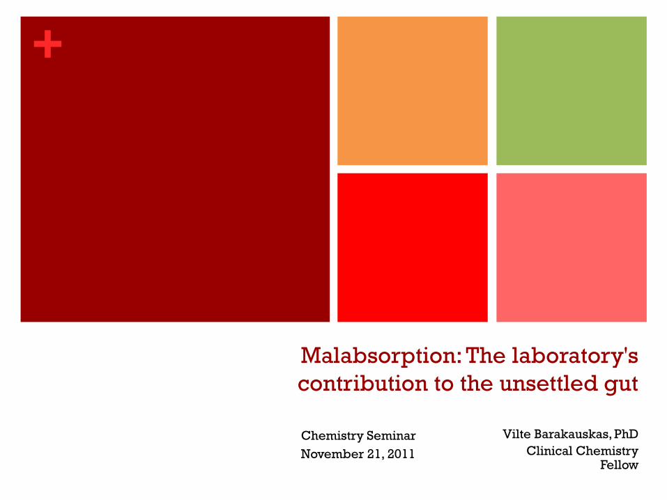
Malabsorption: The laboratory's
contribution to the unsettled gut
Vilte Barakauskas, PhD
Clinical Chemistry Fellow
Chemistry Seminar
November 21, 2011
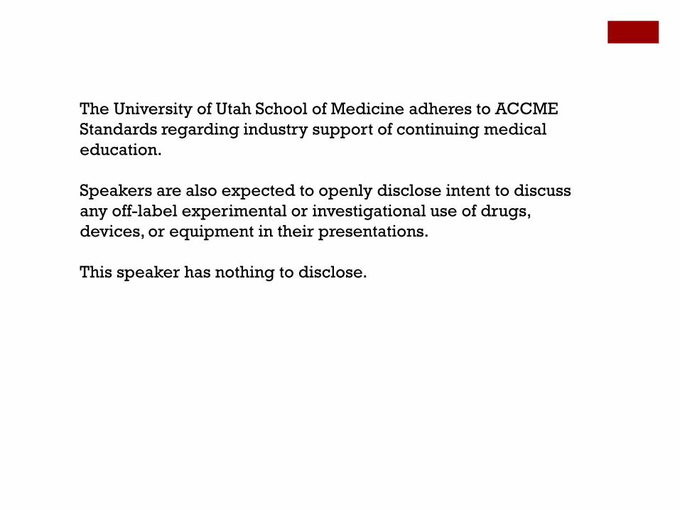
The University of Utah School of Medicine adheres to ACCME
Standards regarding industry support of continuing medical
education.
Speakers are also expected to openly disclose intent to discuss
any off-label experimental or investigational use of drugs,
devices, or equipment in their presentations.
This speaker has nothing to disclose.
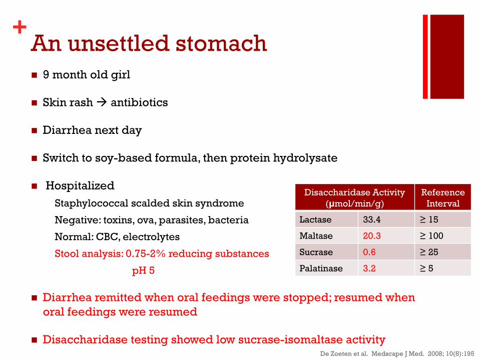
+ An unsettled stomach
9 month old girl
Skin rash antibiotics
Diarrhea next day
Switch to soy-based formula, then protein
hydrolysate
Hospitalized
De Zoeten et al. Medscape J Med. 2008; 10(8):195
+ Learning Objectives
Recall the anatomic location and
physiological processes of digestive organs
List several causes of malabsorption
Suggest appropriate laboratory tests to aid
in the evaluation of suspected
malabsorption
By the end of the session participants should be able
to:
+ Outline
Review of the digestive system
Gastrointestinal anatomy and physiology
Mechanisms of nutrient breakdown
Nutrient absorption
Causes of abnormal function
Malabsorption
Symptoms
Laboratory evaluation
Management

+ Digestion and Absorption
The process by which nutrients are consumed, broken down, absorbed and transported to other parts of the body
Mechanical, chemical processes
Three phases:
Luminal breakdown, solubilization
Mucosal movement of nutrients into GI cells
Transport distribution of nutrients throughout the body
Allows food nutrients to be utilized for energy and growth
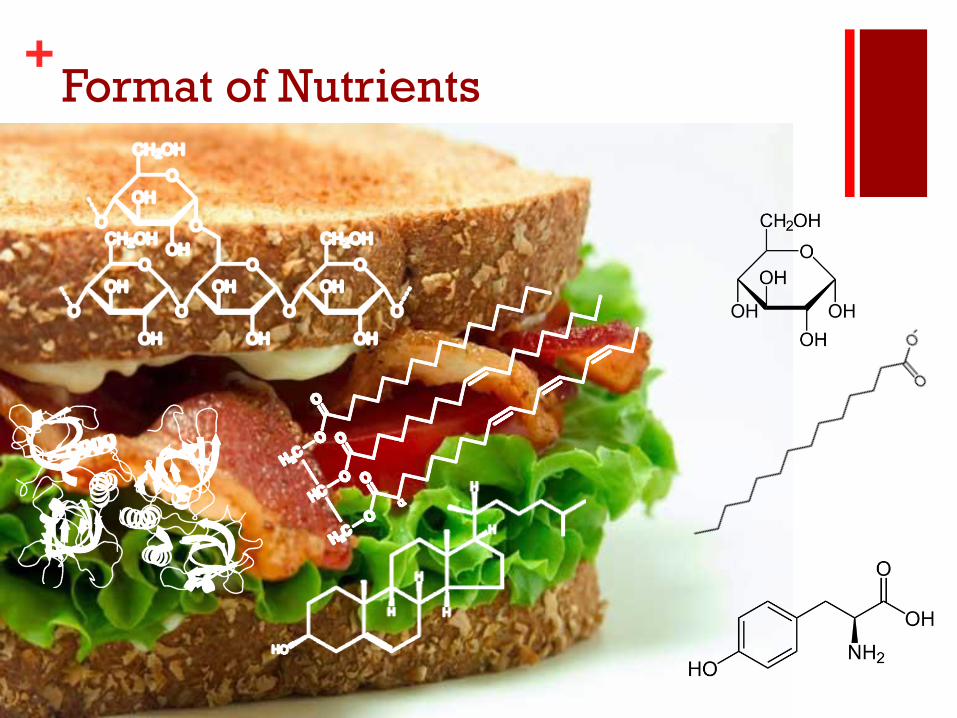
+ Format of Nutrients

+
Th
e D
ige
stive
Sy
stem
Gastrointestinal
system
Long tube
Lumen open to
external
environment
http://commons.wikimedia.org/wiki File:Digestive_system_simplified.svg
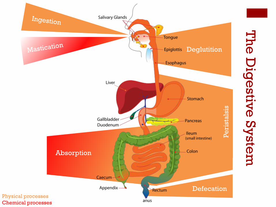
Deglutition
Pe
rista
lsis
Absorption
Defecation Physical processes
Chemical processes
Th
e D
ige
stive
Sy
stem
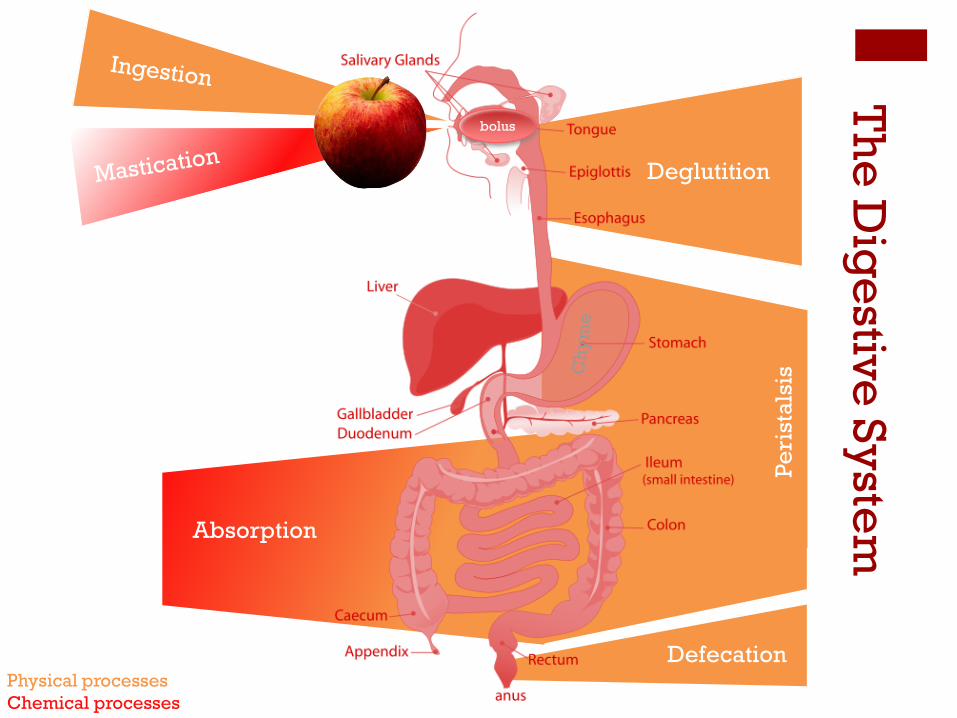
Deglutition
Pe
rista
lsis
Absorption
Defecation Physical processes
Chemical processes
Th
e D
ige
stive
Sy
stem
bolus

Deglutition
Pe
rista
lsis
Bile:
•Produced in liver
•Stored in
gallbladder
•Fat emulsification
Saliva moisture,
amylase
• Pepsinogen, HCl and
hormone production
• Alkaline mucus to
protect lining
• Muscular contraction
to mix contents
Pancreatic secretions:
• Bicarbonate
• Proteases
• Lipases
• Nucleases Breakdown & absorption
3 sections:
• Duodenum
• Jejunum
• Ileum
Produces:
• Mucus
• Enzymes
• Hormones
4 sections:
•Ascending
•Transverse
•Descending
•Sigmoid
Water
Sodium
Storage
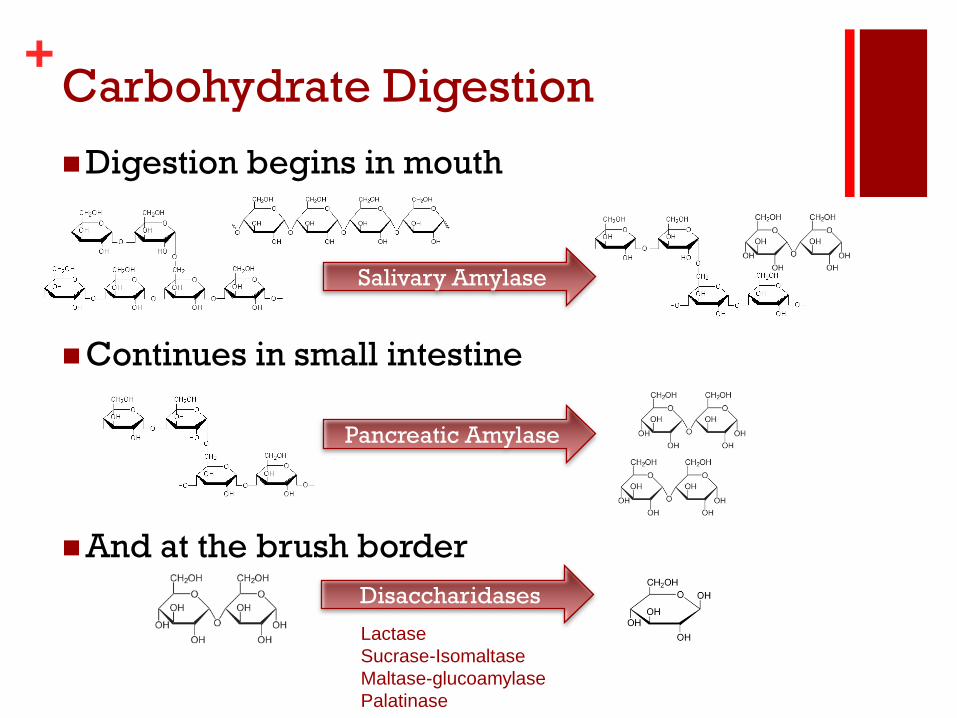
+ Carbohydrate Digestion
Digestion begins in mouth
Continues in small intestine
And at the brush border
Salivary Amylase
Pancreatic Amylase
Disaccharidases
Lactase
Sucrase-Isomaltase
Maltase-glucoamylase
Palatinase
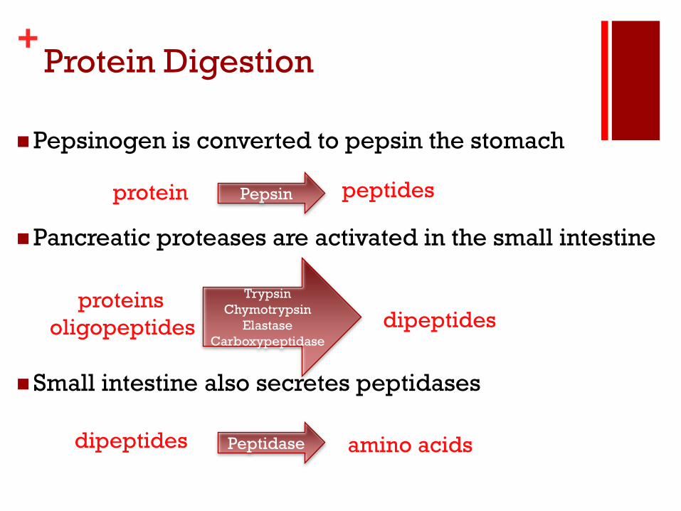
+ Protein Digestion
Pepsinogen is converted to pepsin the stomach
Pancreatic proteases are activated in the small intestine
Small intestine also secretes peptidases
Pepsin protein peptides
Trypsin
Chymotrypsin
Elastase
Carboxypeptidase
proteins
oligopeptides
dipeptides
Peptidase amino acids dipeptides
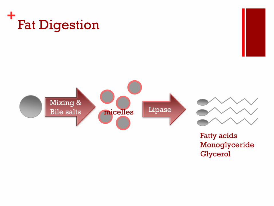
+ Fat Digestion
Mixing &
Bile salts Lipase micelles
Fatty acids
Monoglyceride
Glycerol
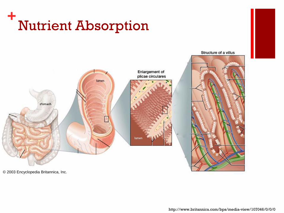
+ Nutrient Absorption
http://www.britannica.com/bps/media-view/107046/0/0/0
© 2003 Encyclopedia Britannica, Inc.
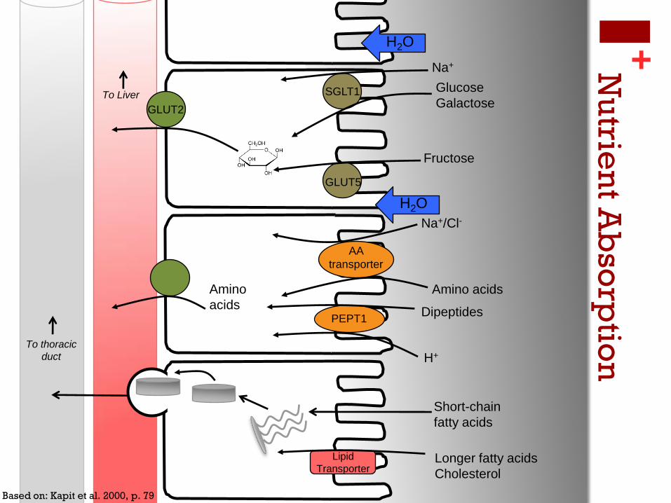
+
To Liver
To thoracic
duct
Nu
trien
t Ab
sorp
tion
SGLT1
GLUT5
GLUT2
Na+
Glucose
Galactose
Fructose
Na+/Cl-
Amino acids
AA
transporter
PEPT1 Dipeptides
H+
Amino
acids
Lipid
Transporter
Short-chain
fatty acids
Longer fatty acids
Cholesterol
H2O
H2O
Based on: Kapit et al. 2000, p. 79
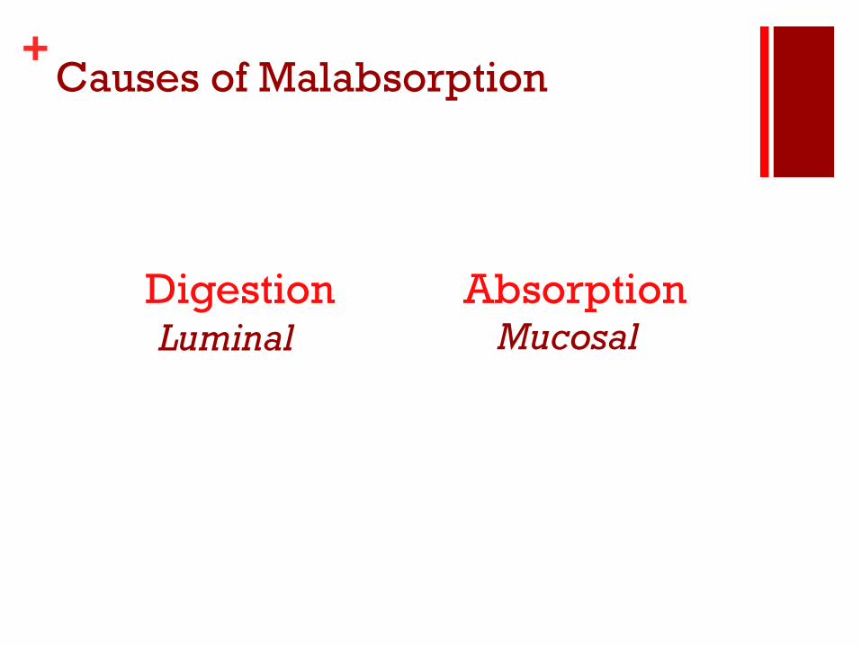
+ Causes of Malabsorption
Digestion Absorption Mucosal Luminal
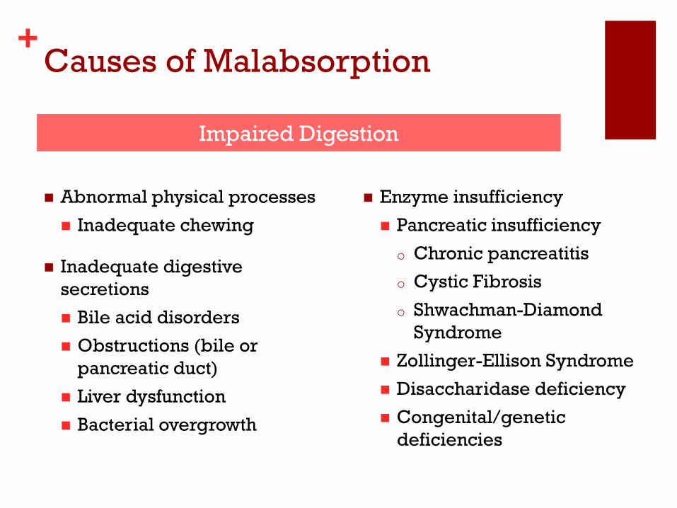
+ Causes of Malabsorption
Abnormal physical processes
Inadequate chewing
Inadequate digestive
secretions
Bile acid disorders
Obstructions (bile or
pancreatic duct)
Liver dysfunction
Bacterial overgrowth
Impaired Digestion
Enzyme insufficiency
Pancreatic insufficiency
o Chronic pancreatitis
o Cystic Fibrosis
o Shwachman-Diamond
Syndrome
Zollinger-Ellison Syndrome
Disaccharidase deficiency
Congenital/genetic
deficiencies
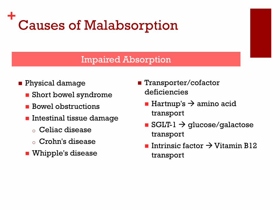
+ Causes of Malabsorption
Physical damage
Short bowel syndrome
Bowel obstructions
Intestinal tissue damage
o Celiac disease
o Crohn's disease
Whipple's disease
Impaired Absorption
Transporter/cofactor
deficiencies
Hartnup's amino acid
transport
SGLT-1 glucose/galactose
transport
Intrinsic factor Vitamin B12
transport

+ Symptoms of Malabsorption
Undigested/unabsorbed molecules in the
GI tract
Na+
Glucose
Galactose
Fructose
Na+/Cl-
Amino
acids
fatty acids
H2O
H2O
H2O
H2O
Osmotic diarrhea
Nutrients reach colon
Excreted in feces
Steatorrhea
Protein
Sugars
Bacterial fermentation
Flatulence
Acid production
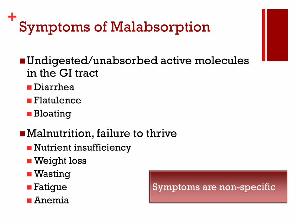
+ Symptoms of Malabsorption
Undigested/unabsorbed active molecules in the GI tract
Diarrhea
Flatulence
Bloating
Malnutrition, failure to thrive
Nutrient insufficiency
Weight loss
Wasting
Fatigue
Anemia
Symptoms are non-specific
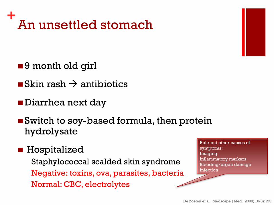
+ An unsettled stomach
9 month old girl
Skin rash antibiotics
Diarrhea next day
Switch to soy-based formula, then protein hydrolysate
Hospitalized
Staphylococcal scalded skin syndrome
Negative: toxins, ova, parasites, bacteria
Normal: CBC, electrolytes
De Zoeten et al. Medscape J Med. 2008; 10(8):195
Rule-out other causes of
symptoms:
Imaging
Inflammatory markers
Bleeding/organ damage
Infection

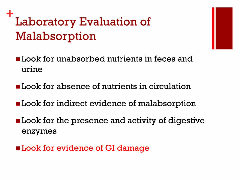
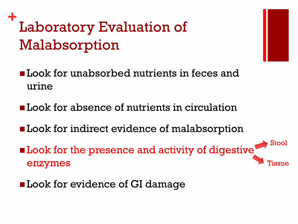
+ Laboratory Evaluation of
Malabsorption
Look for unabsorbed nutrients in feces and urine
Look for absence of nutrients in circulation
Look for indirect evidence of malabsorption
Look for the presence and activity of digestive enzymes
Look for evidence of GI damage
Sugars, fat, protein
Oral glucose load
Stool pH
Hydrogen breath test Fat-soluble vitamin deficiencies
Disaccharidase activity
Fecal elastase, trypsin
Enzyme levels in duodenal aspirates
Xylose absorption test
Endomesial and gliadin antibodies
Inflammatory markers

+ An unsettled stomach
9 month old girl
Skin rash antibiotics
Diarrhea next day
Switch to soy-based formula, then protein hydrolysate
Hospitalized Staphylococcal scalded skin syndrome
Negative: toxins, ova, parasites, bacteria
Normal: CBC, electrolytes
Stool analysis: 0.75-2% reducing substances
pH 5
Diarrhea remitted when oral feedings were stopped; resumed when oral feedings were resumed
De Zoeten et al. Medscape J Med. 2008; 10(8):195
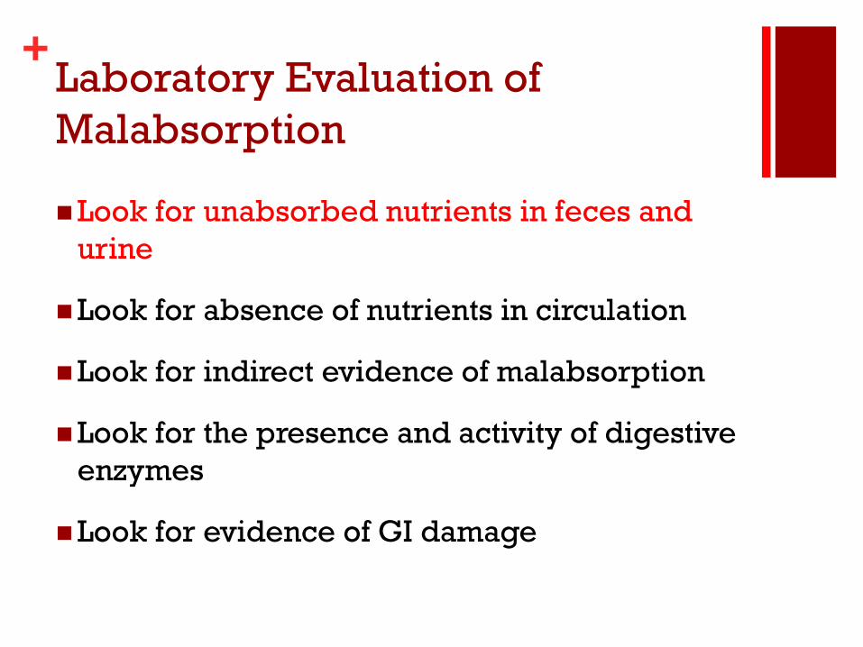
+ Laboratory Evaluation of
Malabsorption
Look for unabsorbed nutrients in feces and
urine
Look for absence of nutrients in circulation
Look for indirect evidence of malabsorption
Look for the presence and activity of digestive
enzymes
Look for evidence of GI damage

+ D-xylose absorption
Pentose monosaccharide passively
absorbed in proximal small bowel
Excreted in urine
Mucosal permeability of small intestine
Procedure:
Overnight fast
5 or 25 g oral dose of D-xylose
5 hour urine collection
(1 or 2 hour blood collection)
xylose + phloroglucinol
acid
heat
product (Abs 540nm)

+
5 hrs
oral xylose
dose
serum serum serum urine
1.7 mM glucose
0.3mM xylose
Eberts et al. 1979
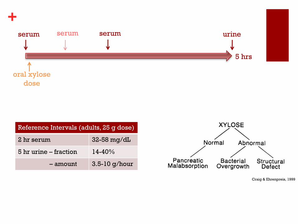
+
5 hrs
oral xylose
dose
serum serum serum urine
Reference Intervals (adults, 25 g dose)
2 hr serum 32-58 mg/dL
5 hr urine – fraction 14-40%
– amount 3.5-10 g/hour
Craig & Ehrenpreis, 1999
+ Laboratory Evaluation of
Malabsorption
Look for unabsorbed nutrients in feces and
urine
Look for absence of nutrients in circulation
Look for indirect evidence of malabsorption
Look for the presence and activity of digestive
enzymes
Look for evidence of GI damage

+ Fecal Fat Testing
Steatorrhea pancreas, bile acid, damage, transport, mixing diagnosing fat malabsorption
"Gold standard" for diagnosis
Treatment monitoring
Method history: solvent extractions, titrimetric or gravimetric, FTIR
Evidence of unabsorbed nutrients
Procedure:
3 day stool collection
Normal (50-150 g/day) fat diet
No barium, charcoal or non-
digestible fat intake
Sample is weighed and dried
Method:
NMR
Calibrated
Quantitation of % fat
Calculated weight/day result
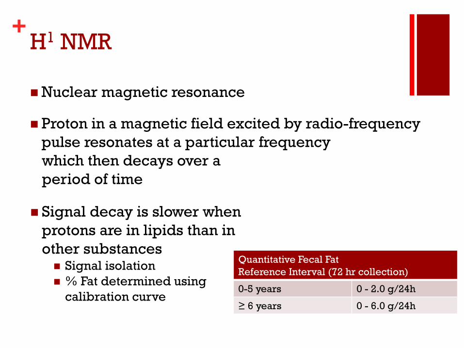
+ H1 NMR
Nuclear magnetic resonance
Proton in a magnetic field excited by radio-frequency
pulse resonates at a particular frequency
Signal decay is slower when
protons are in lipids than in
other substances Signal isolation
% Fat determined using
calibration curve
which then decays over a
period of time
Quantitative Fecal Fat
Reference Interval (72 hr collection)
0-5 years 0 - 2.0 g/24h
≥ 6 years 0 - 6.0 g/24h
+ Laboratory Evaluation of
Malabsorption
Look for unabsorbed nutrients in feces and
urine
Look for absence of nutrients in circulation
Look for indirect evidence of malabsorption
Look for the presence and activity of digestive
enzymes
Look for evidence of GI damage
Stool
Tissue

+ Fecal Elastase
Produced by pancreas
proelastase elastase
Serine protease, hydrolyzes amide and ester bonds
Remains intact and active in the intestine
Concentrated in feces versus duodenal fluid
Pancreatic exocrine function, protease enzymes
elastase
trypsin
duodenum
http://www.worthington-biochem.com/es/default.html

+ Fecal Elastase
Enzyme-linked immunoassay
Stool homogenates
Double-sandwich, signal amplification
Species and tissue-specific antibodies
Y
Pancreatic Elastase (μg/g feces)
Normal 201-500
Mild-moderate
insufficiency
100-200
Severe insufficiency ≤ 99
OD @ 450nm
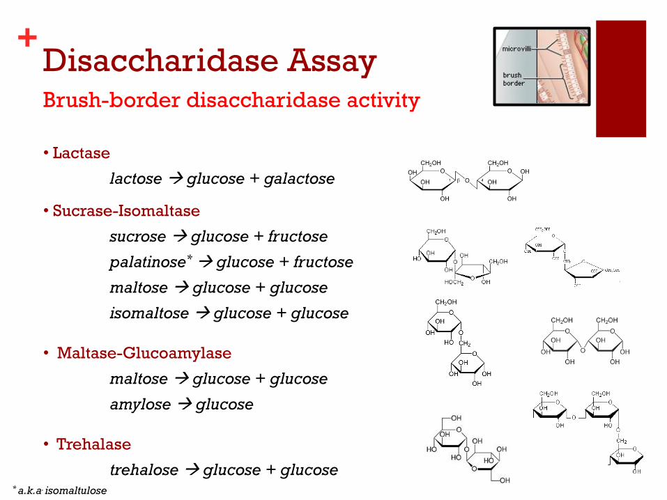
+ Disaccharidase Assay Brush-border disaccharidase activity
• Lactase
lactose glucose + galactose
• Sucrase-Isomaltase
sucrose glucose + fructose
palatinose* glucose + fructose
maltose glucose + glucose
isomaltose glucose + glucose
• Maltase-Glucoamylase
maltose glucose + glucose
amylose glucose
• Trehalase
trehalose glucose + glucose * a.k.a. isomaltulose
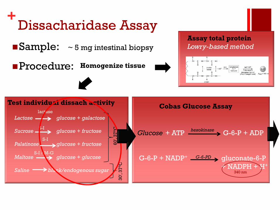
+ Dissacharidase Assay
Sample: ~ 5 mg intestinal biopsy
Procedure: Homogenize tissue
Test individual dissach activity
Lactose glucose + galactose
Sucrose glucose + fructose
Palatinose glucose + fructose
Maltose glucose + glucose
Saline blank/endogenous sugar
lactase
S-I
S-I
S-I / M-G
Assay total protein
Lowry-based method
60
', 3
7°C
3
0',
37
°C
Cobas Glucose Assay
Glucose + ATP G-6-P + ADP
G-6-P + NADP+ gluconate-6-P
+ NADPH + H+
hexokinase
G-6-PD
340 nm
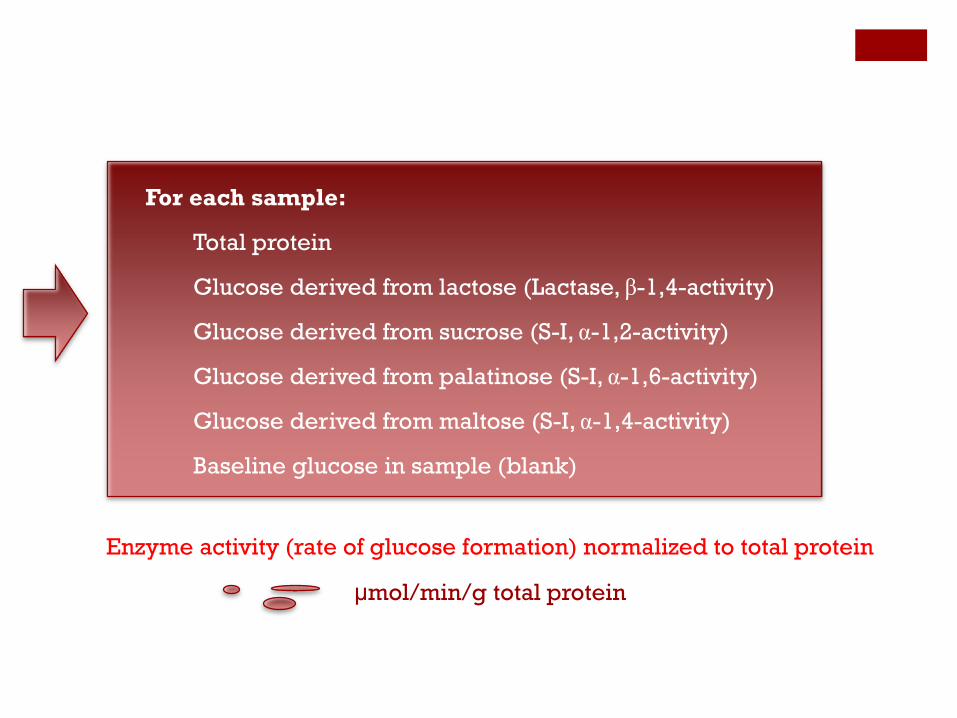
For each sample:
Total protein
Glucose derived from lactose (Lactase, β-1,4-activity)
Glucose derived from sucrose (S-I, α-1,2-activity)
Glucose derived from palatinose (S-I, α-1,6-activity)
Glucose derived from maltose (S-I, α-1,4-activity)
Baseline glucose in sample (blank)
Enzyme activity (rate of glucose formation) normalized to total protein
μmol/min/g total protein

Healthy
Celiac Disease
Upton, Arch Path Lab Med, 2008, 132(10):1594
Reference Intervals (μmol/min/g)
Lactase ≥ 15
Maltase ≥ 100
Sucrase ≥ 25
Palatinase ≥ 5
Primary disaccharidase deficiency can be established only in the absence of intestinal injury
Lactase deficiency Age-dependent onset
80-100% prevalence in some groups
Sucrase-isomaltase deficiency Congenital, gene mutations affect processing
Rare, 0.2-5% prevalence
+ An unsettled stomach 9 month old girl
Skin rash antibiotics
Diarrhea next day
Switch to soy-based formula, then protein hydrolysate
Hospitalized
Staphylococcal scalded skin syndrome
Negative: toxins, ova, parasites, bacteria
Normal: CBC, electrolytes
Stool analysis: 0.75-2% reducing substances
pH 5
Diarrhea remitted when oral feedings were stopped; resumed when
oral feedings were resumed
Disaccharidase testing showed low sucrase-isomaltase activity De Zoeten et al. Medscape J Med. 2008; 10(8):195
Disaccharidase Activity
(μmol/min/g)
Reference
Interval
Lactase 33.4 ≥ 15
Maltase 20.3 ≥ 100
Sucrase 0.6 ≥ 25
Palatinase 3.2 ≥ 5
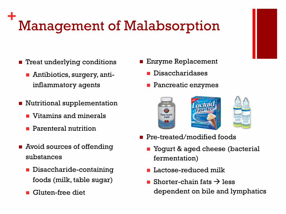
+ Management of Malabsorption
Treat underlying conditions
Antibiotics, surgery, anti-
inflammatory agents
Nutritional supplementation
Vitamins and minerals
Parenteral nutrition
Avoid sources of offending
substances
Disaccharide-containing
foods (milk, table sugar)
Gluten-free diet
Enzyme Replacement
Disaccharidases
Pancreatic enzymes
Pre-treated/modified foods
Yogurt & aged cheese (bacterial
fermentation)
Lactose-reduced milk
Shorter-chain fats less
dependent on bile and lymphatics
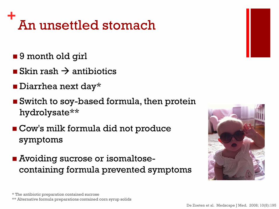
+ An unsettled stomach
9 month old girl
Skin rash antibiotics
Diarrhea next day*
Switch to soy-based formula, then protein
hydrolysate**
De Zoeten et al. Medscape J Med. 2008; 10(8):195
Cow's milk formula did not produce
symptoms
Avoiding sucrose or isomaltose-
containing formula prevented symptoms
* The antibiotic preparation contained sucrose
** Alternative formula preparations contained corn syrup solids
+
Conclusions
Causes of malabsorption may arise
from disruption of physical and/or
chemical processes of digestion as
well as impairments in nutrient
absorption
Laboratory methods employing a
wide variety of methodologies can
help in the evaluation of suspected
malabsorption and help to identify
the underlying causes
Patient management will depend
on the underlying cause and can
include dietary modification as well
as supplementation
Thanks! Any questions?
Special thanks to the Sp Chem and PAFT labs.
+ References
Tietz Textbook of Clinical Chemistry and Molecular Diagnostics,4th Ed. (2006). Burtis CA, Ashwood ER, Bruns DE (Eds) St. Louis, MO: Elsevier Saunders
Henry's Clinical Diagnosis and Management by Laboratory Methods. (2007). McPherson RA, Pincus MR, Henry JB. St. Louis, MO: Elsevier Saunders
The Physiology Coloring Book, 2nd Ed. (2000). Kapit W, Macey RI, Meisami E. San Franciso, CA: Benjamin Cummings
De Zoeten et al. (2008). A 9-month-old girl with chronic diarrhea. Medscape J Med. 10(8):195
Schaum's Outline of Human Anatomy and Physiology (3rd Ed). (2009). Van de Graaff K, Rhees RW, Palmer SL. New York, NY: McGraw-Hill
Ammoury RF, Croffie JM. (2010). Malabsorptive disorders of childhood. Pediatric Rev. 31(10): 407
Corsetti JP et al. (2011). Glycerol as a reference material for fecal fat quantitation using low-resolution time domain 1H NMR spectroscopy. Clin Biochem. 44:1352
Hammer HF. (2010). Pancreatic exocrine insufficiency: diagnostic evaluation and replacement therapy with pancreatic enzymes. Dig Dis. 28:339
Clayton PT. (2011). Disorders of bile acid synthesis. J Inherit Metab Dis. 34:593
Robayo-Torres CC et al. (2006). Disaccharide digestion: clinical and molecular aspects. Clin Gastroenterol Hepatol. 4:276
Erikson RH, Kim YS. (1990). Digestion and absorption of dietary protein. Annu Rev Med. 41:133
The Merck Manual, online: www.merckmanuals.com
ARUP Consult
MasterControl


![Zionism Unsettled - Presbyterian Church [USA]](https://img.pdfslide.net/doc/110x75/577ccf7a1a28ab9e788fd043/zionism-unsettled-presbyterian-church-usa.jpg)