Embed Size (px)
Citation preview
a
Management
of Patients With
ST-ElevationMyocardialInfarction
ACC/AHA Pocket Guideline
Based on the ACC/AHA Guidelines for the Management of Patients With ST-Elevation Myocardial Infarction
July 2004
Learn and Live SM
Management of Patients With
ST-ElevationMyocardial InfarctionJuly 2004
ACC/AHA Writing Committee
Elliott M. Antman, MD, FACC, FAHA, Chair
Daniel T. Anbe, MD, FACC, FAHA
Paul Wayne Armstrong, MD, FACC, FAHA
Eric R. Bates, MD, FACC, FAHA
Lee A. Green, MD, MPH
Mary Hand, MSPH, RN, FAHA
Judith S. Hochman, MD, FACC, FAHA
Harlan M. Krumholz, MD, FACC, FAHA
Frederick G. Kushner, MD, FACC, FAHA
Gervasio A. Lamas, MD, FACC
Charles J. Mullany, MB, MS, FACC
Joseph P. Ornato, MD, FACC, FAHA
David L. Pearle, MD, FACC, FAHA
Michael A. Sloan, MD, FACC
Sidney C. Smith, Jr, MD, FACC, FAHA
Special thanks to
Eli Lilly and Company
supported this
pocket guideline
through an
educational grant.
Eli Lilly and Company
was not involved in the
development of this
publication and in no
way influenced
its contents.
© 2004 American College
of Cardiology Foundation and
American Heart Association, Inc.
The following article was adapted from the
ACC/AHA Guidelines for Management of
Patients with ST-Elevation Myocardial Infarction
(Journal of the American College of Cardiology
2004 ________ ; and Circulation 2004 _________).
For a copy of the full report or published
executive summary, visit our Web sites
at www.acc.org or www.americanheart.org,
or call the ACC Resource Center at
1-800-253-4636, ext 694.
Managem
ent Before STEM
IO
nset of STEMI
Hospital M
anagement
Secondary Prevention
Contents
I. Introduction . . . . . . . . . . . . . . . . . . . . . . . . . . . . . . . . . . . . . . . . . . . . . 5
II. Management Before STEMI . . . . . . . . . . . . . . . . . . . . . . . . . . . . . . 10
A. Recommendations for Identification of Patients at Risk of STEMI . . . . . . 10
B. Recommendations for Patient Education
for Early Recognition and Response to STEMI . . . . . . . . . . . . . . . . . . . . 11
III. Onset of STEMI . . . . . . . . . . . . . . . . . . . . . . . . . . . . . . . . . . . . . . . . 12
A. Prehospital Issues . . . . . . . . . . . . . . . . . . . . . . . . . . . . . . . . . . . . . . . . . 12
B. Initial Recognition and Management in the Emergency Department . . . . 14
IV. Hospital Management . . . . . . . . . . . . . . . . . . . . . . . . . . . . . . . . . . 25
V. Secondary Prevention and Long-Term Management . . . . . . . . . 38
4 5
I. Introduction
The full text of the guidelines is available on the
World Wide Web sites of the American College of
Cardiology (www.acc.org) and the American Heart
Association (www.americanheart.org). The executive
summary is published in the August 4, 2004 issue of
the Journal of the American College of Cardiology and
the August 3, 2004 issue of Circulation.
This pocket guide provides rapid prompts for appro-
priate patient management, which is outlined in
much greater detail in the full-text guidelines. It is
not intended as a replacement for understanding the
caveats and rationales that are stated carefully in the
full-text guidelines. Users should consult the full-text
document for more information.
Scope of the Guidelines
The purpose of these guidelines is to focus on the
numerous advances in the diagnosis and man-
agement of patients with ST-elevation myocardial
infarction (STEMI) since 1999. It is recognized that
there are areas of overlap among these guidelines on
patients with STEMI, the guidelines on patients with
unstable angina/non-STEMI, and other guidelines.
The committee has handled this overlap by reiterat-
ing important concepts and recommendations in the
STEMI guidelines and by providing cross-references
to other guidelines.
6 7
CLASS IIa
Benefit >> RiskAdditional studies withfocused objectives needed
IT IS REASONABLE to per-form procedure/administer treatment
■ Recommendation in favorof treatment or procedurebeing useful/effective
■ Some conflicting evidencefrom multiple randomized trials or meta-analyses
■ Recommendation in favorof treatment or procedurebeing useful/effective
■ Some conflicting evidence from single randomized trial or nonrandomized studies
■ Recommendation in favorof treatment or procedurebeing useful/effective
■ Only diverging expertopinion, case studies, or standard-of-care
CLASS I
Benefit >>> Risk
Procedure/TreatmentSHOULD be performed/administered
■ Recommendation that procedure or treatment is useful/effective
■ Sufficient evidence frommultiple randomized trials or meta-analyses
■ Recommendation that procedure or treatment is useful/effective
■ Limited evidence from single randomized trial ornonrandomized studies
■ Recommendation that procedure or treatment isuseful/effective
■ Only expert opinion, casestudies, or standard-of-care
shouldis recommendedis indicatedis useful/effective/beneficial
LEVEL A
Multiple (3-5) populationrisk strata evaluated*
General consistency ofdirection and magnitudeof effect
LEVEL B
Limited (2-3) populationrisk strata evaluated*
LEVEL C
Very limited (1-2)
population risk strataevaluated*
CLASS IIb
Benefit > RiskAdditional studies with broadobjectives needed; additionalregistry data would be helpful
Procedure/Treatment MAY BE CONSIDERED
■ Recommendation’s usefulness/efficacy less well established
■ Greater conflicting evidence from multiple randomized trials or meta-analyses
■ Recommendation’s usefulness/efficacy less well established
■ Greater conflicting evidence from single randomized trial or nonrandomized studies
■ Recommendation’s usefulness/efficacy less well established
■ Only diverging expert opinion, case studies, orstandard-of-care
may/might be consideredmay/might be reasonableusefulness/effectiveness is
unknown/unclear/uncertain or not well established
CLASS III
Risk > BenefitNo additional studies needed
Procedure/Treatment shouldNOT be performed/adminis-tered SINCE IT IS NOT HELP-FUL AND MAY BE HARMFUL
■ Recommendation that procedure or treatment is not useful/effective and may be harmful
■ Sufficient evidence frommultiple randomized trials or meta-analyses
■ Recommendation that procedure or treatment is not useful/effective and may be harmful
■ Limited evidence from single randomized trial ornonrandomized studies
■ Recommendation that procedure or treatment is not useful/effective and may be harmful
■ Only expert opinion, casestudies, or standard-of-care
is not recommendedis not indicatedshould notis not useful/effective/beneficialmay be harmful
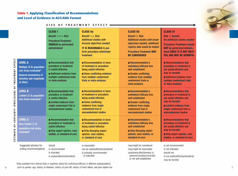
*Data available from clinical trials or registries about the usefulness/efficacy in different subpopulations,
such as gender, age, history of diabetes, history of prior MI, history of heart failure, and prior aspirin use.
Suggested phrases forwriting recommendations
is reasonablecan be useful/effective/beneficialis probably recommended
or indicated
S I Z E O F T R E A T M E N T E F F E C T
Table 1. Applying Classification of Recommendations and Level of Evidence in ACC/AHA Format
ES
TIM
AT
E O
F C
ER
TAIN
TY
(P
RE
CIS
ION
) O
F T
RE
AT
ME
NT
EF
FE
CT
▼
98
The top half of the figure illustrates the chronology of the interfacebetween the patient and the clinician through the progression of plaque formation and the onset and complications of STEMI, along with relevant management considerations at each stage.
Following disruption of a vulnerable or high-risk plaque, patients experience ischemic discomfort resulting from a reduction of flow through the affected epicardial coronary artery. Of patients with ST-segment elevation, most (large red arrow in bottom panel) ultimately develop a Q-wave MI (QwMI), while a few (small red arrow) develop a non-Q-wave MI (NQMI).STEMI, ST-elevation myocardial infarction; Dx, diagnosis; NQMI, non–Q-wave myocardial infarction; QwMI, Q-wave myocardial infarction.
Modified with permission from Libby. Circulation 104:365,2001, Hamm et al.The Lancet 2001;358:1533-1538 and Davies. Heart 2000;83:361-366 with permission from the BMJ Publishing Group.
Pages in the Pocket Guide are
color-coded to correspond to
the chronological interface of
the clinician with the patient.
Pages with yellow tabs refer
to Management Before STEMI,
pages with red tabs refer to the
Onset of STEMI, pages with
orange tabs refer to Hospital
Management, and pages with blue tabs refer to
Secondary Prevention/Long-Term Management.
Management Before STEMI
Hospital Management
Onset of STEMI
Secondary Prevention
Figure 1. Acute Coronary Syndromes
Hospital Management■ Medications■ Arrhythmias■ Complications■ Preparation for discharge
Onset of STEMI■ Prehospital issues■ Initial recognition and management
in the Emergency Department■ Reperfusion
1 2 3 4 5 6
Ischemic Discomfort
Acute Coronary Syndrome
ST Elevation
Presentation
Working Dx
ECG
CardiacBiomarker
Final Dx
▼
▼
▼
▼
No ST Elevation
NQMI QwMIUnstableAngina
Myocardial Infarction
UA
▼ ▼
NSTEMI
Management Before STEMI
Secondary Prevention/Long-Term Management
10 11
II. Management Before STEMI
A. Recommendations for Identification of Patients at Risk of STEMI
Class I 1. Primary care providers should evaluate the
presence and status of control of major risk factors
for coronary heart disease (CHD) for all patients at
regular intervals (approximately every 3 to 5 years).
(Level of Evidence: C)
2. Ten-year risk [National Cholesterol Education
Program (NCEP) global risk] of developing sympto-
matic CHD should be calculated for all patients who
have 2 or more major risk factors to assess the need
for primary prevention strategies. (Level of Evidence: B)
3. Patients with established CHD should be identi-
fied for secondary prevention, and patients with a
CHD risk equivalent (e.g., diabetes mellitus, chronic
kidney disease, or 10-year risk greater than 20% as
calculated by Framingham equations) should
receive equally intensive risk factor intervention as
those with clinically apparent CHD. (Level of Evidence: A)
B. Recommendations for Patient Education for Early Recognition and Response to STEMI
Class I 1. Patients with symptoms of STEMI [chest discom-
fort with or without radiation to the arms(s), back,
neck, jaw, or epigastrium; shortness of breath;
weakness; diaphoresis; nausea; lightheadedness]
should be transported to the hospital by ambulance
rather than by friends or relatives. (Level of Evidence: B)
2. Healthcare providers should actively address the
following issues regarding STEMI with patients and
their families:
a. the patient's heart attack risk (Level of Evidence: C)
b. how to recognize symptoms of STEMI (Level of
Evidence: C)
c. the advisability of calling 9-1-1 if symptoms are
unimproved or worsening after 5 minutes, despite
feelings of uncertainty and fear of potential embar-
rassment (Level of Evidence: C)
d. a plan for appropriate recognition and response
to a potential acute cardiac event that includes the
phone number to access emergency medical ser-
vices (EMS), generally 9-1-1. (Level of Evidence: C)
3. Healthcare providers should instruct patients for
whom nitroglycerin has been prescribed previously
to take ONE nitroglycerin dose sublingually in
response to chest discomfort/pain. If chest discom-
fort/pain is unimproved or worsening 5 minutes
Managem
ent Before STEM
IMan
agem
ent
Befo
re S
TEM
I
12 13
after 1 sublingual nitroglycerin dose has been
taken, it is recommended that the patient or family
member/friend be instructed to call 9-1-1 immedi-
ately to access EMS. (Level of Evidence: C)
III. Onset of STEMI
A. Prehospital Issues
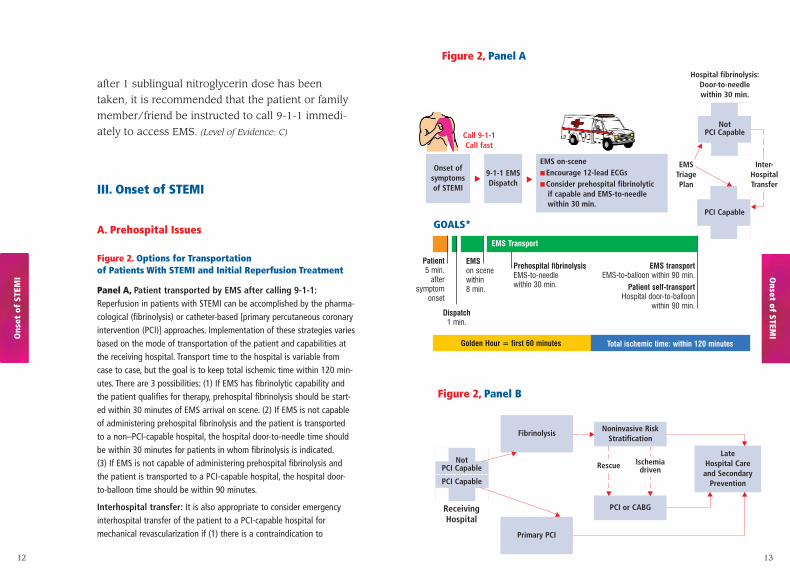
Figure 2. Options for Transportation of Patients With STEMI and Initial Reperfusion Treatment
Panel A, Patient transported by EMS after calling 9-1-1:Reperfusion in patients with STEMI can be accomplished by the pharma-cological (fibrinolysis) or catheter-based [primary percutaneous coronaryintervention (PCI)] approaches. Implementation of these strategies variesbased on the mode of transportation of the patient and capabilities atthe receiving hospital. Transport time to the hospital is variable fromcase to case, but the goal is to keep total ischemic time within 120 min-utes. There are 3 possibilities: (1) If EMS has fibrinolytic capability andthe patient qualifies for therapy, prehospital fibrinolysis should be start-ed within 30 minutes of EMS arrival on scene. (2) If EMS is not capableof administering prehospital fibrinolysis and the patient is transported to a non–PCI-capable hospital, the hospital door-to-needle time shouldbe within 30 minutes for patients in whom fibrinolysis is indicated.(3) If EMS is not capable of administering prehospital fibrinolysis andthe patient is transported to a PCI-capable hospital, the hospital door-to-balloon time should be within 90 minutes.
Interhospital transfer: It is also appropriate to consider emergencyinterhospital transfer of the patient to a PCI-capable hospital formechanical revascularization if (1) there is a contraindication to
Figure 2, Panel A
Figure 2, Panel B
Onset ofsymptomsof STEMI
▲ ▲9-1-1 EMSDispatch
Call 9-1-1Call fast
EMS on-scene■ Encourage 12-lead ECGs■ Consider prehospital fibrinolytic
if capable and EMS-to-needle within 30 min.
PCI Capable
PCI Capable
ReceivingHospital
NotPCI Capable
Hospital fibrinolysis:Door-to-needle within 30 min.
Rescue Ischemia driven
Not PCI Capable
▼
▼
EMS Triage Plan
▼
Inter-HospitalTransfer
Patient5 min.
aftersymptom
onset
EMSon scenewithin8 min.
Dispatch1 min.
GOALS*
EMS Transport
Total ischemic time: within 120 minutes
Prehospital fibrinolysisEMS-to-needle within 30 min.
EMS transportEMS-to-balloon within 90 min.
Patient self-transportHospital door-to-balloon
within 90 min.
Golden Hour = first 60 minutes
PCI or CABG
Primary PCI
Fibrinolysis Noninvasive RiskStratification
▼ ▼
▼
▼
Late Hospital Care
and Secondary Prevention
▼
▼
Onset of STEM
IOns
et o
f ST
EMI
▼
▼
14
fibrinolysis; (2) PCI can be initiated promptly (within 90 minutes afterthe patient presented to the initial receiving hospital or within 60 min-utes compared to when fibrinolysis with a fibrin-specific agent could beinitiated at the initial receiving hospital); or (3) fibrinolysis is adminis-tered and is unsuccessful (i.e., “rescue PCI”). Secondary nonemergencyinterhospital transfer can be considered for recurrent ischemia.
Patient self-transport: Patient self-transportation is discouraged. If the patient arrives at a non–PCI-capable hospital, the door-to-needletime should be within 30 minutes. If the patient arrives at a PCI-capablehospital, the door-to-balloon time should be within 90 minutes. Thetreatment options and time recommendations after first hospital arrivalare the same.
Panel B, For patients who receive fibrinolysis, noninvasive riskstratification is recommended to identify the need for rescue PCI (failed fibrinolysis) or ischemia-driven PCI. Regardless of theinitial method of reperfusion treatment, all patients should receive latehospital care and secondary prevention of STEMI.
EMS = emergency medical system; CABG = coronary artery bypass graft surgery.
*The medical system goal is to facilitate rapid recognition and treatment of patients with STEMIsuch that door-to-needle (or medical contact–to-needle) time for initiation of fibrinolytic therapyis within 30 minutes or that door-to-balloon (or medical contact–to-balloon) time for PCI is within 90 minutes. These goals should not be understood as ideal times but rather as the longesttimes that should be considered acceptable for a given system. Systems that are able to achieveeven more rapid times for treatment of patients with STEMI should be encouraged.
Modified with permission from Armstrong et al. Circulation 2003;107:2533.
Onset of STEMI symptoms
STEMIpatient?
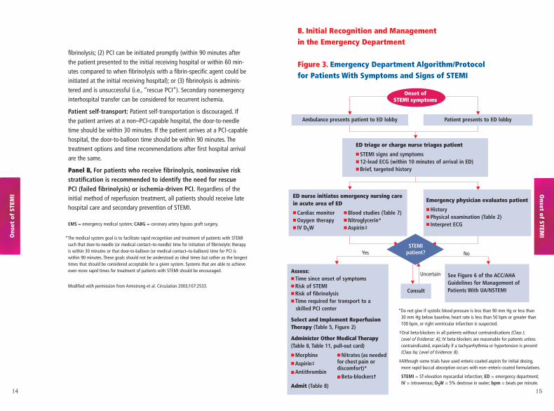
B. Initial Recognition and Management in the Emergency Department
Figure 3. Emergency Department Algorithm/Protocol for Patients With Symptoms and Signs of STEMI
*Do not give if systolic blood pressure is less than 90 mm Hg or less than 30 mm Hg below baseline, heart rate is less than 50 bpm or greater than100 bpm, or right ventricular infarction is suspected.
†Oral beta-blockers in all patients without contraindications (Class I; Level of Evidence: A); IV beta-blockers are reasonable for patients unlesscontraindicated, especially if a tachyarrhythmia or hypertension is present (Class IIa; Level of Evidence: B).
‡Although some trials have used enteric-coated aspirin for initial dosing,more rapid buccal absorption occurs with non–enteric-coated formulations.
STEMI = ST-elevation myocardial infarction; ED = emergency department;IV = intravenous; D5W = 5% dextrose in water; bpm = beats per minute.
15
Ambulance presents patient to ED lobby Patient presents to ED lobby▼ ▼
ED triage or charge nurse triages patient
■ STEMI signs and symptoms■ 12-lead ECG (within 10 minutes of arrival in ED)■ Brief, targeted history
▼
ED nurse initiates emergency nursing carein acute area of ED
■ Cardiac monitor ■ Blood studies (Table 7)■ Oxygen therapy ■ Nitroglycerin*■ IV D5W ■ Aspirin‡
Emergency physician evaluates patient
■ History■ Physical examination (Table 2)■ Interpret ECG
▼ ▼
▼
▼
See Figure 6 of the ACC/AHAGuidelines for Management ofPatients With UA/NSTEMI
Assess:■ Time since onset of symptoms■ Risk of STEMI■ Risk of fibrinolysis■ Time required for transport to a
skilled PCI center
Select and Implement ReperfusionTherapy (Table 5, Figure 2)
Administer Other Medical Therapy(Table 8, Table 11, pull-out card)
■ Morphine ■ Nitrates (as needed■ Aspirin‡ for chest pain or
■ Antithrombindiscomfort)*
■ Beta-blockers†
Admit (Table 8)
Consult
Yes No
Uncertain
▼ ▼
▼
▼
Onset of STEM
IOns
et o
f ST
EMI
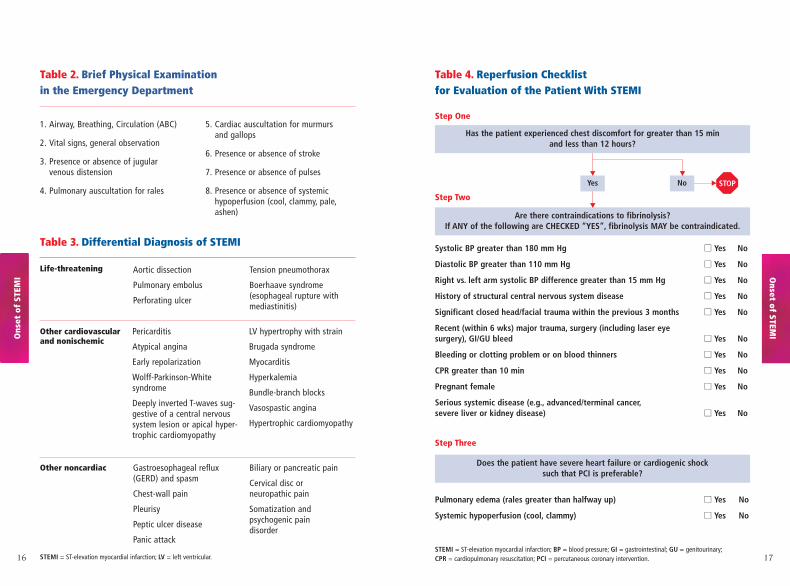
Aortic dissection
Pulmonary embolus
Perforating ulcer
Tension pneumothorax
Boerhaave syndrome(esophageal rupture withmediastinitis)
Gastroesophageal reflux(GERD) and spasm
Chest-wall pain
Pleurisy
Peptic ulcer disease
Panic attack
Biliary or pancreatic pain
Cervical disc or neuropathic pain
Somatization and psychogenic pain disorder
16 17
Table 2. Brief Physical Examination in the Emergency Department
Table 3. Differential Diagnosis of STEMI
Life-threatening
Other cardiovascular and nonischemic
Other noncardiac
Pericarditis
Atypical angina
Early repolarization
Wolff-Parkinson-White syndrome
Deeply inverted T-waves sug-gestive of a central nervoussystem lesion or apical hyper-trophic cardiomyopathy
LV hypertrophy with strain
Brugada syndrome
Myocarditis
Hyperkalemia
Bundle-branch blocks
Vasospastic angina
Hypertrophic cardiomyopathy
1. Airway, Breathing, Circulation (ABC)
2. Vital signs, general observation
3. Presence or absence of jugular venous distension
4. Pulmonary auscultation for rales
5. Cardiac auscultation for murmurs and gallops
6. Presence or absence of stroke
7. Presence or absence of pulses
8. Presence or absence of systemic hypoperfusion (cool, clammy, pale,ashen)
Table 4. Reperfusion Checklist for Evaluation of the Patient With STEMI
Has the patient experienced chest discomfort for greater than 15 min and less than 12 hours?
Yes No
Are there contraindications to fibrinolysis?If ANY of the following are CHECKED “YES”, fibrinolysis MAY be contraindicated.
Systolic BP greater than 180 mm Hg ■■ Yes No
Diastolic BP greater than 110 mm Hg ■■ Yes No
Right vs. left arm systolic BP difference greater than 15 mm Hg ■■ Yes No
History of structural central nervous system disease ■■ Yes No
Significant closed head/facial trauma within the previous 3 months ■■ Yes No
Recent (within 6 wks) major trauma, surgery (including laser eye surgery), GI/GU bleed ■■ Yes No
Bleeding or clotting problem or on blood thinners ■■ Yes No
CPR greater than 10 min ■■ Yes No
Pregnant female ■■ Yes No
Serious systemic disease (e.g., advanced/terminal cancer,severe liver or kidney disease) ■■ Yes No
Pulmonary edema (rales greater than halfway up) ■■ Yes No
Systemic hypoperfusion (cool, clammy) ■■ Yes No
STEMI = ST-elevation myocardial infarction; BP = blood pressure; GI = gastrointestinal; GU = genitourinary;CPR = cardiopulmonary resuscitation; PCI = percutaneous coronary intervention.
Does the patient have severe heart failure or cardiogenic shock such that PCI is preferable?
▼
▼
▼
▼ STOP
Step One
Step Two
Step Three
Onset of STEM
IOns
et o
f ST
EMI
STEMI = ST-elevation myocardial infarction; LV = left ventricular.
18 19
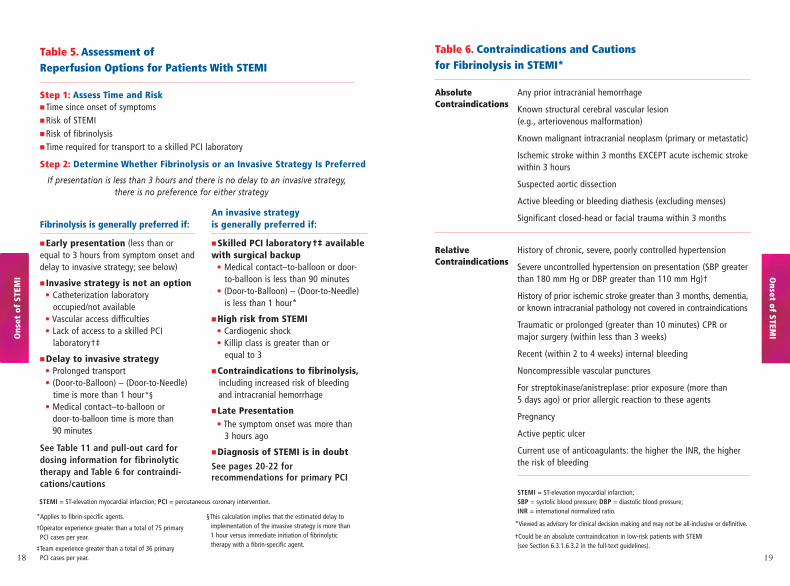
Table 6. Contraindications and Cautions for Fibrinolysis in STEMI*
Absolute Any prior intracranial hemorrhageContraindications Known structural cerebral vascular lesion
(e.g., arteriovenous malformation)
Known malignant intracranial neoplasm (primary or metastatic)
Ischemic stroke within 3 months EXCEPT acute ischemic stroke within 3 hours
Suspected aortic dissection
Active bleeding or bleeding diathesis (excluding menses)
Significant closed-head or facial trauma within 3 months
Relative History of chronic, severe, poorly controlled hypertension Contraindications Severe uncontrolled hypertension on presentation (SBP greater
than 180 mm Hg or DBP greater than 110 mm Hg)†
History of prior ischemic stroke greater than 3 months, dementia,or known intracranial pathology not covered in contraindications
Traumatic or prolonged (greater than 10 minutes) CPR or major surgery (within less than 3 weeks)
Recent (within 2 to 4 weeks) internal bleeding
Noncompressible vascular punctures
For streptokinase/anistreplase: prior exposure (more than 5 days ago) or prior allergic reaction to these agents
Pregnancy
Active peptic ulcer
Current use of anticoagulants: the higher the INR, the higher the risk of bleeding
STEMI = ST-elevation myocardial infarction;SBP = systolic blood pressure; DBP = diastolic blood pressure;INR = international normalized ratio.
*Viewed as advisory for clinical decision making and may not be all-inclusive or definitive.
†Could be an absolute contraindication in low-risk patients with STEMI(see Section 6.3.1.6.3.2 in the full-text guidelines).
Table 5. Assessment of Reperfusion Options for Patients With STEMI
Step 1: Assess Time and Risk■ Time since onset of symptoms■ Risk of STEMI■ Risk of fibrinolysis■ Time required for transport to a skilled PCI laboratory
Step 2: Determine Whether Fibrinolysis or an Invasive Strategy Is Preferred
If presentation is less than 3 hours and there is no delay to an invasive strategy, there is no preference for either strategy
Fibrinolysis is generally preferred if:
■ Early presentation (less than orequal to 3 hours from symptom onset anddelay to invasive strategy; see below)
■ Invasive strategy is not an option• Catheterization laboratory
occupied/not available• Vascular access difficulties• Lack of access to a skilled PCI
laboratory†‡
■ Delay to invasive strategy• Prolonged transport• (Door-to-Balloon) – (Door-to-Needle)
time is more than 1 hour*§
• Medical contact–to-balloon or door-to-balloon time is more than 90 minutes
See Table 11 and pull-out card fordosing information for fibrinolytictherapy and Table 6 for contraindi-cations/cautions
An invasive strategy is generally preferred if:
■ Skilled PCI laboratory†‡ availablewith surgical backup
• Medical contact–to-balloon or door-to-balloon is less than 90 minutes
• (Door-to-Balloon) – (Door-to-Needle) is less than 1 hour*
■ High risk from STEMI• Cardiogenic shock • Killip class is greater than or
equal to 3
■ Contraindications to fibrinolysis,including increased risk of bleeding and intracranial hemorrhage
■ Late Presentation• The symptom onset was more than
3 hours ago
■ Diagnosis of STEMI is in doubt
See pages 20-22 for recommendations for primary PCI
*Applies to fibrin-specific agents.
†Operator experience greater than a total of 75 primaryPCI cases per year.
‡Team experience greater than a total of 36 primary PCI cases per year.
§This calculation implies that the estimated delay toimplementation of the invasive strategy is more than1 hour versus immediate initiation of fibrinolytic therapy with a fibrin-specific agent.
Onset of STEM
IOns
et o
f ST
EMI
STEMI = ST-elevation myocardial infarction; PCI = percutaneous coronary intervention.
20 21
■ greater than 1 hour, fibrinolytic therapy
(fibrin-specific agents) is generally preferred.
(Level of Evidence: B)
c. If symptom duration is greater than 3 hours,
primary PCI should be performed with a medical
contact–to-balloon or door-to-balloon time as brief
as possible, with a goal of within 90 minutes. (Level
of Evidence: B)
d. Primary PCI should be performed for patients less
than 75 years old with ST elevation or LBBB who
develop shock within 36 hours of MI and are suit-
able for revascularization that can be performed
within 18 hours of shock, unless further support is
futile because of the patient's wishes or contraindi-
cations/unsuitability for further invasive care. (Level
of Evidence: A)
e. Primary PCI should be performed in patients with
severe congestive heart failure and/or pulmonary
edema (Killip class 3) and onset of symptoms within
12 hours. The medical contact–to-balloon or door-
to-balloon time should be as short as possible, with
a goal of within 90 minutes. (Level of Evidence: B)
Class IIa 1. Primary PCI is reasonable for selected patients 75
years or older with ST elevation or LBBB who develop
shock within 36 hours of MI and are suitable for
revascularization that can be performed within 18
Recommendations for Primary PCI
See Table 5 regarding additional considerations for
selecting reperfusion therapy.
Class I General Considerations:
1. If immediately available, primary PCI should be
performed in patients with STEMI (including true
posterior MI) or MI with new or presumably new
left bundle-branch block (LBBB) who can undergo
PCI of the infarct artery within 12 hours of symptom
onset, if performed in a timely fashion (balloon
inflation within 90 minutes of presentation) by
persons skilled in the procedure (individuals who
perform more than 75 PCI procedures per year).
The procedure should be supported by experienced
personnel in an appropriate laboratory (one that
performs more than 200 PCI procedures per year, of
which at least 36 are primary PCI for STEMI, and
that has cardiac surgery capability). (Level of Evidence: A)
Specific Considerations:
a. Primary PCI should be performed as quickly as
possible, with the goal of a medical contact–to-
balloon or door-to-balloon time of within 90 minutes.
(Level of Evidence: B)
b. If the symptom duration is within 3 hours and the
expected door-to-balloon time minus the expected
door-to-needle time is:
■ within 1 hour, primary PCI is generally preferred
(Level of Evidence: B)
Onset of STEM
IOns
et o
f ST
EMI
22 23
hours of shock. Patients with good prior functional
status who are suitable for revascularization and
agree to invasive care may be selected for an
invasive strategy. (Level of Evidence: B)
2. It is reasonable to perform primary PCI for
patients with onset of symptoms within the prior
12 to 24 hours and one or more of the following:
a. severe congestive heart failure (Level of Evidence: C)
b. hemodynamic or electrical instability
(Level of Evidence: C)
c. persistent ischemic symptoms. (Level of Evidence: C)
Class IIb 1. The benefit of primary PCI for STEMI patients
eligible for fibrinolysis is not well established when
performed by an operator who performs fewer than
75 PCI procedures per year. (Level of Evidence: C)
Class III 1. Primary PCI should not be performed in a non-
infarct artery in patients without hemodynamic
compromise. (Level of Evidence: C)
2. Primary PCI should not be performed in asympto-
matic patients more than 12 hours after onset of
STEMI if they are hemodynamically and electrically
stable. (Level of Evidence: C)
Pharmacological Support During Primary PCI
Unfractionated No GP IIb/IIIa Inhibitor GP IIb/IIIa Inhibitor UsedHeparin
Bolus: 70-100 U/kg 50-70 U/kg
Target ACT: HemoTec: 250-300 s With either device: 200 s
Hemochron: 300-350 s(Class I: Level of Evidence: C)
Thienopyridine ■ Administer loading dose
Clopidogrel ■ Maintenance dose: 75 mg orally per day
■ Duration:
i) Bare metal stent—1 month minimum
ii) Drug-eluting stent—minimum of 3 months after sirolimus and 6 months after paclitaxel
Continue for 12 months after stent implantation (both types of stents) in patients who are not at risk of bleeding.(Class I: Level of Evidence: B)
GP IIb/IIIa ■ It is reasonable to start abciximab as early as possible beforeInhibitors primary PCI (with or without stenting). The recommended dosage
of abciximab in adults is a 0.25 mg/kg intravenous bolus adminis-tered 10 to 60 minutes before the start of PCI, followed by a con-tinuous intravenous infusion of 0.125 mcg/kg/min (to a maximum of 10 mcg/min) for 12 to 18 hours. (Class IIa; Level of Evidence: B)
■ Treatment with tirofiban (bolus dose of 10 mcg per kilogram of body weight, followed by an infusion of 0.15 mcg/kg/min for 18 to 24 hours) or eptifibatide (for patients with serum creatinine less than 2.0 mg/dL,* an intravenous bolus of 180 mcg/kg admin-istered immediately before the initiation of PCI followed by a continuous infusion of 2.0 mcg/kg/min and a second 180 mcg/kg bolus 10 minutes after the first bolus. Infusion should be continueduntil hospital discharge, or for up to 18 to 24 hours, whichever comes first) may be considered before primary PCI (with or with-out stenting). (Class IIb; Level of Evidence: C)
*For patients with a serum creatinine greater than 2.0 mg/dL, an intravenous bolus of 180 mcg/kg administered immediately before initiation of the procedure, immediately followed by a continuous infusion of 1.0 mcg/kg/min and a second 180 mcg/kg bolus adminis-tered 10 minutes after the first.
GP= glycoprotein; ACT= activated clotting time; U =units; s =seconds.
Onset of STEM
IOns
et o
f ST
EMI
24 25
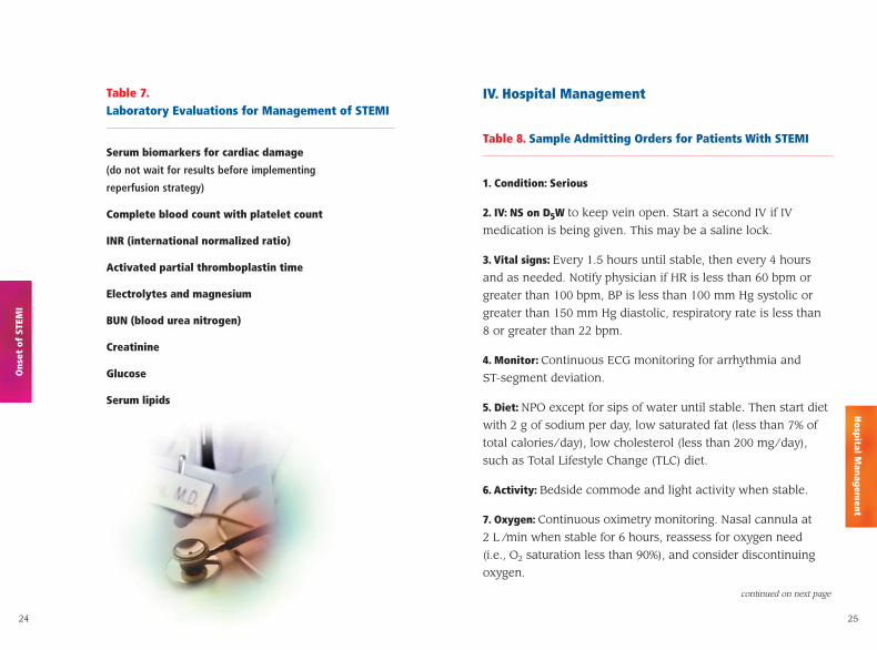
Table 7.Laboratory Evaluations for Management of STEMI
Serum biomarkers for cardiac damage
(do not wait for results before implementing
reperfusion strategy)
Complete blood count with platelet count
INR (international normalized ratio)
Activated partial thromboplastin time
Electrolytes and magnesium
BUN (blood urea nitrogen)
Creatinine
Glucose
Serum lipids
IV. Hospital Management
Table 8. Sample Admitting Orders for Patients With STEMI
1. Condition: Serious
2. IV: NS on D5W to keep vein open. Start a second IV if IV
medication is being given. This may be a saline lock.
3. Vital signs: Every 1.5 hours until stable, then every 4 hours
and as needed. Notify physician if HR is less than 60 bpm or
greater than 100 bpm, BP is less than 100 mm Hg systolic or
greater than 150 mm Hg diastolic, respiratory rate is less than
8 or greater than 22 bpm.
4. Monitor: Continuous ECG monitoring for arrhythmia and
ST-segment deviation.
5. Diet: NPO except for sips of water until stable. Then start diet
with 2 g of sodium per day, low saturated fat (less than 7% of
total calories/day), low cholesterol (less than 200 mg/day),
such as Total Lifestyle Change (TLC) diet.
6. Activity: Bedside commode and light activity when stable.
7. Oxygen: Continuous oximetry monitoring. Nasal cannula at
2 L /min when stable for 6 hours, reassess for oxygen need
(i.e., O2 saturation less than 90%), and consider discontinuing
oxygen.
Ons
et o
f ST
EMI
Hospital M
anagement
continued on next page
26 27
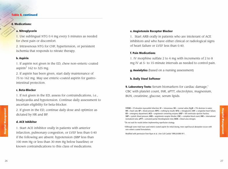
8. Medications:
a. Nitroglycerin
1. Use sublingual NTG 0.4 mg every 5 minutes as needed
for chest pain or discomfort.
2. Intravenous NTG for CHF, hypertension, or persistent
ischemia that responds to nitrate therapy.
b. Aspirin
1. If aspirin not given in the ED, chew non–enteric-coated
aspirin† 162 to 325 mg.
2. If aspirin has been given, start daily maintenance of
75 to 162 mg. May use enteric-coated aspirin for gastro-
intestinal protection.
c. Beta-Blocker
1. If not given in the ED, assess for contraindications, i.e.,
bradycardia and hypotension. Continue daily assessment to
ascertain eligibility for beta-blocker.
2. If given in the ED, continue daily dose and optimize as
dictated by HR and BP.
d. ACE Inhibitor
1. Start ACE inhibitor orally in patients with anterior
infarction, pulmonary congestion, or LVEF less than 0.40
if the following are absent: hypotension (SBP less than
100 mm Hg or less than 30 mm Hg below baseline) or
known contraindications to this class of medications.
e. Angiotensin Receptor Blocker
1. Start ARB orally in patients who are intolerant of ACE
inhibitors and who have either clinical or radiological signs
of heart failure or LVEF less than 0.40.
f. Pain Medications
1. IV morphine sulfate 2 to 4 mg with increments of 2 to 8
mg IV at 5- to 15-minute intervals as needed to control pain.
g. Anxiolytics (based on a nursing assessment)
h. Daily Stool Softener
9. Laboratory Tests: Serum biomarkers for cardiac damage,*
CBC with platelet count, INR, aPTT, electrolytes, magnesium,
BUN, creatinine, glucose, serum lipids.
STEMI = ST-elevation myocardial infarction; IV = intravenous; NS = normal saline; D5W = 5% dextrose in water;HR = heart rate; BP = blood pressure; NPO = nothing by mouth; NTG = nitroglycerin; CHF = congestive heart failure;ED = emergency department; ACE = angiotensin converting enzyme; LVEF = left ventricular ejection fraction;SBP = systolic blood pressure; ARB = angiotensin receptor blocker; CBC = complete blood count; INR = internationalnormalized ratio; aPTT = activated partial thromboplastin time; BUN = blood urea nitrogen.
*Do not wait for results before implementing reperfusion strategy.
†Although some trials have used enteric-coated aspirin for initial dosing, more rapid buccal absorption occurs with non–enteric-coated formulations.
Modified with permission from Ryan et al. J Am Coll Cardiol 1999;34:890-911.
Hospital M
anagementH
ospi
tal M
anag
emen
t
Table 8, continued
28
▼
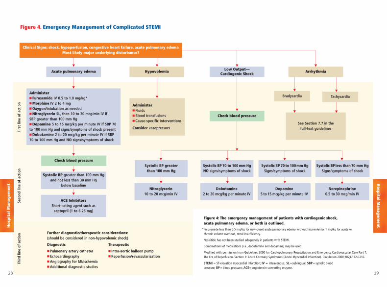
Figure 4. Emergency Management of Complicated STEMI
Acute pulmonary edema Hypovolemia
Nitroglycerin10 to 20 mcg/min IV
Dobutamine2 to 20 mcg/kg per minute IV
Dopamine5 to 15 mcg/kg per minute IV
Norepinephrine0.5 to 30 mcg/min IV
Check blood pressure
Bradycardia TachycardiaAdminister■ Furosemide IV 0.5 to 1.0 mg/kg*■ Morphine IV 2 to 4 mg■ Oxygen/intubation as needed■ Nitroglycerin SL, then 10 to 20 mcg/min IV if SBP greater than 100 mm Hg■ Dopamine 5 to 15 mcg/kg per minute IV if SBP 70to 100 mm Hg and signs/symptoms of shock present■ Dobutamine 2 to 20 mcg/kg per minute IV if SBP70 to 100 mm Hg and NO signs/symptoms of shock
Administer■ Fluids■ Blood transfusions■ Cause-specific interventions
Consider vasopressors
Systolic BP greater than 100 mm Hgand not less than 30 mm Hg
below baseline
Further diagnostic/therapeutic considerations:(should be considered in non-hypovolemic shock)
Diagnostic Therapeutic
■ Pulmonary artery catheter ■ Intra-aortic balloon pump■ Echocardiography ■ Reperfusion/revascularization■ Angiography for MI/ischemia■ Additional diagnostic studies
ACE InhibitorsShort-acting agent such as
captopril (1 to 6.25 mg)
See Section 7.7 in thefull-text guidelines
Firs
t lin
e of
act
ion
Seco
nd li
ne o
f ac
tion
Thir
d lin
e of
act
ion
▼
▼
▼
▼
▼
▼
▼
▼
Systolic BP greater than 100 mm Hg
▼
▼ ▼ ▼
Systolic BPless than 70 mm HgSigns/symptoms of shock
▼
Systolic BP 70 to 100mm HgSigns/symptoms of shock
▼
Systolic BP 70 to 100 mm HgNO signs/symptoms of shock
▼
Arrhythmia
▼
▼ ▼ ▼Low Output—
Cardiogenic Shock
Check blood pressure
▼
▼
▼
Figure 4: The emergency management of patients with cardiogenic shock,acute pulmonary edema, or both is outlined.
*Furosemide less than 0.5 mg/kg for new-onset acute pulmonary edema without hypovolemia; 1 mg/kg for acute orchronic volume overload, renal insufficiency.
Nesiritide has not been studied adequately in patients with STEMI.
Combinations of medications (i.e., dobutamine and dopamine) may be used.
Modified with permission from Guidelines 2000 for Cardiopulmonary Resuscitation and Emergency Cardiovascular Care Part 7:The Era of Reperfusion. Section 1: Acute Coronary Syndromes (Acute Myocardial Infarction). Circulation 2000;102;I-172-I-216.
STEMI = ST-elevation myocardial infarction; IV = intravenous; SL =sublingual; SBP= systolic blood pressure; BP = blood pressure; ACE=angiotensin converting enzyme.
29
Hospital M
anagementH
ospi
tal M
anag
emen
t
Clinical Signs: shock, hypoperfusion, congestive heart failure, acute pulmonary edemaMost likely major underlying disturbance?
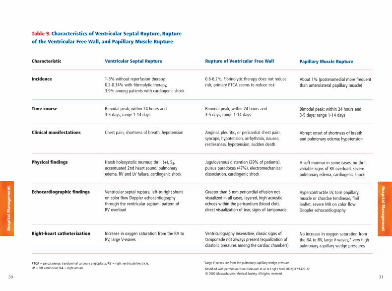
Table 9. Characteristics of Ventricular Septal Rupture, Ruptureof the Ventricular Free Wall, and Papillary Muscle Rupture
30 31
Ventricular Septal Rupture
1-3% without reperfusion therapy,0.2-0.34% with fibrinolytic therapy,3.9% among patients with cardiogenic shock
Bimodal peak; within 24 hours and 3-5 days; range 1-14 days
Chest pain, shortness of breath, hypotension
Harsh holosystolic murmur, thrill (+), S3,accentuated 2nd heart sound, pulmonaryedema, RV and LV failure, cardiogenic shock
Ventricular septal rupture, left-to-right shunton color flow Doppler echocardiographythrough the ventricular septum, pattern of RV overload
Increase in oxygen saturation from the RA toRV, large V-waves
Characteristic
Incidence
Time course
Clinical manifestations
Physical findings
Echocardiographic findings
Right-heart catheterization
Papillary Muscle Rupture
About 1% (posteromedial more frequentthan anterolateral papillary muscle)
Bimodal peak; within 24 hours and 3-5 days; range 1-14 days
Abrupt onset of shortness of breath and pulmonary edema; hypotension
A soft murmur in some cases, no thrill,variable signs of RV overload, severe pulmonary edema, cardiogenic shock
Hypercontractile LV, torn papillary muscle or chordae tendineae, flail leaflet, severe MR on color flow Doppler echocardiography
No increase in oxygen saturation fromthe RA to RV, large V-waves,* very highpulmonary-capillary wedge pressures
Rupture of Ventricular Free Wall
0.8-6.2%, Fibrinolytic therapy does not reducerisk; primary PTCA seems to reduce risk
Bimodal peak; within 24 hours and 3-5 days; range 1-14 days
Anginal, pleuritic, or pericardial chest pain,syncope, hypotension, arrhythmia, nausea,restlessness, hypotension, sudden death
Jugulovenous distention (29% of patients),pulsus paradoxus (47%), electromechanicaldissociation, cardiogenic shock
Greater than 5 mm pericardial effusion notvisualized in all cases, layered, high-acousticechoes within the pericardium (blood clot),direct visualization of tear, signs of tamponade
Ventriculography insensitive, classic signs oftamponade not always present (equalization ofdiastolic pressures among the cardiac chambers)
PTCA = percutaneous transluminal coronary angioplasty; RV = right ventricular/ventricle;LV = left ventricular; RA = right atrium.
*Large V-waves are from the pulmonary capillary wedge pressure.
Modified with permission from Birnbaum et al. N Engl J Med 2002;347:1426-32 © 2002 Massachusetts Medical Society. All rights reserved.
Hospital M
anagementH
ospi
tal M
anag
emen
t
3332
▼
▼
▼▼ ▼
▼
▼
▼
▼
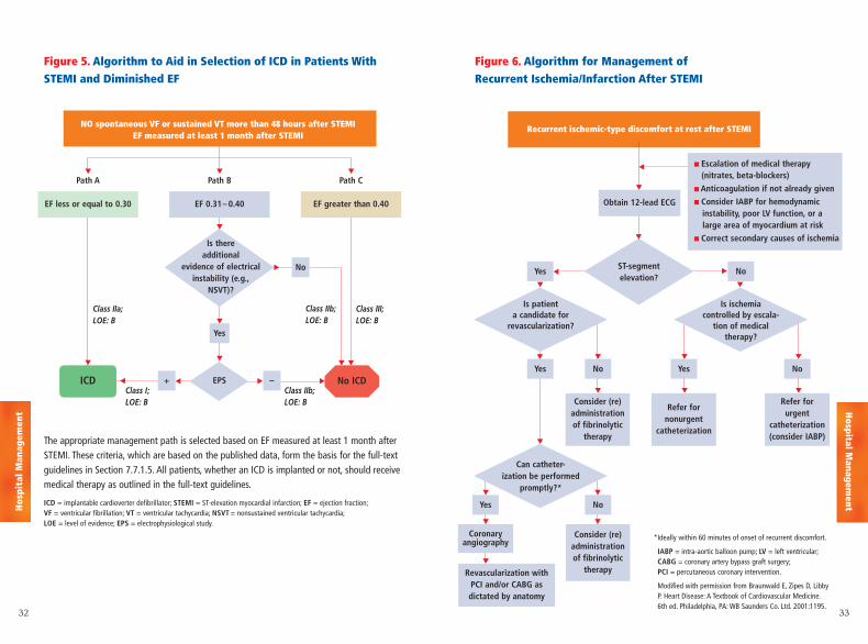
Path B Path CPath A
EF less or equal to 0.30 EF 0.31– 0.40
ICD
NO spontaneous VF or sustained VT more than 48 hours after STEMI EF measured at least 1 month after STEMI
Class IIa; LOE: B
▼▼
Class III; LOE: B
Class IIb; LOE: B
Class IIb; LOE: B
Class I; LOE: B
EPS+
Yes
No
–
EF greater than 0.40
The appropriate management path is selected based on EF measured at least 1 month afterSTEMI. These criteria, which are based on the published data, form the basis for the full-textguidelines in Section 7.7.1.5. All patients, whether an ICD is implanted or not, should receivemedical therapy as outlined in the full-text guidelines.
ICD = implantable cardioverter defibrillator; STEMI = ST-elevation myocardial infarction; EF = ejection fraction;VF = ventricular fibrillation; VT = ventricular tachycardia; NSVT = nonsustained ventricular tachycardia;LOE = level of evidence; EPS = electrophysiological study.
Figure 5. Algorithm to Aid in Selection of ICD in Patients WithSTEMI and Diminished EF
▼▼
▼
▼
▼▼
▼
▼
▼
▼
▼
▼
▼
▼ ▼
▼
▼
▼
▼
Figure 6. Algorithm for Management of Recurrent Ischemia/Infarction After STEMI
Recurrent ischemic-type discomfort at rest after STEMI
Is patient a candidate for
revascularization?
Can catheter-ization be performed
promptly?*
Is ischemia controlled by escala-
tion of medical therapy?
No
NoNo
NoYes
Coronaryangiography
Consider (re)administrationof fibrinolytic
therapyRevascularization with PCI and/or CABG as dictated by anatomy
YesYes
Yes
Obtain 12-lead ECG
Refer forurgent
catheterization(consider IABP)
Refer fornonurgent
catheterization
Consider (re)administrationof fibrinolytic
therapy
■ Escalation of medical therapy (nitrates, beta-blockers)
■ Anticoagulation if not already given
■ Consider IABP for hemodynamicinstability, poor LV function, or a large area of myocardium at risk
■ Correct secondary causes of ischemia
*Ideally within 60 minutes of onset of recurrent discomfort.
IABP = intra-aortic balloon pump; LV = left ventricular;CABG = coronary artery bypass graft surgery;PCI = percutaneous coronary intervention.
Modified with permission from Braunwald E, Zipes D, LibbyP. Heart Disease: A Textbook of Cardiovascular Medicine.6th ed. Philadelphia, PA: WB Saunders Co. Ltd. 2001:1195.
▼
▼
Is there additional
evidence of electrical instability (e.g.,
NSVT)?
▼
No ICD
ST-segmentelevation?
▼
Hospital M
anagementH
ospi
tal M
anag
emen
t
34 35
▼
▼ ▼ ▼ ▼
▼ ▼▼ ▼
▼ ▼
▼ ▼
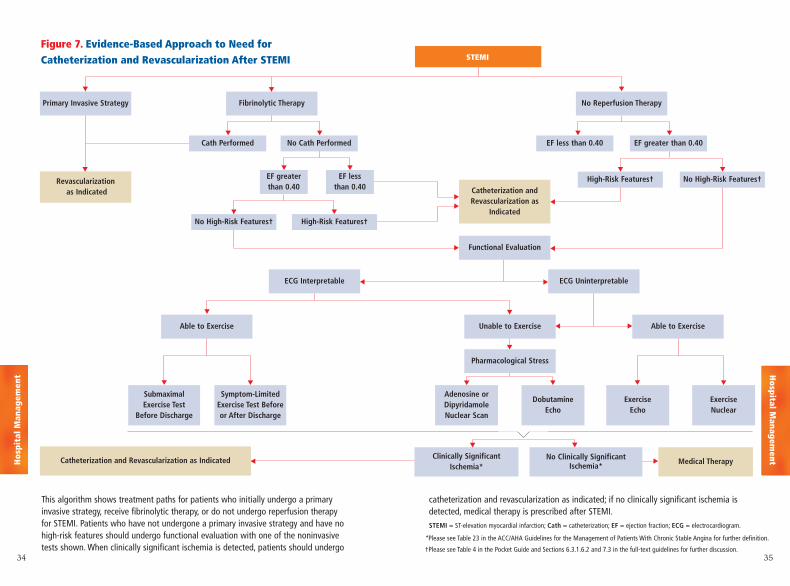
Figure 7. Evidence-Based Approach to Need forCatheterization and Revascularization After STEMI STEMI
Fibrinolytic Therapy
Cath Performed
EF greaterthan 0.40
EF less than 0.40
High-Risk Features† No High-Risk Features†
No High-Risk Features† High-Risk Features†
No Cath Performed EF less than 0.40 EF greater than 0.40
No Reperfusion TherapyPrimary Invasive Strategy
Functional Evaluation
Revascularizationas Indicated
Clinically Significant Ischemia*
No Clinically SignificantIschemia*
Catheterization and Revascularization as Indicated Medical Therapy
Catheterization andRevascularization as
Indicated
▼
▼
▼▼
▼
▼
▼
▼
▼
▼
▼ ▼▼ ▼ ▼ ▼
▼ ▼
▼ ▼
▼
ECG Interpretable
Able to Exercise Able to ExerciseUnable to Exercise
Pharmacological Stress
Adenosine orDipyridamole Nuclear Scan
Dobutamine Echo
Submaximal Exercise Test
Before Discharge
Symptom-LimitedExercise Test Beforeor After Discharge
Exercise Echo
Exercise Nuclear
ECG Uninterpretable
This algorithm shows treatment paths for patients who initially undergo a primary invasive strategy, receive fibrinolytic therapy, or do not undergo reperfusion therapy for STEMI. Patients who have not undergone a primary invasive strategy and have nohigh-risk features should undergo functional evaluation with one of the noninvasivetests shown. When clinically significant ischemia is detected, patients should undergo
catheterization and revascularization as indicated; if no clinically significant ischemia is detected, medical therapy is prescribed after STEMI.STEMI = ST-elevation myocardial infarction; Cath = catheterization; EF = ejection fraction; ECG = electrocardiogram.
*Please see Table 23 in the ACC/AHA Guidelines for the Management of Patients With Chronic Stable Angina for further definition.
†Please see Table 4 in the Pocket Guide and Sections 6.3.1.6.2 and 7.3 in the full-text guidelines for further discussion.
▼
▼
Hospital M
anagementH
ospi
tal M
anag
emen
t
36 37
▼▼▼
▼ ▼
No ASA allergy
No Indications for
Anticoagulation
ASA 75-162 mgClopidogrel 75 mg†
Class: I; LOE: B
Clopidogrel 75 mgClass I, LOE: B
Indications forAnticoagulation
ASA 75-162 mgClopidogrel 75 mg‡
Warfarin (INR 2.0-3.0)§Class: IIb; LOE: C
ASA allergy
No Indications for
Anticoagulation
Indications forAnticoagulation
Clopidogrel 75 mgWarfarin
(INR 2.0-3.0)§Class I, LOE: C
▼
▼
▼
▼ ▼
▼▼▼
▼ ▼
▼ ▼
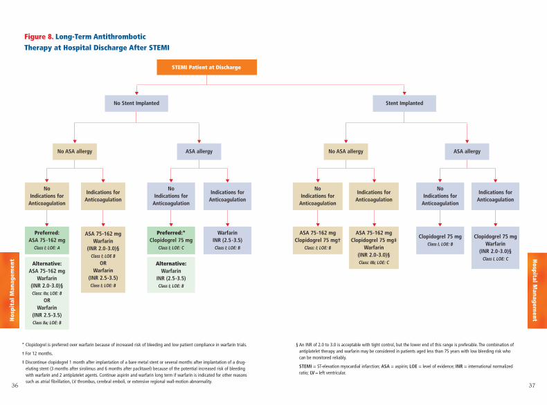
Figure 8. Long-Term Antithrombotic Therapy at Hospital Discharge After STEMI
STEMI Patient at Discharge
Stent ImplantedNo Stent Implanted
No ASA allergy
No Indications for
Anticoagulation
Preferred:ASA 75-162 mg
Class I; LOE: A
Alternative:ASA 75-162 mg
Warfarin(INR 2.0-3.0)§Class: IIa; LOE: B
ORWarfarin
(INR 2.5-3.5)Class IIa; LOE: B
Preferred:*Clopidogrel 75 mg
Class I; LOE: C
Alternative:Warfarin
INR (2.5-3.5)Class I; LOE: B
Indications forAnticoagulation
ASA 75-162 mgWarfarin
(INR 2.0-3.0)§Class I; LOE B
ORWarfarin
(INR 2.5-3.5)Class I; LOE: B
ASA allergy
No Indications for
Anticoagulation
Indications forAnticoagulation
WarfarinINR (2.5-3.5)Class I; LOE: B
▼
▼
▼
▼ ▼
* Clopidogrel is preferred over warfarin because of increased risk of bleeding and low patient compliance in warfarin trials.
† For 12 months.
‡ Discontinue clopidogrel 1 month after implantation of a bare metal stent or several months after implantation of a drug-eluting stent (3 months after sirolimus and 6 months after paclitaxel) because of the potential increased risk of bleedingwith warfarin and 2 antiplatelet agents. Continue aspirin and warfarin long term if warfarin is indicated for other reasonssuch as atrial fibrillation, LV thrombus, cerebral emboli, or extensive regional wall-motion abnormality.
§ An INR of 2.0 to 3.0 is acceptable with tight control, but the lower end of this range is preferable. The combination ofantiplatelet therapy and warfarin may be considered in patients aged less than 75 years with low bleeding risk whocan be monitored reliably.
STEMI = ST-elevation myocardial infarction; ASA = aspirin; LOE = level of evidence; INR = international normalizedratio; LV = left ventricular.
Hospital M
anagementH
ospi
tal M
anag
emen
t
3938
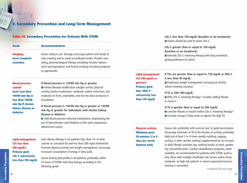
LDL-C less than 100 mg/dL (baseline or on treatment):■ Statins should be used to lower LDL-C.
LDL-C greater than or equal to 100 mg/dL(baseline or on treatment):■ Intensify LDL-C–lowering therapy with drug treatment,giving preference to statins.
If TGs are greater than or equal to 150 mg/dL or HDL-C is less than 40 mg/dL:■ Emphasize weight management and physical activity.Advise smoking cessation.
If TG is 200–499 mg/dL:■ After LDL-C–lowering therapy,† consider adding fibrate or niacin.‡
If TG is greater than or equal to 500 mg/dL:■ Consider fibrate or niacin‡ before LDL-C–lowering therapy.†■ Consider omega-3 fatty acids as adjunct for high TG.
Assess risk, preferably with exercise test, to guide prescription.Encourage minimum of 30 to 60 minutes of activity, preferablydaily but at least 3 or 4 times weekly (walking, jogging,cycling, or other aerobic activity) supplemented by an increasein daily lifestyle activities (eg, walking breaks at work, garden-ing, household work). Cardiac rehabilitation programs, whenavailable, are recommended for patients with STEMI, particu-larly those with multiple modifiable risk factors and/or thosemoderate- to high-risk patients in whom supervised exercisetraining is warranted.
Recommendations
Assess tobacco use. Strongly encourage patient and family tostop smoking and to avoid secondhand smoke. Provide coun-seling, pharmacological therapy (including nicotine replace-ment and bupropion), and formal smoking cessation programsas appropriate.
If blood pressure is 120/80 mm Hg or greater:■ Initiate lifestyle modification (weight control, physical activity, alcohol moderation, moderate sodium restriction, andemphasis on fruits, vegetables, and low-fat dairy products) inall patients.
If blood pressure is 140/90 mm Hg or greater or 130/80mm Hg or greater for individuals with chronic kidneydisease or diabetes:■ Add blood pressure reducing medications, emphasizing theuse of beta-blockers and inhibitors of the renin-angiotensin-aldosterone system.
Start dietary therapy in all patients (less than 7% of total calories as saturated fat and less than 200 mg/d cholesterol).Promote physical activity and weight management. Encourageincreased consumption of omega-3 fatty acids.
Assess fasting lipid profile in all patients, preferably within 24 hours of STEMI. Add drug therapy according to the following guide:
V. Secondary Prevention and Long-Term Management
Table 10. Secondary Prevention for Patients With STEMI
Goals
Smoking
Goal: Complete
cessation
Blood pressure
control
Goal: Less than
140/90 mm Hg or
less than 130/80
mm Hg if chronic
kidney disease or
diabetes
Lipid management
(TG less than
200 mg/dL)
Primary goal:
LDL-C substantially
less than 100 mg/dL
Lipid management
(TG 200 mg/dL or
greater)
Primary goal:
Non–HDL-C*
substantially less
than 130 mg/dL
Physical activity
Minimum goal:
30 minutes 3 to 4
days per week;
Optimal daily
Secondary PreventionSeco
ndar
y Pr
even
tion
continued on next pagecontinued on next page
4140
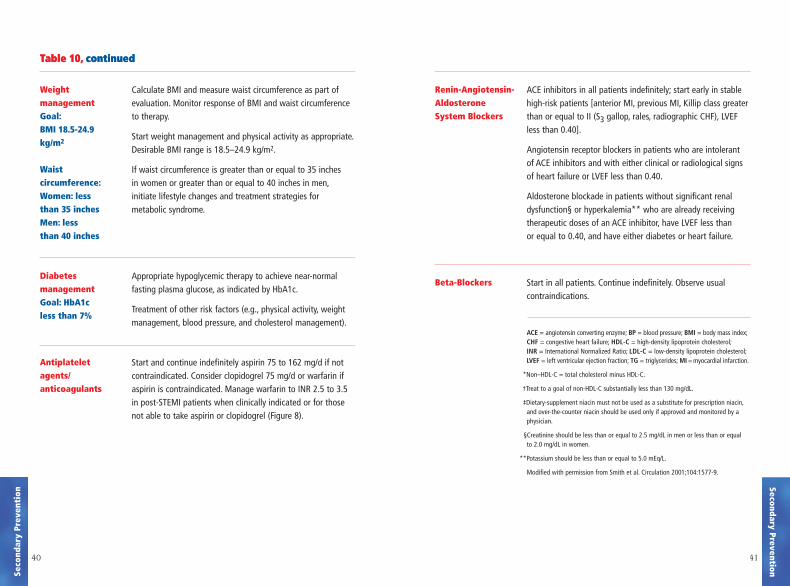
Calculate BMI and measure waist circumference as part ofevaluation. Monitor response of BMI and waist circumferenceto therapy.
Start weight management and physical activity as appropriate.Desirable BMI range is 18.5–24.9 kg/m2.
If waist circumference is greater than or equal to 35 inches in women or greater than or equal to 40 inches in men,initiate lifestyle changes and treatment strategies for metabolic syndrome.
Appropriate hypoglycemic therapy to achieve near-normalfasting plasma glucose, as indicated by HbA1c.
Treatment of other risk factors (e.g., physical activity, weightmanagement, blood pressure, and cholesterol management).
Start and continue indefinitely aspirin 75 to 162 mg/d if notcontraindicated. Consider clopidogrel 75 mg/d or warfarin ifaspirin is contraindicated. Manage warfarin to INR 2.5 to 3.5in post-STEMI patients when clinically indicated or for thosenot able to take aspirin or clopidogrel (Figure 8).
ACE inhibitors in all patients indefinitely; start early in stablehigh-risk patients [anterior MI, previous MI, Killip class greaterthan or equal to II (S3 gallop, rales, radiographic CHF), LVEFless than 0.40].
Angiotensin receptor blockers in patients who are intolerant of ACE inhibitors and with either clinical or radiological signsof heart failure or LVEF less than 0.40.
Aldosterone blockade in patients without significant renal dysfunction§ or hyperkalemia** who are already receivingtherapeutic doses of an ACE inhibitor, have LVEF less than or equal to 0.40, and have either diabetes or heart failure.
Start in all patients. Continue indefinitely. Observe usual contraindications.
ACE = angiotensin converting enzyme; BP = blood pressure; BMI = body mass index;CHF = congestive heart failure; HDL-C = high-density lipoprotein cholesterol;INR = International Normalized Ratio; LDL-C = low-density lipoprotein cholesterol;LVEF = left ventricular ejection fraction; TG = triglycerides; MI = myocardial infarction.
*Non–HDL-C = total cholesterol minus HDL-C.
†Treat to a goal of non-HDL-C substantially less than 130 mg/dL.
‡Dietary-supplement niacin must not be used as a substitute for prescription niacin,and over-the-counter niacin should be used only if approved and monitored by aphysician.
§Creatinine should be less than or equal to 2.5 mg/dL in men or less than or equal to 2.0 mg/dL in women.
**Potassium should be less than or equal to 5.0 mEq/L.
Modified with permission from Smith et al. Circulation 2001;104:1577-9.
Weight managementGoal:BMI 18.5-24.9kg/m2
Waist circumference:Women: less than 35 inches Men: less than 40 inches
Diabetes managementGoal: HbA1c less than 7%
Antiplateletagents/ anticoagulants
Renin-Angiotensin-AldosteroneSystem Blockers
Beta-Blockers
Secondary PreventionSeco
ndar
y Pr
even
tion
Table 10, continuedTable 10, continued
4342
return to work, resumption of sexual activity,
and travel, including driving and flying. A table
describing the metabolic equivalent (MET) values
for various activities can be found in the full-text
guidelines. (Level of Evidence: C)
7. Patients and their families should be asked if they
are interested in CPR (cardiopulmonary resuscita-
tion) training after the patient is discharged from the
hospital. (Level of Evidence: C)
8. Providers should actively review the following
issues with patients and their families:
a. the patient's heart attack risk (Level of Evidence: C)
b. how to recognize symptoms of STEMI
(Level of Evidence: C)
c. the advisability of calling 9-1-1 if symptoms are
unimproved or worsening after 5 minutes, despite
feelings of uncertainty about the symptoms and fear
of potential embarrassment (Level of Evidence: C)
d. a plan for appropriate recognition and response
to a potential acute cardiac event, including the
phone number to access EMS, generally 9-1-1.
(Level of Evidence: C)
9. Cardiac rehabilitation/secondary prevention
programs, when available, are recommended for
patients with STEMI, particularly those with multi-
ple modifiable risk factors and/or those moderate-
to high-risk patients in whom supervised exercise
training is warranted. (Level of Evidence: C)
Recommendations for Follow-Up Visit With a Medical Provider
Class I 1. Follow-up visit should delineate the presence or
absence of cardiovascular symptoms and functional
class. (Level of Evidence: C)
2. The patient’s list of current medications should
be re-evaluated in a follow-up visit, and appropriate
titration of angiotensin converting enzyme (ACE)
inhibitors, beta-blockers, and statins should be
undertaken. (Level of Evidence: C)
3. The predischarge risk assessment and planned
workup should be reviewed and continued (Figure
7). This should include a check of left ventricular
function and possibly Holter monitoring for those
patients whose early post-STEMI ejection fraction
was 0.31 to 0.40 or lower, in consideration of possi-
ble ICD use (Figure 5). (Level of Evidence: C)
4. The healthcare provider should review and
emphasize the principles of secondary prevention
with the patient and family members (Table 10).
(Level of Evidence: C)
5. The psychosocial status of the patient should be
evaluated in follow-up, including inquiries regarding
symptoms of depression, anxiety, or sleep disorders
and the social support environment.
(Level of Evidence: C)
6. In a follow-up visit, the healthcare provider
should discuss in detail issues of physical activity,
Secondary PreventionSeco
ndar
y Pr
even
tion
4544
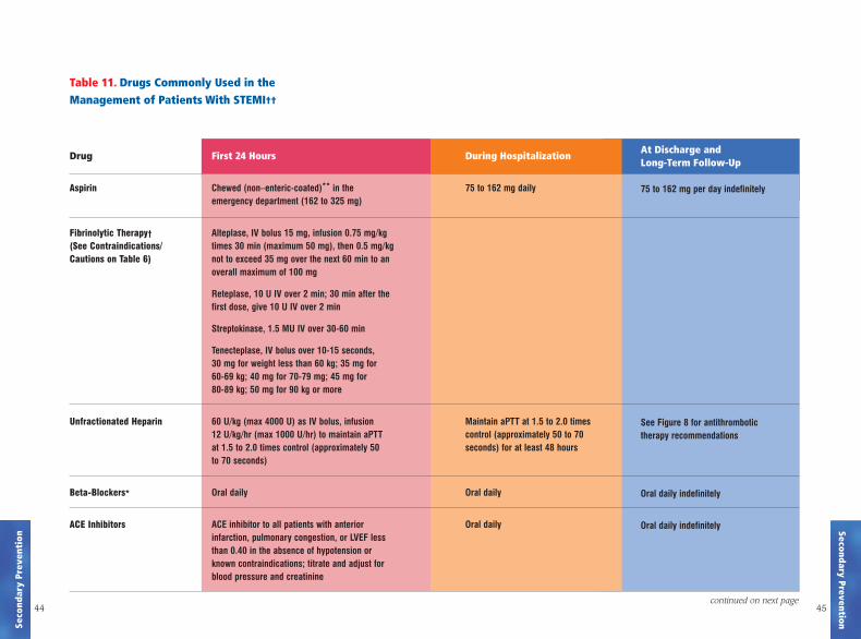
First 24 Hours
Chewed (non–enteric-coated)** in the emergency department (162 to 325 mg)
Alteplase, IV bolus 15 mg, infusion 0.75 mg/kgtimes 30 min (maximum 50 mg), then 0.5 mg/kgnot to exceed 35 mg over the next 60 min to anoverall maximum of 100 mg
Reteplase, 10 U IV over 2 min; 30 min after the first dose, give 10 U IV over 2 min
Streptokinase, 1.5 MU IV over 30-60 min
Tenecteplase, IV bolus over 10-15 seconds, 30 mg for weight less than 60 kg; 35 mg for 60-69 kg; 40 mg for 70-79 mg; 45 mg for 80-89 kg; 50 mg for 90 kg or more
60 U/kg (max 4000 U) as IV bolus, infusion 12 U/kg/hr (max 1000 U/hr) to maintain aPTT at 1.5 to 2.0 times control (approximately 50 to 70 seconds)
Oral daily
ACE inhibitor to all patients with anterior infarction, pulmonary congestion, or LVEF less than 0.40 in the absence of hypotension or known contraindications; titrate and adjust forblood pressure and creatinine
Drug
Aspirin
Fibrinolytic Therapy†
(See Contraindications/Cautions on Table 6)
Unfractionated Heparin
Beta-Blockers*
ACE Inhibitors
At Discharge and Long-Term Follow-Up
75 to 162 mg per day indefinitely
See Figure 8 for antithrombotic therapy recommendations
Oral daily indefinitely
Oral daily indefinitely
Table 11. Drugs Commonly Used in the Management of Patients With STEMI††
continued on next page
Secondary PreventionSeco
ndar
y Pr
even
tion
During Hospitalization
75 to 162 mg daily
Maintain aPTT at 1.5 to 2.0 times control (approximately 50 to 70 seconds) for at least 48 hours
Oral daily
Oral daily
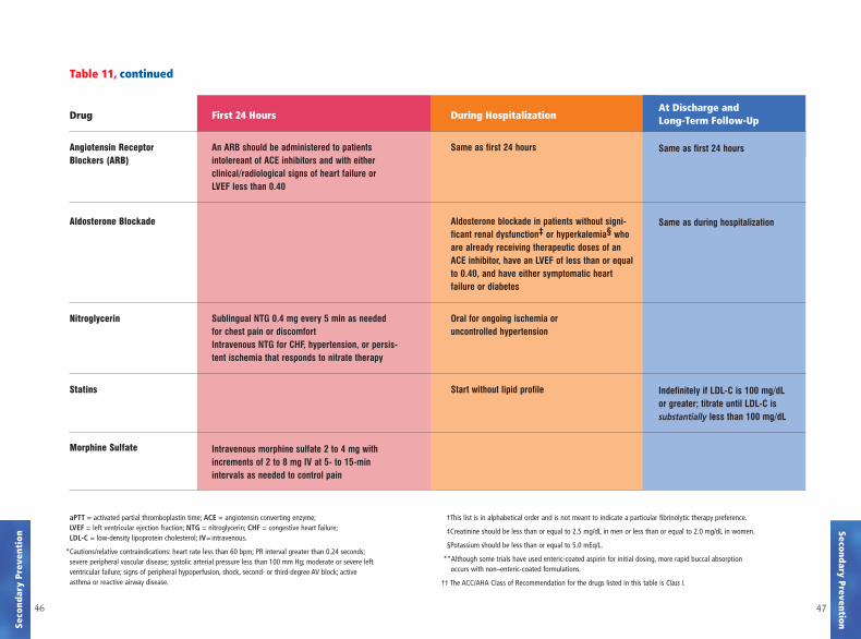
Table 11, continued
4746
First 24 Hours
An ARB should be administered to patients intolereant of ACE inhibitors and with either clinical/radiological signs of heart failure or LVEF less than 0.40
Sublingual NTG 0.4 mg every 5 min as needed for chest pain or discomfortIntravenous NTG for CHF, hypertension, or persis-tent ischemia that responds to nitrate therapy
Intravenous morphine sulfate 2 to 4 mg with increments of 2 to 8 mg IV at 5- to 15-min intervals as needed to control pain
Drug
Angiotensin ReceptorBlockers (ARB)
Aldosterone Blockade
Nitroglycerin
Statins
Morphine Sulfate
At Discharge and Long-Term Follow-Up
Same as first 24 hours
Same as during hospitalization
Indefinitely if LDL-C is 100 mg/dL or greater; titrate until LDL-C is substantially less than 100 mg/dL
During Hospitalization
Same as first 24 hours
Aldosterone blockade in patients without signi-ficant renal dysfunction‡ or hyperkalemia§ who are already receiving therapeutic doses of an ACE inhibitor, have an LVEF of less than or equal to 0.40, and have either symptomatic heart failure or diabetes
Oral for ongoing ischemia or uncontrolled hypertension
Start without lipid profile
aPTT = activated partial thromboplastin time; ACE = angiotensin converting enzyme;LVEF = left ventricular ejection fraction; NTG = nitroglycerin; CHF = congestive heart failure;LDL-C = low-density lipoprotein cholesterol; IV=intravenous.
*Cautions/relative contraindications: heart rate less than 60 bpm; PR interval greater than 0.24 seconds;severe peripheral vascular disease; systolic arterial pressure less than 100 mm Hg; moderate or severe leftventricular failure; signs of peripheral hypoperfusion, shock, second- or third-degree AV block; active asthma or reactive airway disease.
†This list is in alphabetical order and is not meant to indicate a particular fibrinolytic therapy preference.
‡Creatinine should be less than or equal to 2.5 mg/dL in men or less than or equal to 2.0 mg/dL in women.
§Potassium should be less than or equal to 5.0 mEq/L.
**Although some trials have used enteric-coated aspirin for initial dosing, more rapid buccal absorption occurs with non–enteric-coated formulations.
†† The ACC/AHA Class of Recommendation for the drugs listed in this table is Class I.
Secondary PreventionSeco
ndar
y Pr
even
tion
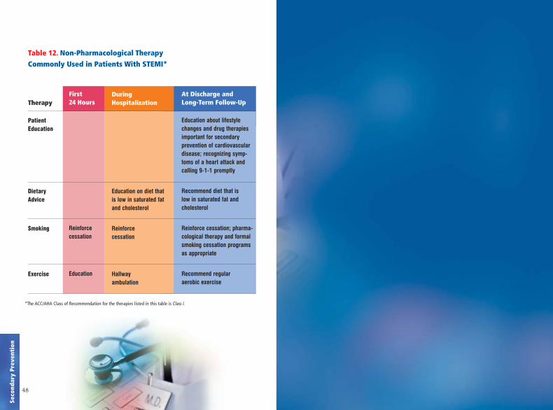
Table 12. Non-Pharmacological Therapy Commonly Used in Patients With STEMI*
48
First 24 Hours
Reinforce cessation
Education
Therapy
Patient Education
Dietary Advice
Smoking
Exercise
During Hospitalization
Education on diet that is low in saturated fatand cholesterol
Reinforce cessation
Hallway ambulation
At Discharge and Long-Term Follow-Up
Education about lifestylechanges and drug therapiesimportant for secondary prevention of cardiovasculardisease; recognizing symp-toms of a heart attack andcalling 9-1-1 promptly
Recommend diet that is low in saturated fat and cholesterol
Reinforce cessation; pharma-cological therapy and formalsmoking cessation programsas appropriate
Recommend regular aerobic exercise
Seco
ndar
y Pr
even
tion
*The ACC/AHA Class of Recommendation for the therapies listed in this table is Class I.


![HIGHLIGHTS OF PRESCRIBING INFORMATION Recent MI, recent ... · – For patients with non–ST-segment elevation ACS (unstable angina [UA]/non–ST-elevation myocardial infarction](https://img.pdfslide.net/doc/110x75/5d5f296288c993e0198b6a74/highlights-of-prescribing-information-recent-mi-recent-for-patients.jpg)