Embed Size (px)
Citation preview
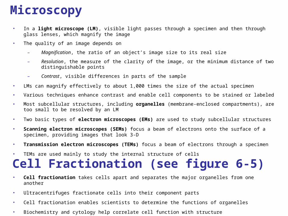
Microscopy• In a light microscope (LM), visible light passes through a specimen and then through glass lenses, which magnify
the image
• The quality of an image depends on
– Magnification, the ratio of an object’s image size to its real size
– Resolution, the measure of the clarity of the image, or the minimum distance of two distinguishable points
– Contrast, visible differences in parts of the sample
• LMs can magnify effectively to about 1,000 times the size of the actual specimen
• Various techniques enhance contrast and enable cell components to be stained or labeled
• Most subcellular structures, including organelles (membrane-enclosed compartments), are too small to be resolved by an LM
• Two basic types of electron microscopes (EMs) are used to study subcellular structures
• Scanning electron microscopes (SEMs) focus a beam of electrons onto the surface of a specimen, providing images that look 3-D
• Transmission electron microscopes (TEMs) focus a beam of electrons through a specimen
• TEMs are used mainly to study the internal structure of cells
Cell Fractionation (see figure 6-5)• Cell fractionation takes cells apart and separates the major organelles from one another
• Ultracentrifuges fractionate cells into their component parts
• Cell fractionation enables scientists to determine the functions of organelles
• Biochemistry and cytology help correlate cell function with structure
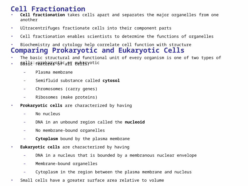
• The basic structural and functional unit of every organism is one of two types of cells: prokaryotic or eukaryotic
Comparing Prokaryotic and Eukaryotic Cells
• Basic features of all cells:
– Plasma membrane
– Semifluid substance called cytosol
– Chromosomes (carry genes)
– Ribosomes (make proteins)
• Prokaryotic cells are characterized by having
– No nucleus
– DNA in an unbound region called the nucleoid
– No membrane-bound organelles
– Cytoplasm bound by the plasma membrane
• Eukaryotic cells are characterized by having
– DNA in a nucleus that is bounded by a membranous nuclear envelope
– Membrane-bound organelles
– Cytoplasm in the region between the plasma membrane and nucleus
• Small cells have a greater surface area relative to volume
Cell Fractionation• Cell fractionation takes cells apart and separates the major organelles from one another
• Ultracentrifuges fractionate cells into their component parts
• Cell fractionation enables scientists to determine the functions of organelles
• Biochemistry and cytology help correlate cell function with structure
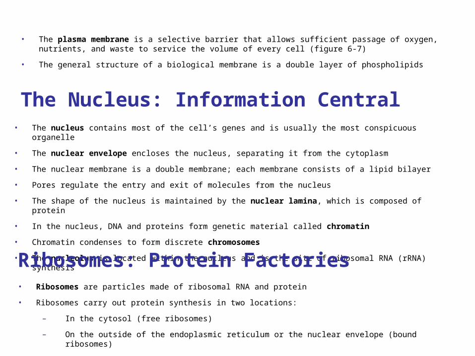
The Nucleus: Information Central• The nucleus contains most of the cell’s genes and is usually the most conspicuous organelle
• The nuclear envelope encloses the nucleus, separating it from the cytoplasm
• The nuclear membrane is a double membrane; each membrane consists of a lipid bilayer
• Pores regulate the entry and exit of molecules from the nucleus
• The shape of the nucleus is maintained by the nuclear lamina, which is composed of protein
• In the nucleus, DNA and proteins form genetic material called chromatin
• Chromatin condenses to form discrete chromosomes
• The nucleolus is located within the nucleus and is the site of ribosomal RNA (rRNA) synthesis
Ribosomes: Protein Factories• Ribosomes are particles made of ribosomal RNA and protein
• Ribosomes carry out protein synthesis in two locations:
– In the cytosol (free ribosomes)
– On the outside of the endoplasmic reticulum or the nuclear envelope (bound ribosomes)
• The plasma membrane is a selective barrier that allows sufficient passage of oxygen, nutrients, and waste to service the volume of every cell (figure 6-7)
• The general structure of a biological membrane is a double layer of phospholipids
The endomembrane system regulates protein traffic and performs metabolic functions in the cell
• Components of the endomembrane system:
– Nuclear envelope
– Endoplasmic reticulum
– Golgi apparatus
– Lysosomes
– Vacuoles
– Plasma membrane
• These components are either continuous or connected via transfer by vesicles
• The endoplasmic reticulum (ER) accounts for more than half of the total membrane in many eukaryotic cells
• The ER membrane is continuous with the nuclear envelope
• There are two distinct regions of ER:
– Smooth ER, which lacks ribosomes
– Synthesizes lipids
– Metabolizes carbohydrates
– Detoxifies poison
– Stores calcium
Rough ER, with ribosomes studding its surface
– Has bound ribosomes, which secrete glycoproteins (proteins covalently bonded to carbohydrates)
– Distributes transport vesicles, proteins surrounded by membranes
– Is a membrane factory for the cell
• The Golgi apparatus consists of flattened membranous sacs called cisternae
• Functions of the Golgi apparatus:
– Modifies products of the ER
– Manufactures certain macromolecules
– Sorts and packages materials into transport vesicles
• Mitochondria are the sites of cellular respiration, a metabolic process that generates ATP
• They have a smooth outer membrane and an inner membrane folded into cristae
• The inner membrane creates two compartments: intermembrane space and mitochondrial matrix
• Some metabolic steps of cellular respiration are catalyzed in the mitochondrial matrix
• Cristae present a large surface area for enzymes that synthesize ATP