Embed Size (px)
Citation preview
Molecular cloning, pharmacological characterization,and histochemical distribution of frog vasotocin andmesotocin receptors
S Acharjee, J-L Do-Rego1, D Y Oh, J S Moon, R S Ahn, K Lee, D G Bai, H Vaudry1,H B Kwon and J Y SeongHormone Research Center, School of Biological Sciences and Technology, Chonnam National University, Gwangju 500-757, Korea
1European Institute for Peptide Research (IFRMP 23), Laboratory of Cellular and Molecular Neuroendocrinology, INSERM U413, University of Rouen,76821 Mont-Saint-Aignan, France
(Requests for offprints should be addressed to J Y Seong; Email: [email protected])
Abstract
The neurohypophysial nonapeptides vasotocin (VT) and mesotocin (MT) are the amphibian counterpartsof arginine vasopressin (AVP) and oxytocin (OT). We have here reported the cloning and functionalcharacterization of the receptors for vasotocin (VTR) and mesotocin (MTR) in two species of frog, Ranacatesbeiana and Rana esculenta. The frog VTR and MTR cDNAs encode proteins of 419 and 384 aminoacids respectively. Frog VTR exhibits a high degree of sequence identity with the mammalian AVP-1a(V1a) receptor while the frog MTR possesses a high degree of sequence identity with the mammalian OTreceptor. Activation of VTR induced both c-fos promoter- and cAMP-responsive element (CRE)-driventranscriptional activities, while activation of MTR induced c-fos promoter-driven transcriptional activity butfailed to evoke CRE-driven transcriptional activity, suggesting differential G protein coupling between VTRand MTR. The VTR exhibited the highest sensitivity for VT followed by OT.AVP<MT, whereas the MTRshowed preferential ligand sensitivity for MT.OT.VT.AVP. A V1a agonist but not V2 and OT agonistssubstantially activated both VTR and MTR with a similar sensitivity. V1a, V2 and OT antagonists inhibitedMT-induced MTR activation but not VT-induced VTR activation. In the frog brain, VTR and MTR mRNAswere found to be widely expressed in the telencephalon, diencephalon and mesencephalon, andexhibited very similar regional distribution. In the pituitary, VTR and MTR were expressed in the distal andintermediate lobes but were virtually absent in the neural lobe. Taken together, these data indicated that,although the distribution of VTR and MTR largely overlaps in the frog brain and pituitary, VT and MT mayplay distinct activities owing to the ligand selectivity and different signaling pathways activated by theirreceptors.
Journal of Molecular Endocrinology (2004) 33, 293–313
Introduction
In mammals, the neurohypophysial hormonesarginine vasopressin (AVP) and oxytocin (OT)exhibit a wide range of biological activities both asneurohormones secreted in the posterior pituitaryand neuromediators released in the central nervoussystem. At the periphery, AVP regulates waterexcretion, vascular tone, and adrenocorticotropin(ACTH) secretion (De Rouffignac et al. 1991, deKeyzer et al. 1996, Szczepanska-Sadowska 1996)while OT stimulates milk ejection and enhances
uterine contractility during parturition (Chibbaret al. 1993, Nishimori et al. 1996). In the centralnervous system, AVP is involved in thermoregula-tion, learning and memory processes, socialrecognition, and aggressive behavior (de Wied et al.1993, Alescio-Lautier & Soumireu-Mourat 1998,Rose & Moore 2002) while OT plays a crucial rolein sexual activity and induces maternal behavior(Pedersen & Prange 1979, Ivell et al. 1997). Theeffects of AVP and OT are mediated through fourtypes of G protein-coupled receptors (GPCRs),i.e. the AVP receptors V1aR, V2R and V1bR (also
293
Journal of Molecular Endocrinology (2004) 33, 293–3130952–5041/04/033–293 © 2004 Society for Endocrinology Printed in Great Britain
Online version via http://www.endocrinology.org
called V3R) (Lolait et al. 1992, Morel et al. 1992,Sugimoto et al. 1994), and the OT receptor (OTR)(Kimura et al. 1992). These various membranereceptor subtypes differ in their tissue distribution,their relative affinity for a broad range of syntheticanalogs, and their signaling mechanisms (Barberiset al. 1998, Gimpl & Fahrenholz 2001). At theperiphery, V1aR is primarily located on vascularsmooth muscle cells and hepatocytes (Morel et al.1992), V1bR is exclusively present in pituitary corti-cotrophs (Sugimoto et al. 1994, de Keyzer et al.1996), V2R is found in the kidney (Lolait et al. 1992),and OTR is expressed in smooth muscle cells of theuterus (Kimura et al. 1992). All four receptor typesare also expressed in the central nervous system(Burbach et al. 1995). Stimulation of V1aR, V1bRand OTR activates the phospholipase C/proteinkinase C (PLC/PKC) signaling pathway, whilestimulation of V2R activates the adenylyl cyclase/protein kinase A (AC/PKA) pathway (Liu & Wess1996, Gimpl & Fahrenholz 2001).
AVP and OT belong to a large family ofnonapeptides that encompasses at least 12 mem-bers (Acher et al. 1995). In most non-mammalianvertebrates including lungfish, amphibians, reptilesand birds, the AVP counterpart is vasotocin (VT;[Ile3]AVP) and the OT counterpart is mesotocin(MT; [Ile8]OT). In the frog, VT and MT areexpressed in both hypothalamic and extrahypotha-lamic cell bodies (González & Smeets 1992),indicating that, in amphibians as in mammals, VTand MT may act both as neurohormones andneurotransmitters. Indeed, it is firmly establishedthat both VT and MT regulate osmotic andelectrolyte balance, VT being an anti-diuretichormone while MT acts as a diuretic agent (Pang &Sawyer 1978, Warburg 1995). Concurrently, it hasbeen shown that VT is expressed in chromaffincells in the frog adrenal gland and that syntheticVT stimulates steroid secretion by frog adrenocor-tical cells (Larcher et al. 1989, 1992a, 1992b). Thereis also clear evidence that VT is involved in repro-ductive behavior in amphibians (Moore et al. 2000,Rose & Moore 2002), and sexual dimorphism inthe distribution of VT-containing neurons has beenreported in the frog brain (Boyd et al. 1992).
In spite of the diverse and crucial roles of VTand MT in lower vertebrates, little is knownregarding the structure, pharmacological profile,distribution and regulation of their receptors. Todate, the VT receptor (VTR) has been character-
ized in several non-mammalian species, includingthe white sucker Catostomus commersoni (Mahlmannet al. 1994), the Pacific salmon Oncorhynchus kisutch(Mahlmann et al. 1994), the cave-dwelling fishAstyanax fasciatus (Mahlmann et al. 1994), theeuryhaline flounder Platichthys flesus (Warne 2001),the anuran Xenopus laevis (Mahlmann et al. 1994),and the domestic chicken Gallus gallus (Tan et al.2000). An MT receptor (MTR) has also beencloned in the giant toad Bufo marinus (Akhundovaet al. 1996). However, the sites of expression, thecoupling mechanisms and the regulation of VTRand MTR have not been thoroughly investigated innon-mammalian vertebrates.
In the present study, we have cloned the cDNAsencoding the MTR and the V1a-type VTR in twospecies of frog, the bullfrog Rana catesbeiana and theEuropean green frog Rana esculenta, a hybridbetween Rana ridibunda and Rana lessonae. We havecharacterized the ligand sensitivity and signaltransduction mechanisms of Rana catesbeiana VTRand MTR and we have compared the pharmaco-logical profiles of frog VTR and MTR with thoseof their mammalian counterparts. Further, we havedetermined the distribution of VTR and MTRmRNAs in the brain and pituitary of Rana esculenta.
Materials and methods
Peptides
Synthetic peptides including AVP, OT, VT, MT,the V1 agonist ([Phe2,Orn8]OT), the V2 agonist([deamino-Cys1,Val4,-Arg8]-AVP), the V1aantagonist ([d(CH2)51,Tyr(Me)2]AVP), the V2antagonist ([d(CH2)51,-Ile2,Ile4]AVP), and the OTantagonist ([d(CH2)51,Tyr(Me)2,Thr4,Orn8,des-Gly-NH2
9]VT) were purchased from Bachem(Bubendorf, Switzerland). The OT agonist([Thr4,Gly7]OT) was purchased from Sigma (StLouis, MO, USA).
Plasmids
The pcDNA3 expression vector was purchasedfrom Invitrogen (San Diego, CA, USA). ThepCMV�-Gal vector was obtained from Clontech(Palo Alto, CA, USA). The CRE-luc vector whichcontains four copies of the cyclic AMP-responsiveelement (CRE; TGACGTCA) was from Stratagene(La Jolla, CA, USA). The c-fos-luc vector,containing the �711�+45 sequence of the
S ACHARJEE and others · Frog vasotocin and mesotocin receptors294
www.endocrinology.orgJournal of Molecular Endocrinology (2004) 33, 293–313
human c-fos promoter constructed in the pFLASHvector, was a kind gift from Dr R Prywes,Columbia University, NY, USA.
Animals and tissue preparation
Male and female bullfrogs (R. catesbeiana, 100–120 gbody weight) were obtained from a local supplier(BCPC, Taein, Korea). Male european green frogs(R. esculenta, 30–40 g body weight) were obtainedfrom a commercial source (Couétard, Saint-Hilairede Riez, France). The animals were kept in glasstanks supplied with tap water under simulatednatural conditions. Frogs were killed by cervicaltransection and the tissues were quickly dissected,immediately frozen in liquid nitrogen, and stored at–80 �C until use. Animal manipulations wereperformed according to the recommendations ofthe ethical committees at our institutions and underthe supervision of authorized investigators.
RNA isolation and RT-PCR
Total RNA was extracted from either the forebrainor the pituitary of five male R. catesbeiana and five
male R. esculenta by using Trireagent (Sigma)according to the manufacturer’s instructions.Poly(A)+ RNA was purified from total RNA byusing QIAGEN Oligotex mRNA kit (QIAGEN,Chatsworth, CA, USA). The RNAs from differenttissues (obtained from five male and five femaleR. catesbeiana) were reverse transcribed by using therandom hexamer and MMTV reverse transcriptase(Promega Corp., Madison, WI, USA). The cDNAsserved as templates for subsequent PCR amplifi-cation of partial clones by using several sets offorward (DGF1, DGF2, DGF3) and reverse(DGR1, DGR2, DGR3) degenerate primers(Table 1). PCR conditions were: denaturation at94 �C for 5 min, followed by 35 cycles at 94 �C for30 s, 55 �C for 30 s, and 72 �C for 50 s. PCRproducts of expected sizes were excised, purifiedand subcloned into pGEM-T easy vector(Promega). Positive clones were isolated andpurified by using a QIAGEN Plasmid MiniprepKit (QIAGEN). Plasmids containing the properinserts were analyzed for their DNA sequenceby the dideoxy chain-termination method, usingthe DNA sequencing kit (USB Corp., Cleveland,OH, USA).
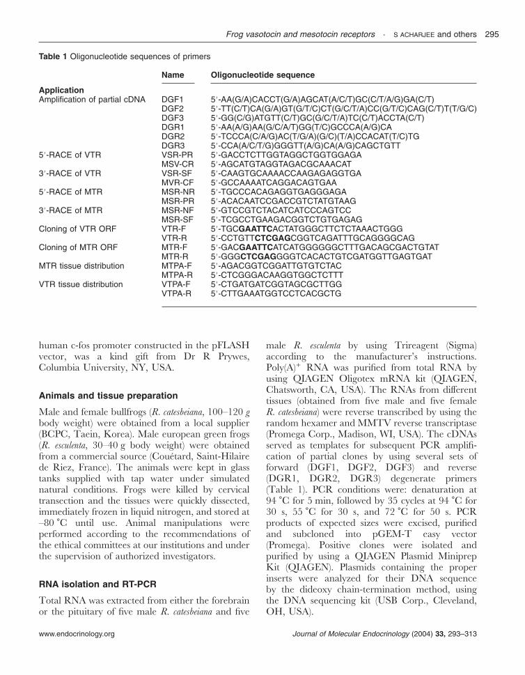
Table 1 Oligonucleotide sequences of primers
Name Oligonucleotide sequence
ApplicationAmplification of partial cDNA DGF1 58-AA(G/A)CACCT(G/A)AGCAT(A/C/T)GC(C/T/A/G)GA(C/T)
DGF2 58-TT(C/T)CA(G/A)GT(G/T/C)CT(G/C/T/A)CC(G/T/C)CAG(C/T)T(T/G/C)DGF3 58-GG(C/G)ATGTT(C/T)GC(G/C/T/A)TC(C/T)ACCTA(C/T)DGR1 58-AA(A/G)AA(G/C/A/T)GG(T/C)GCCCA(A/G)CADGR2 58-TCCCA(C/A/G)AC(T/G/A)(G/C)(T/A)CCACAT(T/C)TGDGR3 58-CCA(A/C/T/G)GGGTT(A/G)CA(A/G)CAGCTGTT
58-RACE of VTR VSR-PR 58-GACCTCTTGGTAGGCTGGTGGAGAMSV-CR 58-AGCATGTAGGTAGACGCAAACAT
38-RACE of VTR VSR-SF 58-CAAGTGCAAAACCAAGAGAGGTGAMVR-CF 58-GCCAAAATCAGGACAGTGAA
58-RACE of MTR MSR-NR 58-TGCCCACAGAGGTGAGGGAGAMSR-PR 58-ACACAATCCGACCGTCTATGTAAG
38-RACE of MTR MSR-NF 58-GTCCGTCTACATCATCCCAGTCCMSR-SF 58-TCGCCTGAAGACGGTCTGTGAGAG
Cloning of VTR ORF VTR-F 58-TGCGAATTCACTATGGGCTTCTCTAAACTGGGVTR-R 58-CCTGTTCTCGAGCGGTCAGATTTGCAGGGGCAG
Cloning of MTR ORF MTR-F 58-GACGAATTCATCATGGGGGGCTTTGACAGCGACTGTATMTR-R 58-GGGCTCGAGGGGTCACACTGTCGATGGTTGAGTGAT
MTR tissue distribution MTPA-F 58-AGACGGTCGGATTGTGTCTACMTPA-R 58-CTCGGGACAAGGTGGCTCTTT
VTR tissue distribution VTPA-F 58-CTGATGATCGGTAGCGCTTGGVTPA-R 58-CTTGAAATGGTCCTCACGCTG
Frog vasotocin and mesotocin receptors · S ACHARJEE and others 295
www.endocrinology.org Journal of Molecular Endocrinology (2004) 33, 293–313
Cloning of the full-length cDNAs by rapidamplification of cDNA ends (RACE)
Two sets of gene-specific primers (GSPs) weredesigned for each VTR and MTR on the basis ofthe partial sequences of the cDNAs obtained byRT-PCR. To obtain the full-length VTR cDNA,the primers sets VSR-PR and MVR-CR (for5�-RACE), and VSR-SF and MVR-CF (for3�-RACE) were used (Table 1). For the MTR, theprimer sets MSR-NR and MSR-PR (for 5�-RACE),and MSR-NF and MSR-SF (for 3�-RACE) wereused (Table 1). Poly(A)-rich RNA purified from theforebrain and pituitary was used to synthesizeadapter-ligated double-stranded cDNA by usingthe Marathon cDNA Amplification Kit (Clontech).Next, 5�- and 3�-RACE was performed by using theGSPs in combination with the adapter primers AP1and AP2 respectively. RACE products were clonedin pGEM-T easy vector (Promega). After obtainingthe proper 5�- and 3�-RACE products, gene specificforward and reverse primers were designed toobtain the full-length cDNAs which were insertedinto the pcDNA3 expression vector at the EcoRIand XhoI enzyme sites. The rat V1aR and V2RcDNAs were amplified through PCR from rat liverand kidney tissues respectively, and inserted at theEcoRI and XhoI sites of the pcDNA3. The humanOTR cloned in the PRK5 plasmid was a kind giftfrom Dr Thierry Durroux (INSERM U469,Montpellier, France). The human OTR was cut atBamHI and XhoI sites and reinserted into thepcDNA3 expression vector.
Luciferase assays
CV-1 cells were maintained in Dulbecco’s modifiedEagle’s medium (DMEM) in the presence of 10%fetal bovine serum. For luciferase assays, cells wereplated in 24-well plates 1 day before transfectionand transfected with SuperFect reagent (QIAGEN)according to the manufacturer’s instructions.Approximately 48 h after transfection, cells weretreated with the respective ligands for 6 h. For c-fospromoter-driven luciferase assay, cells were main-tained in serum-free DMEM 16–18 h beforetreatment with the ligand as previously described(Seong et al. 2003). Cells were harvested 6 h afterligand treatment and luciferase activity in cellextracts was determined using a luciferase assaysystem according to the standard method in a
Lumat LB9501 (EB & G, Berhold, Germany). Theluciferase values were normalized by the�-galactosidase values. Transfection experimentswere performed in duplicate and repeated atleast three times. All data are presented asmeans�S.E.M.
VTR and MTR expression in peripheral tissues
Tissues were collected from five male R. catesbeiana(five females for the oviduct) and stored at �80 �C.The first-strand cDNA was prepared using therandom hexamer and MMTV reverse transcriptase(Promega). To determine the tissue distribution ofVTR and MTR, the primer sets MTPA-F andMTPA-R, and VTPA-F and VTPA-R respectivelywere used (Table 1). PCR conditions were:denaturation at 94 �C for 3 min, followed by 30cycles at 94 �C for 30 s, 60 �C for 30 s and 72 �Cfor 50 s.
In situ hybridization histochemistry
In situ hybridization was performed as previouslydescribed (Alexandre et al. 1999). Briefly, six adultmale frogs, R. esculenta, were anesthetized andperfused transcardially with 50 ml 0·1 M phosphatebuffer (PB, pH 7·4) containing 4% paraformalde-hyde. The brain with the attached pituitary wasrapidly dissected and post-fixed in the same fixativefor 24 h at 4 �C. The tissues were rinsed for 12 h inPB containing 15% sucrose and 24 h in PBcontaining 30% sucrose. The brains were placed inan embedding medium (O.C.T. Tissue Teck;Leica, Nussloch, Germany) and frozen at �80 �C.Frontal sections (12 µm thick) were cut in a cryo-stat (Frigocut 2800E; Reichert-Jung, Nussloch,Germany) and mounted on poly--lysine- andgelatin-coated slides. Partial VTR (nt 424–934)and MTR (nt 271–884) cDNA obtained from R.esculenta were subcloned into the pGME-T vectorbetween SpeI and NcoI sites, and sense andantisense riboprobes were generated by in vitro tran-scription using T7 and Sp6 RNA polymerases inthe presence of [35S]UTP (Combination Systems;Promega). Sections were incubated for 10 min in0·1 M triethanolamine/0·9% NaCl (pH 8·0)/0·25acetic anhydride, rinsed in 2�SSC, and incubatedfor 60 min with prehybridization buffer (pH 7·5)containing 50% formamide, 0·6 M NaCl, 10 mMTris–HCl (pH 7·5), 1�Denhart, 0·02% Ficoll,
S ACHARJEE and others · Frog vasotocin and mesotocin receptors296
www.endocrinology.orgJournal of Molecular Endocrinology (2004) 33, 293–313
0·02% polyvinylpyrrolidone, 0·02% bovine serumalbumin, 1 mM EDTA (pH 8·0), 550 µg/mldenatured salmon sperm DNA, and 50 µg/ml yeasttRNA. Hybridization was performed overnight at55 �C in the same buffer (except for salmon spermDNA whose concentration was reduced to 60 µg/ml) supplemented with 10 mM dithiothreitol, 10%dextran sulfate, and 1·5�107 c.p.m./ml heat-denatured RNA riboprobes. Tissue sections werethen washed in 2�SSC at 55 �C and treated withribonuclease A (50 µg/ml) for 60 min at 37 �C.High stringency washes were performed in0·01�SSC containing 14 mM �-mercaptoethanoland 0·05% sodium pyrophosphate at 60 �C. Thetissue sections were dehydrated in ethanol andexposed onto Hyperfilm �-max (Amersham Phar-macia Biotech, Orsay, France) for 2 weeks. Theoptic density of the autoradiograms was quantifiedby means of a computer-assisted image analyzer(SAMBA Autoradio 4·10; SAMBA Technologies,Meylan, France). To identify anatomical structures,the sections were stained with hematoxylin/eosin.Nomenclature of frog brain structures was based onthe atlas of Neary & Northcutt (1983).
Results
Cloning of full-length VTR and MTR
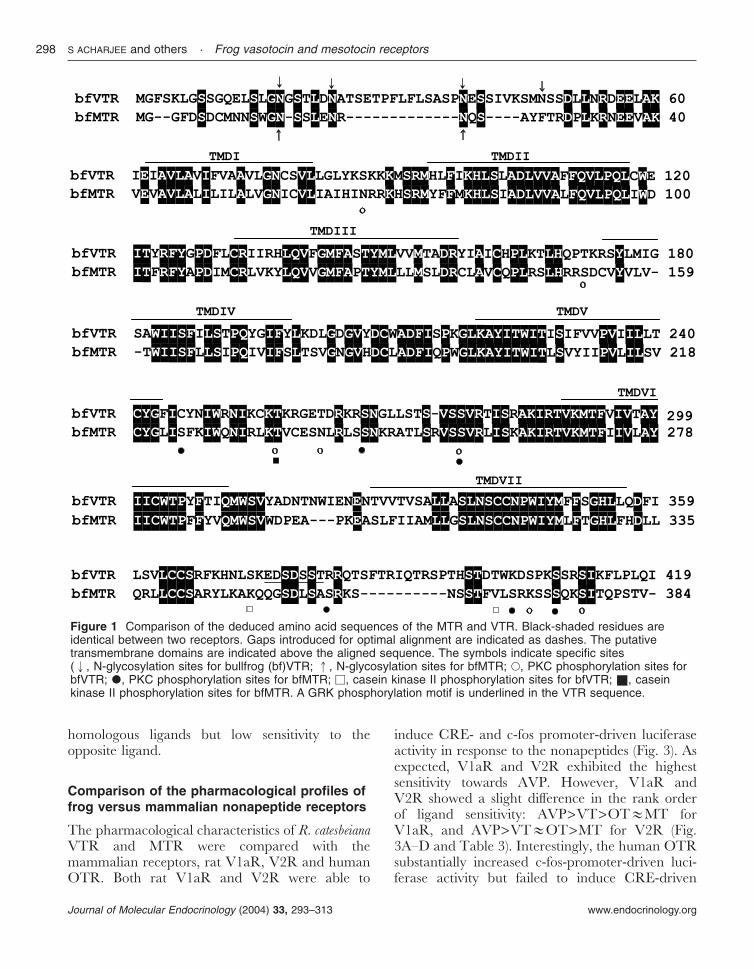
Using degenerate primers we obtained two partialPCR fragments for R. catesbeiana VTR and MTRwith 520 bp and 630 bp respectively. The 5�- and3�-cDNA end sequences for each receptor wereobtained by performing RACE using gene-specificprimers. The full-length bullfrog VTR and MTRconsisted of 1257 and 1152 nucleotides thatencoded proteins of 419 and 384 amino acidsrespectively (Fig. 1). We also identified full-lengthsequences for the R. esculenta VTR having the sameamino acid sequence as the bullfrog VTR butdiffering by five nucleotides. We cloned a partialsequence for R. esculenta MTR that showed 98%sequence similarity with the bullfrog MTR. Thehydropathy analysis of frog VTR and MTRrevealed the presence of seven stretches ofhydrophobic amino acid residues corresponding tothe seven transmembrane domains. Potential sitesfor N-linked glycosylation and phosphorylationwere present in both the VTR and MTR. Severalputative sites for phosphorylation by PKC, casein
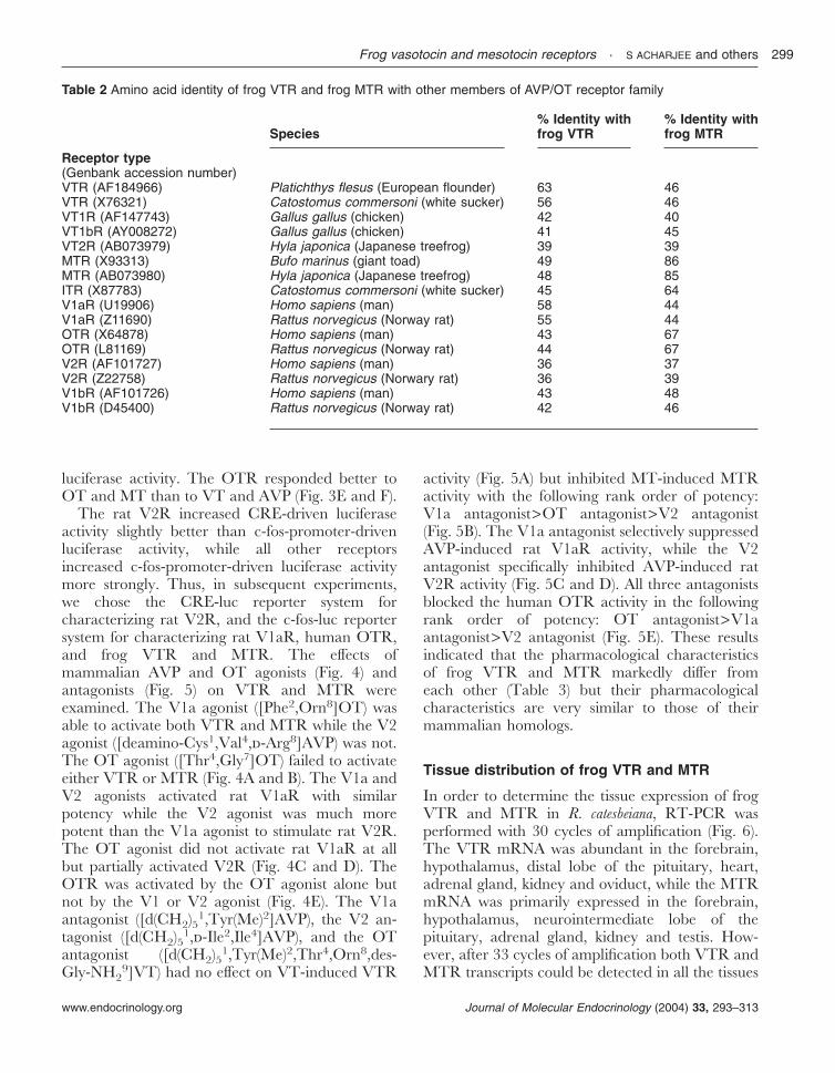
kinase II, and G protein-coupled receptor kinase(GRK) were seen in the intracellular loops andC-terminal tails of the receptors (Fig. 1). The frogVTR showed the highest sequence identity (63%)with the flounder VTR and relatively highsequence identity (58%) with the human V1aR.The frog MTR exhibited the highest sequenceidentity (85%) with the giant toad MTR and arelatively high degree of homology (67%) with thehuman OTR. The VTR and MTR shared 47%sequence identity with each other (Table 2).
Ligand selectivity and signal transductionpathways of frog VTR and MTR
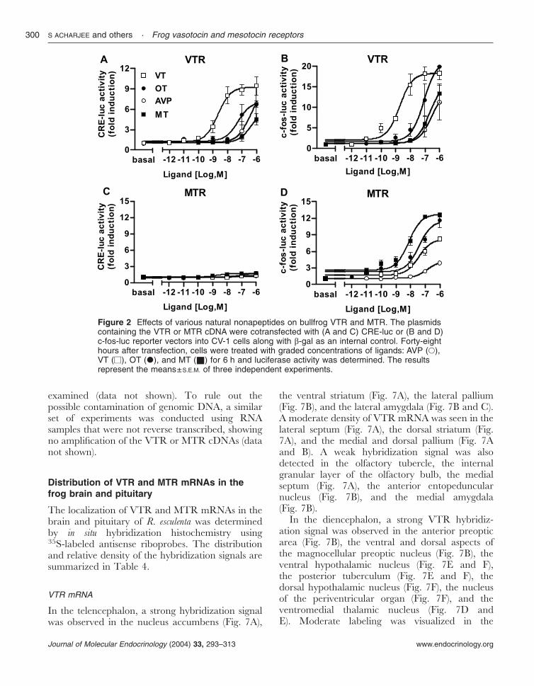
The open reading frames of the R. catesbeiana VTRand MTR were subcloned in the pcDNA3, amammalian expression vector, and the VTR andMTR cDNAs were transiently transfected in CV-1cells in combination with the CRE-luc or c-fos-lucreporter vectors. We have previously demonstratedthat CRE-luc and c-fos-luc reporter systems areuseful tools to discriminate AC/PKA and PLC/PKC signaling pathways respectively (Oh et al.2003, Seong et al. 2003). In the absence of frogVTR or MTR, neither CRE- nor c-fos-promoter-driven transcriptional activity in CV-1 cells wasinduced by the nonapeptide ligands VT, AVP, MTand OT, indicating that CV-1 cells do not naturallyexpress the endogenous receptors for these ligands(data not shown).
In CV-1 cells transfected with the VTR cDNA,VT induced a concentration-dependent increase ofboth CRE- and c-fos-promoter-driven luciferaseactivity (Fig. 2A and B). The other nonapeptideligands tested also stimulated VTR activity with thefollowing rank order of potency: OT>AVP�MT(Fig. 2A and B). In CV-1 cells transfected with theMTR cDNA, the nonapeptide ligands did notinduce CRE-driven luciferase activity (Fig. 2C)but substantially increased c-fos-promoter-drivenluciferase activity with the following rank order ofpotency: MT>OT>VT>AVP (Fig. 2D). Thesedata indicated that frog VTR and MTR havedifferent functional characteristics with respectto signal transduction and ligand sensitivity: frogVTR couples to both the PLC/PKC and AC/PKA pathways, while frog MTR preferentiallycouples to the PLC/PKC pathway but margin-ally to the AC/PKA pathway. Frog VTR andMTR exhibited the highest sensitivity to the
Frog vasotocin and mesotocin receptors · S ACHARJEE and others 297
www.endocrinology.org Journal of Molecular Endocrinology (2004) 33, 293–313
homologous ligands but low sensitivity to theopposite ligand.
Comparison of the pharmacological profiles offrog versus mammalian nonapeptide receptors
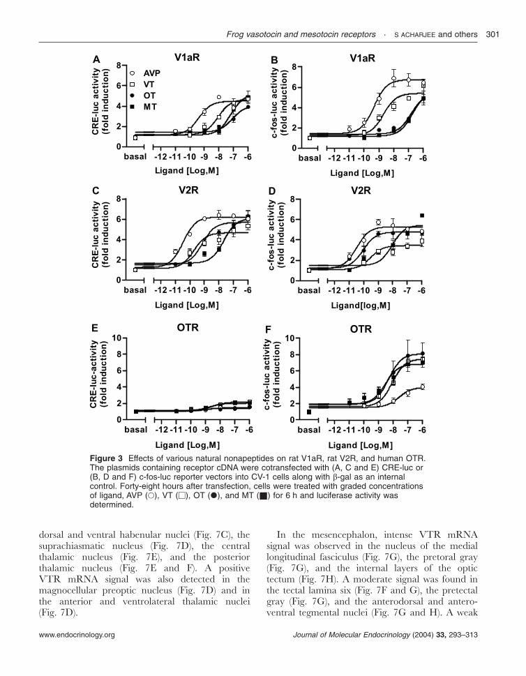
The pharmacological characteristics of R. catesbeianaVTR and MTR were compared with themammalian receptors, rat V1aR, V2R and humanOTR. Both rat V1aR and V2R were able to
induce CRE- and c-fos promoter-driven luciferaseactivity in response to the nonapeptides (Fig. 3). Asexpected, V1aR and V2R exhibited the highestsensitivity towards AVP. However, V1aR andV2R showed a slight difference in the rank orderof ligand sensitivity: AVP>VT>OT�MT forV1aR, and AVP>VT�OT>MT for V2R (Fig.3A–D and Table 3). Interestingly, the human OTRsubstantially increased c-fos-promoter-driven luci-ferase activity but failed to induce CRE-driven
Figure 1 Comparison of the deduced amino acid sequences of the MTR and VTR. Black-shaded residues areidentical between two receptors. Gaps introduced for optimal alignment are indicated as dashes. The putativetransmembrane domains are indicated above the aligned sequence. The symbols indicate specific sites(", N-glycosylation sites for bullfrog (bf)VTR; !, N-glycosylation sites for bfMTR; •, PKC phosphorylation sites forbfVTR; d, PKC phosphorylation sites for bfMTR; M, casein kinase II phosphorylation sites for bfVTR; ", caseinkinase II phosphorylation sites for bfMTR. A GRK phosphorylation motif is underlined in the VTR sequence.
S ACHARJEE and others · Frog vasotocin and mesotocin receptors298
www.endocrinology.orgJournal of Molecular Endocrinology (2004) 33, 293–313
luciferase activity. The OTR responded better toOT and MT than to VT and AVP (Fig. 3E and F).
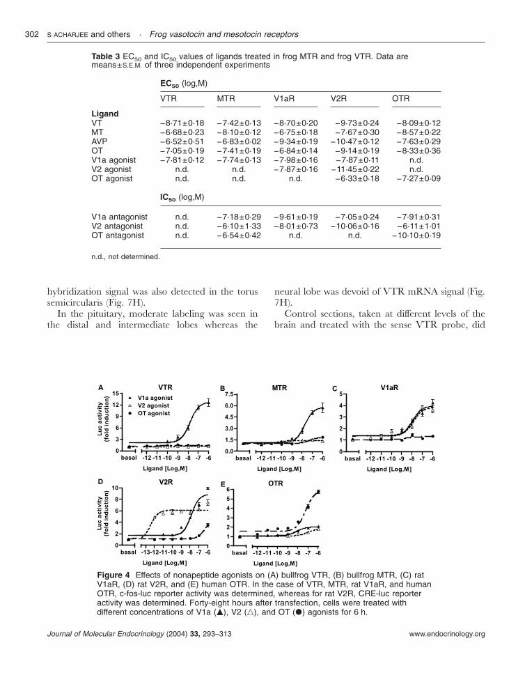
The rat V2R increased CRE-driven luciferaseactivity slightly better than c-fos-promoter-drivenluciferase activity, while all other receptorsincreased c-fos-promoter-driven luciferase activitymore strongly. Thus, in subsequent experiments,we chose the CRE-luc reporter system forcharacterizing rat V2R, and the c-fos-luc reportersystem for characterizing rat V1aR, human OTR,and frog VTR and MTR. The effects ofmammalian AVP and OT agonists (Fig. 4) andantagonists (Fig. 5) on VTR and MTR wereexamined. The V1a agonist ([Phe2,Orn8]OT) wasable to activate both VTR and MTR while the V2agonist ([deamino-Cys1,Val4,-Arg8]AVP) was not.The OT agonist ([Thr4,Gly7]OT) failed to activateeither VTR or MTR (Fig. 4A and B). The V1a andV2 agonists activated rat V1aR with similarpotency while the V2 agonist was much morepotent than the V1a agonist to stimulate rat V2R.The OT agonist did not activate rat V1aR at allbut partially activated V2R (Fig. 4C and D). TheOTR was activated by the OT agonist alone butnot by the V1 or V2 agonist (Fig. 4E). The V1aantagonist ([d(CH2)51,Tyr(Me)2]AVP), the V2 an-tagonist ([d(CH2)51,-Ile2,Ile4]AVP), and the OTantagonist ([d(CH2)51,Tyr(Me)2,Thr4,Orn8,des-Gly-NH2
9]VT) had no effect on VT-induced VTR
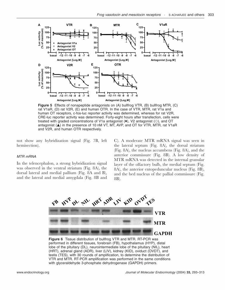
activity (Fig. 5A) but inhibited MT-induced MTRactivity with the following rank order of potency:V1a antagonist>OT antagonist>V2 antagonist(Fig. 5B). The V1a antagonist selectively suppressedAVP-induced rat V1aR activity, while the V2antagonist specifically inhibited AVP-induced ratV2R activity (Fig. 5C and D). All three antagonistsblocked the human OTR activity in the followingrank order of potency: OT antagonist>V1aantagonist>V2 antagonist (Fig. 5E). These resultsindicated that the pharmacological characteristicsof frog VTR and MTR markedly differ fromeach other (Table 3) but their pharmacologicalcharacteristics are very similar to those of theirmammalian homologs.
Tissue distribution of frog VTR and MTR
In order to determine the tissue expression of frogVTR and MTR in R. catesbeiana, RT-PCR wasperformed with 30 cycles of amplification (Fig. 6).The VTR mRNA was abundant in the forebrain,hypothalamus, distal lobe of the pituitary, heart,adrenal gland, kidney and oviduct, while the MTRmRNA was primarily expressed in the forebrain,hypothalamus, neurointermediate lobe of thepituitary, adrenal gland, kidney and testis. How-ever, after 33 cycles of amplification both VTR andMTR transcripts could be detected in all the tissues
Table 2 Amino acid identity of frog VTR and frog MTR with other members of AVP/OT receptor family
Species% Identity withfrog VTR
% Identity withfrog MTR
Receptor type(Genbank accession number)VTR (AF184966) Platichthys flesus (European flounder) 63 46VTR (X76321) Catostomus commersoni (white sucker) 56 46VT1R (AF147743) Gallus gallus (chicken) 42 40VT1bR (AY008272) Gallus gallus (chicken) 41 45VT2R (AB073979) Hyla japonica (Japanese treefrog) 39 39MTR (X93313) Bufo marinus (giant toad) 49 86MTR (AB073980) Hyla japonica (Japanese treefrog) 48 85ITR (X87783) Catostomus commersoni (white sucker) 45 64V1aR (U19906) Homo sapiens (man) 58 44V1aR (Z11690) Rattus norvegicus (Norway rat) 55 44OTR (X64878) Homo sapiens (man) 43 67OTR (L81169) Rattus norvegicus (Norway rat) 44 67V2R (AF101727) Homo sapiens (man) 36 37V2R (Z22758) Rattus norvegicus (Norwary rat) 36 39V1bR (AF101726) Homo sapiens (man) 43 48V1bR (D45400) Rattus norvegicus (Norway rat) 42 46
Frog vasotocin and mesotocin receptors · S ACHARJEE and others 299
www.endocrinology.org Journal of Molecular Endocrinology (2004) 33, 293–313
examined (data not shown). To rule out thepossible contamination of genomic DNA, a similarset of experiments was conducted using RNAsamples that were not reverse transcribed, showingno amplification of the VTR or MTR cDNAs (datanot shown).
Distribution of VTR and MTR mRNAs in thefrog brain and pituitary
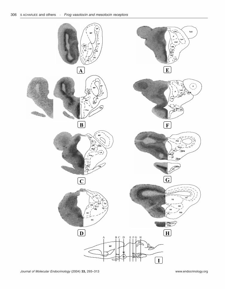
The localization of VTR and MTR mRNAs in thebrain and pituitary of R. esculenta was determinedby in situ hybridization histochemistry using35S-labeled antisense riboprobes. The distributionand relative density of the hybridization signals aresummarized in Table 4.
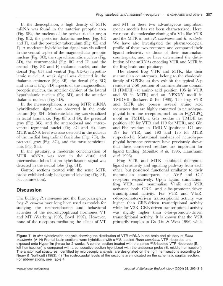
VTR mRNA
In the telencephalon, a strong hybridization signalwas observed in the nucleus accumbens (Fig. 7A),
the ventral striatum (Fig. 7A), the lateral pallium(Fig. 7B), and the lateral amygdala (Fig. 7B and C).A moderate density of VTR mRNA was seen in thelateral septum (Fig. 7A), the dorsal striatum (Fig.7A), and the medial and dorsal pallium (Fig. 7Aand B). A weak hybridization signal was alsodetected in the olfactory tubercle, the internalgranular layer of the olfactory bulb, the medialseptum (Fig. 7A), the anterior entopeduncularnucleus (Fig. 7B), and the medial amygdala(Fig. 7B).
In the diencephalon, a strong VTR hybridiz-ation signal was observed in the anterior preopticarea (Fig. 7B), the ventral and dorsal aspects ofthe magnocellular preoptic nucleus (Fig. 7B), theventral hypothalamic nucleus (Fig. 7E and F),the posterior tuberculum (Fig. 7E and F), thedorsal hypothalamic nucleus (Fig. 7F), the nucleusof the periventricular organ (Fig. 7F), and theventromedial thalamic nucleus (Fig. 7D andE). Moderate labeling was visualized in the
Figure 2 Effects of various natural nonapeptides on bullfrog VTR and MTR. The plasmidscontaining the VTR or MTR cDNA were cotransfected with (A and C) CRE-luc or (B and D)c-fos-luc reporter vectors into CV-1 cells along with �-gal as an internal control. Forty-eighthours after transfection, cells were treated with graded concentrations of ligands: AVP (•),VT (M), OT (d), and MT (") for 6 h and luciferase activity was determined. The resultsrepresent the means±S.E.M. of three independent experiments.
S ACHARJEE and others · Frog vasotocin and mesotocin receptors300
www.endocrinology.orgJournal of Molecular Endocrinology (2004) 33, 293–313
dorsal and ventral habenular nuclei (Fig. 7C), thesuprachiasmatic nucleus (Fig. 7D), the centralthalamic nucleus (Fig. 7E), and the posteriorthalamic nucleus (Fig. 7E and F). A positiveVTR mRNA signal was also detected in themagnocellular preoptic nucleus (Fig. 7D) and inthe anterior and ventrolateral thalamic nuclei(Fig. 7D).
In the mesencephalon, intense VTR mRNAsignal was observed in the nucleus of the mediallongitudinal fasciculus (Fig. 7G), the pretoral gray(Fig. 7G), and the internal layers of the optictectum (Fig. 7H). A moderate signal was found inthe tectal lamina six (Fig. 7F and G), the pretectalgray (Fig. 7G), and the anterodorsal and antero-ventral tegmental nuclei (Fig. 7G and H). A weak
Figure 3 Effects of various natural nonapeptides on rat V1aR, rat V2R, and human OTR.The plasmids containing receptor cDNA were cotransfected with (A, C and E) CRE-luc or(B, D and F) c-fos-luc reporter vectors into CV-1 cells along with �-gal as an internalcontrol. Forty-eight hours after transfection, cells were treated with graded concentrationsof ligand, AVP (•), VT (M), OT (d), and MT (") for 6 h and luciferase activity wasdetermined.
Frog vasotocin and mesotocin receptors · S ACHARJEE and others 301
www.endocrinology.org Journal of Molecular Endocrinology (2004) 33, 293–313
hybridization signal was also detected in the torussemicircularis (Fig. 7H).
In the pituitary, moderate labeling was seen inthe distal and intermediate lobes whereas the
neural lobe was devoid of VTR mRNA signal (Fig.7H).
Control sections, taken at different levels of thebrain and treated with the sense VTR probe, did
Table 3 EC50 and IC50 values of ligands treated in frog MTR and frog VTR. Data aremeans±S.E.M. of three independent experiments
EC50 (log,M)
VTR MTR V1aR V2R OTR
LigandVT −8·71±0·18 −7·42±0·13 −8·70±0·20 −9·73±0·24 −8·09±0·12MT −6·68±0·23 −8·10±0·12 −6·75±0·18 −7·67±0·30 −8·57±0·22AVP −6·52±0·51 −6·83±0·02 −9·34±0·19 −10·47±0·12 −7·63±0·29OT −7·05±0·19 −7·41±0·19 −6·84±0·14 −9·14±0·19 −8·33±0·36V1a agonist −7·81±0·12 −7·74±0·13 −7·98±0·16 −7·87±0·11 n.d.V2 agonist n.d. n.d. −7·87±0·16 −11·45±0·22 n.d.OT agonist n.d. n.d. n.d. −6·33±0·18 −7·27±0·09
IC50 (log,M)
V1a antagonist n.d. −7·18±0·29 −9·61±0·19 −7·05±0·24 −7·91±0·31V2 antagonist n.d. −6·10±1·33 −8·01±0·73 −10·06±0·16 −6·11±1·01OT antagonist n.d. −6·54±0·42 n.d. n.d. −10·10±0·19
n.d., not determined.
Figure 4 Effects of nonapeptide agonists on (A) bullfrog VTR, (B) bullfrog MTR, (C) ratV1aR, (D) rat V2R, and (E) human OTR. In the case of VTR, MTR, rat V1aR, and humanOTR, c-fos-luc reporter activity was determined, whereas for rat V2R, CRE-luc reporteractivity was determined. Forty-eight hours after transfection, cells were treated withdifferent concentrations of V1a (m), V2 (n), and OT (d) agonists for 6 h.
S ACHARJEE and others · Frog vasotocin and mesotocin receptors302
www.endocrinology.orgJournal of Molecular Endocrinology (2004) 33, 293–313
not show any hybridization signal (Fig. 7B, lefthemisection).
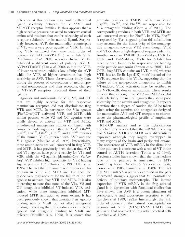
MTR mRNA
In the telencephalon, a strong hybridization signalwas observed in the ventral striatum (Fig. 8A), thedorsal lateral and medial pallium (Fig. 8A and B),and the lateral and medial amygdala (Fig. 8B and
C). A moderate MTR mRNA signal was seen inthe lateral septum (Fig. 8A), the dorsal striatum(Fig. 8A), the nucleus accumbens (Fig. 8A), and theanterior commissure (Fig. 8B). A low density ofMTR mRNA was detected in the internal granularlayer of the olfactory bulb, the medial septum (Fig.8A), the anterior entopeduncular nucleus (Fig. 8B),and the bed nucleus of the pallial commissure (Fig.8B).
Figure 5 Effects of nonapeptide antagonists on (A) bullfrog VTR, (B) bullfrog MTR, (C)rat V1aR, (D) rat V2R, (E) and human OTR. In the case of VTR, MTR, rat V1a andhuman OT receptors, c-fos-luc reporter activity was determined, whereas for rat V2R,CRE-luc reporter activity was determined. Forty-eight hours after transfection, cells weretreated with graded concentrations of V1a antagonist (♦), V2 antagonist (e), and OTantagonist (m) in the presence of 10 nM VT, MT, AVP, and OT for VTR, MTR, rat V1aRand V2R, and human OTR respectively.
Figure 6 Tissue distribution of bullfrog VTR and MTR. RT-PCR wasperformed in different tissues, forebrain (FB), hypothalamus (HYP), distallobe of the pituitary (DL), neurointermediate lobe of the pituitary (NIL), heart(HRT), adrenal gland (ADR), liver (LIV), kidney (KID), oviduct (OVDT), andtestis (TES), with 30 rounds of amplification, to determine the distribution ofVTR and MTR. RT-PCR amplification was performed in the same conditionswith glyceraldehyde 3-phosphate dehydrogenase (GAPDH) primers.
Frog vasotocin and mesotocin receptors · S ACHARJEE and others 303
www.endocrinology.org Journal of Molecular Endocrinology (2004) 33, 293–313
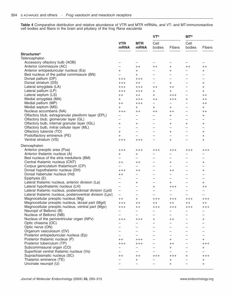
Table 4 Comparative distribution and relative abundance of VTR and MTR mRNAs, and VT- and MT-immunoreactivecell bodies and fibers in the brain and pituitary of the frog Rana esculenta
VTRmRNA
MTRmRNA
VTb MTb
Cellbodies Fibers
Cellbodies Fibers
Structuresa
TelencephalonAccessory olfactory bulb (AOB) − − − − − −Anterior commissure (AC) − ++ ++ + ++ ++Anterior entopeduncular nucleus (Ea) + + − + − +Bed nucleus of the pallial commissure (BN) − + − − − −Dorsal pallium (DP) +++ +++ − − − −Dorsal striatum (DS) +++ ++ − + − +Lateral amygdala (LA) +++ +++ ++ ++ − +Lateral pallium (LP) +++ +++ + + − +Lateral septum (LS) ++ ++ + +++ − ++Medial amygdala (MA) + +++ ++ +++ + ++Medial pallium (MP) ++ +++ − − − ++Medial septum (MS) + + + + − +Nucleus accumbens (NA) +++ ++ ++ ++ − ++Olfactory blub, extragranular plexiform layer (EPL) − − − + − +Olfactory blub, glomerular layer (GL) − − − − − −Olfactory bulb, internal granular layer (IGL) + + − + + +Olfactory bulb, mitral cellular layer (ML) − − − − − −Olfactory tubercle (TO) + − − + − +Postolfactory eminence (PE) + − − − − +Ventral striatum (VS) +++ +++ − + − +
DiencephalonAnterior preoptic area (Poa) +++ +++ +++ +++ +++ +++Anterior thalamic nucleus (A) + + − − − −Bed nucleus of the stria medullaris (BM) − − − − − −Central thalamic nucleus (CNT) ++ ++ − + − +Corpus geniculatum thalamicum (CP) − − − − − −Dorsal hypothalamic nucleus (DH) +++ ++ − ++ − +Dorsal habenular nucleus (Hd) ++ − − − − −Epiphysis (E) − − − − − −Lateral thalamic nucleus, anterior division (La) − + − + − +Lateral hypothalamic nucleus (LH) − − + +++ − ++Lateral thalamic nucleus, posterodorsal division (Lpd) − − − − − −Lateral thalamic nucleus, posteroventral division (Lpv) − − − − − −Magnocellular preoptic nucleus (Mg) ++ + +++ +++ +++ +++Magnocellular preoptic nucleus, dorsal part (Mgd) +++ ++ ++ ++ ++ ++Magnocellular preoptic nucleus, ventral part (Mgv) +++ ++ +++ +++ +++ +++Neuropil of Bellonci (B) − − − − − −Nucleus of Bellonci (NB) − − − − − −Nucleus of the periventricular organ (NPv) +++ +++ + ++ − +Optic chiasma (OC) − − − − − −Optic nerve (ON) − − − − − −Organum vasculosum (OV) − − − − − −Posterior entopeduncular nucleus (Ep) − − − − − −Posterior thalamic nucleus (P) ++ +++ − + − +Posterior tuberculum (TP) +++ +++ − ++ − +++Subcommissural organ (CO) − − − + − +Superficial ventral thalamic nucleus (Vs) − − − − − −Suprachiasmatic nucleus (SC) ++ ++ +++ +++ + +++Thalamic eminence (TE) − + − + − +Uncinate neuropil (U) − − − + − +
S ACHARJEE and others · Frog vasotocin and mesotocin receptors304
www.endocrinology.orgJournal of Molecular Endocrinology (2004) 33, 293–313
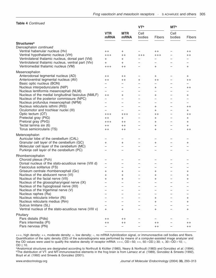
Table 4 Continued
VTRmRNA
MTRmRNA
VTb MTb
Cellbodies Fibers
Cellbodies Fibers
Structuresa
Diencephalon continuedVentral habenular nucleus (Hv) ++ + − ++ − ++Ventral hypothalamic nucleus (VH) +++ ++ +++ +++ − ++Ventrolateral thalamic nucleus, dorsal part (Vld) + + − − − −Ventrolateral thalamic nucleus, ventral part (Vlv) + + − − − −Ventromedial thalamic nucleus (VM) +++ ++ − + − +
MesencephalonAnterodorsal tegmental nucleus (AD) ++ ++ − + − +Anterioventral tegmental nucleus (AV) ++ ++ + ++ − ++Basic optic nucleus (BON) − − − − − −Nucleus interpeduncularis (NIP) − − − + − ++Nucleus lentiformis mesencephali (NLM) − − − − − −Nucleus of the medial longitudinal fasiculus (NMLF) ++ + − + − +Nucleus of the posterior commissure (NPC) − − − − − −Nucleus profundus mesencephali (NPM) − − − − − −Nucleus reticularis isthmi (RIS) − − − + − ++Oculomotor and trochlear nuclei (III) − − − + − +Optic tectum (OT) +++ +++ − ++ − ++Pretectal gray (PtG) ++ + − + − +Pretoral gray (PtrG) +++ ++ − + − +Tectal lamina six (6) ++ ++ − + − +Torus semicircularis (TS) ++ ++ − + − ++
MetencephalonAuricular lobe of the cerebellum (CAL) − − − − − −Granular cell layer of the cerebellum (GC) + + − + − +Molecular cell layer of the cerebellum (MC) − − − − − −Purkinje cell layer of the cerebellum (PC) − − − − − −
RhombencephalonChoroid plexus (Pch) − − − − − −Dorsal nucleus of the stato-acusticus nerve (VIII d) − − − − − −Fasciculus solitarius (FS) − − − − − −Griseum centrale rhombencephali (Gc) + + − + − +Nucleus of the abducent nerve (VI) + + − + − +Nucleus of the facial nerve (VII) + + − + − +Nucleus of the glossopharyngeal nerve (IX) − − − − − −Nucleus of the hypoglossal nerve (XII) − − − − − −Nucleus of the trigeminal nerve (V) − − − − − −Nucleus raphes (Ra) − − − − − −Nucleus reticularis inferior (Ri) − − − + − +Nucleus reticularis medius (Rm) − − − + − +Sulcus limitans (SL) − − − − − −Ventral nucleus of the stato-acusticus nerve (VIII v) + + − + − +
PituitaryPars distalis (Pdis) ++ ++ − − − −Pars intermedia (PI) ++ ++ − ++ − ++Pars nervosa (PN) − − − ++ − ++
+++, high density; ++, moderate density; +, low density; −, no mRNA hybridization signal, or immunoreactive cell bodies and fibers.Quantification of the optic density (OD) of the autoradiograms was performed by means of a computer-assisted image analyzer andthe OD values were used to qualify the relative density of receptor mRNA: +++, OD.50; ++, 50.ODR30; +, 30.OD.10; −,OD%10.aAnatomical structures are designated according to Northcutt & Kicliter (1980), Neary & Northcutt (1983) and Gonzalez et al. (1994).bThe distribution of VT- and MT-immunoreactive elements in the frog brain is from Lamacz et al. (1989), Gonzalez & Smeets (1992),Boyd et al. (1992) and Smeets & Gonzalez (2001).
Frog vasotocin and mesotocin receptors · S ACHARJEE and others 305
www.endocrinology.org Journal of Molecular Endocrinology (2004) 33, 293–313
S ACHARJEE and others · Frog vasotocin and mesotocin receptors306
www.endocrinology.orgJournal of Molecular Endocrinology (2004) 33, 293–313
In the diencephalon, a high density of MTRmRNA was found in the anterior preoptic area(Fig. 8B), the nucleus of the periventricular organ(Fig. 8E), the posterior thalamic nucleus (Fig. 8Eand F), and the posterior tuberculum (Fig. 8E andF). A moderate hybridization signal was visualizedin the ventral aspect of the magnocellular preopticnucleus (Fig. 8C), the suprachiasmatic nucleus (Fig.8D), the ventromedial (Fig. 8C and D) and thecentral (Fig. 8E and F) thalamic nuclei, and thedorsal (Fig. 8F) and ventral (Fig. 8E–G) hypotha-lamic nuclei. A weak signal was detected in thethalamic eminence (Fig. 8B), the dorsal (Fig. 8C)and central (Fig. 8D) aspects of the magnocellularpreoptic nucleus, the anterior division of the lateralhypothalamic nucleus (Fig. 8D), and the anteriorthalamic nucleus (Fig. 8D).
In the mesencephalon, a strong MTR mRNAhybridization signal was observed in the optictectum (Fig. 8H). Moderate labeling was visualizedin tectal lamina six (Fig. 8F and G), the pretectalgray (Fig. 8G), and the anterodorsal and antero-ventral tegmental nuclei (Fig. 8G and H). LowMTR mRNA level was also detected in the nucleusof the medial longitudinal fasciculus (Fig. 7G), thepretectal gray (Fig. 8G), and the torus semicircu-laris (Fig. 8H).
In the pituitary, a moderate concentration ofMTR mRNA was seen in the distal andintermediate lobes but no hybridization signal wasdetected in the neural lobe (Fig. 8H).
Control sections treated with the sense MTRprobe exhibited only background labeling (Fig. 8F,left hemisection).
Discussion
The bullfrog R. catesbeiana and the European greenfrog R. esculenta have long been used as models forstudying the neuroendocrine and behavioralactivities of the neurohypophysial hormones VTand MT (Warburg 1995, Boyd 1997). However,none of the receptors mediating the effects of VT
and MT in these two advantageous amphibianspecies models has yet been characterized. Here,we report the molecular cloning of a V1a-like VTRand the MTR in both R. catesbeiana and R. esculenta.We have also investigated the pharmacologicalprofile of these two receptors and compared theirligand selectivity to those of their mammaliancounterparts, and we have determined the distri-bution of the mRNAs encoding VTR and MTR inthe frog brain and pituitary.
The cloned frog VTR and MTR, like theirmammalian counterparts, belong to the rhodopsinfamily of GPCRs as they exhibit the typical Aspresidue at 2·50 position of transmembrane domainII (TMDII) (at amino acid position 105 in VTRand 85 in MTR) and an NPXXY motif inTMDVII (Bockaert & Pin 1999). The frog VTRand MTR also possess several amino acidsequences that are highly conserved in neurohypo-physial hormone receptors, such as an FQVLPQmotif in TMDII, a Gln residue in TMDII (atposition 139 for VTR and 119 for MTR), and Glnand Phe residues in TMDIV (positions 171 and197 for VTR, and 193 and 175 for MTRrespectively). Mutational analyses of neurohypo-physial hormone receptors have previously shownthat these conserved residues are important forligand binding (Mouillac et al. 1995, Hausmannet al. 1996).
Frog VTR and MTR exhibited differentialligand sensitivity and signaling pathway from eachother, but possessed functional similarity to theirmammalian counterparts, i.e. AVP and OTreceptors respectively. Upon ligand stimulation,frog VTR, and mammalian V1aR and V2Ractivated both CRE- and c-fos-promoter-driventranscriptional activity. For VTR and V1aR,c-fos-promoter-driven transcriptional activity washigher than CRE-driven transcriptional activitywhile for V2R, CRE-driven transcriptional activitywas slightly higher than c-fos-promoter-driventranscriptional activity. It is known that the V2Rprimarily couples to Gs (Liu & Wess 1996) while
Figure 7 In situ hybridization analysis showing the distribution of VTR mRNA in the brain and pituitary of Ranaesculenta. (A–H) Frontal brain sections were hybridized with a 35S-labeled Rana esculenta VTR riboprobe andexposed onto Hyperfilm �-max for 2 weeks. A control section treated with the sense 35S-labeled VTR riboprobe (B,left hemisection) is compared with a consecutive section hybridized with the antisense probe (B, middle hemisection).The anatomical structures, identified by microscopic analysis, are designated on the right hemisections according toNeary & Northcutt (1983). (I) The rostrocaudal levels of the sections are indicated on the schematic sagittal section.For abbreviations, see Table 4.
Frog vasotocin and mesotocin receptors · S ACHARJEE and others 307
www.endocrinology.org Journal of Molecular Endocrinology (2004) 33, 293–313
S ACHARJEE and others · Frog vasotocin and mesotocin receptors308
www.endocrinology.orgJournal of Molecular Endocrinology (2004) 33, 293–313
our study showed that the rat V2R could activateboth the CRE- and c-fos-luc activity. Our findingscan be supported by a previous report showing thatoverexpression (100 000 sites/cell) of the V2R canstimulate phospholipase C, which explains the dualsignaling potential of the V2R (Zhu et al. 1994).Alternatively, the reporter system used in this studyis more sensitive than the second messenger assaysystem which allows us to detect amplified signalsthrough multiple signaling cascades. These obser-vations indicated that AVP/VT receptors couple toboth PKA- and PKC-mediated signaling pathways,but that VTR and V1aR preferentially couple toPKC-linked signaling, while V2R preferentiallycouple to PKA-linked signaling, which is in goodagreement with previous reports (Liu & Wess1996). Concurrently, frog MTR and OTRtriggered only c-fos-promoter-driven transcrip-tional activity, indicating that MTR and OTRexclusively couple to the PKC-mediated signalingpathway. We found that VTR, like V1aR, containsa highly conserved triplet of residues (Asp-Arg-Tyr,DRY) at the boundary between TMDIII andintracellular loop 2 (ICL2), which is a commonfeature of the rhodopsin family of GPCRs(Bockaert & Pin 1999). Interestingly, in MTR, theDRY motif was changed to DRC as found in theOTR (Gimpl & Fahrenholz 2001), while this motifis modified to DRH in V2R (Lolait et al. 1992). TheDRY motif plays a pivotal role in G proteincoupling and is important for receptor activationand internalization (Arora et al. 1997, Gether &Kobilka 1998, Mitchell et al. 1998). Substitutionof the Tyr residue in the DRY motif may thusaccount for the differential signaling betweenVT/AVP receptors and MT/OT receptors. Alter-natively, differential signaling may be ascribed toamino acid substitutions in ICLs. It has beendemonstrated that chimeric receptors having theV1aR sequence in ICL2 are able to activate thephosphatidylinositol pathway with high efficiency,while hybrid receptors containing the V2R
sequence in ICL3 are capable of efficientlystimulating cAMP production (Liu & Wess 1996).Another motif at the junction between TMDV andICL3, CYGLISF/YKIW, is highly conserved inMTR and OTR, but not in VTR/V1aR and inV2R. The lack of conservation of this motif mayalso explain the differential signal transductionactivated by VT/AVP receptors and MT/OTreceptors. Recently, VTR and MTR have beencloned from the Japanese treefrog Hyla japonica(Kohno et al. 2003). This VTR differs considerablyfrom the bullfrog VTR. The Japanese treefrogVTR shows high homology with the mammalianV2R subtype and couples to Gs. So, it is likely thatthe amphibians possess two different kinds of VTR,one resembling the mammalian V1a subtype andthe other representing the mammalian V2 subtype.
With regard to ligand sensitivity, the VT/AVPreceptor family responded better to VT than MT,while MT/OT receptors were more sensitive toMT than VT. This finding indicated that thespecific amino acids responsible for ligand selec-tivity are conserved within the VT/AVP andMT/OT receptor families. It is known that theTyr115 residue in extracellular loop 1 (ECL1) of thehuman V1aR is crucial for agonist selectivity (Chiniet al. 1995). Interestingly, this residue is substitutedto Asp in the V2R and to Phe in the OTRrespectively. It has been suggested that amino acidat position 8 (Arg for VT/AVP and Leu forMT/OT) in neurohypophysial nonapeptides mayinteract with the amino acid at this position (Tyr115
for V1aR, Asp103 for V2R, and Phe103 for OTR).AVP and VT that possess an Arg8 residue exhibitsimilar sensitivity to V1aR, V2R and OTR,whereas OT and AVP with an Arg8�Leu sub-stitution exhibit highest sensitivity to OTR and aremarkable decrease in sensitivity to V1aR andV2R (Chini et al. 1995). Like V1aR, frog VTRcontains a Tyr residue at this position, while frogMTR has a Phe residue as has the OTR. Theseobservations suggest that a single amino acid
Figure 8 In situ hybridization analysis showing the distribution of MTR mRNA in the brain and pituitary of Ranaesculenta. (A–H) Frontal brain sections were hybridized with a 35S-labeled Rana esculenta MTR riboprobe andexposed onto Hyperfilm �-max for 2 weeks. A control section treated with the sense 35S-labeled MTR riboprobe (F,left hemisection) is compared with a consecutive section hybridized with the antisense probe (F, central hemisection).The anatomical structures, identified by microscopic analysis, are designated on the right hemisections according toNeary & Northcutt (1983). (I) The rostrocaudal levels of the sections are indicated on the schematic sagittal section.For abbreviations, see Table 4.
Frog vasotocin and mesotocin receptors · S ACHARJEE and others 309
www.endocrinology.org Journal of Molecular Endocrinology (2004) 33, 293–313
difference at this position may confer differentialligand selectivity between the VT/AVP andMT/OT receptor families. It thus appears that ahigh selective pressure has acted to conserve crucialamino acid residues that confer selectivity of eachreceptor subfamily for its natural ligand. Surpris-ingly however, AVP, the mammalian counterpartof VT, was a very poor agonist of VTR. In fact,frog VTR exhibited the same rank order ofpotency (VT>OT>AVP�MT) as a fish VTR(Mahlmann et al. 1994), whereas chicken VT1Rexhibited a different order of potency, AVT�AVP>OT�MT (Tan et al. 2000) so that the VTRof lower vertebrates exhibits low affinity for AVPwhile the VTR of higher vertebrates has highsensitivity to AVP. These observations imply that,during the process of co-evolution of neurohypo-physial nonapeptides and their receptors, changesof VT/AVP receptors preceded those of theirligands.
Agonists and antagonists for V1a, V2, and OTthat are highly selective for the respectivemammalian receptors did not discriminate frogVTR and MTR. In particular, the V1a agonist[Phe2,Orn8]OT activated VTR and MTR withsimilar potency while V2 and OT agonists weretotally devoid of activity on VTR and MTR.Site-directed mutagenesis studies combined withcomputer modeling indicate that the Asp97, Gln104,Gln108, Lys128, Gln131, Gln185, and Gln311 residuesof the human V1aR interact with AVP and theV1a agonist (Mouillac et al. 1995). Interestingly,these amino acids are well conserved in frog VTRand MTR. It has previously been shown that AVPand V1a agonist have poor selectivity for V1a andV2R, while the V2 agonist [deamino-Cys1,Val4,-Arg8]AVP exhibits high specificity for V2R havingAsp at position 103 (Chini et al. 1995, Ufer et al.1995). The fact that the amino acid residues at thisposition in VTR and MTR are Tyr and Pherespectively may account for the failure of the V2agonist to activate frog VTR and MTR. It is alsointeresting to note that none of the V1a, V2 andOT antagonists inhibited VT-induced VTR acti-vation, while these antagonists inhibited MT-induced MTR activation to some extent. It hasbeen previously shown that mutations in agonist-binding sites of V1aR do not affect antagonistbinding, indicating that the residues responsible foragonist and antagonist binding to V1aR aredifferent (Mouillac et al. 1995). It is known that
aromatic residues in TMDVI of human V1aR(Trp304, Phe307, and Phe308) are responsible forV1a antagonist binding (Cotte et al. 2000). Thecorresponding residues in both VTR and MTR arewell conserved except for Phe307. In VTR, Phe307
is replaced by Tyr, suggesting that this substitutionmay account for the insensitivity of the nonapep-tide antagonist towards VTR even though VTRand V1aR show a high degree of sequence identity.Another motif in TMDIII (Leu-Val-Lys, LVK forOTR and Val-Val-Lys, VVK for V1aR) hasrecently been found to be responsible for bindingcyclic peptide antagonist (Breton et al. 2001). LikeOTR, frog MTR contains an LVK motif, whereasVTR has an Ile-Ile-Lys (IIK) motif instead of theVVK sequence found in V1aR, suggesting that thefailure of the nonapeptide antagonists to inhibitVT-induced VTR activation may be ascribed tothe VVK�IIK double substitution. These resultsindicate that although frog VTR and MTR exhibithigh selectivity for natural ligands they show poorselectivity for the agonist and antagonist. It appearstherefore that a degree of caution should be takenwhen using the agonists and antagonists designedfor mammalian AVP and OT receptors to charac-terize the pharmacological profile of amphibianVTR and MTR.
RT-PCR analysis and in situ hybridizationhistochemistry revealed that the mRNAs encodingfrog V1a-type VTR and MTR were differentiallyexpressed although they largely overlapped inmany regions of the brain and peripheral organs.The occurrence of VTR mRNA in the distal lobeof the pituitary is consistent with a role of VT in thecontrol of ACTH secretion (Tonon et al. 1986).Previous studies have shown that the intermediatelobe of the pituitary is innervated by MT-containing fibers (Dierickx & Vandesande 1976,Tonon et al. 1985, Lamacz et al. 1989). The factthat MTR mRNA is actively expressed in the parsintermedia strongly suggests that MT controls theactivity of pituitary melanotrophs. The intenseexpression of VTR mRNA in the frog adrenalgland is in agreement with functional studies thathave shown that AVP is a potent stimulator ofcorticosterone and aldosterone secretion in vitro(Larcher et al. 1989, 1992a). Interestingly, the rankorder of potency of the natural nonapeptides onrecombinant VTR (VT>OT>AVP�MT) wassimilar to that observed on frog adrenocortical cells(Larcher et al. 1992a).
S ACHARJEE and others · Frog vasotocin and mesotocin receptors310
www.endocrinology.orgJournal of Molecular Endocrinology (2004) 33, 293–313
In the brain, the distribution of VTR and MTRlargely overlapped in the telencephalon, dien-cephalon, and mesencephalon (Table 4). In a fewregions, however, one of the receptors was moreintensely expressed than the other. For example, inthe medial amygdala, MTR mRNA was far moreabundant than VTR mRNA whereas in the dorsalaspect of the magnocellular preoptic nucleus, VTRmRNA was predominant.
A strong correlation was observed between thelocalization of VT- and MT-immunoreactive fibersand the distribution of VTR and MTR mRNAsrespectively (Table 4). For instance, several areassuch as the lateral septum, the accumbens nucleus,the anterior preoptic area, the dorsal hypothalamicnucleus, the ventral part of the magnocellularpreoptic nucleus, and the ventral hypothalamicnucleus contained high density of VT fibers and adense accumulation of VTR mRNA. In amphib-ians, VT administration enhances reproductivebehavior, i.e. vocalization, male amplectic claspingbehavior, and female sexual receptivity (Moore1992, Boyd 1997). In this respect, the densenetwork of VT-containing fibers and the intenseexpression of VTR mRNA in the anterior preopticarea is of particular interest since this nucleus isclearly involved in the regulation of male callingbehavior (Boyd 1997) and sexual activity (Mooreet al. 2000).
However, in a few regions of the brain,mismatching was observed between the localizationof VT- or MT-immunoreactive fibers and VTR orMTR mRNAs respectively (Table 4). For example,the dorsal pallium, the ventral striatum, and thenucleus of the periventricular organ exhibitedintense MTR mRNA expression but containedvery few MT-positive processes. The receptorsexpressed in these areas may be influenced bynonapeptides released at a distance, i.e. throughvolume transmission (Fuxe & Agnati 1991,MacMillan et al. 1998). Reciprocally, the lateralhypothalamic nucleus that is innervated by a highdensity of VT fibers did not express VTR mRNA,suggesting that these VT-positive processes projecttowards distant areas.
In conclusion, we have cloned and functionallycharacterized two different neurohypophysial pep-tide receptors in the frog. Although the distributionof VTR and MTR mRNAs overlapped in manybrain regions and in some peripheral tissues, thesereceptors may play distinct roles as they showed
differential ligand sensitivity and signal transduc-tion mechanisms. This study will provide importantinformation for the development of new analogs forVT/AVP and MT/OT families.
Acknowledgements
This work was supported in part by grants from theKorea Research Foundation Grant (KRF-2002-070-C007), the Korea Science and EngineeringFoundation (KOSEF) through the HormoneResearch Center (HRC-2002-G0102), INSERM(U413) and a STAR exchange program to H B K,J Y S and H V. D Y O was supported by a BrainKorea 21 research fellowship from the KoreaMinistry of Education.
Footnote
The sequences reported in this paper have beendeposited in the GenBank data base (accession nos.AY277924 for bullfrog VTR and AY277925 forbullfrog MTR).
References
Acher R, Chauvet J & Chauvet MT 1995 Man and the chimaera.Selective versus neutral oxytocin evolution. Advances in ExperimentalMedicine and Biology 395 615–627.
Akhundova A, Getmanova E, Gorbulev V, Carnazi E, Eggena P &Fahrenholz F 1996 Cloning and functional characterization of theamphibian mesotocin receptor, a member of the oxytocin/vasopressin receptor superfamily. European Journal of Biochemistry237 759–767.
Alescio-Lautier B & Soumireu-Mourat B 1998 Role of vasopressin inlearning and memory in the hippocampus. Progress in Brain Research119 501–502.
Alexandre D, Anouar Y, Jegou S, Fournier A & Vaudry H 1999 Acloned frog vasoactive intestinal polypeptide/pituitary adenylatecyclase-activating polypeptide receptor exhibits pharmacologicaland tissue distribution characteristics of both VPAC1 and VPAC2receptors in mammals. Endocrinology 140 1285–1293.
Arora KK, Cheng Z & Catt KJ 1997 Mutations of the conservedDRS motif in the second intracellular loop of the gonadotropin-releasing hormone receptor affect expression, activation, andinternalization. Molecular Endocrinology 11 1203–1212.
Barberis C, Mouillac B & and Durroux T 1998 Structural bases ofvasopressin/oxytocin receptor function. Journal of Endocrinology 156223–229.
Bockaert J & Pin JP 1999 Molecular tinkering of G protein-coupledreceptors: an evolutionary success. EMBO Journal 18 1723–1729.
Boyd SK 1997 Brain vasotocin pathways and the control of sexualbehaviors in the bullfrog. Brain Research Bulletin 44 345–350.
Boyd SK, Tyler CJ & De Vries GJ 1992 Sexual dimorphism in thevasotocin system of the bullfrog (Rana catesbeiana). Journal ofComparative Neurology 325 313–325.
Frog vasotocin and mesotocin receptors · S ACHARJEE and others 311
www.endocrinology.org Journal of Molecular Endocrinology (2004) 33, 293–313
Breton C, Chellil H, Kabbaj-Benmansour M, Carnazzi E, Seyer R,Phalipou S, Morin D, Durroux T, Zingg H, Barberis C &Mouillac B 2001 Direct identification of human oxytocinreceptor-binding domains using a photoactivatable cyclic peptideantagonist: comparison with the human V1a vasopressin receptor.Journal of Biological Chemistry 276 26931–26941.
Burbach JPH, Adan RAH, Lolait SJ, van Leeuwen FW, Mezey E,Palkovits M & Barberis C 1995 Molecular neurobiology andpharmacology of vasopressin/oxytocin receptor family. Cellular andMolecular Neurobiology 15 573–595.
Chibbar R, Miller FD & Mitchell BF 1993 Synthesis of oxytocin inamnion, chorion, and decidua may influence the timing of humanparturition. Journal of Clinical Investigation 91 185–192.
Chini B, Mouillac B, Ala Y, Balestre MN, Trumpp-Kallmeyer S,Hoflack J, Elands J, Hibert M, Manning M, Jard S & Barberis C1995 Tyr115 is the key residue for determining agonist selectivityin the V1a vasopressin receptor. EMBO Journal 14 2176–2182.
Cotte N, Balestre MN, Aumelas A, Mahe E, Phalipou S, Morin D,Hibert M, Manning M, Durroux T, Barberis C & Mouillac B2000 Conserved aromatic residues in the transmembrane regionVI of the V1a vasopressin receptor differentiate agonist vs.antagonist ligand binding. European Journal of Biochemistry 2674253–4263.
De Rouffignac C, Di Stefano A, Wittner M, Roinel N & Elalouf JM1991 Consequences of differential effects of ADH and otherpeptide hormones on thick ascending limb of mammalian kidney.American Journal of Physiology 260 R1023–R1035.
Dierickx K & Vandesande F 1976 Immuno-enzyme cytochemicaldemonstration of mesotocinergic nerve fibers in the parsintermedia of the amphibian hypophysis. Cell and Tissue Research174 25–53.
Fuxe K & Agnati LF 1991 Volume transmission in the brain. Novelmechanisms for neuronal transmission. In Advances in Neurosciences,pp 1–9. Eds K Fuxe & LF Agnati New York: Raven Press.
Gether U & Kobilka BK 1998 G protein-coupled receptors. II.Mechanism of agonist activation. Journal of Biological Chemistry 27317979–17982.
Gimpl G & Fahrenholz F 2001 The oxytocin receptor system:structure, function, and regulation. Physiological Reviews 81629–683.
González A & Smeets WJAJ 1992 Comparative analysis of thevasotocinergic and mesotocinergic cells and fibers in the brain oftwo amphibians, the anuran Rana ridibunda and the urodelePleurodeles waltlii. Journal of Comparative Neurology 315 53–73.
González A, Marin O, Tuinhof R & Smeets WJ 1994 Ontogeny ofcatecholamine systems in the central nervous system of anuranamphibians: an immunohistochemical study with antibodiesagainst tyrosine hydroxylase and dopamine. Journal of ComparativeNeurology 346 63–79.
Hausmann H, Richters A, Kreienkamp HJ, Meyerhof W, Mattes H,Lederis K, Zwiers H & Richter D 1996 Mutational analysis andmolecular modeling of the nonapeptide hormone binding domainsof the [Arg8]vasotocin receptor. PNAS 93 6907–6912.
Ivell R, Balvers M, Rust W, Bathgate R & Einspanier A 1997Oxytocin and male reproductive function. Advances in ExperimentalMedicine and Biology 424 253–264.
de Keyzer Y, Lenne F, Auzan C, Jegou S, Rene P, Vaudry H,Kuhn JM, Luton JP, Clauser E & Bertagna X 1996 The pituitaryV3 vasopressin receptor and the corticotroph phenotype inectopic ACTH syndrome. Journal of Clinical Investigation 971311–1318.
Kimura T, Tanizawa O, Mori K, Brownstein MJ & Okayama H1992 Structure and expression of a human oxytocin receptor.Nature 356 526–529.
Kohno S, Kamishima Y & Iguchi T 2003 Molecular cloning of ananuran V(2) type [Arg(8)] vasotocin receptor and mesotocinreceptor: functional characterization and tissue expression in the
Japanese tree frog (Hyla japonica). General and ComparativeEndocrinology 132 485–498.
Lamacz M, Hindelang C, Tonon MC, Vaudry H & Stoeckel ME1989 Three distinct thyrotropin-releasing hormone-immunoreactive axonal systems project in the medianeminence-pituitary complex of the frog Rana ridibunda.Immunocytochemical evidence for co-localization ofthyrotropin-releasing hormone and mesotocin in fibers innervatingpars intermedia cells. Neuroscience 32 451–462.
Larcher A, Delarue C, Idres S, Lefebvre H, Feuilloley M,Vandesande F, Pelletier G & Vaudry H 1989 Identification ofVT-like immunoreactivity in chromaffin cells of the frog adrenalgland: effect of VT on corticosteroid secretion. Endocrinology 1252691–2700.
Larcher A, Delarue C, Homo-Delarche F, Kikuyama S,Kupryszewski G & Vaudry H 1992a Pharmacologicalcharacterization of VT stimulation of phosphoinositide turnover infrog adrenal gland. Endocrinology 130 475–483.
Larcher A, Lamacz M, Delarue C & Vaudry H 1992b Effect ofvasotocin on cytosolic free calcium concentrations in frogadrenocortical cells in primary culture. Endocrinology 1311087–1093.
Liu J & Wess J 1996 Different single receptor domains determine thedistinct G protein coupling profiles of members of the vasopressinreceptor family. Journal of Biological Chemistry 271 8772–8778.
Lolait SJ, O’Carroll AM, McBride OW, Konig M, Morel A &Brownstein MJ 1992 Cloning and characterization of avasopressin V2 receptor and possible link to nephrogenic diabetesinsipidus. Nature 357 336–339.
MacMillan MA, Mark MA & Duan AW 1998 The release of�-endorphin and the neuropeptide-receptor mismatch in thebrain. Brain Research 794 127–136.
Mahlmann S, Meyerhof W, Hausmann H, Heierhorst J, SchonrockC, Zwiers H, Lederis K & Richter D 1994 Structure, function,and phylogeny of [Arg8]VT receptors from teleost fish and toad.PNAS 91 1342–1345.
Mitchell R, McCulloch D, Lutz E, Johnson M, MacKenzie C,Fennell M, Fink G, Zhou W & Sealfon SC 1998Rhodopsin-family receptors associate with small G proteins toactivate phospholipase D. Nature 392 411–414.
Moore FL 1992 Evolutionary precedents for behavioral actions ofoxytocin and vasopressin. Annals of the New York Academy of Sciences652 156–165.
Moore FL, Richardson C & Lowry CA 2000 Sexual dimorphism innumbers of vasotocin-immunoreactive neurons in brain areasassociated with reproductive behaviors in the roughskin newt.General and Comparative Endocrinology 117 281–298.
Morel A, O’Carroll AM, Brownstein MJ & Lolait SJ 1992 Molecularcloning and expression of a rat V1a arginine vasopressin receptor.Nature 356 523–526.
Mouillac B, Chini B, Balestre MN, Elands J, Trumpp-Kallmeyer S,Hoflack J, Hibert M, Jard S & Barberis C 1995 The binding siteof neuropeptide vasopressin V1a receptor. Evidence for a majorlocalization within transmembrane regions. Journal of BiologicalChemistry 270 25771–25777.
Neary TJ & Northcutt RG 1983 Nuclear organization of the bullfrogdiencephalon. Journal of Comparative Neurology 213 262–278.
Nishimori K, Young LJ, Guo Q, Wang Z, Insel TR & Matzuk MM1996 Oxytocin is required for nursing but is not essential forparturition or reproductive behavior. PNAS 93 11699–11704.
Northcutt RG & Kicliter E 1980 Organization of the amphibiantelencephalon. In Comparative Neurology of the Telencephalon, pp203–255. Ed. SOE Ebbeson. New York: Plenum Press.
Oh da Y, Wang L, Ahn RS, Park JY, Seong JY & Kwon HB 2003Differential G protein coupling preference of mammalian andnonmammalian gonadotropin-releasing hormone receptors.Molecular and Cellular Endocrinology 205 89–98.
S ACHARJEE and others · Frog vasotocin and mesotocin receptors312
www.endocrinology.orgJournal of Molecular Endocrinology (2004) 33, 293–313
Pang PK & Sawyer WH 1978 Renal and vascular responses of thebullfrog (Rana catesbeiana) to mesotocin. American Journal of Physiology235 F151–F155.
Pedersen CA & Prange AJ Jr 1979 Induction of maternal behaviorin virgin rats after intracerebroventricular administration ofoxytocin. PNAS 76 6661–6665.
Rose JD & Moore FL 2002 Behavioral neuroendocrinology ofvasotocin and vasopressin and the sensorimotor processinghypothesis. Frontiers in Neuroendocrinology 23 317–341.
Seong JY, Wang L, Oh DY, Yun O, Maiti K, Li JH, Soh JM, ChoiHS, Kim K, Vaudry H & Kwon HB 2003 Ala/Thr(201) inextracellular loop 2 and Leu/Phe(290) in transmembrane domain6 of type 1 frog gonadotropin-releasing hormone receptor conferdifferential ligand sensitivity and signal transduction. Endocrinology144 454–466.
Sugimoto T, Saito M, Mochizuki S, Watanabe Y, Hashimoto S &Kawashima H 1994 Molecular cloning and functional expressionof a cDNA encoding the human V1b vasopressin receptor. Journalof Biological Chemistry 269 27088–27092.
Szczepanska-Sadowska E 1996 Interaction of vasopressin andangiotensin II in central control of blood pressure and thirst.Regulatory Peptides 66 65–71.
Tan FL, Lolait SJ, Brownstein MJ, Saito N, MacLeod V, BaeyensDA, Mayeux PR, Jones SM & Cornett LE 2000 Molecularcloning and functional characterization of a VT receptor subtypethat is expressed in the shell gland and brain of the domesticchicken. Biology of Reproduction 62 8–15.
Tonon MC, Burlet A, Lauber M, Cuet P, Jegou S, Gouteux L, LingN & Vaudry H 1985 Immunohistochemical localization and
radioimmunoassay of corticotropin-releasing factor in theforebrain and hypophysis of the frog Rana ridibunda.Neuroendocrinology 40 109–119.
Tonon MC, Cuet P, Lamacz M, Jegou S, Cote J, Gouteux L, LingN, Pelletier G & Vaudry H 1986 Comparative effects ofcorticotropin-releasing factor, arginine vasopressin, and relatedneuropeptides on the secretion of ACTH and alpha-MSH by froganterior pituitary cells and neurointermediate lobes in vitro. Generaland Comparative Endocrinology 61 438–445.
Ufer E, Postina R, Gorbulev V & Fahrenholz F 1995 Anextracellular residue determines the agonist specificity of V2vasopressin receptors. FEBS Letters 362 19–23.
Warburg MR 1995 Hormonal effect on the osmotic, electrolyte andnitrogen balance in terrestrial amphibia. Zoological Science 12 1–11.
Warne JM 2001 Cloning and characterization of an arginine VTreceptor from the euryhaline flounder Platichthys flesus. General andComparative Endocrinology 122 312–319.
de Wied D, Diamant M & Fodor M 1993 Central nervous systemeffects of the neurohypophyseal hormones and related peptides.Frontiers in Neuroendocrinology 14 251–302.
Zhu X, Gilbert S, Birnbaumer M & Birnbaumer L 1994 Dualsignaling potential is common among Gs-coupled receptorsand dependent on receptor density. Molecular Pharmacology 46460–469.
Received 19 January 2004Accepted 1 June 2004Made available online as an Accepted Preprint
Frog vasotocin and mesotocin receptors · S ACHARJEE and others 313
www.endocrinology.org Journal of Molecular Endocrinology (2004) 33, 293–313