Embed Size (px)
Citation preview

Neoplasia 2018lecture 3
Dr Heyam Awad
MD, FRCPath

ILOS
• 1. list types of genes mutated or altered during carcinogenesis.
• 2. differentiate between oncogenes and tumor suppressor genes.
• 3. understand the mutational and non mutational genetic changes responsible for carcinogenesis.
INTRO
• In this lecture we will start discussing how neoplasms, mainly malignant ones, occur.
• As you know cancer is caused by mutations and DNA changes.. But not any mutation will cause cancer.
• In this lecture we will take a broad look at the types of DNA changes that can cause cancer and in the coming lectures we will discuss specific mutations in details.

Molecular basis of cancer
• Neoplasms are caused by nonlethal, genetic damage, which causes uncontrolled cellular proliferation.
• Nonlethal: so cells can still multiply!
• Genetic damage: mutations or non-mutational damages (details later in this lecture)
• uncontrolled proliferation… not all genetic damages produce tumors, they only do so if they result in a crazy cell that can multiply continuously in an uncontrolled, uninhibited fashion!
Tumor clonality
• Because tumor cells originate from one single genetically damaged crazy cell, they are clonal
• What does a clone mean?.. Refer to lecture 2!
• Note: tumors start as a clone, but with time they acquire several mutations in some of the cells.. They become heterogeneous. This is because some cells develop mutations that make them acquire characteristics like: ability to invade, to metastasize.. etc
Tumor clonality
• So: malignant cells originate from one single transformed cell that acquires a mutation allowing it to proliferate in an uncontrolled manner.
• This cell keeps proliferating forming a clone.
• But the proliferating cells acquire additional mutations, that helps the tumor mass to grow further or to avoid death, or to metastasize ..etc.
• Each cell with a new mutation proliferates forming a sub-clone.
• The end result is a tumor mass where each cell has the original mutation in the parent cell plus extra mutations that differ between the sub-clones.
• This is referred to as: branched evolution ( see pic on next slide)
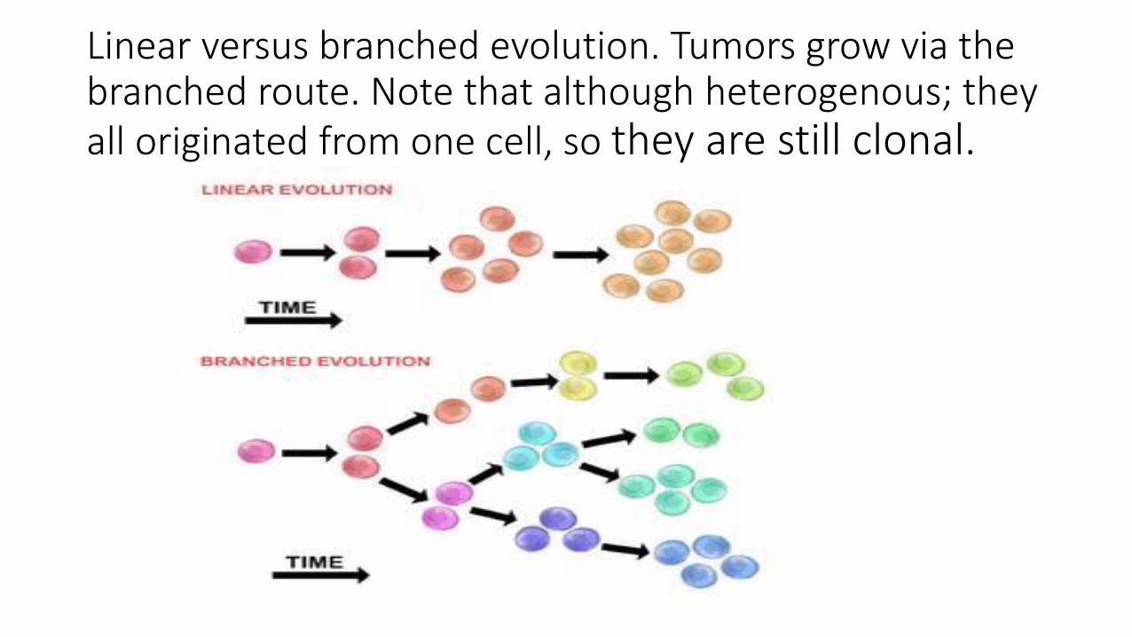
Linear versus branched evolution. Tumors grow via the branched route. Note that although heterogenous; they all originated from one cell, so they are still clonal.

Carcinogenesis is a multistep process

What are the genetic damages that can transform cells?• For a genetic damage to transform a cell, it has to cause uncontrolled
proliferation.
• The majority of our cells proliferate continuously. This proliferation is regulated by certain genes. There is a balance between genes that stimulate growth and those inhibiting it. Loosing this balance can cause uncontrolled proliferation.
• So : for cancer to occur there is stimulation of genes that cause cell proliferation, or downregulation of genes that inhibit proliferation.

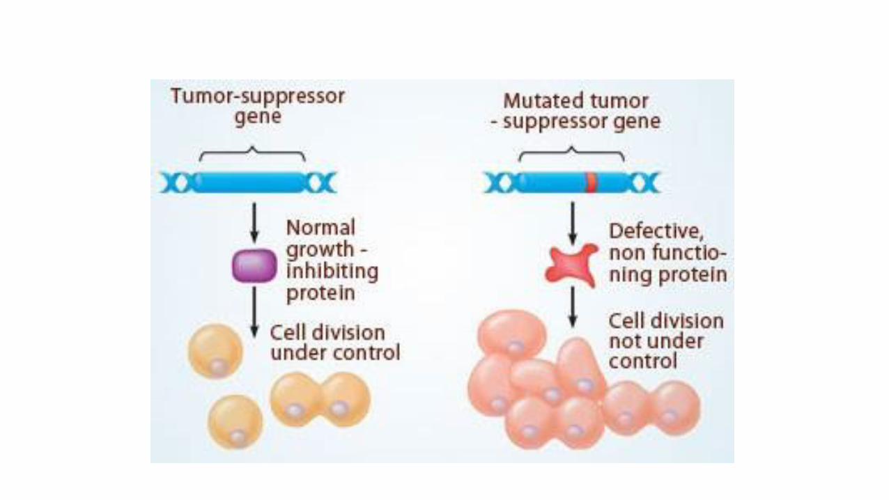
Cell cycle is regulated by a balance between growth stimulating genes = protooncogenes and growth inhibiting genes= tumor suppressor genes.
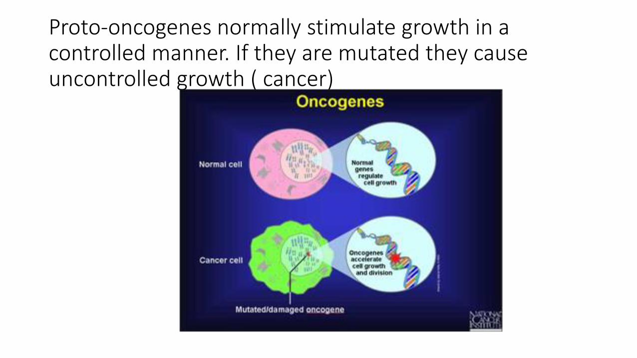
Proto-oncogenes normally stimulate growth in a controlled manner. If they are mutated they cause uncontrolled growth ( cancer)

Tumor suppressor genes counteract the function of the oncogenes. If they are inhibited by a mutation then cells can proliferate without this “breaking” effect of the tumor suppressor genes.
Other genes involved in cancer
• Besides oncogenes and tumor suppressor genes, there are other types that are involved in transforming cells:
• - Genes that regulate apoptosis: these are very important because if the damaged cell dies by apoptosis then no proliferation is possible. So these genes are frequently mutated in cancers to keep cells alive and block apoptotic messages.
• - DNA repair genes also play a role in carcinogenesis. If DNA damages are repaired, then no cancer will occur. If DNA repair genes become nonfunctioning then there is a chance of DNA damages to accumulate in cells.
• Recently, genes that affect the interaction between tumour cells and host cells ( surrounding normal cells) are thought to play a role in carcinogenesis.. Especially genes which affect immune response of the host to cancer cells.
Genetic damages in neoplasms
So: five types of regulatory genes are mainly affected:
• 1. growth promoting proto-oncogenes
• 2. growth inhibiting tumor suppressor genes
• 3. genes that regulate apoptosis
• 4. genes involved in DNA repair.
• 5.genes that regulate interactions between tumor cells and host cells . Particularly important are genes that enhance or inhibit recognition of tumors cells by the host immune system.

note
• Normal genes that cause cell proliferation are traditionally called: proto-oncogenes.
• When they are mutated, they are called oncogenes.

oncogenes
• Normally: our cells have proto-oncogenes. These cause cell proliferation in a regulated manner
• If the proto-oncogenes are mutated or overexpressed: they are called oncogenes
• Proto-oncogenes encode for proteins: proto-oncoproteins, or oncoproteins
• These oncoproteins include: transcription factors, growth regulating proteins, proteins involved in cell survival.
oncogenes
• Oncogenes cause overexpression of proteins involved in cell growth.
• If one allele is mutated or overexpressed: there will be increase in the growth proteins, which is enough to increase cell growth
• So oncogenes are dominant .
• Important oncogenes : RAS and ABL
How oncogenes overexpressed??
• 1. point mutation resulting in activation
• 2. amplification : increased number of copies of the oncogenes
• 3. translocations
• 4.Epigenetic modification
• Details will follow . Don’t worry

Tumor suppressor genes
• They normally inhibit cell growth
• If mutated or lost: loss of growth inhibition : so tumor
• Both alleles need to be lost or mutated for the tumors to develop…. Because if only one allele is lost , the other can compensate!
• So they are recessive genes
• in some cases loss of one allele is enough for transformation… haploinsufficiency ( see next slide for definition)
Haploinsufficiency
• Haploinsufficiency: A situation in which the total level of a gene product (a particular protein) produced by the cell is about half of the normal level and that is not sufficient to permit the cell to function normally.
• Another way to define haploinsufficiency is as a condition that arises when the normal phenotype requires the protein product of both alleles, and reduction of 50% of gene function results in an abnormal phenotype
Tumor suppressor genes
• Most important examples:
• 1. RB gene(retinoblastoma gene) .. Governor: controls growth and puts a brake in cellular proliferation
• 2. TP53 gene … guardian of the genome… sense genetic damage. So if there is damage it causes cessation of proliferation or if the damage cannot be repaired it causes apoptosis.
TP53
• Mutations in TP53 do not directly transform cells. They permit and accelerate acquisition of mutations in oncogenes or tumor suppressor genes.
• Same rule applies for mutations in genes that are responsible for DNA repair
• Ability of a cell to accumulate mutations = mutator phenotype
note
• Genes that regulate apoptosis or DNA repair may be dominant or recessive.
Genetic lesions in cancer
• We now know the types of genes that should be damaged for cancer to occur. But how they are damaged?
• They can be damaged by Mutational or non-mutational damages.
• Mutations: 1.subtle: point mutations, insertions, point deletions
:or 2. large, karyotypic change: translocations, large deletions, gene amplification, aneuploidy
• Non mutational: MicroRNAs and epigenetic modifications

Genetic lesions in cancer
Point mutations
• These are single changes in nucleotides
• Point mutations that stimulate an oncogene or inhibit both alleles of a tumor suppressor gene can result in cancer.

Balanced translocations
• Translocations can cause cancer if they increase expression of a proto-oncogene.
• This can happen by two mechanisms:
• 1. removing the proto-oncogene from its normal, regulated locus to a new position where it becomes under influence of a highly active promoter.
• 2. translocation forms a new fusion gene that encodes a novel protein.
translocations
• Occur mainly in haematogenous neoplasms ; why ??
• Because lymphoid cells make DNA breaks during antibody or T cell receptor recombination. ( loads of cutting and rearrangements of the genes… so there is more chance that a gene that was cut will be “pasted “ in a new locus!
translocations
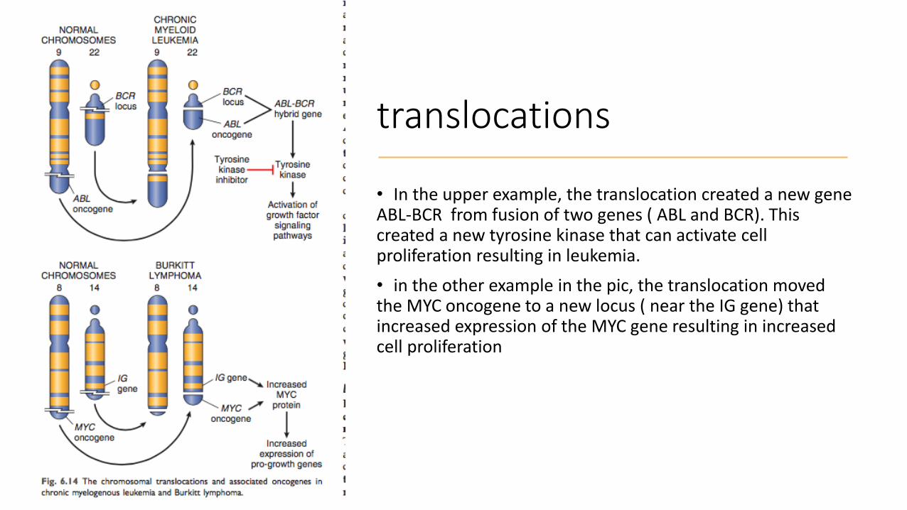
• In the upper example, the translocation created a new gene ABL-BCR from fusion of two genes ( ABL and BCR). This created a new tyrosine kinase that can activate cell proliferation resulting in leukemia.
• in the other example in the pic, the translocation moved the MYC oncogene to a new locus ( near the IG gene) that increased expression of the MYC gene resulting in increased cell proliferation
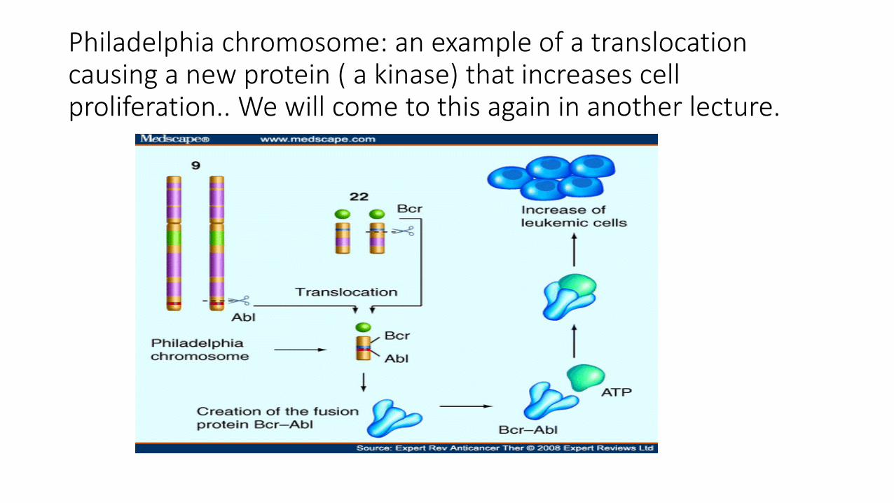
Philadelphia chromosome: an example of a translocation causing a new protein ( a kinase) that increases cell proliferation.. We will come to this again in another lecture.
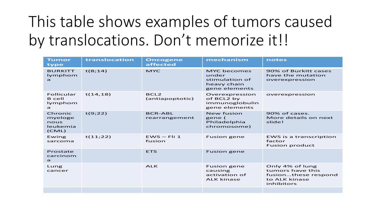
This table shows examples of tumors caused by translocations. Don’t memorize it!!
Tumortype
translocation Oncogeneaffected
mechanism notes
BURKITT lymphoma
t(8;14) MYC MYC becomes under stimulation of
heavy chain gene elements
90% of Burkitt cases have the mutationoverexpression
Follicular B cell lymphoma
t(14,18) BCL2 (antiapoptotic)
Overexpression of BCL2 by immunoglobulin gene elements
overexpression
Chronic myelogenous
leukemia (CML)
t(9;22) BCR-ABL rearrangement
New fusion gene ( Philadelphia
chromosome)
90% of cases.More details on next slide!
Ewing sarcoma
t(11;22) EWS – Fli 1 fusion
Fusion gene EWS is a transcriptionfactorFusion product
Prostate carcinoma
ETS Fusion gene
Lung cancer
ALK Fusion gene causingactivation of
ALK kinase
Only 4% of lung tumors have this fusion…these respond
to ALK kinase inhibitors

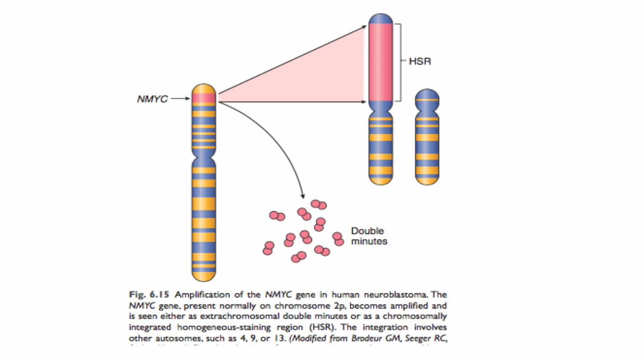
Gene amplifications
• Proto-oncogenes can be amplified and overexpressed .. Converted to oncogenes.
• This is seen in karyotyping as two patterns :1.homogenously stained region (HSR) = increased copies of the gene present within the chromosome
:2.Double minutes: extra copies of the gene separated from the chromosome.
deletions
• More in non-hematopoietic solid tumors
• Second most common karyotypic abnormality.
• Result in loss of tumor suppressor genes
• 2 copies of the tumor suppressor gene need to be lost, usually one by point mutation and another by deletion
aneuploidy
• = number of chromosomes not multiple of the haploid state (23).
• Result from errors of the mitotic checkpoint
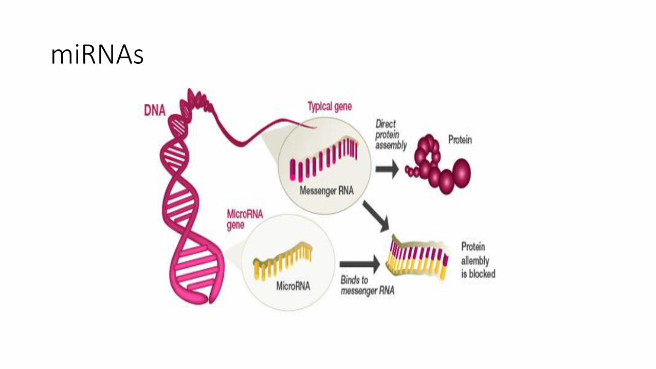
microRNAs (miRNAs)
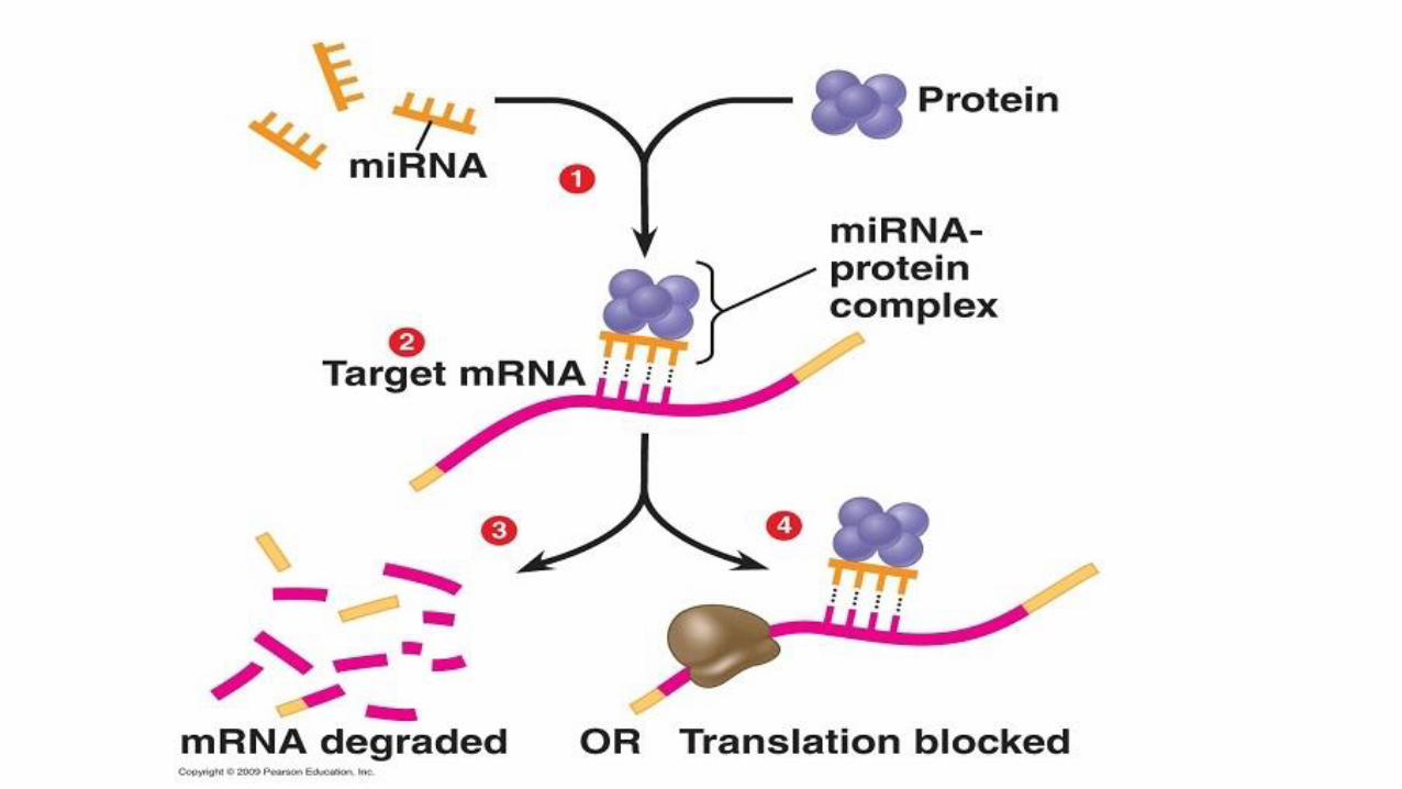
• Noncoding, micro RNA segments (22 nucleotides) that are negative regulators of the genes.
• They inhibit gene expression post-transcriptionally = repress translation or cleave mRNA.
• SO: transcription occurs = messenger RNA formed.. But mRNA is not translated to a protein.
• microRNA can inhibit translation or cleave the messenger ( tears the message before it is read)
miRNA
• Cause cancer by increasing oncogene expression or decreasing tumor suppressor gene expression.
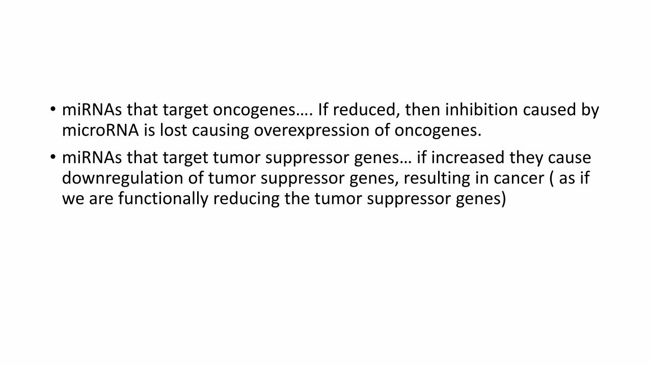
• miRNAs that target oncogenes…. If reduced, then inhibition caused by microRNA is lost causing overexpression of oncogenes.
• miRNAs that target tumor suppressor genes… if increased they cause downregulation of tumor suppressor genes, resulting in cancer ( as if we are functionally reducing the tumor suppressor genes)
miRNAs
epigenetics
• Epigenetics are reversible, heritable changes in gene expression that occur without mutation.
Epigenetic mutations
• functionally relevant changes to the genome that do not involve a change in the nucleotide sequence. Examples of mechanisms that produce such changes are DNA methylation and histone modification, each of which alters how genes are expressed without altering the underlying DNA sequence.
Epigenetic modifications
• Reversible , heritable changes in gene expression without mutation.
• Two types: Histone modifications and DNA methylation.

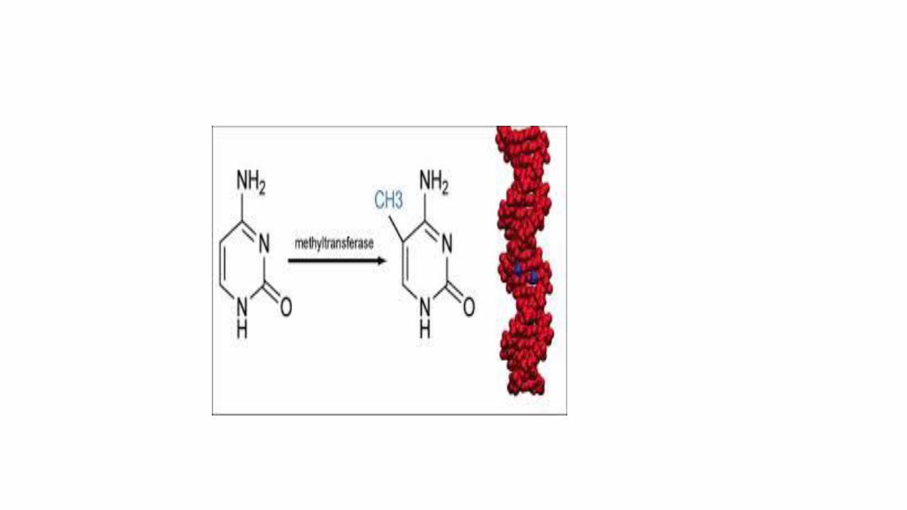
Epigenetics and cancer
• Gene expression is silenced by DNA methylation= more methyl groups lead to more silencing.
In cancer cells:
• 1.Global DNA hypo methylation : increases expression of genes. Also causes chromosomal instability
• 2.Selective promoter hyper methylation of tumor suppressor genes: silenced
Case study: Application of today’s lecture
• A 66 year old lady had a breast lump.
• A biopsy was taken and examined histologically.
• The biopsy reported as follows: the breast biopsy shows infiltration by glandular structures lined by epithelial cells showing large hyperchromatic nuclei.
• Question: Does this description indicate a benign or malignant tumor?
• Answer: definitely malignant.. The presence of infiltration and the anaplastic features described mean its malignant.
• Question: can you indicate the type of this malignancy from the description??
• Answer: the report describes glandular structures, so this is an adenocarcinoma.
• Note: adenocarcinoma in the breast is called invasive ductal carcinoma.
• Ok: the pathologist then mentions that they did a stain for EGFR( epidermal growth factor receptor) which is an epidermal growth receptor. And in this patient the EGFR was positive.
• What does this mean????
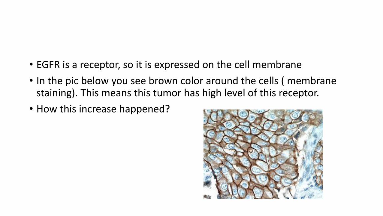
• EGFR is a receptor, so it is expressed on the cell membrane
• In the pic below you see brown color around the cells ( membrane staining). This means this tumor has high level of this receptor.
• How this increase happened?
• EGFR is a protein.. So in this tumor its production is increased.
• This increase was found to be due to amplification of the gene encoding this protein (HER2/neu)
• Patients with this mutation can be treated by a drug that targets and inhibits this gene which will decrease the EGFR production. This will deprive the tumor cells from the receptor which increases the proliferation.
• This is an example of why we need to know the genetic mutations in cancers.. We can develop specific treatments that target the mutation.

Summary 1/2
• Cancer starts as a clone from a transformed cell but acquires extra mutations during its progression forming several subclones each with a phenotype that aids tumor survival.
• Neoplasms are caused by nonlethal, genetic damage, which causes uncontrolled cellular proliferation
• Genetic alterations in 5 gene types can cause cancer: oncogenes that increase proliferation and are dominant, tumor suppressor genes that suppress proliferation and are recessive, genes that regulate apoptosis, DNA repair genes and genes regulating tumor-host response.
Summary 2/2
• Changes in those genes can be mutational ( point mutations, deletions, amplifications, translocations) or non mutational ( epigenetic changes or microRNA changes)
• microRNAs are posttranscriptional inhibitors of gene expression.
• Epigenetic changes are changes that do not affect the DNA sequence but change gene expression through stimulating or inhibiting gene promotors.