Embed Size (px)
Citation preview
In: Sudden Death ISBN: 978-1-62618-825-9
Editors: Jiashin Wu and Jessica Wu © 2013 Nova Science Publishers, Inc.
Chapter VIII
Neoplasm-related Sudden Death:
Causes and Clinicopathological
Characteristics
Katsuya Chinen * Department of Pathology, Nerima General Hospital,
Nerima, Tokyo, Japan
Abstract
Patients with neoplastic diseases, especially malignancies, are at greatly elevated risk
of sudden death, because they may suffer from a variety of neoplasm-, and/or treatment-
related clinical conditions including myelosuppression, immunosuppression, coagulation
abnormalities, metabolic disorders, and malnutrition. Cancer patients die suddenly of
miscellaneous etiologies, and about one third of sudden death in cancer patients is
attributed to neoplasm-related complications.
In the literature, the majority of neoplasm-related sudden deaths (NSDs) are caused
by neoplasms affecting critical organs such as the heart and brain. In addition, neoplasms
damaging respiratory system or causing acute blood loss are responsible for sudden
death. Mechanisms are miscellaneous and most are directly related to the anatomical
location of the neoplasm. On the other hand, regardless of the site, specific types of
neoplasm (e.g. hormone-producing tumors) sometimes cause sudden death by
mechanisms which a pathophysiological function peculiar to the tumor induces critical
manifestations of vital organs such as the heart.
As mentioned above, causes of NSD is classified as follows; (1) cardiac tumors, (2)
brain tumors, (3) pulmonary tumor embolism, (4) asphyxia, (5) massive exsanguinations,
and (6) sudden death due to neoplasm-specific pathophysiology. The latter includes
amyloidosis, hyperviscosity syndrome, and hormone-induced complications, and these
diseases can present with a variety of lethal clinical manifestations. In this chapter,
* Katsuya Chinen, MD, PhD, Department of Pathology, Nerima General Hospital, 1-24-1 Asahigaoka, Nerima,
Tokyo 176-8530 Japan; Tel: 81-3-5988-2200; Fax: 81-3-5988-2250; E-mail; [email protected].
No part of this digital document may be reproduced, stored in a retrieval system or transmitted commercially in any form or by any means. The publisher has taken reasonable care in the preparation of this digital document, but makes no expressed or implied warranty of any kind and assumes no responsibility for any errors or omissions. No liability is assumed for incidental or consequential damages in connection with or arising out of information contained herein. This digital document is sold with the clear understanding that the publisher is not engaged in rendering legal, medical or any other professional services.

Katsuya Chinen 152
clinical and pathological characteristics of NSDs are described based on this
classification with a review of the literature.
Introduction
Patients with neoplastic diseases, especially malignancies, are at greatly elevated risk of
sudden death, because they may suffer from a variety of neoplasm-, and/or treatment-related
clinical conditions including myelosuppression, immunosuppression, coagulation
abnormalities, metabolic disorders, and malnutrition. Cancer patients die suddenly of
miscellaneous etiologies, and about one third of sudden death in cancer patients is attributed
to neoplasm-related complications [1].
The cardiovascular and central nervous systems are crucial for life, and any damage
affecting them can be life-threatening. In the literature, the majority of neoplasm-related
sudden deaths (NSDs) are caused by neoplasms affecting critical organs such as the heart and
brain. In addition, neoplasms damaging respiratory system or causing acute blood loss are
responsible for sudden death. Mechanisms are miscellaneous and most are directly related to
the anatomical location of the neoplasm. On the other hand, regardless of the site, specific
types of neoplasm (e.g. hormone-producing tumors) sometimes cause sudden death by
mechanisms which a pathophysiological function peculiar to the tumor induces critical
manifestations of vital organs such as the heart.
An etiological classification of NSD is presented in Table 1. In this chapter, clinical and
pathological characteristics of NSDs are described following this classification with a review
of the literature.
Table 1. Etiological Classification of Neoplasm-Related Sudden Death
1) Cardiac tumors
2) Brain tumors
3) Pulmonary tumor embolism
4) Asphyxia
5) Massive exsanguination
6) Sudden death due to neoplasm-specific pathophysiology
a) Amyloidosis
b) Hyperviscosity syndrome
c) Hormone-induced complications
1. Cardiac Tumors
Cardiac tumors, regardless of their nature (i.e., primary or metastatic, benign or
malignant) constitute risks for sudden death by themselves. The pathophysiology is
Neoplasm-related Sudden Death 153
miscellaneous, including cardiac ischemia, arrhythmia, pump failure, outflow obstruction,
valvular dysfunction, and cardiac tamponade.
1.1. Primary Cardiac Tumors
In the literature, there have been a large number of cases of sudden death attributed to
primary cardiac neoplasms. Cina et al. [2], in their review, indicated that in most cases the
tumors are histologically benign but their intracardiac location precipitates conductive and
hemodynamic abnormalities.
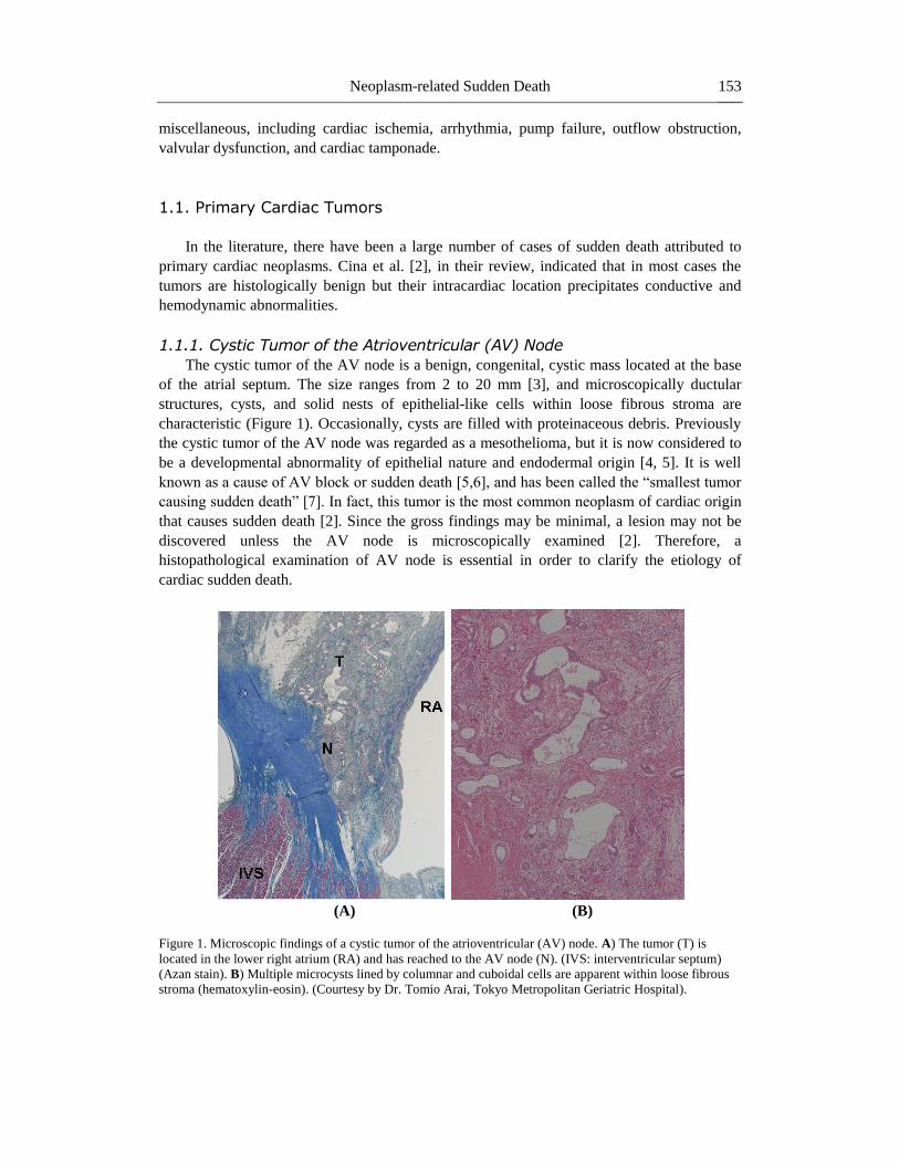
1.1.1. Cystic Tumor of the Atrioventricular (AV) Node
The cystic tumor of the AV node is a benign, congenital, cystic mass located at the base
of the atrial septum. The size ranges from 2 to 20 mm [3], and microscopically ductular
structures, cysts, and solid nests of epithelial-like cells within loose fibrous stroma are
characteristic (Figure 1). Occasionally, cysts are filled with proteinaceous debris. Previously
the cystic tumor of the AV node was regarded as a mesothelioma, but it is now considered to
be a developmental abnormality of epithelial nature and endodermal origin [4, 5]. It is well
known as a cause of AV block or sudden death [5,6], and has been called the “smallest tumor
causing sudden death” [7]. In fact, this tumor is the most common neoplasm of cardiac origin
that causes sudden death [2]. Since the gross findings may be minimal, a lesion may not be
discovered unless the AV node is microscopically examined [2]. Therefore, a
histopathological examination of AV node is essential in order to clarify the etiology of
cardiac sudden death.
(A) (B)
Figure 1. Microscopic findings of a cystic tumor of the atrioventricular (AV) node. A) The tumor (T) is
located in the lower right atrium (RA) and has reached to the AV node (N). (IVS: interventricular septum)
(Azan stain). B) Multiple microcysts lined by columnar and cuboidal cells are apparent within loose fibrous
stroma (hematoxylin-eosin). (Courtesy by Dr. Tomio Arai, Tokyo Metropolitan Geriatric Hospital).

Katsuya Chinen 154
1.1.2. Fibroma
Fibroma, the most commonly resected cardiac tumor of children, constitutes the second
most frequent causing sudden death [2]. The cardiac sites of fibromas are, in order of
decreasing frequency, the interventricular septum, left ventricular free wall, right ventricle,
and atria [3].
The location has been noted as a determining factor in the presentation and outcome of
affected patients. Fibromas arising in the interventricular septum are inoperable, but those of
the ventricular free wall may be removed surgically.
It seems that sudden death occurs due to compression of the conducting system (bundle
of His) or triggering of ventricular fibrillation. In one study [8], more than half of the cases
presenting with sudden death had tumors in the interventricular septum, where compression
of the conduction system is more likely.
1.1.3. Myxoma
Among primary cardiac tumors, myxoma is the most common in adults and is the third
most frequent neoplasm causing sudden death [2].
It usually develops in the atria; about 75% originate in the left atrium, and 15 to 20 % in
the right atrium.
If the tumor is sufficiently large, soft, and easily deformable, and if it has a long stalk,
obstruction of the orifice of the mitral or tricuspid valve may occur [9]. Sudden death may be
the result of either acute disturbance in cardiac hemodynamics or systemic embolization by
tumor fragments.
The lethal potential of this tumor can be attributed to both its location (usually in the left
atrium) and its configuration [2].
1.1.4. Rhabdomyoma
Rhabdomyoma is the most common primary cardiac tumor in children. It may occur as
solitary, multiple or diffuse lesions of the myocardium, and is considered to be a
malformation tumor (hamartoma), rather than a true neoplasm [10]. Sudden cardiac death
may result from rhythm disturbance, outflow obstruction and valvular distortion [11]. Cardiac
rhabdomyoma is also frequently associated with tuberous sclerosis, an autosomal dominant
inherited disease.
If a cardiac rhabdomyoma is identified in patients presenting with sudden death, other
lesions characteristic of this syndrome such as adrenal angiomyolipoma and subependymal
giant cell astrocytoma, may further be present and mortality may in fact be due to
complications of tuberous sclerosis [2, 11-13].
1.1.5. Papillary Fibroelastoma
The papillary fibroelastoma is a benign endocardial papilloma predominantly affecting
the cardiac valves, especially the aortic valve (Figure 2), where it has been implicated in
sudden death because of obstruction of the ostium of the right or left coronary artery. In one
review of reported cases of papillary fibroelastoma [14], sudden death was found in 21 of 725
patients (2.9%).

Neoplasm-related Sudden Death 155
(A) (B)
Figure 2. Papillary fibroelastoma of the aortic valve. A) Macroscopically, an expophytic tumor with thin
stalk, measuring 5 mm in diameter, is apparent on the right coronary cusp of the aortic valve (arrow). B)
Microscopic examination shows a papillary endocardial tumor with hyalinized central core. Fibrin clot and
bacterial colonization are also evident. (hematoxylin-eosin, original magnification x40).
1.1.6. Hemangioma
Hemangiomas are proliferations of endothelial cells forming vascular channels of
variable size. The cardiac hemangioma occurs in patients of all ages and there is a male
predominance [15]. It appears most lethal when disrupting the conducting system or
involving the atrioventricular node. Very rarely, this tumor can induce cardiac tamponade
resulting in sudden death [16, 17].
1.1.7. Angiosarcoma
Angiosarcoma is the most frequent primary malignant cardiac neoplasm and most
commonly arises in the right atrium [18]. Extension into the pericardial sac or atrial chamber
often leads to hemopericardium, cardiac tamponade, or obstruction of blood flow.
Widespread metastasis (especially pulmonary) is common and may be related to causes of
death [19]. However, the number of patients with cardiac angiosarcoma presenting with
sudden death is limited [2, 20, 21].
1.1.8. Miscellaneous Primary Cardiac Tumors
Many other kinds of primary cardiac tumors have been reported as causes of sudden
death, although their incidences are very low. They include the rhabdomyosarcoma,
malignant lymphoma, undifferentiated sarcoma, fibrosarcoma, malignant nerve sheath tumor,
teratoma, lipoma, angiomyoma, cardiac inflammatory myofibroblastic tumor, low-grade
myofibroblastic sarcoma, and coronary artery intimal sarcoma [2, 22-25].
1.2. Metastatic Cardiac Tumors
Metastatic tumors in the heart are far more frequent than primary neoplasms, by at least a
30 to 1 ratio [26]. The most common lesions with cardiac metastatic potential are carcinomas
of the lung, breast, esophagus and thyroid gland, leukemia and malignant lymphomas [27].
Metastatic cardiac tumors also cause sudden death due to impaired cardiac function, including

Katsuya Chinen 156
cardiac tamponade, conduction disturbance and outflow obstruction [28-30]. In the literature,
metastatic cardiac tumors responsible for sudden death have been derived from a variety of
organs, such as the lung [30], esophagus [31], thyroid gland [32], pancreas [33] and colon
[28], as well as soft tissue [34]. T-cell lymphomas, when compared with B-cell lymphomas,
metastasize more frequently and are more likely to give rise to cardiac manifestations,
including sudden death (Figure 3) [35].
Figure 3. An example of cardiac involvement by malignant lymphoma in a 74 year-old man presenting with
sudden death. Note diffuse interstitial infiltration of lymphoma cells (hematoxylin-eosin, original
magnification x200).
2. Brain Tumors
Brain tumors, like cardiac tumors, are etiological factors for NSD. Although the actual
proportion of sudden deaths from this cause is very low (0.02% to 2.1%) [36], many
anecdotal cases have been reported. Any kind of brain tumors, regardless of their nature (i.e.
primary or metastatic, benign or malignant), can cause sudden death. In the literature,
however, the astrocytoma-glioblastoma category has predominated [37]. Other examples
include oligodendroglioma, medulloblastoma, subependymoma, microglioma, meningioma,
colloid cyst, lymphoma, teratoma, hemangiopericytoma and metastatic brain tumors [37-45].
Of the metastatic intracranial tumors, bronchial carcinoma, choriocarcinoma and melanoma
are the most common [46].
Sudden death in patients with brain tumors is caused mainly by rapid functional
deterioration of a vital focus, such as the brainstem, which controls circulatory or respiratory
functions. Mechanisms of sudden death have been explained as detailed below.
2.1. Raised Intracranial Pressure
The most common mechanism is raised intracranial pressure as a result of an intracranial
expanding (space-occupying) lesion. Hemorrhage from a tumor often causes rapid increase of
the tumor mass. Raised intracranial pressure finally causes cerebral herniation and brainstem
compression with a fatal outcome.

Neoplasm-related Sudden Death 157
2.2. Acute Obstructive Hydrocephalus
Tumors that can obstruct cerebrospinal fluid flow are responsible for acute
hydrocephalus. It is the site of tumor, rather than its nature, that is of importance. If there is a
small tumor in a crucial site adjacent to a ventricular foramen, then it is of much greater
importance than a large expanding tumor in a frontal or occipital lobe. For example, tumors
of the third ventricle can cause acute obstructive hydrocephalus by blocking the foramen of
Munro or the posterior third ventricle and cerebral aqueduct. Primary third-ventricle tumors
include colloid cysts (the most frequent type), astrocytomas, ependymomas, and choroid
plexus papillomas, and these tumors have been reported as causes of sudden death [47].
Leptomeningeal seeding of tumor cells, such as lymphoma cells, may also be responsible for
acute obstructive hydrocephalus [48]. Most patients with acute hydrocephalus are not
diagnosed antemortem as having brain tumors, because clinical manifestations of acute
hydrocephalus, including vomiting, headache and lethargy, often lead to a misdiagnosis as a
viral illness such as gastroenteritis [47].
2.3. Neoplastic Involvement of the Respiratory and/or Cardiac Centers
In infratentorial tumors, direct involvement with the respiratory and/or cardiac centers in
the brainstem can cause sudden death. In the literature, oligodendroglioma [42,43],
ganglioglioma [49,50], and mycosis fungoides [51] have been reported as responsible lesions.
Mechanical compression, without direct invasion, caused by tumors neighboring the
brainstem may be involved [39].
2.4. Epilepsy
The incidence of epileptic seizures for all intracranial tumors is about 20-50%, but
sudden death caused by epilepsy is rare [52]. In one review article [52], tumors causing fatal
epilepsy were miscellaneous, including low-grade astrocytoma, anaplastic astrocytoma,
oligodendroglioma, meningioma, subependymal giant cell astrocytoma, anaplastic
oligodendroglioma, gangliocytoma, ependymoma, subependymoma, and teratoma. The most
common localization was the frontal lobe and the thalamus. It is interesting that there was no
tumor solely in the parietal lobe, which is reported to be the most common location for
tumors causing seizures. The mechanism of epilepsy-induced sudden death remains
controversial. The most widely accepted theory proposes that seizure activity results in
autonomic nervous system discharge, which ultimately triggers lethal cardiac arrhythmias or
respiratory arrest [12, 52].
2.5. Neurogenic Pulmonary Edema
Some patients with brain tumor die suddenly of neurogenic pulmonary edema [45]. Not
only brain tumors but also subarachnoid and intraventricular hemorrhage may be responsible.
Katsuya Chinen 158
The exact pathophysiology is unclear, but it may feature an adrenergic response induced by
cerebral insult, which leads to increased pulmonary hydrostatic pressure and lung capillary
permeability. It is known that an increase of cerebrospinal fluid pressure, infusion of
thrombin and fibrinogen into the cistern magna, and intravenous or intrathecal infusion of
massive amounts of catecholamines can cause neurogenic pulmonary edema [45].
3. Pulmonary Tumor Embolism
Autopsy studies estimate that the incidence of pulmonary tumor embolism (PTE) is
between 3% and 26% among patients with solid tumors. Primary tumors for PTE have been
reported to be, in order of decreasing frequency, carcinomas of the breast, stomach, lung,
liver, prostate, and pancreas [53]. Pulmonary hypertension due to PTE is considered a major
contributory cause of death, which may occur in as much as 8% of cases [54]. Although
comprehensive studies are still lacking, many anecdotal case reports dealing with PTE-
induced sudden death have been published. Sudden deaths are caused by tumors that have
high angioinvasive potential, such as renal cell carcinomas (Figure 4) [1], hepatocellular
carcinomas [55], testicular germ cell tumors [56], and Wilms tumors [57,58]. Primary tumors
causing PTE are often undiagnosed before sudden death, which is the first clinical
presentation in many cases. Tumors originating from the vessel wall of large veins or
pulmonary artery can also obstruct the pulmonary arterial system and cause mortality, for
example with leiomyosarcoma of the pulmonary artery [59] and intravenous leiomyoma [60].
Strictly speaking, when tumors in the pulmonary artery are contiguous with the original
tumor site, they are not “PTE” because PTE is defined as “the presence of isolated cells or
clusters of tumor cells within the pulmonary arterial system, which are not contiguous with
primary or metastatic foci” [61]. However, because these types of tumors can cause sudden
death by the same mechanism as true “PTE”, it may be reasonable to describe here as
“sudden death due to tumorous obstruction of the pulmonary circulation system.”
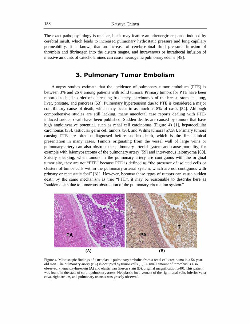
(A) (B)
Figure 4. Microscopic findings of a neoplastic pulmonary embolus from a renal cell carcinoma in a 54-year-
old man. The pulmonary artery (PA) is occupied by tumor cells (T). A small amount of thrombus is also
observed. (hematoxylin-eosin (A) and elastic van Gieson stain (B), original magnification x40). This patient
was found in the state of cardiopulmonary arrest. Neoplastic involvement of the right renal vein, inferior vena
cava, right atrium, and pulmonary truncus was grossly observed.
Neoplasm-related Sudden Death 159
3.1. Pulmonary Tumor Thrombotic Microangiopathy
Recently, a distinct form of PTE, designated as “pulmonary tumor thrombotic
microangiopathy (PTTM)”, has become recognized as a cause of sudden death.
Clinicopathological characteristics of PTTM are quite different from those of
conventional PTE in many points, therefore it seems better to document PTTM as a single
disease entity. PTTM is in fact a rare clinicopathologic condition that causes severe clinical
manifestations including pulmonary hypertension, right-sided heart failure and sudden death.
It is characterized by wide spread tumor emboli in the small arteries and arterioles of the lung,
associated with thrombus formation and fibrocellular and fibromuscular intimal proliferation
[62]. It differs from conventional PTE in that metastatic tumors do not cause simple
mechanical obstruction of the pulmonary vasculature but rather stimulate vessel reactions
(Figure 5). Apparent clinicopathologic features [63,64] include:
1) adenocarcinomas as the predominant histology, especially of poorly differentiated
type including the signet-ring cell carcinoma;
2) the stomach as the most frequent primary site, PTTM often being due to clinically
undetected, occult gastric carcinoma;
3) no age dependence, both the old and young being affected; and
4) a difficult antemortem diagnosis with frequent misdiagnosis as primary pulmonary
hypertension.
In cases of sudden death with unexplained pulmonary hypertension or cor pulmonale, the
possibility of PTTM should be considered and special attention should be paid in postmortem
examination. Since gross findings of the lung are often unremarkable, microscopic
examination of the lung is required for the diagnosis. Although neoplasms of various organs
can cause PTTM [65], the most frequent neoplasm is, as mentioned above, the gastric
carcinoma [62]. Therefore, in cases of PTTM with obscure primary neoplasm, the stomach
should be examined carefully. Many serial step sections of the stomach may be necessary for
detection of the primary lesion because PTTM is sometimes caused by a tiny early cancer
limited to the mucosal or submucosal layer [66, 67].
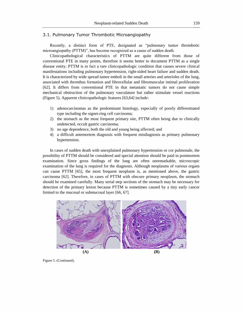
(A) (B)
Figure 5. (Continued).
Katsuya Chinen 160
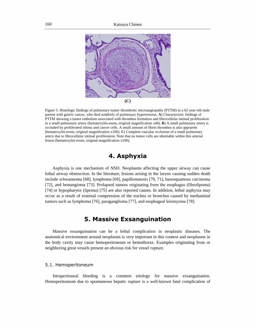
(C)
Figure 5. Histologic findings of pulmonary tumor thrombotic microangiopathy (PTTM) in a 62 year-old male
patient with gastric cancer, who died suddenly of pulmonary hypertension. A) Characteristic findings of
PTTM showing a tumor embolism associated with thrombus formation and fibrocellular intimal proliferation
in a small pulmonary artery (hematoxylin-eosin, original magnification x40). B) A small pulmonary artery is
occluded by proliferated intima and cancer cells. A small amount of fibrin thrombus is also apprarent
(hematoxylin-eosin, original magnification x100). C) Complete vascular occlusion of a small pulmonary
artery due to fibrocellular intimal proliferation. Note that no tumor cells are identiable within this arterial
lesion (hematoxylin-eosin, original magnification x100).
4. Asphyxia
Asphyxia is one mechanism of NSD. Neoplasms affecting the upper airway can cause
lethal airway obstruction. In the literature, lesions arising in the larynx causing sudden death
include schwannoma [68], lymphoma [69], papillomatosis [70, 71], basosquamous carcinoma
[72], and hemangioma [73]. Prolapsed tumors originating from the esophagus (fibrolipoma)
[74] or hypopharynx (lipoma) [75] are also reported causes. In addition, lethal asphyxia may
occur as a result of external compression of the trachea or bronchus caused by mediastinal
tumors such as lymphoma [76], paraganglioma [77], and esophageal leiomyoma [78].
5. Massive Exsanguination
Massive exsanguination can be a lethal complication in neoplastic diseases. The
anatomical environment around neoplasms is very important in this context and neoplasms in
the body cavity may cause hemoperitoneum or hemothorax. Examples originating from or
neighboring great vessels present an obvious risk for vessel rupture.
5.1. Hemoperitoneum
Intraperitoneal bleeding is a common etiology for massive exsanguination.
Hemoperitoneum due to spontaneous hepatic rupture is a well-known fatal complication of
Neoplasm-related Sudden Death 161
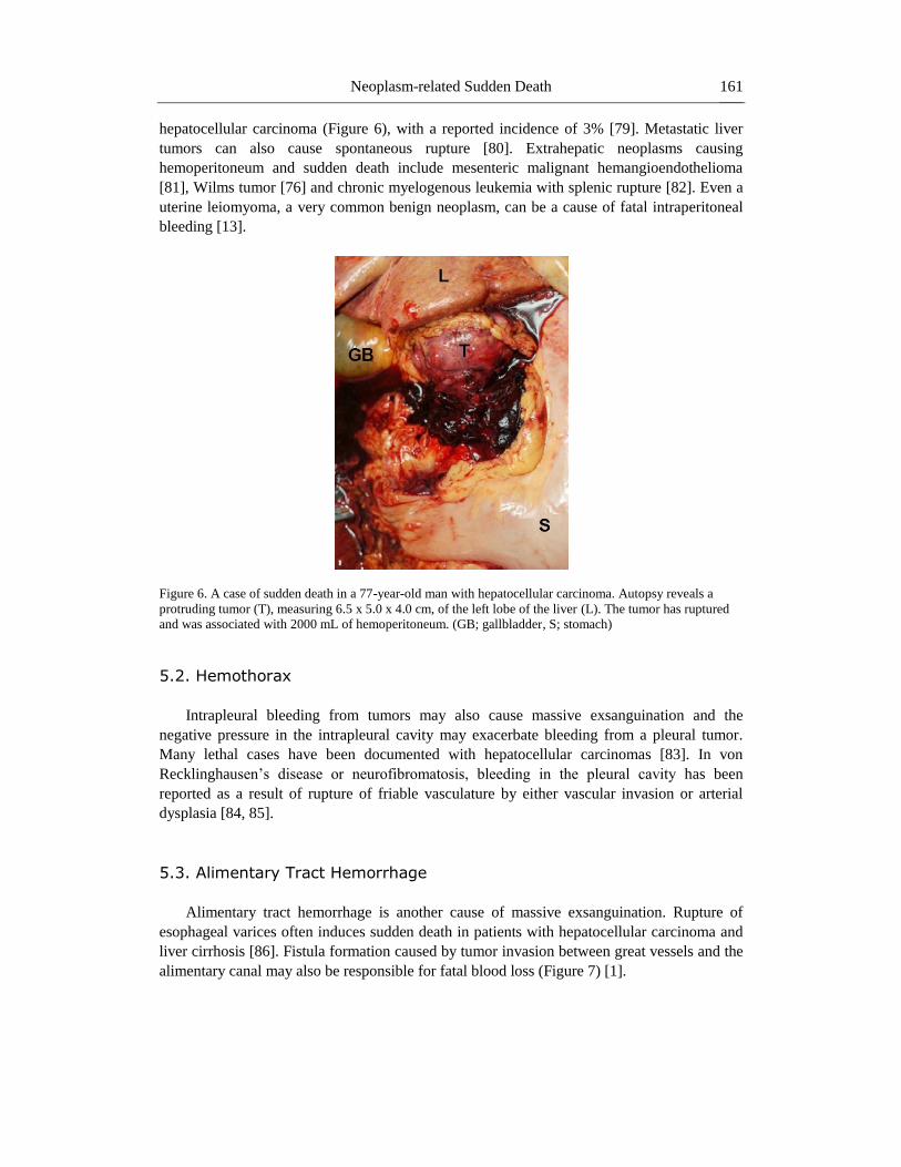
hepatocellular carcinoma (Figure 6), with a reported incidence of 3% [79]. Metastatic liver
tumors can also cause spontaneous rupture [80]. Extrahepatic neoplasms causing
hemoperitoneum and sudden death include mesenteric malignant hemangioendothelioma
[81], Wilms tumor [76] and chronic myelogenous leukemia with splenic rupture [82]. Even a
uterine leiomyoma, a very common benign neoplasm, can be a cause of fatal intraperitoneal
bleeding [13].
Figure 6. A case of sudden death in a 77-year-old man with hepatocellular carcinoma. Autopsy reveals a
protruding tumor (T), measuring 6.5 x 5.0 x 4.0 cm, of the left lobe of the liver (L). The tumor has ruptured
and was associated with 2000 mL of hemoperitoneum. (GB; gallbladder, S; stomach)
5.2. Hemothorax
Intrapleural bleeding from tumors may also cause massive exsanguination and the
negative pressure in the intrapleural cavity may exacerbate bleeding from a pleural tumor.
Many lethal cases have been documented with hepatocellular carcinomas [83]. In von
Recklinghausen’s disease or neurofibromatosis, bleeding in the pleural cavity has been
reported as a result of rupture of friable vasculature by either vascular invasion or arterial
dysplasia [84, 85].
5.3. Alimentary Tract Hemorrhage
Alimentary tract hemorrhage is another cause of massive exsanguination. Rupture of
esophageal varices often induces sudden death in patients with hepatocellular carcinoma and
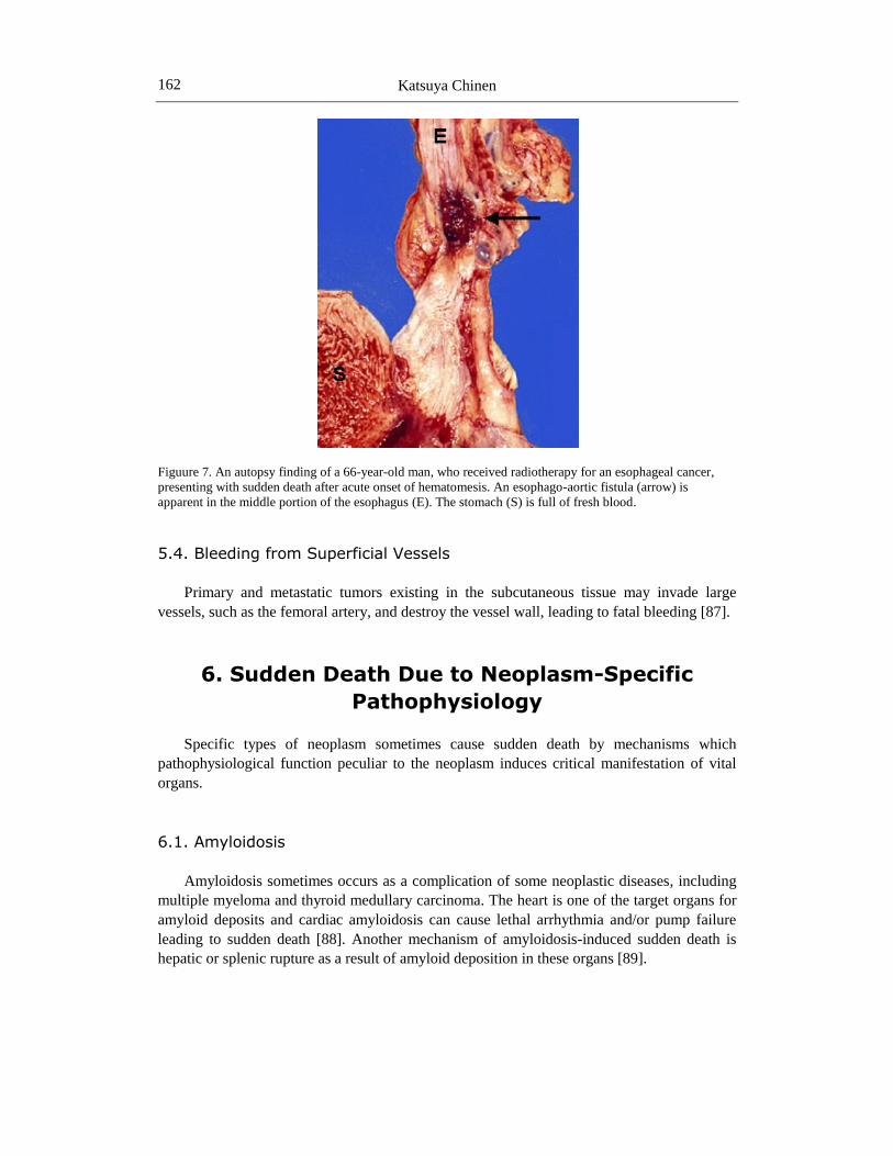
liver cirrhosis [86]. Fistula formation caused by tumor invasion between great vessels and the
alimentary canal may also be responsible for fatal blood loss (Figure 7) [1].
Katsuya Chinen 162
Figuure 7. An autopsy finding of a 66-year-old man, who received radiotherapy for an esophageal cancer,
presenting with sudden death after acute onset of hematomesis. An esophago-aortic fistula (arrow) is
apparent in the middle portion of the esophagus (E). The stomach (S) is full of fresh blood.
5.4. Bleeding from Superficial Vessels
Primary and metastatic tumors existing in the subcutaneous tissue may invade large
vessels, such as the femoral artery, and destroy the vessel wall, leading to fatal bleeding [87].
6. Sudden Death Due to Neoplasm-Specific
Pathophysiology
Specific types of neoplasm sometimes cause sudden death by mechanisms which
pathophysiological function peculiar to the neoplasm induces critical manifestation of vital
organs.
6.1. Amyloidosis
Amyloidosis sometimes occurs as a complication of some neoplastic diseases, including
multiple myeloma and thyroid medullary carcinoma. The heart is one of the target organs for
amyloid deposits and cardiac amyloidosis can cause lethal arrhythmia and/or pump failure
leading to sudden death [88]. Another mechanism of amyloidosis-induced sudden death is
hepatic or splenic rupture as a result of amyloid deposition in these organs [89].
Neoplasm-related Sudden Death 163
6.2. Hyperviscosity Syndrome
The hyperviscosity syndrome is one of the lethal complications in patients with
hematological malignancies. Embolic events due to hyperviscosity have been documented as
a cause of sudden death in multiple myeloma [90] and leukemia [76].
6.3. Hormone-Induced Complications
6.3.1. Hypercalcemia
Hypercalcemia is one critical complication in cancer patients. Metastatic bone tumors
destroy bony structures and induce hypercalcemia. In addition, over-production of
parathyroid hormone (PTH) or PTH-related peptide (PTH-rP) by specific types of neoplasm
can cause a lethal clinical condition as a result of hypercalcemia. Adult T-cell
leukemia/lymphoma (ATLL) is a well-known malignancy inducing lethal hypercalcemia.
Metastatic calcification of the heart is reported as a cause of sudden death in patients with
ATLL [91] and parathyroid adenoma [92].
6.3.2. Hypokalemia
Hypokalemia may cause severe clinical symptoms such as cardiac arrhythmia and
respiratory failure due to muscle weakness. Hypokalemia and consequent lethal arrhythmia
are documented to constitute a fatal pathophysiology in patients with primary aldosteronism
[93] and Cushing syndrome [94].
6.3.3. Pheochromocytoma
Pheochromocytoma can cause miscellaneous severe clinical symptoms. In addition to
critical arterial hypertension, severe cardiac arrhythmia must be recognized as a life-
threatening associated complication [95-97]. Catecholamine-induced cardiomyopathy due to
pheochromocytoma may also elevate the risk of cardiac sudden death [98].
6.3.4. Insulinoma
Hypoglycemia is itself a lethal clinical condition, and increased sympathetic nerve
activity followed by hypoglycemia can be responsible for lethal cardiac arrhythmia. In the
literature, sudden death due to hypoglycemia-induced cardiac arrhythmia has been reported in
a patient with a duodenal insulinoma [99].
References
[1] Chinen, K., Kurosumi, M., Ohkura, Y., et al (2006) Sudden unexpected death in
patients with malignancy: A clinicopathologic study of 28 autopsy cases. Pathol Res
Pract 202:867-875.
[2] Cina, S.J., Smialek, J.E., Burke, A.P., et al (1996) Primary cardiac tumors causing
sudden death: a review of the literature. Am J Forensic Med Pathol 17:271-281.
Katsuya Chinen 164
[3] Burke, A. & Virmani, R. Tumors of the heart and the great vessels. Atlas of Tumor
Pathology, Third series, Fascicle 16. Washington DC, Armed Forces Institute of
Pathology, 1996.
[4] Duray, P.H., Mark, E.J., Barwick, K.W., et al (1985) Congenital polycystic tumor of the
atrioventricular node: autopsy study with immunohistochemical findings suggesting
endodermal derivation. Arch Pathol Lab Med 109:30-34.
[5] Burke, M.A,P., Anderson, P.G., Vermani, R., et al (1990) Tumor of the atrioventricular
nodal region: a clinical and immunohistochemical study. Arch Pathol Lab Med
114:1057-1062.
[6] Law, K.B., Feng, T., Nair, V., et al (2012) Cystic tumor of the atrioventricular node:
rare antemortem diagnosis. Cardiovasc Pathol 21: 120-127.
[7] Wolf, P.L. & Bing, R. (1965) The smallest tumor which causes sudden death. JAMA
194:204-205.
[8] Amr, S.S., Al-Ragheb, S.Y.A., Soleiman, N.A., et al (1987) Sudden, unexpected death
due to cardiac fibroma: report of a case and review of the literature. Am J Forensic Med
Pathol 8:142-147.
[9] Reynen, K. (1995) Cardiac myxomas. New Engl J Med 333:1610-1617.
[10] Böhm, N. & Krebs, G. (1980) Solitary rhabdomyoma of the heart: clinically silent case
with sudden, unexpected deatn in an 11-month-old boy. Eur J Pediatr 134:167-172.
[11] Byard. R.W., Blumbergs, P.C., James, R.A. (2003) Mechanisms of unexpected death in
tuberous sclerosis. J Forensic Sci 48:172-176.
[12] Prahlow, J.A., Teot, L.A., Lantz, P.E., et al (1995) Sudden death in epilepsy due to an
isolated subependymal giant cell astrocytoma of the septum pellucidum. Am J Forensic
Med Pathol 16:30-37.
[13] Ihama, Y., Miyazaki, T., Fuke, C. (2008) Hemoperitoneum due to rupture of a
subserosal vein overlying a uterine leiomyoma. Am J Forensic Med Pathol 29:177-180.
[14] Gowda, R.M., Khan, I., Nair, C.K., et al (2003) Cardiac papillary fibroelastoma: a
complihensive analysis of 725 cases. Am Heart J 146:404-410.
[15] Burke, A.P., Johns, J.P., Virmani, R. (1990) Hemangiomas of the heart: a
clinicopathologic study of ten cases. Am J Cardiovasc Pathol 3:283-290.
[16] Kaminsky, R.A. (2001) Pathologic quiz case: an exceedingly rare cause of sudden
cardiac death. Arch Pathol Lab Med 125:573-574.
[17] Patel, J. & Sheppard M.N. (2011) Sudden death owing to right atrial hemangioma. J
Forensic Sci 56: 529-530.
[18] Burke, A.P., Cowan, D., Vermani, R. (1992) Primary sarcomas of the heart. Cancer
69:387-395.
[19] Herrmann, M.A., Shankerman, R.A., Edwards, W.D., et al (1992) Primary cardiac
angiosarcoma: a clinicopathologic study of six cases. J Thorac Cardiovasc Surg
103:655-664.
[20] Yoshida, A., Kanda, T., Sakamoto, H., et al (1999) Sudden death with malignant
hemangioendothelioma originating in the pericardium: a case report. Angiology 50:607-
611.
[21] Kamiyoshihara, M., Ishikawa, S., Morishita, Y. (2001) Sudden death due to rupture of
an omental metastatic tumor arising from cardiac angiosarcoma: a case report. J
Cardiovasc Surg 42:495-497.
Neoplasm-related Sudden Death 165
[22] Burke, A., Li, L., Kling, E., et al (2007) Cardiac inflammatory myofibroblastic tumor: a
“benign” neoplasm that may result in syncope, myocardial infarction, and sudden death.
Am J Surg Pathol 31:1115-1122.
[23] Eisenstat, J., Gilson, T., Reimann, J., et al (2008) Low-grade myofibroblastic sarcoma
of the heart causing sudden death. Cardiovasc Pathol 17:55-59.
[24] Jurek, T., Czuba, M., Smigiel, R., et al (2011) Giant heart tumors in infants leading to
sudden, unexpected death: description of two cases. Pediatr Int 53: 1090-1093.
[25] Jiang, W.X., Gao, C.R., Sun, J.H., et al (2009) Sudden cardiac death caused by a
primary intimal sarcoma of the left coronary artery. Int J Legal Med 123: 503-506.
[26] Roberts, W.C. (1997) Primary and secondary neoplasms of the heart. Am J Cardiol
80:671-682.
[27] Reynen, K., Köckeritz, U., Strasser, R.H. (2004) Metastases to the heart. Ann Oncol
15:375-381.
[28] Norell, M.S., Sarvasvaran, R., Sutton, G.C. (1984) Solitary tumor metastasis: a rare
cause of right ventricular outflow tract obstruction and sudden death. Eur Heart J
5:684-688.
[29] Ottaviani, G., Matturri, L., Rossi, L., et al (2003) Sudden death due to lymphomatous
infiltration of the cardiac conduction system. Cardiovasc Pathol 12:77-81.
[30] Altun, G., Bilgi, A., Altun, A. (2005) Sudden death due to cardiac tamponade caused by
metastasis of squamous cell carcinoma of the lung. Cardiology 103:53-54.
[31] Kataoka, M., Shigemitsu, K., Tanabe, S., et al (2005) Sudden death from metastatic
esophageal cancer to the ventricular septum. Jpn J Thorac Cardiovasc Surg 53:365-
368.
[32] Fukuda, A., Saito, T., Imai, M., et al (2000) Metastatic cardiac papillary carcinoma
originating from the thyroid in both ventricles with a mobile right ventricular
pedunculated tumor. Jpn Circ J 64:890-892.
[33] Ottaviani, G., Rossi, L., Matturri, L. (2002) Histopathology of the cardiac conduction
system in a case of metastatic pancreatic ductal adenocarcinoma. Anticancer Res
22:3029-3032.
[34] Hallahan, D.E., Vogelzang, N.J., Borow, K.M., et al (1986) Cardiac metastases from
soft-tissue sarcomas. J Clin Oncol 4:1662-1669.
[35] Chinen, K. & Izumo, T. (2005) Cardiac involvement by malignant lymphoma: a
clinicopathologic study of 25 autopsy cases based on the WHO classification. Ann
Hematol 84:498-505.
[36] Vougiouklakis, T,, Mitselou, A., Agnantis, N.J. (2006) Sudden death due to primary
intraclanial neoplasms: a forensic study. Anticancer Res 26:2463-2466.
[37] DiMaio, S.M., DiMaio, V.J.M., Kirkpatrick, J.B. (1980) Sudden, unexpected death due
to primary intracranial neoplasms. Am J Forensic Med Pathol 1:29-45.
[38] Abu Al Ragheb, S.Y., Koussous, K.J., Amr, S.S. (1986) Intracranial neoplasms
associated with sudden death: a report of seven cases and a review of the literature. Med
Sci Law 26:270-272.
[39] Mørk, S.J., Morild, I., Giertsen, J.C. (1986) Subependymoma and unexpected death.
Forensic Sci Int 30:275-280.
[40] Schwarz, K.O., Perper, J.A., Rozin, L. (1987) Sudden, unexpected death due to fourth
ventricular subependymoma. Am J Forensic Med Pathol 8:153-157.
Katsuya Chinen 166
[41] Byard, R.W., Bourne, A.J., Hanieh, A. (1991-1992) Sudden and unexpected death due
to hemorrhage from occult central nervous system lesions. Pediatr Neurosurg 17:88-94.
[42] Rajs, J., Råsten-Almquvist, P., Nennesmo, I. (1997) Unexpected death in young infants
mimics SIDS: autopsies demonstrate tumors of medulla and heart. Am J Forensic Med
Pathol 18:384-390.
[43] Ozkul, A., Meteoglu, I., Tataroglu, C., et al (2007) Primary diffuse leptomeningeal
oligodendrogliomatosis causing sudden death. J Neurooncol 81:75-79.
[44] Sanchez-Hermosillo, E., Sikirica, M., Carter, D., et al (1998) Sudden death due to
undetected mediastinal germ cell tumor. Am J Forensic Med Pathol 19:69-71.
[45] Bunai, Y., Akaza, K., Tsujinaka, M., et al (2008) Sudden death due to undiagnosed
intracranial hamagiopericytoma. Am J Forensic Med Pathol 29:170-172.
[46] Black, M. & Graham, D.I. (2002) Sudden unexplained death in adults caused by
intracranial pathology. J Clin Pathol 55:44-50.
[47] Shemie, S., Jay, V., Rutka, J., et al (1997) Acute obstructive hydrocephalus and sudden
death in children. Ann Emerg Med 29:524-528.
[48] Chen, H.S., Shen, M.C., Tien, H.F., et al (1996) Leptomeningeal seeding with acute
hydrocephalus – unusual central nervous system presentation during chemotherapy in
Ki-1-positive anaplastic large-cell lymphoma. Acta Haematol 95:135-139.
[49] Nelson, J., Frost, J.L., Schochet, S.S. Jr. (1987) Sudden, unexpected death in a 5-year-
old boy with an unusual primary intracranial neoplasm: ganglioglioma of the medulla.
Am J Forensic Med Pathol 8:148-152.
[50] Gleckman, A.M. & Smith, T.W. (1998) Sudden unexpected death from primary
posterior fossa tumors. Am J Forensic Med Pathol 19:303-308.
[51] Downs, A.M.R., Love, S., Kennedy, C.C.T. (1999) Sudden death secondary to mycosis
funoides of the midbrain. Acta Derm Venereol 79:475.
[52] Büttner, A., Gall, C., Mall, G., et al (1999) Unexpected death in persons with
symptomatic epilepsy due to glial brain tumors: a report of two cases and review of the
literature. Forensic Sci Int 100:127-136.
[53] Roberts, K.E., Hamela-Bena, D.. Saqi, A., et al (2003) Pulmonary tumor embolism: a
review of the literature. Am J Med 115:228-232.
[54] Shields, D.J. & Edwards, W.D. (1992) Pulmonary hypertension attributable to
neoplastic emboli: an autopsy study of 20 cases and a review of literature. Cardiovasc
Pathol 1:279-287.
[55] Chan, G.S.W., Ng, W.K., Ng, I.O.L., et al (2000) Sudden death from massive
pulmonary tumor embolism due to hapatocellular carcinoma. Forensic Sci Int 108:215-
221.
[56] Dada, M.A. & van Velden, D.J.J. (1995) Sudden death caused by testicular germ cell
tumor. Med Sci Law 35: 357-359.
[57] Zakowski, M.F., Edwards, R.H., McDonough, E.T. (1990) Wilms’ tumor presenting as
sudden death due to tumor embolism. Arch Pathol Lab Med 114:605-608.
[58] van den Heuvel-Eibrink, M.M., Lankhorst, B., Egeler, R.M., et al (2008) Sudden death
due to pulmonary embolism as presenting symptom of renal tumors. Pediatr Blood
Cancer 50: 1062-1064.
[59] Marchetti, D., La Monaca, G., Ranalletta, D. (1996) Death due to a leiomyosarcoma of
the pulmonary artery. Am J Forensic Med Pathol 17:315-318.
Neoplasm-related Sudden Death 167
[60] Roman, D.A. & Mirchandani, H. (1987) Intravenous leiomyoma in intracardiac
extension causing sudden death. Arch Pathol Lab Med 111:1176-1178.
[61] Bassiri, A.G., Haghighi, B., Doyle, R.L., et al (1997) Pulmonary tumor embolism. Am J
Respir Crit Care Med 155: 2089-2095.
[62] von Herbay, A., Illes, A., Waldherr, R., et al (1990) Pulmonary tumor thrombotic
microangiopathy with pulmonary hypertension. Cancer 66:587-592.
[63] Chinen, K., Kazumoto, T., Ohkura, Y., et al (2005) Pulmonary tumor thrombotic
microangiopathy caused by a gastric carcinoma expressing vascular endothelial growth
factor and tissue factor. Pathol Int 55: 27-31.
[64] Chinen, K., Tokuda, Y., Fujiwara, M., et al (2010) Pulmonary tumor thrombotic
microangiopathy in patients with gastric carcinoma: an analysis of 6 autopsy cases and
review of the literature. Pathol Res Pract 206: 682-689.
[65] Chinen, K., Fujino, T., Horita, A., et al (2009) Pulmonary tumor thrombotic
microangiopathy caused by an ovarian cancer expressing tissue factor and vascular
endothelial growth factor. Pathol Res Pract 205: 63-68.
[66] Igarashi, S., Kawaguchi, T., Hoshi, N. (1997) Early gastric carcinoma with widespread
metastasis and pulmonary tumor thrombotic microangiopathy (PTTM): report of an
autopsy case. I to Cho (Stomach and Intestine) 32:861-865 (in Japanese with English
abstract).
[67] Hara, A., Ichinoe, M., Ogawa, T., et al (2005) A microscopic adenocarcinoma of the
stomach with pulmonary tumor thrombotic microanigopathy in a 17-year-old male.
Pathol Res Pract 201:457-461.
[68] Gardner, P.M., Jentzen, J.M., Komorowski, R.A., et al (1997) Asphyxial death caused
by a lanynegal schwannoma: a case report. J Laryngol Otol 111:1171-1173.
[69] Donnelly, S.C., Hogan, J.M., Bredin, C. (1991) Sudden death from primary B-cell non-
Hodgkin’s lymphoma of the larynx. Respir Med 85:77-79.
[70] Sperry, K. (1994) Lethal asphyxiating juvenile laryngeal papillomatosis: a case report
with human papillomavirus in situ hybridization analysis. Am J Forensic Med Pathol
15:146-150.
[71] Balažic, J., Masera, A., Poljak, M. (1997) Sudden death caused by laryngeal
papillomatosis. Acta Otolaryngol Suppl 527:111-113.
[72] Belding, R.C. & Sabet, Z. (2002) Basosquamous carcinoma of the supraglottic larynx
with sudden death from asphyxia. South Med J 95:765-767.
[73] Porzionato, A., Macchi, V., Rodriguez, D., et al (2007) Airway obstruction by laryngeal
masses in sudden and homicidal deaths. Forensic Sci Int 171:e15-e20.
[74] Taff, M.L., Schwartz, I.S., Boglioli, L.R. (1991) Sudden asphyxia death due to a
prolapsed esophageal fibrolipoma. Am J Forensic Med Pathol 12:85-88.
[75] Fyfe, B. & Mittleman, R.E. (1991) Hypopharyngeal lipoma as a cause for sudden
asphyxial death. Am J Forensic Med Pathol 12:82-84.
[76] Somers, G.R., Smith, C.R., Perrin, D.G., et al (2006) Sudden unexpected death in
infancy and childhood due to undiagnosed neoplasia: an autopsy study. Am J Forensic
Med Pathol 27:64-69.
[77] Hutchins, K.D., Dickson, D., Hameed, M., et al (1999) Sudden death in a child due to
an intrathoracic paraganglioma. Am J Forensic Med Pathol 20:338-342.
[78] Peacock, J.A., Saleem, S.R., Becker, S.M. (1985) Sudden asphyxia death due to an
esophageal leiomyoma. Am J Forensic Med Pathol 6:159-161.

Katsuya Chinen 168
[79] Kaczynski, J., Hansson, G., Wallerstedt, S. (2005) Clinical features in hepatocellular
carcinoma and the impact of autopsy on diagnosis: a study of 530 cases from a low-
endemicity area. Hepatogastroenterology 52:1798-1802.
[80] Mittleman, R.E. (1987) Hepatic rupture due to metastatic lung carcinoma. Am J Clin
Pathol 88:506-509.
[81] Perrot, L.J. (1997) Malignant hemanigioendothelioma: a rare case of sudden
unexpected death in infancy. Am J Forensic Med Pathol 18:96-99.
[82] Nestok, B.R., Goldstein, J.D., Lipkovic, P. (1988) Splenic rupture as a cause of sudden
death in undiaghosed chronic myelogenous leukemia. Am J Forensic Med Pathol
9:241-245.
[83] Sohara, N., Takagi, H., Yamada, T., et al (2000) Hepatocellular carcinoma complicated
by hemothorax. J Gastroenterol 35:240-244.
[84] Griffiths, A.P., White, J., Dawson, A. (1998) Spontaneous haemothorax: a cause of
sudden death in von Recklinghausen’s disease. Postgrad Med J 74:679-681.
[85] Baldó, X., Ortiz, M.R., Sebastián, F., et al (2003) Fatal right spontaneous haemothorax
in Von Recklinghausen’s disease. Interact Cardiovasc Thorac Surg 2:35-37.
[86] Tsokos, M. & Türk, E.E. (2002) Esophageal variceal hemorrhage presenting as sudden
death in outpatients: a study of 45 medicolegal autopsy cases. Arch Pathol Lab Med
126:1197-1200.
[87] Türk, E.E., Lockemann, U., Tsokos, M. (2007) Fatal bleeding from major femoral
vessels: three case reports. Int J Legal Med 121:220-222.
[88] Skadberg, B.T., Bruserud, Ø., Karwinski, W., et al (1988) Sudden death caused by heart
block in a patient with multiple myeloma and cardiac amyloidosis. Acta Med Scand
223:379-383.
[89] Okazaki, K., Moriyasu, F., Shiomura, T., et al (1986) Spontaneous rupture of the spleen
and liver in amyloidosis: a case report and review of the literature. Gastroenterol Jpn
21:518-524.
[90] Arai, N., Nakata, M., Yamazaki, J., et al (1992) Sudden death due to bilateral
pulmonary thromoboembolism in a patient with multiple myeloma: an autopsy case
report. Nihon Kyobu Shikkan Gakkai Zasshi 30:1756-1760 (in Japanese with English
abstract).
[91] Furukawa, Y., Tanaka, K., Hasuike T et al (1991) Adult T-cell leukemia lymphoma
with metastatic calcification. Rinsho Byori 39:886-890 (in Japanese with English
abstract)
[92] Al-Daraji, W.I, & Prescott, R.J. (2005) Heterotopic cardiac calcification: a rare
presentation of multiple endocrine neoplasia. Histopathology 47:643-652.
[93] Geist, M., Dorian, P., Davies, T., et al (1996) Hyperaldosteronism and sudden cardiac
death. Am J Cardial 78:605-606.
[94] Scott, B.D. & Friday, K.J. (1988) Cushing’s syndrome presenting as sudden death. Am
Heart J 115:487-488.
[95] Primhak, R.A., Spicer, R.D., Variend, S. (1986) Sudden death after minor abdominal
trauma: an unusual presentation of phaemochromocytoma. Br Med J 292:95-96.
[96] Preuβ, J., Woenckhaus, C., Schwesinger, G., et al (2006) Non-diagnosed
pheochromocytoma as a cause of sudden death in a 49-year-old man: a case report with
medico-legal implications. Forensic Sci Int 156:223-228.

Neoplasm-related Sudden Death 169
[97] Methe, H., Hinterseer, M., Wilbert-Lampen, U., et al (2007) Torsades de Pointes: a rare
complication of an extra-adrenal pheochromocytoma. Hypertens Res 30:1263-1266.
[98] Kassim, T.A., Clarke, D.D., Mai, V.Q., et al (2008) Catecholamine-induced
cardiomyopathy. Endocr Pract 14:1137-49.
[99] Iscovich, A.L. (1983) Sudden cardiac death due to hypoglycemia. Am J Emerg Med
1:28-29.
![Papillary fibroelastoma of the aortic valve in association with ......PFE is a rare, benign cardiac neoplasm with an estimated frequency of 0.021 % in autopsy series [3]. PFE is the](https://img.pdfslide.net/doc/110x75/60a9c74216e6de6baa24e873/papillary-fibroelastoma-of-the-aortic-valve-in-association-with-pfe-is-a.jpg)