Embed Size (px)
Citation preview
Copyright © 2002 by Lipid Research, Inc.
1908 Journal of Lipid Research
Volume 43, 2002
This article is available online at http://www.jlr.org
Niemann-Pick type C disease: mutations of NPC1 gene and evidence of abnormal expression of some mutant alleles in fibroblasts
Patrizia Tarugi,* Giorgia Ballarini,
§
Bruno Bembi,
§
Carla Battisti,
†
Silvia Palmeri,
†
Francesca Panzani,* Enza Di Leo,* Cristina Martini,
§
Antonio Federico,
†
and Sebastiano Calandra
1,
*
Dipartimento di Scienze Biomediche,* Università di Modena e Reggio Emilia, Modena, Italy; Istituto di Scienze Neurologiche,
†
Università di Siena, Siena, Italy; and Istituto Pediatrico “Burlo Garofolo,”
§
Trieste, Italy
Abstract We analyzed Niemann-Pick type C disease 1(NPC1) gene in 12 patients with Niemann-Pick type C dis-ease by sequencing both cDNA obtained from fibroblastsand genomic DNA. All the patients were compound het-erozygotes. We found 15 mutations, eight of which previ-ously unreported. The comparison of cDNA and genomicDNA revealed discrepancies in some subjects. In two unre-lated patients carrying the same mutations (P474L and nt2972del2) only one mutant allele (P474L), was expressed infibroblasts. The mRNA corresponding to the other allelewas not detected even in cells incubated with cyclohexi-mide. The promoter variants (
�
1026T/G and
�
1186T/C or
�
238 C/G), found to be in linkage with 2972del2 allele donot explain the lack of expression of this allele, as theywere also found in control subjects. In another patient,(N1156S/Q922X) the N1156S allele was expressed in fibro-blasts while the expression of the other allele was hardly de-tectable. In a fourth patient cDNA analysis revealed a pointmutation in exon 20 (P1007A) and a 56 nt deletion in exon22 leading to a frameshift and a premature stop codon. Thefirst mutation was confirmed in genomic DNA; the second
turned out to be a T
→
G transversion in exon 22, predictedto cause a missense mutation (V1141G). In fact, this trans-version generates a donor splice site in exon 22, whichcauses an abnormal pre-mRNA splicing leading to a partialdeletion of this exon. In some NPC patients, therefore,the comparison between cDNA and genomic DNA may re-veal an unexpected expression of some mutant alleles ofNPC1 gene.
—Tarugi, P., G. Ballarini, B. Bembi, C. Battisti,S. Palmeri, F. Panzani, E. Di Leo, C. Martini, A. Federico,and S. Calandra.
Niemann-pick type C disease: mutationsof NPC1 gene and evidence of abnormal expression of
some mutant alleles in fibroblasts.
J. Lipid Res.
2002.
43:
1908–1919.
Supplementary key words
monoallelic expression
•
abnormal mRNAdecay
•
splicing defects
•
alternative splicing
Niemann-Pick type C disease (NPC, OMIM 257220) isa fatal autosomal recessive neuro-visceral disease charac-terized by progressive neurological deterioration andhepato-splenomegaly, with varying age of onset and ensu-ing course (1). One of the hallmarks of the NPC disease isthe intracellular accumulation of unesterified cholesteroland other lipids in various tissues (2). In NPC fibroblasts,delayed homeostatic responses towards the regulatory ef-fects of plasma LDL-derived cholesterol have been dem-onstrated (3). In these cells, the movement of LDL-derived cholesterol from the cytoplasm to the cellmembrane is defective and, as a consequence, unesterifiedcholesterol accumulates in the lysosomes (2, 3). Comple-mentation analysis using cultured skin fibroblasts indi-cated the presence of two groups of NPC:
i
) Niemann-Picktype C disease 1 (NPC1) (the major group that comprises
�
90% of NPC patients) and
ii
) Niemann-Pick type Cdisease 2 (NPC2) (the minor group) (4, 5). The first generesponsible for NPC, referred to as the Niemann-PickC1 gene (NPC1 gene), encodes a membrane proteinwhich has 13 predicted membrane-spanning domains (1,2), five of which share sequence homology with the puta-tive sterol-sensing domains identified in other integralmembrane proteins that respond to cell cholesterol con-tent (1, 2, 6). Although the function of the NPC1 proteinis not completely understood, several lines of evidence in-dicate that it is required for the intracellular trafficking ofLDL derived cholesterol and endogenously synthesisedcholesterol, as well as plasma membrane-derived glycolip-ids (7–9). More than 50 mutations of NPC1 gene havebeen reported in the last couple of years in NPC1 patientsof different ethnic groups (10–16). A second gene, desig-nated HE1, was found to be responsible for NPC2 (17). It
Abbreviations: NPC1, Niemann-Pick type C disease 1; NPC2, Nie-mann-Pick type C disease 2; RT-PCR, reverse transcription and PCRamplification.
1
To whom correspondence should be addressed.e-mail: [email protected]
Manuscript received 21 May 2002 and in revised form 6 August 2002.Published, JLR Papers in Press, August 16, 2002.DOI 10.1194/jlr.M200203-JLR200
by guest, on February 19, 2019
ww
w.jlr.org
Dow
nloaded from
Tarugi et al.
Mutations of
NPC1
gene 1909
encodes a 151 amino acid lysosomal glycoprotein which ispresent in many tissues (17). Several mutations of thisgene have been reported in NPC patients of different eth-nic groups (18). The function of this protein in the intra-cellular cholesterol traffic and its functional relationshipwith the NPC1 protein are unknown at present.
In the present study we report the survey of mutationsof the NPC1 gene in a group of Italian NPC1 patients anddescribe in detail some mutations characterised by a dis-crepancy between cDNA and genomic DNA sequence,which are associated with an unexpected expression ofthe mutant alleles in cultured fibroblasts.
MATERIALS AND METHODS
Patients
We studied 12 patients (seven males and five females) inwhom the diagnosis of Niemann-Pick type C disease was made atan age ranging from 4 months to 33 years. The diagnosis of Nie-mann-Pick type C disease was based on well established clinicaland biochemical criteria (1). Classification of patients with re-spect to their clinical and biochemical characteristics was madeas reported by Vanier et al. (19) and Millat et al. (12).
All the patients’ families were unrelated and no consanguin-ity was reported. According to the type and age of onset of thefirst neurological symptoms, patients were stratified into infan-tile form (onset at age
�
2 years), late infantile (onset at age 5–16years), and adult form (onset at the age
�
16 years). The classicalbiochemical phenotype refers to patients with a massive accumu-lation of free cholesterol in lysosomes, as revealed by a stronglypositive filipin staining. The variant biochemical phenotype re-fers to patients who, despite the presence of classical neurologi-cal symptoms, showed a weakly positive filipin stain.
Informed consent was obtained from all subjects, and, in thecase of children, from their parents. The study protocol was ap-proved by the human investigation committee of each participat-ing institution.
A skin biopsy was taken from all probands and from somehealthy control subjects after informed consent. Explants werecultured in 25 cm
2
flasks in DMEM (Dulbecco’s modification ofEagle’s medium), 100 IU/ml of penicillin, and 50
�
g/ml of
streptomycin, 2 mM glutamine, 15% fetal calf serum, and 95%air-5% CO
2
as previously reported (20).
Reverse transcription and PCR amplification
Total cellular RNA was isolated from cultured fibroblasts byextraction with RNAzol
TM
B (Tel-Test, Inc. Friendswood, TX) ac-cording to the manufacturer’s instructions. Total RNA (5
�
g)from cultured fibroblasts was retro-transcribed in a 20
�
l reac-tion mixture containing 0.5
�
g of oligo (dT)
12-18
and 200 units ofRNase H Reverse Transcriptase (Super Script
TM
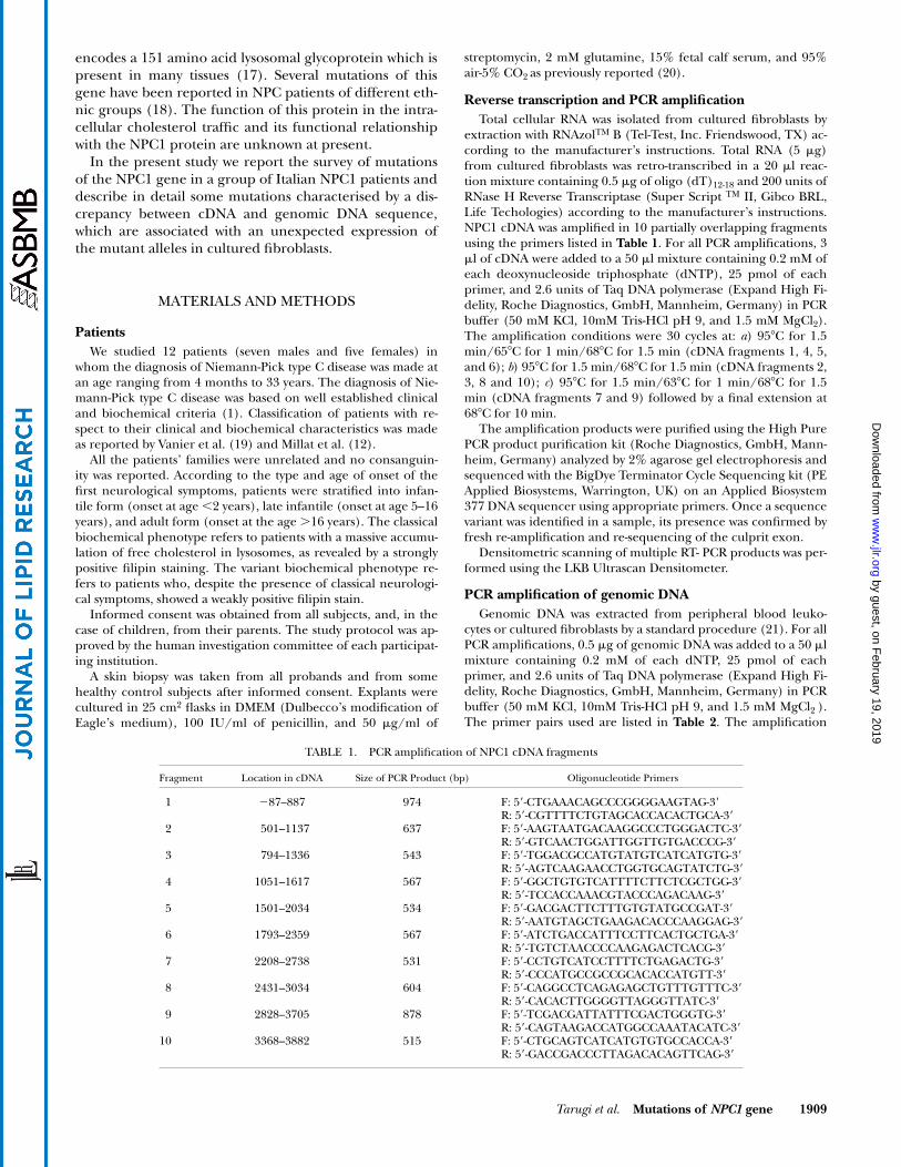
II, Gibco BRL,Life Techologies) according to the manufacturer’s instructions.NPC1 cDNA was amplified in 10 partially overlapping fragmentsusing the primers listed in
Table 1
.
For all PCR amplifications, 3
�
l of cDNA were added to a 50
�
l mixture containing 0.2 mM ofeach deoxynucleoside triphosphate (dNTP), 25 pmol of eachprimer, and 2.6 units of Taq DNA polymerase (Expand High Fi-delity, Roche Diagnostics, GmbH, Mannheim, Germany) in PCRbuffer (50 mM KCl, 10mM Tris-HCl pH 9, and 1.5 mM MgCl
2
).The amplification conditions were 30 cycles at:
a
) 95
�
C for 1.5min/65
�
C for 1 min/68
�
C for 1.5 min (cDNA fragments 1, 4, 5,and 6);
b
) 95
�
C for 1.5 min/68
�
C for 1.5 min (cDNA fragments 2,3, 8 and 10);
c
) 95
�
C for 1.5 min/63
�
C for 1 min/68
�
C for 1.5min (cDNA fragments 7 and 9) followed by a final extension at68
�
C for 10 min.The amplification products were purified using the High Pure
PCR product purification kit (Roche Diagnostics, GmbH, Mann-heim, Germany) analyzed by 2% agarose gel electrophoresis andsequenced with the BigDye Terminator Cycle Sequencing kit (PEApplied Biosystems, Warrington, UK) on an Applied Biosystem377 DNA sequencer using appropriate primers. Once a sequencevariant was identified in a sample, its presence was confirmed byfresh re-amplification and re-sequencing of the culprit exon.
Densitometric scanning of multiple RT- PCR products was per-formed using the LKB Ultrascan Densitometer.
PCR amplification of genomic DNA
Genomic DNA was extracted from peripheral blood leuko-cytes or cultured fibroblasts by a standard procedure (21). For allPCR amplifications, 0.5
�
g of genomic DNA was added to a 50
�
lmixture containing 0.2 mM of each dNTP, 25 pmol of eachprimer, and 2.6 units of Taq DNA polymerase (Expand High Fi-delity, Roche Diagnostics, GmbH, Mannheim, Germany) in PCRbuffer (50 mM KCl, 10mM Tris-HCl pH 9, and 1.5 mM MgCl
2
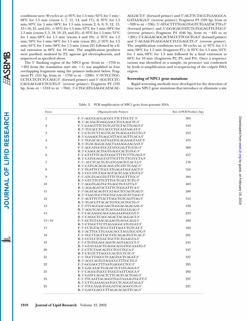
).The primer pairs used are listed in
Table 2
. The amplification
TABLE 1. PCR amplification of NPC1 cDNA fragments
Fragment Location in cDNA Size of PCR Product (bp) Oligonucleotide Primers
1
�
87–887 974 F: 5
�
-CTGAAACAGCCCGGGGAAGTAG-3
�
R: 5
�
-CGTTTTCTGTAGCACCACACTGCA-3
�
2 501–1137 637 F: 5
�
-AAGTAATGACAAGGCCCTGGGACTC-3
�
R: 5
�
-GTCAACTGGATTGGTTGTGACCCG-3
�
3 794–1336 543 F: 5
�
-TGGACGCCATGTATGTCATCATGTG-3
�
R: 5
�
-AGTCAAGAACCTGGTGCAGTATCTG-3
�
4 1051–1617 567 F: 5
�
-GGCTGTGTCATTTTCTTCTCGCTGG-3
�
R: 5
�
-TCCACCAAACGTACCCAGACAAG-3
�
5 1501–2034 534 F: 5
�
-GACGACTTCTTTGTGTATGCCGAT-3
�
R: 5
�
-AATGTAGCTGAAGACACCCAAGGAG-3
�
6 1793–2359 567 F: 5
�
-ATCTGACCATTTCCTTCACTGCTGA-3
�
R: 5
�
-TGTCTAACCCCAAGAGACTCACG-3
�
7 2208–2738 531 F: 5
�
-CCTGTCATCCTTTTCTGAGACTG-3
�
R: 5
�
-CCCATGCCGCCGCACACCATGTT-3
�
8 2431–3034 604 F: 5
�
-CAGGCCTCAGAGAGCTGTTTGTTTC-3
�
R: 5
�
-CACACTTGGGGTTAGGGTTATC-3
�
9 2828–3705 878 F: 5
�
-TCGACGATTATTTCGACTGGGTG-3
�
R: 5
�
-CAGTAAGACCATGGCCAAATACATC-3
�
10 3368–3882 515 F: 5
�
-CTGCAGTCATCATGTGTGCCACCA-3
�
R: 5
�
-GACCGACCCTTAGACACAGTTCAG-3
�
by guest, on February 19, 2019
ww
w.jlr.org
Dow
nloaded from
1910 Journal of Lipid Research
Volume 43, 2002
conditions were 30 cycles at:
a
) 95
�
C for 1.5 min/65
�
C for 1 min/68
�
C for 1.5 min (exons 1, 7, 11, 14, and 17);
b
) 95
�
C for 1.5min/63
�
C for 1 min/68
�
C for 1.5 min (exons 2, 6, 8, 9, 12, 13,15
�
16, 21, and 24);
c
) 95
�
C for 1.5 min/67
�
C for 1 min/68
�
C for1.5 min (exons 3, 5, 18, 19, 23, and 25);
d
) 95
�
C for 1.5 min/51
�
Cfor 1 min/68
�
C for 1.5 min (exons 4 and 10);
e
) 95
�
C for 1.5min/59
�
C for 1 min/68
�
C for 1.5 min (exon 20);
f
) 95
�
C for 1.5min/61
�
C for 1 min/68
�
C for 1.5 min (exon 22) followed by a fi-nal extension at 68
�
C for 10 min. The amplification productswere purified, analysed by 2% agarose gel electrophoresis, andsequenced as specified above.
The 5
�
flanking region of the NPC1 gene (from nt
�1750 to�201 from the translation start site �1) was amplified in fouroverlapping fragments using the primers indicated below. Frag-ment P1 (551 bp, from nt �1750 to nt �1200): 5�-TCTCCTGC-CCTCCTGTCTCCAGC-3� (forward primer) and 5�-AGGTCCTC-CACGACGACCTCGTG-3� (reverse primer); Fragment P2 (535bp, from nt �1318 to nt �784): 5�-CTGCATGAAGGCATACAC-
AGGACT-3� (forward primer) and 5�-AGTTCTACGTGAAGGCA-GATAAAGA-3� (reverse primer); Fragment P3 (593 bp, from nt�928 to nt �336): 5�-ATGCTTTTGAGTGGTCTGAATACTTG-3�(forward primer) and 5�-GGAGAGTATCTGTGGAGTTGTGC-3�(reverse primer); Fragment P4 (646 bp, from nt �445 to nt�201): 5�-CAGAACAGCACTACCCTTCGCTGA-3� (forward primer)and 5�-ACAAGTGAGGAACCTCCGAGCTC-3� (reverse primer).The amplification conditions were 30 cycles at: a) 95�C for 1.5min/68�C for 1.5 min (fragment P1); b) 95�C for 1.5 min/65�Cfor 1 min/68�C for 1.5 min followed by a final extension at68�C for 10 min (fragments P2, P3, and P4). Once a sequencevariant was identified in a sample, its presence was confirmedby fresh re-amplification and re-sequencing of the culprit DNAregion.
Screening of NPC1 gene mutationsRapid screening methods were developed for the detection of
four new NPC1 gene mutations that introduce or eliminate a site
Table 2. PCR amplification of NPC1 gene from genomic DNA
Exon Oligonucleotide Primers Size of PCR Product (bp)
1 F: 5�-AGCCGACGACGCCTTCTTCCTT- 3�R: 5�-ACAAGTGAGGAACCTCCGAGCTC-3�
383
2 F: 5�-GAAGTTTCTGTGATTGTACTTGAGT-3�R: 5�-TCCACCTCCACCCTGCAATAACAT-3�
310
3 F: 5�-GTGTCTTAGTTCACTGAGGAATGTTG-3�R: 5�-GAAAGCTGAGCATTACCAGTTCACA-3�
253
4 F: 5�-TGGACACAATAAATGCAGAAAGTAAT-3�R: 5�-TGACAGGACAACTAAAAGGAACAAT-3�
475
5 F: 5�-AGCATGGTGCATATGGAGTTCGTG-3�R: 5�-CAAGCACTGGTGAGCCACTGTGC-3�
369
6 F: 5�-GTATTTCAGTGGGCTTTTCTTTGAGT-3�R: 5�-CATGGAGGTATTTGTTTCTTGTCCTA-3�
457
7 F: 5�- ACCTCACTGTGATGAAGTCCACTA-3�R: 5�-CATGACAGACAGCATCATCTGAAC-3�
178
8 F: 5�-TGATTCCTGCCATGAGATAGCAACT-3�R: 5�-CCCATCTAGCAGTAGTCAACATGTA-3�
556
9 F: 5�-ATGTGACGTGTTTCTGGGTTTGC-3�R: 5�-GTCTTGTTGTTTGCTCACCTCTG-3�
384
10 F: 5�-AGGTGAGTGCTGAGCTGTATTA-3�R: 5�-AGGAGATACTATTCTGGGATTCA-3�
403
11 F: 5�-AGATACAGTCCATAGCTCCAGTGAG-3�R: 5�-TAAGTGCTTGCTGCAAGTGTCTAGC-3�
288
12 F: 5�-AGTTTCTTACTTAGCTGTCAGTTAG-3�R: 5�-TGACGTTACACTGTGCACTGCTG-3�
312
13 F: 5�-TTTAGTAACAAGTGGGACAGACAAC-3�R: 5�-AGGTCACACTCACGAATGCGGAG-3�
339
14 F: 5�-CACAAGGCAGCAAGAAATGGCGT-3�R: 5�-CAGGCTCAGCAGACTACAGGACT-3�
233
15�16 F: 5�-GCTGTAAACAGAAGTGACGCAGA-3�R: 5�-CTGGCTTCTTAGAAGGCATGTGAT-3�
480
17 F: 5�-CCTGTACTCCCTATTAGCCTGTCAT-3�R: 5�-ACTTGCTTGAAACACCTACGTGCATG-3�
322
18 F: 5�-TGCTTAGTTACTATCAGAGTGTTCAC-3�R: 5�-CCTCCTCCGCTGCTTCTGAAGTA-3�
291
19 F: 5�-CTGTGGAGCAGGTCAGTAACCCT-3�R: 5�-GTATAAACTGAGGCACGATGCAAATG-3�
245
20 F: 5�-CTTCTAACAGTCCTCCCTGCA-3�R: 5�-CTGTCTTAGCCCAGTCCTCTC-3�
247
21 F: 5�-TGCTTAGCCTCAAGTGCTCAGAT-3�R: 5�-ACCCAGTGTAGGCCCTTTGCTG-3�
337
22 F: 5�-GCGAGCTTTAATGAGGCCTCC-3�R: 5�-GACAAACTGAGACTGTATGAGGA-3�
295
23 F: 5�-CAGGGTGCCCTGGGTAATTAGCA-3�R: 5�-GATCCAGACTCTTCAGTCACTGAG-3�
292
24 F: 5�-TTCAATTACAGGTTGGTAAAAGTGGTT-3�R: 5�-CTTGAAAAGAATGCCTCAGGATAGA-3�
297
25 F: 5�-TTCCAAAGTGGGATTACAGGCGTG-3�R: 5�-GACCGACCCTTAGACACAGTTCAG-3�
221
by guest, on February 19, 2019
ww
w.jlr.org
Dow
nloaded from
Tarugi et al. Mutations of NPC1 gene 1911
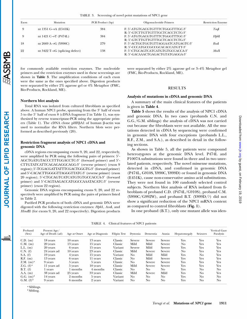
for commonly available restriction enzymes. The nucleotideprimers and the restriction enzymes used in these screenings areshown in Table 3. The amplification conditions of each exonwere the same as the ones specified above. Digestion productswere separated by either 2% agarose gel or 4% Metaphor (FMC,Bio-Products, Rockland, ME).
Northern blot analysisTotal RNA was isolated from cultured fibroblasts as specified
above. A human NPC1 probe, spanning from the 3� half of exon5 to the 5� half of exon 8 (cDNA fragment 2 in Table 1), was syn-thesized by reverse transcriptase-PCR using the appropriate prim-ers (Table 1). The cDNA clone pHFA1 of human -actin wasused to normalize the RNA filters. Northern blots were per-formed as described previously (20).
Restriction fragment analysis of NPC1 cDNA andgenomic DNA
cDNA regions encompassing exons 9, 20, and 22, respectively,were amplified by PCR using the following pairs of primers: 5�-AGCTGATGTACCCTTTGGACCTC-3� (forward primer) and 5�-CTTGTATCATTCAGAGAGGCAGG-3� (reverse primer) (exon 9region); 5�-TCGACGATTATTTCGACTGGGTG-3� (forward primer)and 5�-CACACTTGGGGTTAGGGTTATC-3� (reverse primer) (exon20 region); 5�-CTGCAGTCATCATGTGTGCCACCA-3� (forwardprimer) and 5�-CAGTAAGACCATGGCCAAATACATC-3� (reverseprimer) (exon 22 region).
Genomic DNA regions encompassing exons 9, 20, and 22 re-spectively were amplified by PCR using the pairs of primers listedin Table 2.
Purified PCR products of both cDNA and genomic DNA weredigested with the following restriction enzymes: Hph1, AvaI, andHindII (for exons 9, 20, and 22 respectively). Digestion products
were separated by either 2% agarose gel or 3–4% Metaphor gel(FMC, Bio-Products, Rockland, ME).
RESULTS
Analysis of mutations in cDNA and genomic DNAA summary of the main clinical features of the patients
is given in Table 4.Table 5 shows the results of the analysis of NPC1 cDNA
and genomic DNA. In two cases (probands C.N. andG.G./G.M. siblings) the analysis of cDNA was not carriedout because the fibroblasts were not available. All the mu-tations detected in cDNA by sequencing were confirmedin genomic DNA with four exceptions (probands L.L.,R.F., Z.M., and S.A.), as described in detail in the follow-ing sections.
As shown in Table 5, all the patients were compoundheterozygotes at the genomic DNA level. P474L andP1007A substitutions were found in three and in two unre-lated patients, respectively. The novel missense mutations,detected in cDNA, and confirmed in genomic DNA(P474L, G910S, Y890C, Y899D) or found in genomic DNA(E451K), cause non-conservative amino acid substitutions.They were not found in 100 randomly selected controlsubjects. Northern blot analysis of RNA isolated from fi-broblasts of proband C.D. (P474L/G910S), proband C.M.(Y890C/G992W), and proband B.T. (Y899D/?) did notshow a significant reduction of the NPC1 mRNA contentas compared to control fibroblasts (Fig. 1).
In one proband (B.T.), only one mutant allele was iden-
TABLE 3. Screening of novel point mutations of NPC1 gene
Exon Mutation PCR Product (bp) Oligonucleotide Primers Restriction Enzyme
9 nt 1351 G→A (E541K) 384 F: 5�-ATGTGACGTGTTTCTGGGTTTGC-3�R: 5�-GTCTTGTTGTTTGCTCACCTCTG-3�
TaqI
9 nt 1421 C→T (P474L) 384 F: 5�-ATGTGACGTGTTTCTGGGTTTGC-3�R: 5�-GTCTTGTTGTTTGCTCACCTCTG-3�
HphI
18 nt 2669 A→G (Y890C) 279 F: 5�-ACTGGTGGTGTTAGGAATCATGAGTC-3�R: 5�-CCCATGCCGCCGCACACCATGTT-3�
RsaI
22 nt 3422 T→G (splicing defect) 138 F: 5�-CTGCAGTCATCATGTGTGCCACCA-3�R: 5�-GACAAACTGAGACTGTATGAGGA-3�
MnlI
TABLE 4. Clinical features of NPC1 patients
Proband (Sex)
Present Age/Age of Death (ad) Age at Onset Age at Diagnosis Filipin Test Dystonia Dementia Ataxia Hepatomegaly Seizures
Vertical GazeParalysis
C.D. (m) 18 years 10 years 13 years Classic Severe Severe Severe Yes Yes YesC.M. (m) 20 years 13 years 15 years Classic Mild Mild Severe No Yes YesL.L. (m) 20 years 6 years 15 years Variant Severe Mild Severe Yes Yes YesC.N. (f) 24 years ad 10 years 23 years Classic Mild Severe Severe No Yes YesS.A. (f) 19 years 4 years 15 years Variant No Mild Mild Yes No YesR.F. (m) 13 years 6 years 11 years Classic No Mild Severe Yes Yes YesZ.M. (m)a 9 years 5 years 5 years Classic No Severe Severe Yes Yes YesZ.G. (f)a 11 years ad 5 years 10 years Classic Mild Severe Severe Yes Yes YesB.T. (f) 1 years 1 months 4 months Classic No No No Yes No NoA.A. (m) 38 years ad 25 years 33 years Classic Mild Severe Mild Yes No YesG.G. (m)b 14 years 2 months 5 years Variant No No No Yes No NoG.M. (f)b 9 years 6 months 2 years Variant No No No Yes No No
a Siblings.b Sibling.
by guest, on February 19, 2019
ww
w.jlr.org
Dow
nloaded from
1912 Journal of Lipid Research Volume 43, 2002
tified in cDNA and genomic DNA. Both NPC1 alleles ofthis proband were expressed in cultured fibroblasts, asdemonstrated by the presence of heterozygosity for somecommon polymorphisms in the coding region: C1926G(I642M), A2572G (I858V), and C2793T (N931N) (see below).
Sequence polymorphisms detected in cDNA andgenomic DNA
The following common polymorphisms were found incDNA (and confirmed in genomic DNA) of both NPC1patients and controls: T387C (Y129Y), A644G (H215R),C1926G (I642M), A2572G (I858V), C2793T (N931N), andG3797A (R1266Q). All of them were previously reported(16, 22). We found another silent mutation, C540T (D180D),previously unreported. The following unreported se-quence variants were observed in the 5� flanking region
(designated promoter region) in patients and controlsubjects: �238C/G, �887C/T, �1026T/G, �1186T/C,�1435A/T.
Discrepancy between cDNA and genomic DNA sequenceMonoallelic expression of some mutant alleles in fibroblastsProband L.L. The analysis of cDNA showed that pro-
band L.L. was homozygous for two nucleotide substitu-tions: i) C1421T leading to the rare mutation P474L (Ta-ble 5); ii) C1926G leading to the common amino acidvariant I642M (22). The analysis of genomic DNA re-vealed that the patient was in fact heterozygous for: i)both these substitutions; ii) two additional common poly-morphisms [A644G (H215R) and C2793T (N931N)]; iii)a dinucleotide deletion in exon 20 [AG deletion at posi-tion 2972-2973, indicated as 2972(del2)]. The latter muta-
TABLE 5. Mutations in NPC1 cDNA and NPC1 gene
Patient ExonMutation inNPC1 cDNA Effect on NPC1 Protein
Mutation inNPC1 Gene
PreviouslyReported
C.D. 9 C1421T ht P474L ht C1421T ht New 18 G2728A ht G910S ht G2728A ht New
C.M. 18 A2669G ht Y890C ht A2669G ht New 20 G2974T ht G992W ht G2974T ht ref. 10
L.L. 9 C1421T hz P474L hz C1421T ht New20 none nt 2972(del2) (AG) ht
(Fs. from codon 991→Stop 1005)ref. 11
C.N. 19 NA C2861T ht (S954L ht) ref. 11 24 NA nt 3734(del2) (CT) ht
(Fs. from codon 1245→Stop 1256)New
S.A. 20 C3019G ht P1007A ht C3019G ht ref. 11 22 nt 3422–3477 (del56) ht Fs. from codon
1141→Stop 1238 htT3422G ht (V1141G) New
R.F. 9 C1421T hz P474L hz C1421T ht New 20 none nt 2972(del2) (AG) ht
(Fs. from codon 991 →Stop 1005)ref. 11
Z.M./Z.G. Siblings 18 none C2764T ht (Q922X) New 22 A3467G hz N1156S hz A3467G ht refs. 10, 15
B.T. 18 T2695G ht?
Y899D ht?
T2695G ht?
New
A.A. 20 C3019G ht P1007A ht C3019G ht ref. 11 21 T3182C ht I1061T ht T3182C ht ref. 11
G.G./G.M. Siblings 9 NA G1351A ht (E451K) New 20 NA G2974T ht (G992W) ref. 11
NA, cDNA not available; ht, heterozyote; hz, homozygote. The effect on the NPC1 protein predicted on the basis of the mutations found in ge-nomic DNA (NPC1 gene) is indicated in italics.
Fig. 1. Northern blot analysis of Niemann-Pick type C disease 1 (NPC1) mRNA isolated from fibroblasts.Lane 1, proband C.D. (P474L/G910S); lane 2, proband C.M. (Y890C/G992W); lane 3, proband B.T. (Y899D/?);lanes 4 and 5, control subjects.
by guest, on February 19, 2019
ww
w.jlr.org
Dow
nloaded from
Tarugi et al. Mutations of NPC1 gene 1913
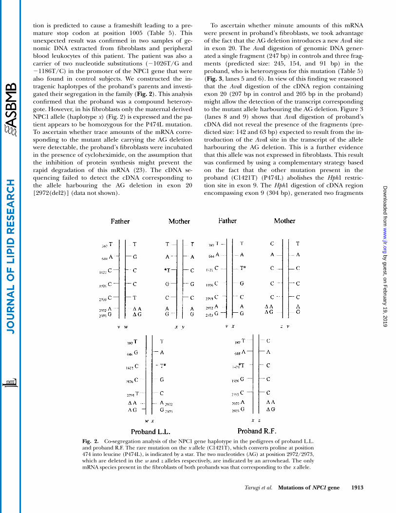
tion is predicted to cause a frameshift leading to a pre-mature stop codon at position 1005 (Table 5). Thisunexpected result was confirmed in two samples of ge-nomic DNA extracted from fibroblasts and peripheralblood leukocytes of this patient. The patient was also acarrier of two nucleotide substitutions (�1026T/G and�1186T/C) in the promoter of the NPC1 gene that werealso found in control subjects. We constructed the in-tragenic haplotypes of the proband’s parents and investi-gated their segregation in the family (Fig. 2). This analysisconfirmed that the proband was a compound heterozy-gote. However, in his fibroblasts only the maternal derivedNPC1 allele (haplotype x) (Fig. 2) is expressed and the pa-tient appears to be homozygous for the P474L mutation.To ascertain whether trace amounts of the mRNA corre-sponding to the mutant allele carrying the AG deletionwere detectable, the proband’s fibroblasts were incubatedin the presence of cycloheximide, on the assumption thatthe inhibition of protein synthesis might prevent therapid degradation of this mRNA (23). The cDNA se-quencing failed to detect the cDNA corresponding tothe allele harbouring the AG deletion in exon 20[2972(del2)] (data not shown).
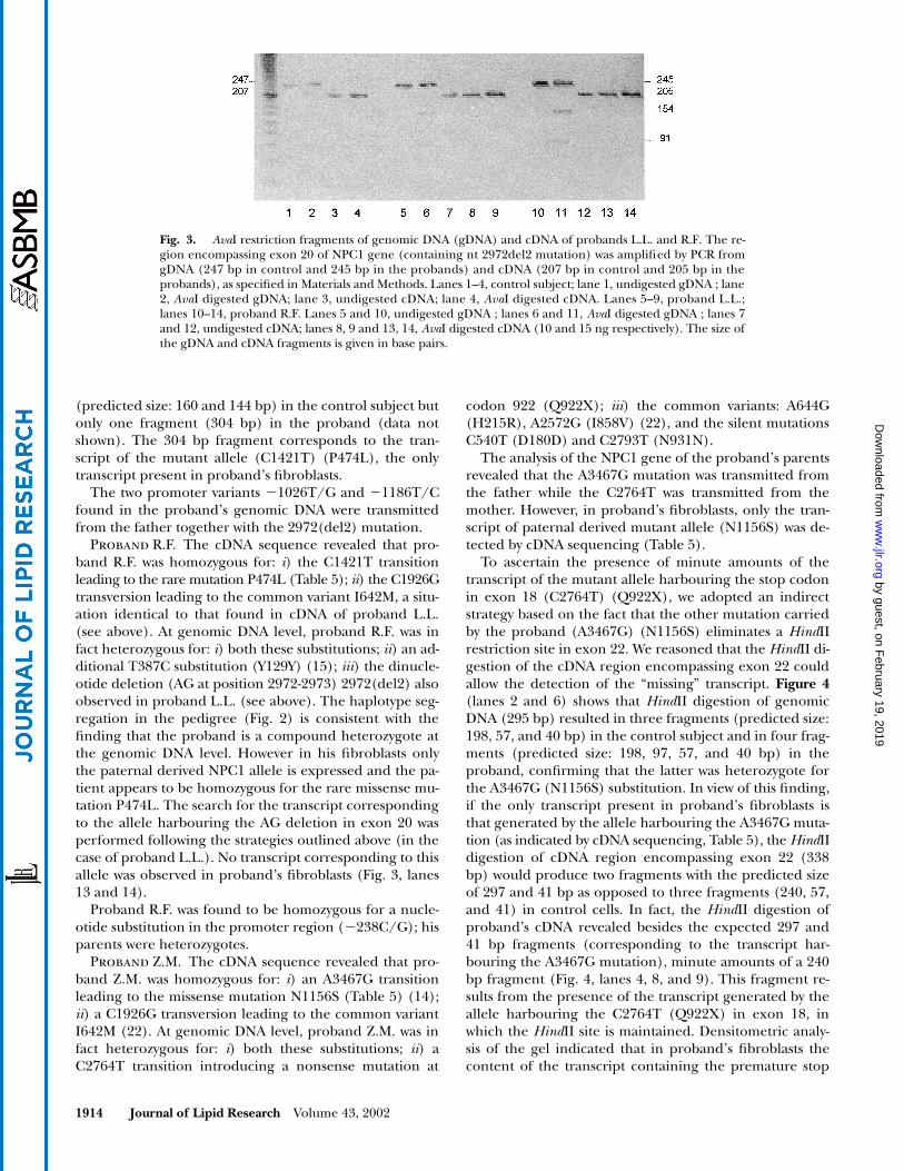
To ascertain whether minute amounts of this mRNAwere present in proband’s fibroblasts, we took advantageof the fact that the AG deletion introduces a new AvaI sitein exon 20. The AvaI digestion of genomic DNA gener-ated a single fragment (247 bp) in controls and three frag-ments (predicted size: 245, 154, and 91 bp) in theproband, who is heterozygous for this mutation (Table 5)(Fig. 3, lanes 5 and 6). In view of this finding we reasonedthat the AvaI digestion of the cDNA region containingexon 20 (207 bp in control and 205 bp in the proband)might allow the detection of the transcript correspondingto the mutant allele harbouring the AG deletion. Figure 3(lanes 8 and 9) shows that AvaI digestion of proband’scDNA did not reveal the presence of the fragments (pre-dicted size: 142 and 63 bp) expected to result from the in-troduction of the AvaI site in the transcript of the alleleharbouring the AG deletion. This is a further evidencethat this allele was not expressed in fibroblasts. This resultwas confirmed by using a complementary strategy basedon the fact that the other mutation present in theproband (C1421T) (P474L) abolishes the Hph1 restric-tion site in exon 9. The Hph1 digestion of cDNA regionencompassing exon 9 (304 bp), generated two fragments
Fig. 2. Co-segregation analysis of the NPC1 gene haplotype in the pedigrees of proband L.L.and proband R.F. The rare mutation on the x allele (C1421T), which converts proline at position474 into leucine (P474L), is indicated by a star. The two nucleotides (AG) at position 2972/2973,which are deleted in the w and z alleles respectively, are indicated by an arrowhead. The onlymRNA species present in the fibroblasts of both probands was that corresponding to the x allele.
by guest, on February 19, 2019
ww
w.jlr.org
Dow
nloaded from
1914 Journal of Lipid Research Volume 43, 2002
(predicted size: 160 and 144 bp) in the control subject butonly one fragment (304 bp) in the proband (data notshown). The 304 bp fragment corresponds to the tran-script of the mutant allele (C1421T) (P474L), the onlytranscript present in proband’s fibroblasts.
The two promoter variants �1026T/G and �1186T/Cfound in the proband’s genomic DNA were transmittedfrom the father together with the 2972(del2) mutation.
Proband R.F. The cDNA sequence revealed that pro-band R.F. was homozygous for: i) the C1421T transitionleading to the rare mutation P474L (Table 5); ii) the C1926Gtransversion leading to the common variant I642M, a situ-ation identical to that found in cDNA of proband L.L.(see above). At genomic DNA level, proband R.F. was infact heterozygous for: i) both these substitutions; ii) an ad-ditional T387C substitution (Y129Y) (15); iii) the dinucle-otide deletion (AG at position 2972-2973) 2972(del2) alsoobserved in proband L.L. (see above). The haplotype seg-regation in the pedigree (Fig. 2) is consistent with thefinding that the proband is a compound heterozygote atthe genomic DNA level. However in his fibroblasts onlythe paternal derived NPC1 allele is expressed and the pa-tient appears to be homozygous for the rare missense mu-tation P474L. The search for the transcript correspondingto the allele harbouring the AG deletion in exon 20 wasperformed following the strategies outlined above (in thecase of proband L.L.). No transcript corresponding to thisallele was observed in proband’s fibroblasts (Fig. 3, lanes13 and 14).
Proband R.F. was found to be homozygous for a nucle-otide substitution in the promoter region (�238C/G); hisparents were heterozygotes.
Proband Z.M. The cDNA sequence revealed that pro-band Z.M. was homozygous for: i) an A3467G transitionleading to the missense mutation N1156S (Table 5) (14);ii) a C1926G transversion leading to the common variantI642M (22). At genomic DNA level, proband Z.M. was infact heterozygous for: i) both these substitutions; ii) aC2764T transition introducing a nonsense mutation at
codon 922 (Q922X); iii) the common variants: A644G(H215R), A2572G (I858V) (22), and the silent mutationsC540T (D180D) and C2793T (N931N).
The analysis of the NPC1 gene of the proband’s parentsrevealed that the A3467G mutation was transmitted fromthe father while the C2764T was transmitted from themother. However, in proband’s fibroblasts, only the tran-script of paternal derived mutant allele (N1156S) was de-tected by cDNA sequencing (Table 5).
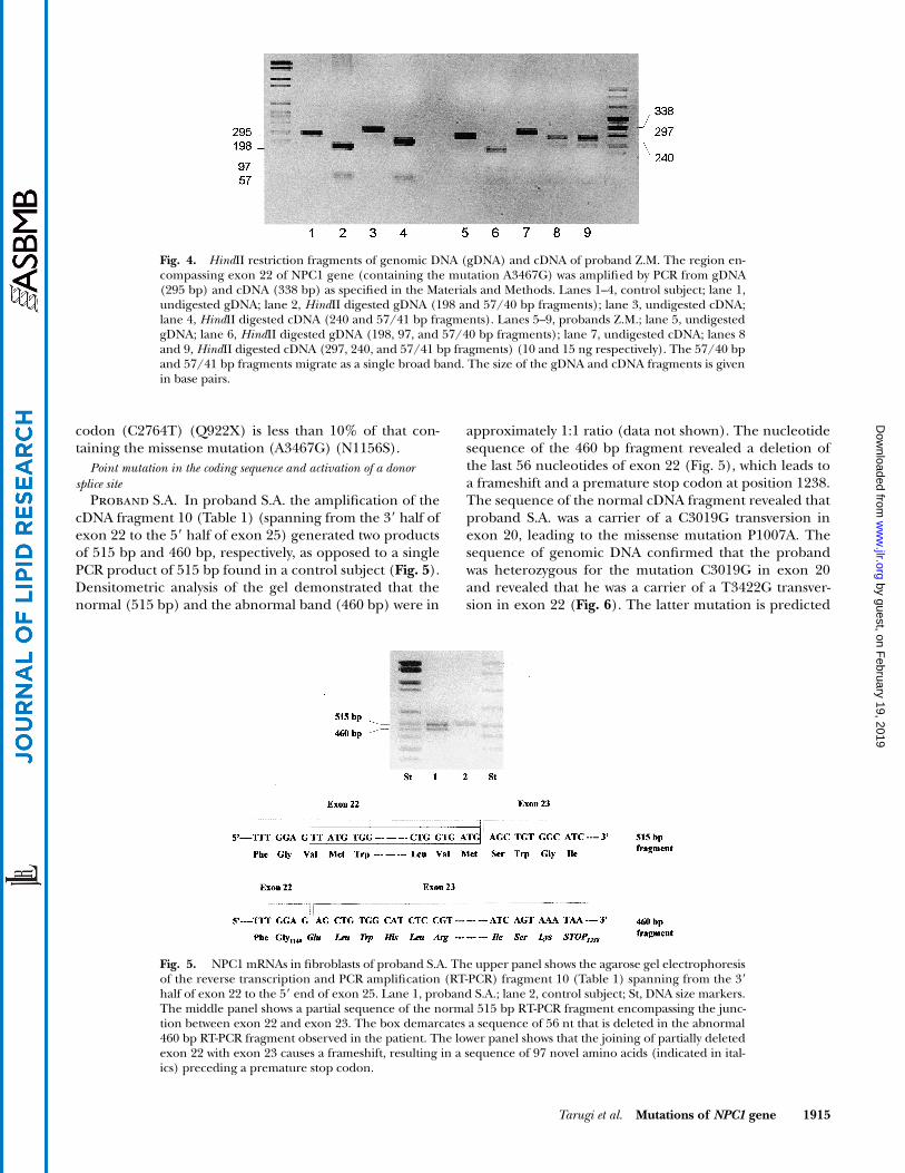
To ascertain the presence of minute amounts of thetranscript of the mutant allele harbouring the stop codonin exon 18 (C2764T) (Q922X), we adopted an indirectstrategy based on the fact that the other mutation carriedby the proband (A3467G) (N1156S) eliminates a HindIIrestriction site in exon 22. We reasoned that the HindII di-gestion of the cDNA region encompassing exon 22 couldallow the detection of the “missing” transcript. Figure 4(lanes 2 and 6) shows that HindII digestion of genomicDNA (295 bp) resulted in three fragments (predicted size:198, 57, and 40 bp) in the control subject and in four frag-ments (predicted size: 198, 97, 57, and 40 bp) in theproband, confirming that the latter was heterozygote forthe A3467G (N1156S) substitution. In view of this finding,if the only transcript present in proband’s fibroblasts isthat generated by the allele harbouring the A3467G muta-tion (as indicated by cDNA sequencing, Table 5), the HindIIdigestion of cDNA region encompassing exon 22 (338bp) would produce two fragments with the predicted sizeof 297 and 41 bp as opposed to three fragments (240, 57,and 41) in control cells. In fact, the HindII digestion ofproband’s cDNA revealed besides the expected 297 and41 bp fragments (corresponding to the transcript har-bouring the A3467G mutation), minute amounts of a 240bp fragment (Fig. 4, lanes 4, 8, and 9). This fragment re-sults from the presence of the transcript generated by theallele harbouring the C2764T (Q922X) in exon 18, inwhich the HindII site is maintained. Densitometric analy-sis of the gel indicated that in proband’s fibroblasts thecontent of the transcript containing the premature stop
Fig. 3. AvaI restriction fragments of genomic DNA (gDNA) and cDNA of probands L.L. and R.F. The re-gion encompassing exon 20 of NPC1 gene (containing nt 2972del2 mutation) was amplified by PCR fromgDNA (247 bp in control and 245 bp in the probands) and cDNA (207 bp in control and 205 bp in theprobands), as specified in Materials and Methods. Lanes 1–4, control subject; lane 1, undigested gDNA ; lane2, AvaI digested gDNA; lane 3, undigested cDNA; lane 4, AvaI digested cDNA. Lanes 5–9, proband L.L.;lanes 10–14, proband R.F. Lanes 5 and 10, undigested gDNA ; lanes 6 and 11, AvaI digested gDNA ; lanes 7and 12, undigested cDNA; lanes 8, 9 and 13, 14, AvaI digested cDNA (10 and 15 ng respectively). The size ofthe gDNA and cDNA fragments is given in base pairs.
by guest, on February 19, 2019
ww
w.jlr.org
Dow
nloaded from
Tarugi et al. Mutations of NPC1 gene 1915
codon (C2764T) (Q922X) is less than 10% of that con-taining the missense mutation (A3467G) (N1156S).
Point mutation in the coding sequence and activation of a donor splice site
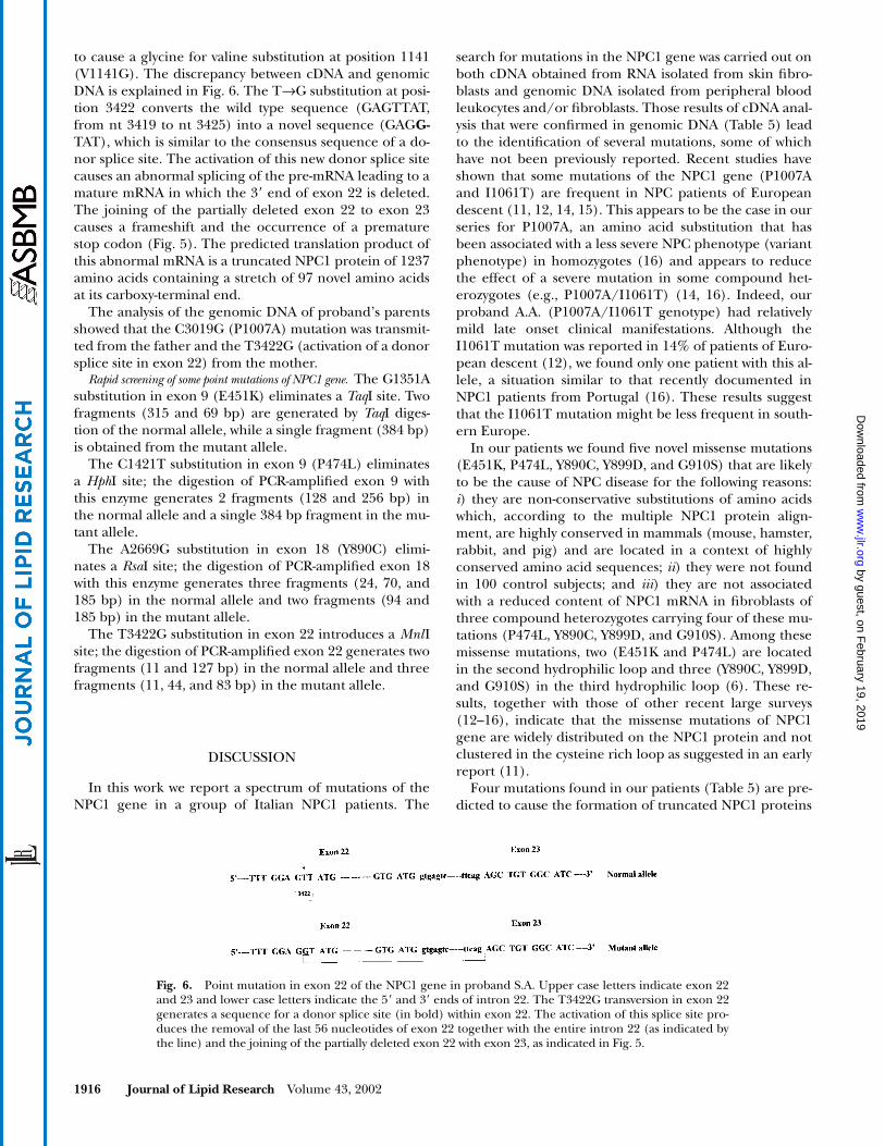
Proband S.A. In proband S.A. the amplification of thecDNA fragment 10 (Table 1) (spanning from the 3� half ofexon 22 to the 5� half of exon 25) generated two productsof 515 bp and 460 bp, respectively, as opposed to a singlePCR product of 515 bp found in a control subject (Fig. 5).Densitometric analysis of the gel demonstrated that thenormal (515 bp) and the abnormal band (460 bp) were in
approximately 1:1 ratio (data not shown). The nucleotidesequence of the 460 bp fragment revealed a deletion ofthe last 56 nucleotides of exon 22 (Fig. 5), which leads toa frameshift and a premature stop codon at position 1238.The sequence of the normal cDNA fragment revealed thatproband S.A. was a carrier of a C3019G transversion inexon 20, leading to the missense mutation P1007A. Thesequence of genomic DNA confirmed that the probandwas heterozygous for the mutation C3019G in exon 20and revealed that he was a carrier of a T3422G transver-sion in exon 22 (Fig. 6). The latter mutation is predicted
Fig. 4. HindII restriction fragments of genomic DNA (gDNA) and cDNA of proband Z.M. The region en-compassing exon 22 of NPC1 gene (containing the mutation A3467G) was amplified by PCR from gDNA(295 bp) and cDNA (338 bp) as specified in the Materials and Methods. Lanes 1–4, control subject; lane 1,undigested gDNA; lane 2, HindII digested gDNA (198 and 57/40 bp fragments); lane 3, undigested cDNA;lane 4, HindII digested cDNA (240 and 57/41 bp fragments). Lanes 5–9, probands Z.M.; lane 5, undigestedgDNA; lane 6, HindII digested gDNA (198, 97, and 57/40 bp fragments); lane 7, undigested cDNA; lanes 8and 9, HindII digested cDNA (297, 240, and 57/41 bp fragments) (10 and 15 ng respectively). The 57/40 bpand 57/41 bp fragments migrate as a single broad band. The size of the gDNA and cDNA fragments is givenin base pairs.
Fig. 5. NPC1 mRNAs in fibroblasts of proband S.A. The upper panel shows the agarose gel electrophoresisof the reverse transcription and PCR amplification (RT-PCR) fragment 10 (Table 1) spanning from the 3�half of exon 22 to the 5� end of exon 25. Lane 1, proband S.A.; lane 2, control subject; St, DNA size markers.The middle panel shows a partial sequence of the normal 515 bp RT-PCR fragment encompassing the junc-tion between exon 22 and exon 23. The box demarcates a sequence of 56 nt that is deleted in the abnormal460 bp RT-PCR fragment observed in the patient. The lower panel shows that the joining of partially deletedexon 22 with exon 23 causes a frameshift, resulting in a sequence of 97 novel amino acids (indicated in ital-ics) preceding a premature stop codon.
by guest, on February 19, 2019
ww
w.jlr.org
Dow
nloaded from
1916 Journal of Lipid Research Volume 43, 2002
to cause a glycine for valine substitution at position 1141(V1141G). The discrepancy between cDNA and genomicDNA is explained in Fig. 6. The T→G substitution at posi-tion 3422 converts the wild type sequence (GAGTTAT,from nt 3419 to nt 3425) into a novel sequence (GAGG-TAT), which is similar to the consensus sequence of a do-nor splice site. The activation of this new donor splice sitecauses an abnormal splicing of the pre-mRNA leading to amature mRNA in which the 3� end of exon 22 is deleted.The joining of the partially deleted exon 22 to exon 23causes a frameshift and the occurrence of a prematurestop codon (Fig. 5). The predicted translation product ofthis abnormal mRNA is a truncated NPC1 protein of 1237amino acids containing a stretch of 97 novel amino acidsat its carboxy-terminal end.
The analysis of the genomic DNA of proband’s parentsshowed that the C3019G (P1007A) mutation was transmit-ted from the father and the T3422G (activation of a donorsplice site in exon 22) from the mother.
Rapid screening of some point mutations of NPC1 gene. The G1351Asubstitution in exon 9 (E451K) eliminates a TaqI site. Twofragments (315 and 69 bp) are generated by TaqI diges-tion of the normal allele, while a single fragment (384 bp)is obtained from the mutant allele.
The C1421T substitution in exon 9 (P474L) eliminatesa HphI site; the digestion of PCR-amplified exon 9 withthis enzyme generates 2 fragments (128 and 256 bp) inthe normal allele and a single 384 bp fragment in the mu-tant allele.
The A2669G substitution in exon 18 (Y890C) elimi-nates a RsaI site; the digestion of PCR-amplified exon 18with this enzyme generates three fragments (24, 70, and185 bp) in the normal allele and two fragments (94 and185 bp) in the mutant allele.
The T3422G substitution in exon 22 introduces a MnlIsite; the digestion of PCR-amplified exon 22 generates twofragments (11 and 127 bp) in the normal allele and threefragments (11, 44, and 83 bp) in the mutant allele.
DISCUSSION
In this work we report a spectrum of mutations of theNPC1 gene in a group of Italian NPC1 patients. The
search for mutations in the NPC1 gene was carried out onboth cDNA obtained from RNA isolated from skin fibro-blasts and genomic DNA isolated from peripheral bloodleukocytes and/or fibroblasts. Those results of cDNA anal-ysis that were confirmed in genomic DNA (Table 5) leadto the identification of several mutations, some of whichhave not been previously reported. Recent studies haveshown that some mutations of the NPC1 gene (P1007Aand I1061T) are frequent in NPC patients of Europeandescent (11, 12, 14, 15). This appears to be the case in ourseries for P1007A, an amino acid substitution that hasbeen associated with a less severe NPC phenotype (variantphenotype) in homozygotes (16) and appears to reducethe effect of a severe mutation in some compound het-erozygotes (e.g., P1007A/I1061T) (14, 16). Indeed, ourproband A.A. (P1007A/I1061T genotype) had relativelymild late onset clinical manifestations. Although theI1061T mutation was reported in 14% of patients of Euro-pean descent (12), we found only one patient with this al-lele, a situation similar to that recently documented inNPC1 patients from Portugal (16). These results suggestthat the I1061T mutation might be less frequent in south-ern Europe.
In our patients we found five novel missense mutations(E451K, P474L, Y890C, Y899D, and G910S) that are likelyto be the cause of NPC disease for the following reasons:i) they are non-conservative substitutions of amino acidswhich, according to the multiple NPC1 protein align-ment, are highly conserved in mammals (mouse, hamster,rabbit, and pig) and are located in a context of highlyconserved amino acid sequences; ii) they were not foundin 100 control subjects; and iii) they are not associatedwith a reduced content of NPC1 mRNA in fibroblasts ofthree compound heterozygotes carrying four of these mu-tations (P474L, Y890C, Y899D, and G910S). Among thesemissense mutations, two (E451K and P474L) are locatedin the second hydrophilic loop and three (Y890C, Y899D,and G910S) in the third hydrophilic loop (6). These re-sults, together with those of other recent large surveys(12–16), indicate that the missense mutations of NPC1gene are widely distributed on the NPC1 protein and notclustered in the cysteine rich loop as suggested in an earlyreport (11).
Four mutations found in our patients (Table 5) are pre-dicted to cause the formation of truncated NPC1 proteins
Fig. 6. Point mutation in exon 22 of the NPC1 gene in proband S.A. Upper case letters indicate exon 22and 23 and lower case letters indicate the 5� and 3� ends of intron 22. The T3422G transversion in exon 22generates a sequence for a donor splice site (in bold) within exon 22. The activation of this splice site pro-duces the removal of the last 56 nucleotides of exon 22 together with the entire intron 22 (as indicated bythe line) and the joining of the partially deleted exon 22 with exon 23, as indicated in Fig. 5.
by guest, on February 19, 2019
ww
w.jlr.org
Dow
nloaded from

Tarugi et al. Mutations of NPC1 gene 1917
of 921, 1004, 1237, and 1255 amino acids. These changes,which are expected to disrupt several trans-membrane do-mains of NPC1 protein (2, 6), probably prevent theproper integration of this peptide in the membrane of theendoplasmic reticulum as well as its localisation in late en-dosomes. It is also possible that these truncated proteinsare either synthesised at a reduced rate (as mRNAs har-bouring a premature stop codon may be degraded morerapidly than their normal counterparts) (see below) or arerapidly degraded in the cell shortly after their synthesis.
One unexpected finding of the present study was thediscrepancy between the results of the sequence analysisof cDNA and that of genomic DNA we observed in fourpatients. Two types of discrepancies emerged: i) the ex-pression of a single mutant allele (monoallelic expression)in fibroblasts of three patients who are compound het-erozygotes; ii) the mutant mRNA and the correspondingmutant allele in genomic DNA bear different mutations.
Monoallelic expressionIn two unrelated compound heterozygotes carrying the
same mutations (P474L and nt 2972del2), cDNA se-quence revealed that only the P474L allele was expressedin fibroblasts. Furthermore, we failed to demonstrate the“missing” transcript after specific PCR amplification andrestriction enzyme digestion of the cDNA region harbour-ing the AG deletion (nt 2972del2) in exon 20 (Fig. 3).One simple explanation for this lack of expression is thatthe nt 2972del2 mutation is in linkage with another muta-tion in the promoter of the NPC1 gene, which preventsthe transcription of the deletion carrying allele. The ob-servation that one of these patients (proband L.L.) was acarrier of two single nucleotide substitutions in the pro-moter (�1186T/C and �1026T/G), found to be in link-age with the nt 2972del2 mutation, might support this hy-pothesis. However, it is unlikely that these substitutions inthe promoter abolish the expression of the mutant allelesince they: i) represent common polymorphisms found inrandomly selected healthy subjects; and ii) are not presentin the promoter of the same mutant allele (nt 2972del2),which is not expressed in the fibroblasts of the second pa-tient (proband R.F.) carrying this specific mutation. Ofcourse, it cannot be excluded that in both patients nt2972del2 is linked to another mutation elsewhere in thegene, outside the regions we have sequenced.
The third patient (proband Z.M.), in whom the cDNAsequence revealed the presence of the transcript of onlyone mutant allele (N1156S), was found to be a compoundheterozygote for two mutations (N1156S and Q922X) dif-ferent from the ones found in probands L.L. and R.F. Inthe case of proband Z.M. however, we were able to detecttrace amounts of the transcript containing the prematurestop codon by using PCR –amplification of the appropri-ate cDNA region followed by restriction enzyme digestion(Fig. 4). We found no sequence variations in the pro-moter in this proband that were in linkage with the mu-tant allele (Q922X).
The common feature of the three mutant alleles foundin these three probands, which are not expressed or are
expressed at a very low level in fibroblasts, appears to bethe presence of a premature termination codon in thecorresponding mRNA. It is possible that these mRNAscontaining a premature termination codon (PTC) un-dergo a rapid degradation (nonsense mediated RNA de-cay, NMD) as reported in other genetic disorders (24).NMD appears to be an ancient and evolutionary con-served surveillance strategy to protect cells from muta-tions that could yield truncated, potentially dangerousproteins. Several genes have been identified in human ge-nome which are involved in this process (24–26). The cellcompartment where mRNA harbouring PTC is destroyedis still a matter of intense investigation as there is evidencethat it might occur in the cytoplasm (site of translation) aswell as in the nucleus or close to the nuclear pore (24, 27–29). It is likely that the recognition of the PTC containingpre-mRNA and its destruction is linked with the splicingprocess. If the destruction occurs before the abnormaltranscript leaves the nucleus, no mature mRNA is ex-pected to accumulate in the cytoplasm. Not all pre-mRNAs (or mature mRNAs) of NPC1 gene that containPTC are targeted for destruction. Notably, in the presentstudy we found that in the fibroblasts of proband S.A. car-rying a splicing defect (see below), the mature mRNAcontaining PTC was present in approximately the sameconcentration as the wild type mRNA (Fig. 5). This sug-gests that pre-mRNAs containing PTC loose their stabilityonly when PTC has some specific location with respect tosome recognition signal (for example the last intron, as sug-gested in the case of human triosephosphate isomeraseand -globin mRNA containing PTC) (30–31).
Regardless of the mechanism underlying the lack (orthe substantial reduction) of the expression of some mu-tant alleles of NPC1 gene, it would be interesting to knowwhether this process is confined to fibroblasts or ispresent in other tissues more directly involved in the NPCsyndrome (i.e., neurons, hepatocytes, etc.). It is temptingto speculate that the coexistence of bi-allelic and mono-allelic expression in different tissues (if present) might af-fect the clinical expression of the disease and contributeto the phenotypic variability observed among NPC pa-tients.
Activation of a new donor splice site by anexonic mutation
The other discrepancy between cDNA and genomicDNA is related to the presence of a point mutation(T3422G) in the middle of exon 22, which, instead of re-sulting in a missense mutation as predicted, causes theformation of a new donor splice site whose activation pro-duces an abnormal pre-mRNA splicing (Fig. 5 and 6). Theobservation that the content of normally and abnormallyspliced mRNA in the proband’s fibroblasts was similar(Fig. 5) suggests that this new donor splice site in exon 22completely supersedes the normal donor site in intron 22.It is unknown why the normal donor splice site in intron22 is blocked while the novel site in exon 22 is mostly usedby the spliceosome. The eight nucleotide sequence of thenormal donor splice site in intron 22 (TGgtgagt) gives a
by guest, on February 19, 2019
ww
w.jlr.org
Dow
nloaded from
1918 Journal of Lipid Research Volume 43, 2002
Shapiro and Senapathy’s score (a system to find potentialsplice sites in a given sequence) of 88.8 (32). The splicesite activated by the T3422G transversion in exon 22 (AG-gtatgt) has a Shapiro and Senaphaty’s score of 88.6. In thisrespect the two donor sites should manifest the same activ-ity, unless the presence of specific nucleotides at somecrucial position (e.g., the last two nucleotides at the 3� endof an exon) plays a key role in governing the splicing effi-ciency. The last two nucleotides of normal exon 22 (T andG at position �2 and �1 with respect to the first nucle-otide of intron 22 indicated as �1) are present in 15%and 78% respectively of the cases of primate gene splicejunctions. The nucleotides A and G present in mutantexon 22 at position �2 and �1 with respect to the new do-nor splice site, are present in 58% and 78% of the cases.This difference might increase the affinity of the new do-nor splice site for the spliceosomes.
In conclusion, the unexpected molecular findings inthese four NPC1 probands underscore the fact that, insome instances, the predictions of the type of mRNA andprotein changes made on the basis of the mutation foundby sequencing genomic DNA should be taken with cau-tion (33). A more systematic comparison between cDNAand genomic DNA might reveal mono-allelic expressionor the presence of abnormal mRNA species not easily pre-dictable from the mutations discovered in genomic DNA(33).
This work was supported by a grant of Italian Ministry ofHealth to B.B. (Ricerca Finalizzata rf 97/51), a grant of theUniversity of Siena to S.P. (research project URP), and a finan-cial support of the Consorzio Interuniversitario Biotecnologie(CIB) to S.C.
REFERENCES
1. Patterson, M. C., M. T. Vanier, K. Suzuki, J. A. Morris, E. Carstea,E. B. Neufeld, E. J. Blanchette Mackie, and P. G. Pentchev. 2001.Niemann-Pick disease type-C: a lipid trafficking disorder. In TheMetabolic and Molecular Bases of Inherited Disease, 8th edition.Vol. 3. C. R. Scriver, A. L. Beaudet, W. S. Sly, D. B. Valle, B. Childs,K. W., Kinzler, B. Vogelstein, editors. McGraw-Hill, New York, NY.3611–3634.
2. Blanchette-Mackie, E. J. 2000. Intracellular cholesterol trafficking:role of the NPC1 protein. Biochim. Biophys. Acta. 1486: 171–183.
3. Pentchev, P. G., R. O. Brady, E. J. Blanchette-Mackie, M. T. Vanier,E. D. Carstea, C. C. Parker, E. Goldin, and C. F. Roff. 1994. The Nie-mann-Pick C lesion and its relationship to the intracellular distri-bution and utilisation of cholesterol. Biochim. Biophys. Acta. 1225:235–243.
4. Steinberg, S. J., D. Mondal, and A. H. Fensom. 1996. Co-cultiva-tion of Niemann-Pick disease type C fibroblasts belonging to com-plementation groups alpha and beta stimulates LDL-derived cho-lesterol esterification. J. Inherit. Metab. Dis. 19: 769–774.
5. Vanier, M. T., S. Duthel, C. Rodriguez-Lafrasse, P. Pentchev, andE. D. Carstea. 1996. Genetic heterogeneity in Niemann-Pick dis-ease: a study using somatic cell hybridisation and linkage analysis.Am. J. Hum. Genet. 58: 118–125.
6. Davies, J. P., and Y. A. Ioannou. 2000. Topological analysis of Nie-mann-Pick C1 protein reveals that the membrane orientation ofthe putative sterol-sensing domain is identical to those of 3-hydroxy-3methylglutaryl-CoA reductase and sterol regulatory elementbinding protein cleavage-activating protein. J. Biol. Chem. 275:24367–24374.
7. Cruz, J. C., S. Sugii, Y. Chunjiang, and T-Y. Chang. 2000. Role of Nie-mann-Pick type C1 protein in intracellular trafficking of low den-sity lipoprotein-derived cholesterol. J. Biol. Chem. 275: 4013–4021.
8. Cruz, J. C., and T. Y. Chang. 2000. Fate of endogenously synthe-sised cholesterol in Niemann-Pick type C1 cells. J. Biol. Chem. 275:41309–41316.
9. Zhang, M., N. K. Dwyer, E. B. Neufeld, D. C. Lowe, A. Cooney, M.Comly, S. Patel, H. Watari, J. F. Strauss III, P. G. Pentchev, J. A.Hanover, and E. J. Blanchette-Mackie. 2001. Sterol-modulated gly-colipid sorting occurs in Niemann-Pick C1 late endosomes. J. Biol.Chem. 276: 3417–3425.
10. Carstea, E. G., J. A. Morris, K. G. Coleman, S. K. Loftus, D. Zhang,C. Cummings, J. Gu, M. A. Rosenfeld, W. J. Pavan, D. B. Krizman,J. Nagle, M. H. Polymeropoulos, S. L. Sturley, Y. A. Ioannuou, M. E.Higgins, M. Comly, A. Cooney, A. Brown, C. R. Kaneski, E. J.Blanchette-Mackie, N. K. Dwer, E. B. Neufeld, T-Y. Chang, L. Liscum,J. F. Strauss III, K. Ohno, M. Zeigler, R. Carmi, J. Sokol, D. Markie,R. R. O’Neill, O. P. van Diggelen, M. Elleder, M. C. Patterson,R. O. Brady, M. T. Vanier, P. G. Pentchev, and D. A. Tagle. 1999. Nie-mann-Pick C1 disease gene: homology to mediators of cholesterolhomeostasis. Science. 277: 228–231.
11. Greer, W. L., M. J. Dobson, G. S. Girouard, D. M. Byers, D. C. Rid-dell, and P. E. Neumann. 1999. Mutations in NPC1 highlight aconserved NPC1-specific cysteine- rich domain. Am. J. Hum. Genet.65: 1252–1260.
12. Millat, G., C. Marcais, M. A. Rafi, T. Yamamoto, J. A. Morris, P. G.Pentchev, K. Ohno, D. Wenger, and M. T. Vanier. 1999. Niemann-Pick C1 disease: the I1061T substitution is a frequent mutant allelein patients of western European descent and correlates with a clas-sic juvenile phenotype. Am. J. Hum. Genet. 65: 1321–1329.
13. Yamamoto, T., H. Ninomiya, M. Matsumoto, Y. Ohta, E. Namba, Y.Tsutsumi, K. Yamakawa, G. Millat, M. T. Vanier, P. G. Pentchev, andK. Ohno. 2000. Genotype-phenotype relationship of Niemann-Pick disease type C: a possible correlation between clinical onsetand levels of NPC1 protein in isolated skin fibroblasts. J. Med.Genet. 37: 707–711.
14. Millat, G., C. Marcais, C. Tomasetto, K. Chikh, A. H. Fensom, K.Harzer, D. A. Wenger, K. Ohno, and M. T. Vanier. 2001. Niemann-Pick C1 disease: correlations between NPC1 mutations, levels ofNPC1 protein, and phenotypes emphasise the functional signifi-cance of the putative sterol-sensing domain and of the cyteine-richluminal loop. Am. J. Hum. Genet. 68: 1373–1385.
15. Sun, X., D. L. Marks, W. D. Park, C. L. Wheatley, V. Puri, J. F.O’Brien, D. L. Kraft, P. A. Lundquist, M. C. Patterson, R. E. Pa-gano, and K. Snow. 2001. Niemann-Pick C variant detection by al-tered sphingolipid trafficking and correlation with mutationswithin a specific domain of NPC1. Am. J. Hum. Genet. 68: 1361–1372.
16. Ribeiro, I., A. Marcao, O. Aamaral, M. C. Sa Miranda, M. T. Vanier,and G. Millat. 2001. Niemann-Pick type C disease: NPC1 mutationsassociated with severe and mild cellular cholesterol trafficking al-terations. Hum. Genet. 109: 24–32.
17. Naureckiene, S., D. E. Sleat, H. Lackland, A. Fensom, M. T. Vanier,R. Wattiaux, J. M. Jadot, and P. Lobel. 2000. Identification of HE1as the second gene of Niemann-Pick Type C disease. Science. 290:2298–2301.
18. Millat, G., K. Chikh, S. Naureckiene, D. E. Sleat, A. H. Fensom, K.Higaki, M. Elleder, P. Lobel, and M. T. Vanier. 2001. Niemann-Pickdisease type C: spectrum of HE1 mutations and genotype/pheno-type correlations in the NPC2 group. Am. J. Hum. Genet. 69: 1013–1021.
19. Vanier, M. T., and K. Suzuki. 1998. Recent advances in elucidatingNiemann-Pick C Disease. Brain Pathol. 89: 163–174.
20. Lelli, N., M. Ghisellini, R. Gualdi, R. Tiozzo, S. Calandra, A. Gaddi,A. Ciarrocchi, M. Arca, S. Fazio, and S. Bertolini. 1991. Characteri-sation of three mutations of the low density lipoprotein receptorgene in Italian patients with familial hypercholesterolemia. Arterio-scler. Thromb. 11: 234–243.
21. Sambrook, J. E., F. Fritsch, and T. Maniatis. 1989. Molecular clon-ing. A Laboratory Manual. Cold Spring Harbor Laboratory Press,Cold Spring Harbor, N.Y.
22. Morris, J. A., D. Zhang, K. G. Coleman, J. Nagle, P. G. Pentchev,and E. D. Carstea. 1999. The genomic organization and polymor-phism analysis of the human Niemann-Pick C1 gene. Biochem. Bio-phys. Res. Commun. 261: 493–498.
23. Carter, M. S., J. Doskow, P. Morris, S. Li, R. P. Nhim, S. Sandstedt,and M. F. Wilkinson. 1995. A regulatory mechanism that detects
by guest, on February 19, 2019
ww
w.jlr.org
Dow
nloaded from
Tarugi et al. Mutations of NPC1 gene 1919
premature nonsense codons in T-cell receptor transcripts in vivo isreversed by protein synthesis inhibitors in vitro. J. Biol. Chem. 270:28995–29003.
24. Byers, P. H. 2002. Killing the messenger: new insights into non-sense-mediated mRNA. J. Clin. Invest. 109: 3–6.
25. Lykke-Andersen, J., M-D. Shu, and J. A. Steitz. 2000. Human Upfproteins target an mRNA for nosense-mediated decay when bounddownstream of a termination codon. Cell. 103: 1121–1131.
26. Bhattacharya, A., K. Czaplinski, P. Trifillis, F. He, and A. Jacobson.2000. Characterisation of the biochemical properties of the hu-man Upf1 gene product that is involved in nonsense-mediatedmRNA decay. RNA. 9: 1226–1235.
27. Ishigaki, Y., X. Li, G. Serin, and L. E. Maquat. 2001. Evidence for apioneer round of mRNA translation: mRNA subject to nonsense-mediated decay in mammalian cells are bound by CBP80 andCBP20. Cell. 106: 607–617.
28. Lykke-Andersen, J., M-D. Shu, and J. A. Steitz. 2001. Communica-
tion of the position of exon-exon junctions to the mRNA surveil-lance machinery by the protein RNPS1. Science. 293: 1836–1838.
29. Iborra, F. J., D. A. Jackson, and P. R. Cook. 2001. Coupled tran-scription and translation within nuclei of mammalian cells. Science.293: 1139–1142.
30. Cheng, J., P. Belgrader, X. Zhou, and L. E. Maquat. 1994. Intronsare cis-effectors of the nonsense-codon-mediated reduction in nu-clear mRNA abundance. Mol. Cell. Biol. 14: 6317–6325.
31. Carter, M. S., S. Li, and M. L. Wilkinson. 1996. A splicing-depen-dent regulatory mechanism that detects translation signals. EMBOJ. 15: 5965–5975.
32. Shapiro, M. B., and P. Senapathy. 1987. RNA splice junctions ofdifferent classes of eukaryotes: sequence statistics and functionalimplications in gene expression. Nucleic Acids Res. 15: 7155–7175.
33. Cartegni, L., S. L. Chew, and A. R. Krainer. 2002. Listening to si-lence and understanding nonsense: exonic mutations that affectsplicing. Nat. Rev. Genet. 3: 285–298.
by guest, on February 19, 2019
ww
w.jlr.org
Dow
nloaded from


![GÖQ - tip.kocaeli.edu.trtip.kocaeli.edu.tr/.../NIEMANN-PICKTIPC.pdf · x Genetik inceleme Niemann Pick d] Z ofRfvf }R µo v fX. Niemann Pick , ofRf (NPH) Niemann Pick , ofRfV ol](https://img.pdfslide.net/doc/110x75/5c6c994f09d3f2fe088b4cea/goeq-tip-x-genetik-inceleme-niemann-pick-d-z-ofrfvf-r-o-v-fx-niemann.jpg)