Embed Size (px)
Citation preview
High-Throughput Acoustofluidic Fabrication of Tumor Spheroids
Bin Chen,1,2,6 Yue Wu,1,6 Zheng Ao,1 Hongwei Cai,1 Asael Nunez,1 Yunhua Liu,3,5 John Foley,4,5
Kenneth Nephew,4,5 Xiongbin Lu,3,5 and Feng Guo1,5*
1. Department of Intelligent Systems Engineering, Indiana University, Bloomington, IN 47405,United States
2. Department of Laboratory Medicine, Guangzhou First People's Hospital, School of Medicine,South China University of Technology, Guangzhou 510180, China.
3. Department of Medical and Molecular Genetics, Indiana University School of Medicine,Indianapolis, IN 46202, United States
4. Cell, Molecular and Cancer Biology Program, Medical Sciences, Indiana University School ofMedicine, Bloomington, IN 47405, United States
5. Melvin and Bren Simon Cancer Center, Indiana University School of Medicine, Indianapolis, IN46202, United States
6. These authors contributed equally to this work
*Corresponding email: [email protected]
Abstract. Three-dimensional (3D) culture of multicellular spheroids, offering a desirable
biomimetic microenvironment, is appropriate for recapitulating tissue cellular adhesive complexity
and revealing a more realistic drug response. However, current 3D culture methods are suffering
from low-throughput, poor controllability, intensive-labor, and variation in spheroid size, thus not
ready for many high-throughput screening applications including drug discovery and toxicity
testing. Herein, we developed a high-throughput multicellular spheroid fabrication method using
acoustofluidics. By acoustically-assembling cancer cells with low-cost and disposable devices,
our method can produce more than 12,000 multicellular aggregates within several minutes and
allow us to transfer these aggregates into ultra-low attachment dishes for long-term culture. This
method can generate more than 6,000 tumor spheroids per operation, and reduce tumor spheroid
formation time to one day. Our platform has advantages in forming spheroids with high throughput,
short time, and long-term effectiveness, and is easy-to-operation. This acoustofluidic spheroid
assembly method provides a simple and efficient way to produce large numbers of uniform-sized
spheroids for biomedical applications in translational medicine, pharmaceutical industry and basic
life science research.
Key Words: 3D Cell Culture; Tumor Spheroids; Acoustic Assembly; Acoustofluidics;
____________________________________________________
This is the author's manuscript of the article published in final edited form as:
Chen, B., Wu, Y., Ao, Z., Cai, H., Nunez, A., Liu, Y., Foley, J., Nephew, K., Lu, X., & Guo, F. (2019). High-throughput acoustofluidic fabrication of tumor spheroids. Lab on a Chip, 19(10), 1755–1763. https://doi.org/10.1039/C9LC00135B
1. Introduction
Traditional in vitro drug screening, normally based on 2D cell cultures on plastic surfaces, 1,2
cannot recapitulate the in vivo physiological conditions, such as the complex microenvironment
due to the lack of appropriate cell–cell and cell–matrix interactions and absence of tissue-specific
architecture, mechanical, and chemical cues that are essential for unique functions of tissues and
organs in the body.3 The lack of physical-biochemical characteristics of the cell monolayer
generally leads to greater efficiency of drugs reaching specific molecular targets, which in turn
contributes to the high failure rate of compounds at the latter stages of dug development. To
overcome this limitation, 3D cell culture methods have been developed to better represent the in
vivo microenvironment and mimic the physiological functions of living tissues when investigating
the response to therapeutic agents.4, 5
3D tumor spheroid culture is considered as an improved in vitro model to mimic biological
properties of poorly vascularized regions of tumors and non-vascularized micro-metastases since
they retain the architecture and many morphological and physiological characteristics of their
tumor counterparts. 6-8 Spheroid formation is one of the most well characterized models for 3D
culture and screening due to its simplicity, reproducibility, and similarity to physiological tissues
as compared to other scaffold-based methods.9, 10 Spheroids are self-assembled spherical
clusters of cell colonies cultured in environments where cell-cell interactions dominate over cell-
substrate interactions, and they naturally mimic avascular tumors with inherent metabolic (oxygen)
and proliferative (nutrient) gradients.11-16
Although tumor spheroids have been widely recognized and served as excellent physiologic
tumor models17, it has been difficult to scale up spheroid culture for screening and testing.
Traditional spheroid formation and culture systems used hanging drops18, non-adherent
surfaces19, spinner flask cultures20, scaffold structures21, 22 or micro/nanostructures23. However,
these methods require complex set-up, are time-consuming, low-throughput, and low-yielded,
and produce spheres of irregular size leading to many difficulties in scale up. Recently, various
microfluidic devices have been developed to increase spheroid formation efficiency and simplify
handling procedures.24-31 The significant advantages of microfluidic devices include providing
controlled mixing, chemical concentration gradients, lower reagent consumption, continuous
perfusion and precision control of shear stress on cells.32-39 Many of these techniques, however,
still suffer from problems such as inability to support long-term culture and incompatibility with
high-throughput drug screening.
Acoustics provides a promising alternative approach for spheroid fabrication, benefiting from its
excellent biocompatibility, flexibility, and contactless and label-free manipulation of cells while
preserving the cells’ native state.40-51 In this paper, we describe a high-throughput acoustic
spheroid fabrication method in a simple disposable polydimethylsiloxane (PDMS) device. In
comparison with conventional 3D spheroid formation methods, the developed platform has the
following advantages: (i) high generation yield and reproducibility, (ii) fast spheroid assembly with
controllable size, (iii) long term culture that maintained high viability, (iv) applicability to a wide
range of cells lines, and (v) high-throughput drug screening with close-to-physiological medium
flow conditions.
2. Results and discussion
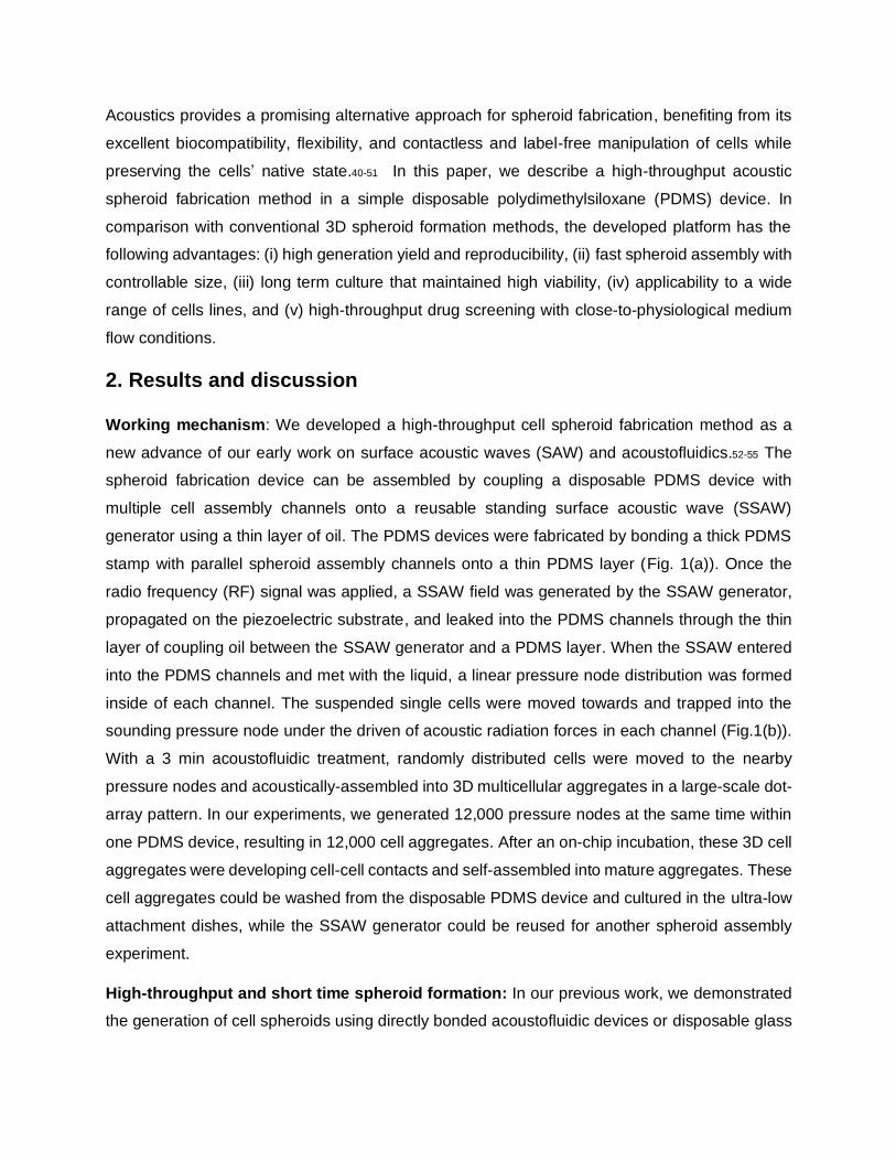
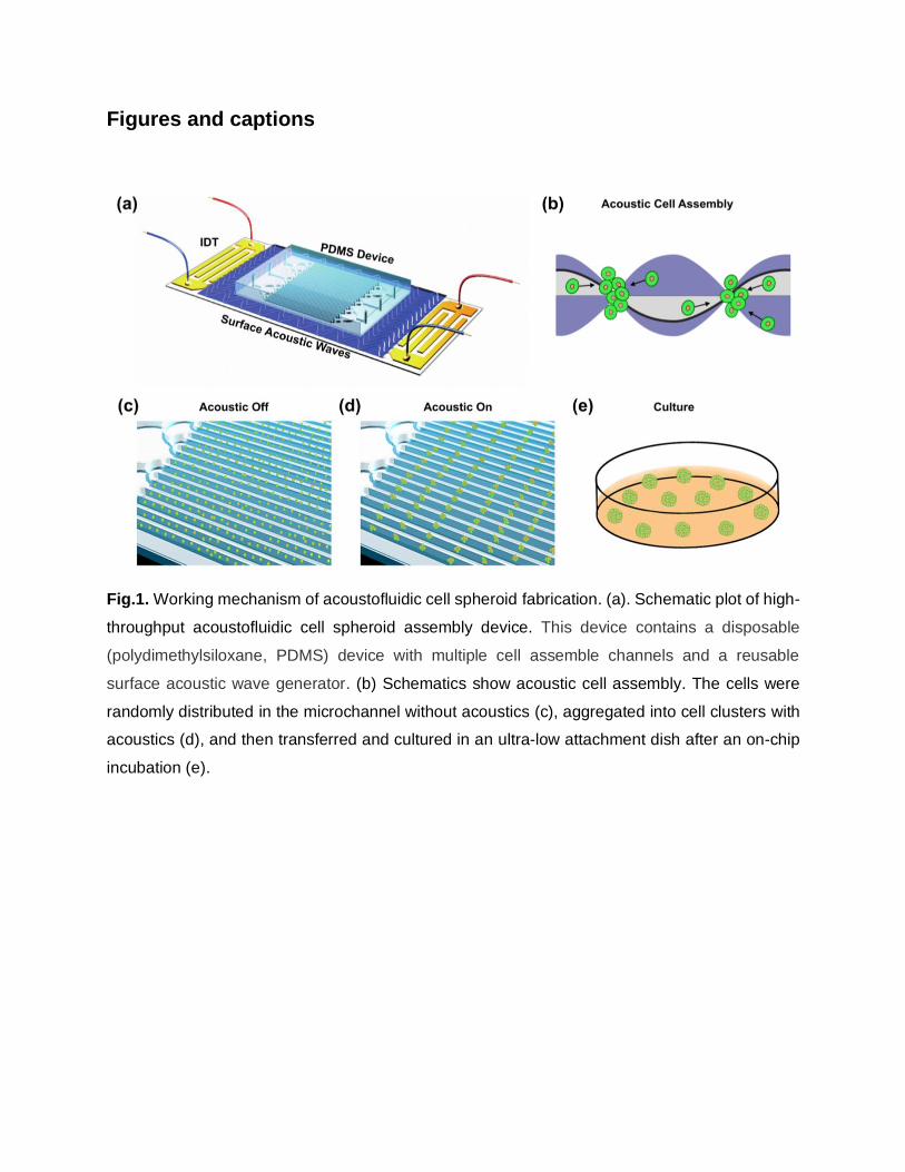
Working mechanism: We developed a high-throughput cell spheroid fabrication method as a
new advance of our early work on surface acoustic waves (SAW) and acoustofluidics.52-55 The
spheroid fabrication device can be assembled by coupling a disposable PDMS device with
multiple cell assembly channels onto a reusable standing surface acoustic wave (SSAW)
generator using a thin layer of oil. The PDMS devices were fabricated by bonding a thick PDMS
stamp with parallel spheroid assembly channels onto a thin PDMS layer (Fig. 1(a)). Once the
radio frequency (RF) signal was applied, a SSAW field was generated by the SSAW generator,
propagated on the piezoelectric substrate, and leaked into the PDMS channels through the thin
layer of coupling oil between the SSAW generator and a PDMS layer. When the SSAW entered
into the PDMS channels and met with the liquid, a linear pressure node distribution was formed
inside of each channel. The suspended single cells were moved towards and trapped into the
sounding pressure node under the driven of acoustic radiation forces in each channel (Fig.1(b)).
With a 3 min acoustofluidic treatment, randomly distributed cells were moved to the nearby
pressure nodes and acoustically-assembled into 3D multicellular aggregates in a large-scale dot-
array pattern. In our experiments, we generated 12,000 pressure nodes at the same time within
one PDMS device, resulting in 12,000 cell aggregates. After an on-chip incubation, these 3D cell
aggregates were developing cell-cell contacts and self-assembled into mature aggregates. These
cell aggregates could be washed from the disposable PDMS device and cultured in the ultra-low
attachment dishes, while the SSAW generator could be reused for another spheroid assembly
experiment.
High-throughput and short time spheroid formation: In our previous work, we demonstrated
the generation of cell spheroids using directly bonded acoustofluidic devices or disposable glass
capillaries coupled with standing acoustic waves, however, the throughput of cell spheroid
generation was limited at around 300.56, 57 In this work, we developed a new strategy to
significantly increase the throughput of cell spheroid production from 300 to 6,000. In our
experiments, we generated surface acoustic waves with a wavelength of 300 µm. The PDMS
device was designed with 60 parallel channels and each channel with a dimension of 3 cm x 150
µm x 150 µm. Thus, about 12,000 pressure nodes could be formed at the same time within a
PDMS device. The suspended cells in each channel can be moved towards and captured in the
sounding pressure node as a 3D trap with a dimension of 150 µm x 150 µm x 150 µm resulting in
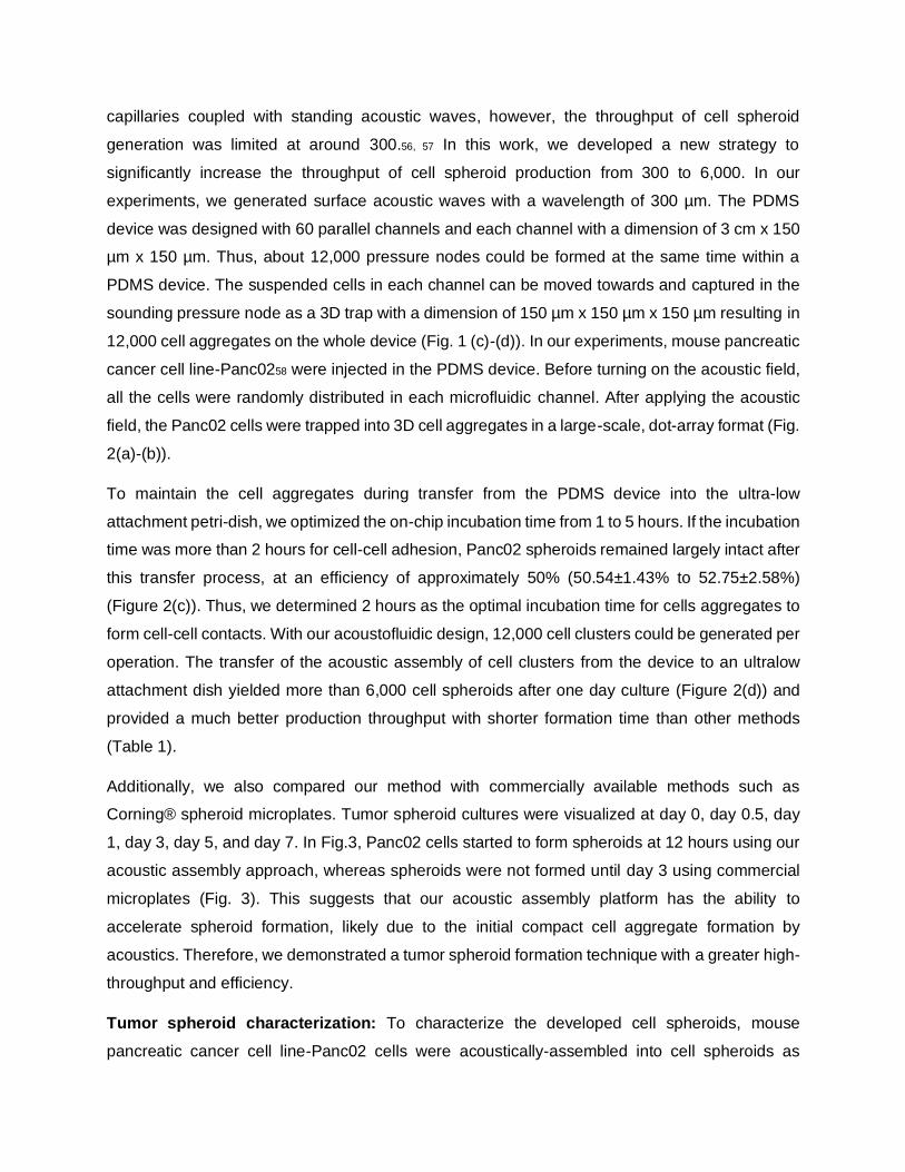
12,000 cell aggregates on the whole device (Fig. 1 (c)-(d)). In our experiments, mouse pancreatic
cancer cell line-Panc0258 were injected in the PDMS device. Before turning on the acoustic field,
all the cells were randomly distributed in each microfluidic channel. After applying the acoustic
field, the Panc02 cells were trapped into 3D cell aggregates in a large-scale, dot-array format (Fig.
2(a)-(b)).
To maintain the cell aggregates during transfer from the PDMS device into the ultra-low
attachment petri-dish, we optimized the on-chip incubation time from 1 to 5 hours. If the incubation
time was more than 2 hours for cell-cell adhesion, Panc02 spheroids remained largely intact after
this transfer process, at an efficiency of approximately 50% (50.54±1.43% to 52.75±2.58%)
(Figure 2(c)). Thus, we determined 2 hours as the optimal incubation time for cells aggregates to
form cell-cell contacts. With our acoustofluidic design, 12,000 cell clusters could be generated per
operation. The transfer of the acoustic assembly of cell clusters from the device to an ultralow
attachment dish yielded more than 6,000 cell spheroids after one day culture (Figure 2(d)) and
provided a much better production throughput with shorter formation time than other methods
(Table 1).
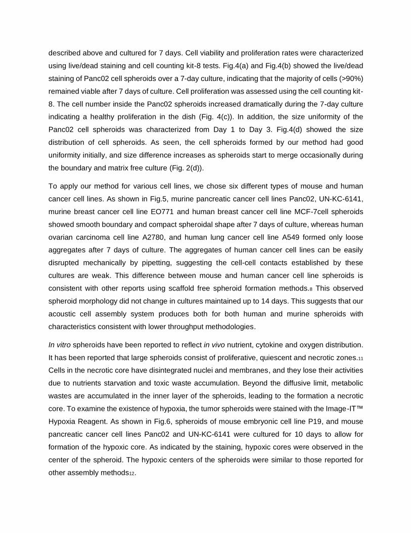
Additionally, we also compared our method with commercially available methods such as
Corning® spheroid microplates. Tumor spheroid cultures were visualized at day 0, day 0.5, day
1, day 3, day 5, and day 7. In Fig.3, Panc02 cells started to form spheroids at 12 hours using our
acoustic assembly approach, whereas spheroids were not formed until day 3 using commercial
microplates (Fig. 3). This suggests that our acoustic assembly platform has the ability to
accelerate spheroid formation, likely due to the initial compact cell aggregate formation by
acoustics. Therefore, we demonstrated a tumor spheroid formation technique with a greater high-
throughput and efficiency.
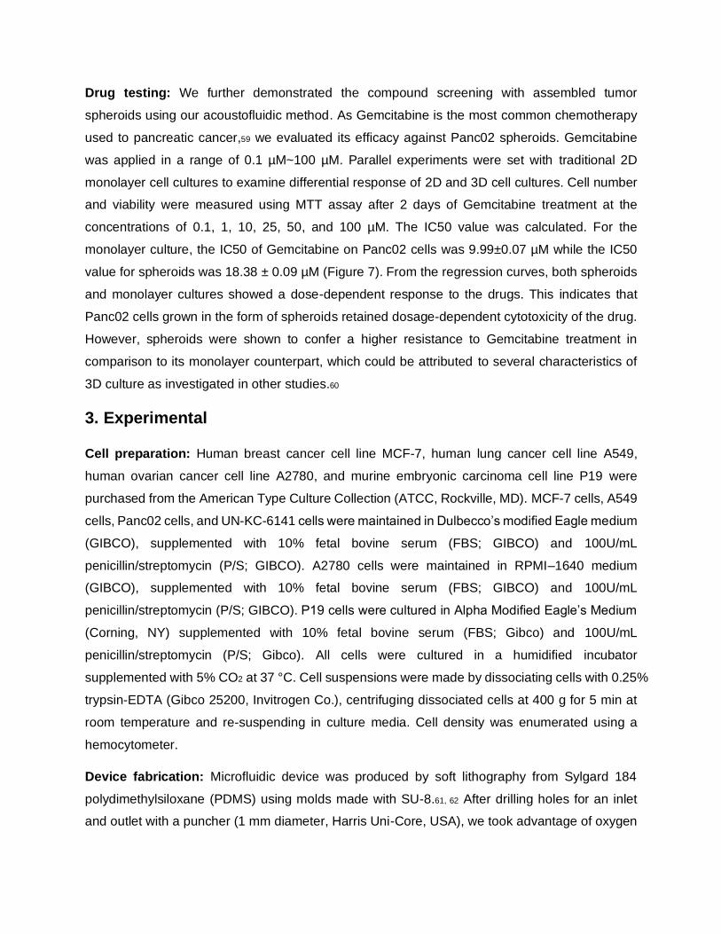
Tumor spheroid characterization: To characterize the developed cell spheroids, mouse
pancreatic cancer cell line-Panc02 cells were acoustically-assembled into cell spheroids as
described above and cultured for 7 days. Cell viability and proliferation rates were characterized
using live/dead staining and cell counting kit-8 tests. Fig.4(a) and Fig.4(b) showed the live/dead
staining of Panc02 cell spheroids over a 7-day culture, indicating that the majority of cells (>90%)
remained viable after 7 days of culture. Cell proliferation was assessed using the cell counting kit-
8. The cell number inside the Panc02 spheroids increased dramatically during the 7-day culture
indicating a healthy proliferation in the dish (Fig. 4(c)). In addition, the size uniformity of the
Panc02 cell spheroids was characterized from Day 1 to Day 3. Fig.4(d) showed the size
distribution of cell spheroids. As seen, the cell spheroids formed by our method had good
uniformity initially, and size difference increases as spheroids start to merge occasionally during
the boundary and matrix free culture (Fig. 2(d)).
To apply our method for various cell lines, we chose six different types of mouse and human
cancer cell lines. As shown in Fig.5, murine pancreatic cancer cell lines Panc02, UN-KC-6141,
murine breast cancer cell line EO771 and human breast cancer cell line MCF-7cell spheroids
showed smooth boundary and compact spheroidal shape after 7 days of culture, whereas human
ovarian carcinoma cell line A2780, and human lung cancer cell line A549 formed only loose
aggregates after 7 days of culture. The aggregates of human cancer cell lines can be easily
disrupted mechanically by pipetting, suggesting the cell-cell contacts established by these
cultures are weak. This difference between mouse and human cancer cell line spheroids is
consistent with other reports using scaffold free spheroid formation methods.8 This observed
spheroid morphology did not change in cultures maintained up to 14 days. This suggests that our
acoustic cell assembly system produces both for both human and murine spheroids with
characteristics consistent with lower throughput methodologies.
In vitro spheroids have been reported to reflect in vivo nutrient, cytokine and oxygen distribution.
It has been reported that large spheroids consist of proliferative, quiescent and necrotic zones.11
Cells in the necrotic core have disintegrated nuclei and membranes, and they lose their activities
due to nutrients starvation and toxic waste accumulation. Beyond the diffusive limit, metabolic
wastes are accumulated in the inner layer of the spheroids, leading to the formation a necrotic
core. To examine the existence of hypoxia, the tumor spheroids were stained with the Image-IT™
Hypoxia Reagent. As shown in Fig.6, spheroids of mouse embryonic cell line P19, and mouse
pancreatic cancer cell lines Panc02 and UN-KC-6141 were cultured for 10 days to allow for
formation of the hypoxic core. As indicated by the staining, hypoxic cores were observed in the
center of the spheroid. The hypoxic centers of the spheroids were similar to those reported for
other assembly methods12.
Drug testing: We further demonstrated the compound screening with assembled tumor
spheroids using our acoustofluidic method. As Gemcitabine is the most common chemotherapy
used to pancreatic cancer,59 we evaluated its efficacy against Panc02 spheroids. Gemcitabine
was applied in a range of 0.1 µM~100 µM. Parallel experiments were set with traditional 2D
monolayer cell cultures to examine differential response of 2D and 3D cell cultures. Cell number
and viability were measured using MTT assay after 2 days of Gemcitabine treatment at the
concentrations of 0.1, 1, 10, 25, 50, and 100 µM. The IC50 value was calculated. For the
monolayer culture, the IC50 of Gemcitabine on Panc02 cells was 9.99±0.07 µM while the IC50
value for spheroids was 18.38 ± 0.09 µM (Figure 7). From the regression curves, both spheroids
and monolayer cultures showed a dose-dependent response to the drugs. This indicates that
Panc02 cells grown in the form of spheroids retained dosage-dependent cytotoxicity of the drug.
However, spheroids were shown to confer a higher resistance to Gemcitabine treatment in
comparison to its monolayer counterpart, which could be attributed to several characteristics of
3D culture as investigated in other studies.60
3. Experimental
Cell preparation: Human breast cancer cell line MCF-7, human lung cancer cell line A549,
human ovarian cancer cell line A2780, and murine embryonic carcinoma cell line P19 were
purchased from the American Type Culture Collection (ATCC, Rockville, MD). MCF-7 cells, A549
cells, Panc02 cells, and UN-KC-6141 cells were maintained in Dulbecco’s modified Eagle medium
(GIBCO), supplemented with 10% fetal bovine serum (FBS; GIBCO) and 100U/mL
penicillin/streptomycin (P/S; GIBCO). A2780 cells were maintained in RPMI–1640 medium
(GIBCO), supplemented with 10% fetal bovine serum (FBS; GIBCO) and 100U/mL
penicillin/streptomycin (P/S; GIBCO). P19 cells were cultured in Alpha Modified Eagle’s Medium
(Corning, NY) supplemented with 10% fetal bovine serum (FBS; Gibco) and 100U/mL
penicillin/streptomycin (P/S; Gibco). All cells were cultured in a humidified incubator
supplemented with 5% CO2 at 37 °C. Cell suspensions were made by dissociating cells with 0.25%
trypsin-EDTA (Gibco 25200, Invitrogen Co.), centrifuging dissociated cells at 400 g for 5 min at
room temperature and re-suspending in culture media. Cell density was enumerated using a
hemocytometer.
Device fabrication: Microfluidic device was produced by soft lithography from Sylgard 184
polydimethylsiloxane (PDMS) using molds made with SU-8.61, 62 After drilling holes for an inlet
and outlet with a puncher (1 mm diameter, Harris Uni-Core, USA), we took advantage of oxygen
plasma treatment (PDC001, Harrick Plasma, USA) to bind the molded PDMS chamber to a thin
layer of PDMS film (25 μm thickness) that was spin coated on a surface polished silicon wafer.
The SSAW generator was fabricated by a standard soft lithography and lift-off process. A 7-μm-
thick photoresist layer (S1813, MicroChem, USA) was spin-coated on the piezoelectric substrate
(a 500-μm-thick, double-side polished, 128° YX-propagation LiNbO3 wafer). Then, the designed
IDT patterns of 40 electrode pairs with the 75 μm finger width and periodic spacing were
transferred from the plastic mask (Kunshan Kaisheng Electronics Co., Ltd, China) to the substrate
by UV exposure. The IDT patterns were developed in a photoresist developer (MF CD-26,
Microposit, USA) and deposited with double metal layers (Cr/50 Å, Au/600 Å) by a thermal
evaporation (JSD-350, Anhui Jiaoshuo Vacuum Technology Co., Ltd, China). IDTs on the
piezoelectric substrate were finally obtained after a standard lift-off process. Then the resonant
frequency of the fabricated SSAW generator was measured at around 13.13-13.41 MHz using a
network analyzer (E8362C, Agilent, USA).
Prior to the cell assembly, the PDMS device was sterilized by autoclaving at 121 °C for 30 min.
During experiment manipulation, the disposable PDMS device was assembled on top of a SSAW
substrate with a layer of coupling oil. The mineral oil (SLBX1961, SIGMA, USA) was chosen as
the coupling material to introduce the acoustic waves from the substrate to the above device. In
comparison with water or olive oil or other coupling materials, mineral oil can offer both a lower
evaporation rate and a better coupling performance with causing no harm to cell viability.
High-throughput acoustic cell assembly: In the cell patterning experiment, cells were
aggregated into linear assembly arrays in PDMS channels by applying a radio frequency signal
produced by a function generator (AFG3102C, Tektronix, USA) and modulated with an amplifier
(25A100A, Amplifier Research, USA) to the IDT pair. The movement of cells was monitored and
recorded by a microscope (IX-81, Olympus, Japan) with a CMOS camera (ORCA-Flash 4.0,
HAMAMATSU, Japan) connected to a computer (Cellsens) The input voltages on the devices
were from 10 to 60 Vpp. Different kinds of cells were resuspended in type I collagen (Life
Technologies, USA) and injected after the acoustic field was formed. The whole acoustic cell
aggregation process took one minute. To form cell spheroids, the cell clusters were incubated in
PDMS device for a short period and subsequently transferred into ultra-low attachment plates
(Corning, 3471, USA) with fresh cell culture medium. The cell cluster plate was incubated and
maintained at 5% CO2 and 37 °C. These cell clusters were imaged and recorded every day for
several days until spheroids like structure formed.
Spheroid formation by Corning® spheroid microplates: Panc02 cells used in this study were
harvested as single cell suspension with 0.25% Trypsin-EDTA solution (Gibco 25200, Invitrogen
Co) for 2 min at 37 °C, centrifuged at 400 g for 5 min and resuspended in their respective culture
media. To induce 3D spheroid formation, the harvested cells were seeded at fixed density of 500
cells per well into Corning® spheroid microplates (Corning, USA), spun down at 400 g for 3 min
and incubated at 37 °C, 5% CO2. Spheroid cultures were visualized at 0~7 days using a
microscope.
Tumor spheroid proliferation and viability tests: Cell proliferation was assessed using the cell
counting kit-8 (CCK-8; Sigma-Aldrich, St. Louis, MO, USA) according to the manufacturer’s
instructions. Control cells were exposed to an equivalent amount of vehicle. Tumor spheroids or
cultured tumor cells were incubated in the CCK-8 solution for 4 h, and the supernatants were
transferred to 96-well plates. Cell proliferation was assessed by measuring the absorbance at 450
nm using the Epoch™ microplate spectrophotometer (Bio-Tek Instruments, USA). The viability
testing was achieved by the live/dead stain assay (live/dead viability kit, L3224, Thermo Fisher
Scientific Inc.). Tumor spheroids were stained with a mixture of 2 μM Calcein AM and 4 μM
ethidium homodimer-1 to stain for live and dead cells, respectively. Brightfield and fluorescent
images were captured using a microscope (IX83, Olympus, Japan).
Tumor spheroids hypoxia characterization: To examine hypoxia, the tumor spheroids were
stained with the Image-IT™ Hypoxia Reagent (Invitrogen™, USA) according to the
manufacturer’s instruction. The reagent was added to the spheroids at a final concentration of 10
μM and incubate at 37 °C for 48 hours. The spheroids were then imaged on the fluorescence
microscope with excitation/emission of 490/610 nm.
Cytotoxicity assay: The cytotoxic effect of the chemotherapeutic drug Gemcitabine (LC
Laboratories, Woburn, MA, USA) on Panc02 cell spheroids culture was compared to the cytotoxic
effect on monolayer culture. For the cytotoxic assay, 1×10⁴ cells suspended in complete medium
were seeded in each well of a 96-well plate. The 4-day-old Panc02 spheroids were transferred to
a new 96 ultra-low attachment well plate. The next day, the cells and spheroids were treated with
different concentrations of Gemcitabine solution (0, 0.1, 1, 10, 25, 50, 100 µM) in sextuplicate
wells. After incubating the cells with Gemcitabine for 48 h, 20 µl of MTT solution (5 mg/ml) was
added into each well, and the cells were incubated for 4 hours. While the monolayer culture was
left untouched in the original plate, the content of each well containing the tumor spheroids culture
was transferred to a new, flat bottom 96-well plate before the plate was centrifuged at 200×g for
5 minutes. Then, 100 µl of media was aspirated from each well from the plates containing the
monolayer and spheroids cultures. The plates were then blotted dry on paper towels, followed by
the addition of 100 µl of DMSO. Finally, absorbance was recorded at 570 nm using the Epoch™
microplate spectrophotometer (Bio-Tek Instruments, USA). The assay was carried out with 3
replicates for each culture.
Statistical analysis: Data presented in this study are representative of at least three independent
experiments. All values are expressed as arithmetic mean ± standard deviation (SD). Statistical
difference between experimental groups was determined using Student’s t test, when P
values<0.05 were considered statistically significant.
4. Conclusions
Our study presents a proof-of-concept in improving the cell spheroid formation throughout and
time using acoustofluidics, for the purpose of drug efficacy, metabolism and toxicity studies.
Compared to the traditional spheroid formation and culture methods, our approach generates a
larger number of spheroids in a short time. Like other spheroid methods, the scaffold-free nature
of the presented approach has the potential to eliminate the effects associated with stiffness,
roughness and chemical composition of these substrates that affect cell behavior and phenotype.
We expect that the acoustofluidic cell assembly technique can be used as a powerful tool for high-
throughput screening in new drug development processes.
5. Conflicts of interest
The authors declare no conflicts of interests.
6. Acknowledgments
This project was supported by the departmental start-up fund and the Vice Provost for the
Research through the Faculty Research Support Program (Indiana University Bloomington).
Murine pancreatic cancer cell line Panc02 was a kind grift from Dr. Jill Smith (Georgetown
University), and UN-KC-6141 cell line was a kind gift from Surinder K. Batra (University of
Nebraska)
References
1. W. Mueller-Klieser, American Journal of Physiology-Cell Physiology, 1997, 273, C1109-C1123. 2. A. Abbott, Nature, 2003, 424, 870. 3. F. Pampaloni, E. G. Reynaud and E. H. K. Stelzer, Nat Rev Mol Cell Bio, 2007, 8, 839-845. 4. L. G. Griffith and M. A. Swartz, Nat Rev Mol Cell Bio, 2006, 7, 211. 5. C. S. Shin, B. Kwak, B. Han and K. Park, Molecular Pharmaceutics, 2013, 10, 2167-2175. 6. R. Z. Lin and H. Y. Chang, Biotechnol J, 2008, 3, 1172-1184. 7. J. Friedrich, C. Seidel, R. Ebner and L. A. Kunz-Schughart, Nature Protocols, 2009, 4, 309. 8. B. W. Huang and J. Q. Gao, J Control Release, 2018, 270, 246-259. 9. L. P. Ferreira, V. M. Gaspar and J. F. Mano, Acta Biomater, 2018, 75, 11-34. 10. Y. B. Lee, E. M. Kim, H. Byun, H. K. Chang, K. Jeong, Z. M. Aman, Y. S. Choi, J. Park and H. Shin,
Biomaterials, 2018, 165, 105-120. 11. E. Fennema, N. Rivron, J. Rouwkema, C. van Blitterswijk and J. de Boer, Trends Biotechnol, 2013,
31, 108-115. 12. M. W. Laschke and M. D. Menger, Biotechnol Adv, 2017, 35, 782-791. 13. K. M. Tevis, Y. L. Colson and M. W. Grinstaff, Adv Biosyst, 2017, 1. 14. D. L. Priwitaningrum, J. G. Blonde, A. Sridhar, J. van Baarlen, W. E. Hennink, G. Storm, S. Le Gac
and J. Prakash, J Control Release, 2016, 244, 257-268. 15. M. Vinci, S. Gowan, F. Boxall, L. Patterson, M. Zimmermann, W. Court, C. Lomas, M. Mendiola,
D. Hardisson and S. A. Eccles, BMC Biology, 2012, 10, 29. 16. J. Friedrich, R. Ebner and L. A. Kunz-Schughart, International Journal of Radiation Biology, 2007,
83, 849-871. 17. S. Nath and G. R. Devi, Pharmacology & Therapeutics, 2016, 163, 94-108. 18. Y. C. Tung, A. Y. Hsiao, S. G. Allen, Y. S. Torisawa, M. Ho and S. Takayama, Analyst, 2011, 136,
473-478. 19. A. P. Napolitano, D. M. Dean, A. J. Man, J. Youssef, D. N. Ho, A. P. Rago, M. P. Lech and J. R.
Morgan, Biotechniques, 2007, 43, 494, 496-500. 20. M. Ingram, G. B. Techy, R. Saroufeem, O. Yazan, K. S. Narayan, T. J. Goodwin and G. F. Spaulding,
In Vitro Cellular & Developmental Biology - Animal, 1997, 33, 459-466. 21. S. Yan, J. Wei, Y. Liu, H. Zhang, J. Chen and X. Li, Acta Biomater, 2015, 28, 138-148. 22. R. H. Dosh, A. Essa, N. Jordan-Mahy, C. Sammon and C. L. Le Maitre, Acta Biomaterialia, 2017,
62, 128-143. 23. Y. Yoshii, T. Furukawa, A. Waki, H. Okuyama, M. Inoue, M. Itoh, M. R. Zhang, H. Wakizaka, C.
Sogawa, Y. Kiyono, H. Yoshii, Y. Fujibayashi and T. Saga, Biomaterials, 2015, 51, 278-289. 24. C. T. Kuo, J. Y. Wang, Y. F. Lin, A. M. Wo, B. P. C. Chen and H. Lee, Sci Rep, 2017, 7, 4363. 25. V. Vickerman, J. Blundo, S. Chung and R. Kamm, Lab Chip, 2008, 8, 1468-1477. 26. A. P. Aijian and R. L. Garrell, J Lab Autom, 2015, 20, 283-295. 27. Y. S. Torisawa, B. H. Chueh, D. Huh, P. Ramamurthy, T. M. Roth, K. F. Barald and S. Takayama,
Lab Chip, 2007, 7, 770-776. 28. C. T. Kuo, S. R. Lu, W. M. Chen, J. Y. Wang, S. C. Lee, H. H. Chang, A. M. Wo, B. P. C. Chen and H.
Lee, Lab Chip, 2018, 18, 2453-2465. 29. J. Park, G. H. Lee, J. Yull Park, J. C. Lee and H. C. Kim, Biofabrication, 2017, 9, 045006. 30. L. Y. Yeo, H. C. Chang, P. P. Chan and J. R. Friend, Small, 2011, 7, 12-48. 31. A. Neild, S. Oberti and J. Dual, Sensors and Actuators B: Chemical, 2007, 121, 452-461. 32. S. Hao, L. Ha, G. Cheng, Y. Wan, Y. Xia, D. M. Sosnoski, A. M. Mastro and S. Y. Zheng, Small, 2018,
14, e1702787.
33. C. M. Puleo, H. C. Yeh and T. H. Wang, Tissue Eng, 2007, 13, 2839-2854. 34. D. D. Nalayanda, C. M. Puleo, W. B. Fulton, T. H. Wang and F. Abdullah, Exp Lung Res, 2007, 33,
321-335. 35. J. Friend and L. Y. Yeo, Reviews of Modern Physics, 2011, 83, 647-704. 36. L. Trung-Dung and N. Nam-Trung, Micro and Nanosystems, 2010, 2, 217-225. 37. L. Meng, F. Cai, Q. Jin, L. Niu, C. Jiang, Z. Wang, J. Wu and H. Zheng, Sensors and Actuators B:
Chemical, 2011, 160, 1599-1605. 38. H. D. Xi, H. Zheng, W. Guo, A. M. Ganan-Calvo, Y. Ai, C. W. Tsao, J. Zhou, W. Li, Y. Huang, N. T.
Nguyen and S. H. Tan, Lab Chip, 2017, 17, 751-771. 39. L. Y. Yeo and J. R. Friend, Biomicrofluidics, 2009, 3, 12002. 40. D. J. Collins, B. Morahan, J. Garcia-Bustos, C. Doerig, M. Plebanski and A. Neild, Nat Commun,
2015, 6, 8686. 41. F. Cai, Z. He, Z. Liu, L. Meng, X. Cheng and H. Zheng, Applied Physics Letters, 2011, 99, 253505. 42. D. J. Collins, A. Neild and Y. Ai, Lab Chip, 2016, 16, 471-479. 43. L. Y. Yeo and J. R. Friend, Annual Review of Fluid Mechanics, 2014, 46, 379-406. 44. K. H. Lam, H. S. Hsu, Y. Li, C. Lee, A. Lin, Q. Zhou, E. S. Kim and K. K. Shung, Biotechnol Bioeng,
2013, 110, 881-886. 45. T.-D. Luong, V.-N. Phan and N.-T. Nguyen, Microfluidics and Nanofluidics, 2010, 10, 619-625. 46. L. Meng, F. Cai, J. Chen, L. Niu, Y. Li, J. Wu and H. Zheng, Applied Physics Letters, 2012, 100,
173701. 47. W. Qi, R. Li, T. Ma, J. Li, K. Kirk Shung, Q. Zhou and Z. Chen, Appl Phys Lett, 2013, 103, 103704. 48. Q. Zhou, S. Lau, D. Wu and K. K. Shung, Prog Mater Sci, 2011, 56, 139-174. 49. L. Meng, F. Cai, Z. Zhang, L. Niu, Q. Jin, F. Yan, J. Wu, Z. Wang and H. Zheng, Biomicrofluidics,
2011, 5, 044104. 50. Z. Ma, D. J. Collins and Y. Ai, Analytical Chemistry, 2016, 88, 5316-5323. 51. J. Reboud, R. Wilson, Y. Zhang, M. H. Ismail, Y. Bourquin and J. M. Cooper, Lab on a Chip, 2012,
12, 1268-1273. 52. F. Guo, P. Li, J. B. French, Z. Mao, H. Zhao, S. Li, N. Nama, J. R. Fick, S. J. Benkovic and T. J. Huang,
Proc Natl Acad Sci U S A, 2015, 112, 43-48. 53. F. Guo, Z. M. Mao, Y. C. Chen, Z. W. Xie, J. P. Lata, P. Li, L. Q. Ren, J. Y. Liu, J. Yang, M. Dao, S.
Suresh and T. J. Huang, P Natl Acad Sci USA, 2016, 113, 1522-1527. 54. X. Ding, P. Li, S.-C. S. Lin, Z. S. Stratton, N. Nama, F. Guo, D. Slotcavage, X. Mao, J. Shi, F. Costanzo
and T. J. Huang, Lab on a Chip, 2013, 13, 3626-3649. 55. F. Guo, Y. Xie, S. Li, J. Lata, L. Ren, Z. Mao, B. Ren, M. Wu, A. Ozcelik and T. J. Huang, Lab on a
Chip, 2015, 15, 4517-4523. 56. K. Chen, M. Wu, F. Guo, P. Li, C. Y. Chan, Z. Mao, S. Li, L. Ren, R. Zhang and T. J. Huang, Lab Chip,
2016, 16, 2636-2643. 57. W. Yue, A. Zheng, C. Bin, M. Maram, B. Maria, L. Xiongbin and G. Feng, Nanotechnology, 2018,
29, 504006. 58. J. P. Smith, S. Wang, S. Nadella, S. A. Jablonski and L. M. Weiner, Cancer Immunology,
Immunotherapy, 2018, 67, 195-207. 59. C. J. Campen, T. Dragovich and A. F. Baker, American Journal of Health-System Pharmacy, 2011,
68, 573. 60. M. P. Torres, S. Rachagani, J. J. Souchek, K. Mallya, S. L. Johansson and S. K. Batra, PLoS One,
2013, 8, e80580. 61. D. C. Duffy, J. C. McDonald, O. J. A. Schueller and G. M. Whitesides, Analytical Chemistry, 1998,
70, 4974-4984.
62. P. Abgrall, V. Conedera, H. Camon, A. M. Gue and N. T. Nguyen, Electrophoresis, 2007, 28, 4539-4551.
63. D. A. Close, D. P. Camarco, F. Shan, S. J. Kochanek and P. A. Johnston, in High Content Screening: A Powerful Approach to Systems Cell Biology and Phenotypic Drug Discovery, eds. P. A. Johnston and O. J. Trask, Springer New York, New York, NY, 2018, DOI: 10.1007/978-1-4939-7357-6_20, pp. 355-369.
64. J. H. Lee and W. Hur, Biotechnology and Bioengineering, 2014, 111, 1038-1047. 65. T.-J. Lee, S. H. Bhang, W.-G. La, H. S. Yang, J. Y. Seong, H. Lee, G.-I. Im, S.-H. Lee and B.-S. Kim,
Biotechnology Letters, 2011, 33, 829-836.
Figures and captions
Fig.1. Working mechanism of acoustofluidic cell spheroid fabrication. (a). Schematic plot of high-
throughput acoustofluidic cell spheroid assembly device. This device contains a disposable
(polydimethylsiloxane, PDMS) device with multiple cell assemble channels and a reusable
surface acoustic wave generator. (b) Schematics show acoustic cell assembly. The cells were
randomly distributed in the microchannel without acoustics (c), aggregated into cell clusters with
acoustics (d), and then transferred and cultured in an ultra-low attachment dish after an on-chip
incubation (e).
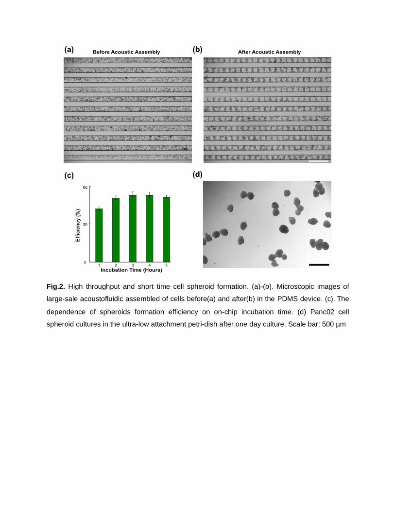
Fig.2. High throughput and short time cell spheroid formation. (a)-(b). Microscopic images of
large-sale acoustofluidic assembled of cells before(a) and after(b) in the PDMS device. (c). The
dependence of spheroids formation efficiency on on-chip incubation time. (d) Panc02 cell
spheroid cultures in the ultra-low attachment petri-dish after one day culture. Scale bar: 500 µm
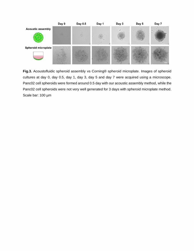
Fig.3. Acoustofluidic spheroid assembly vs Corning® spheroid microplate. Images of spheroid
cultures at day 0, day 0.5, day 1, day 3, day 5 and day 7 were acquired using a microscope.
Panc02 cell spheroids were formed around 0.5 day with our acoustic assembly method, while the
Panc02 cell spheroids were not very well generated for 3 days with spheroid microplate method.
Scale bar: 100 µm
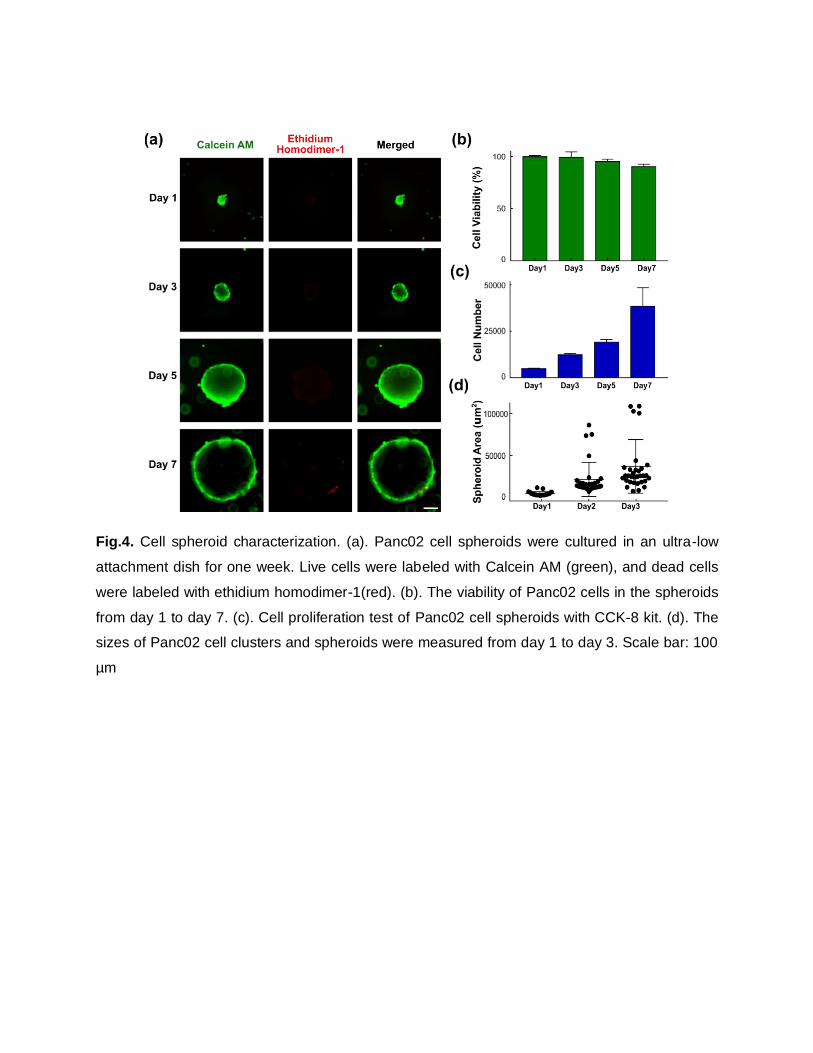
Fig.4. Cell spheroid characterization. (a). Panc02 cell spheroids were cultured in an ultra-low
attachment dish for one week. Live cells were labeled with Calcein AM (green), and dead cells
were labeled with ethidium homodimer-1(red). (b). The viability of Panc02 cells in the spheroids
from day 1 to day 7. (c). Cell proliferation test of Panc02 cell spheroids with CCK-8 kit. (d). The
sizes of Panc02 cell clusters and spheroids were measured from day 1 to day 3. Scale bar: 100
µm
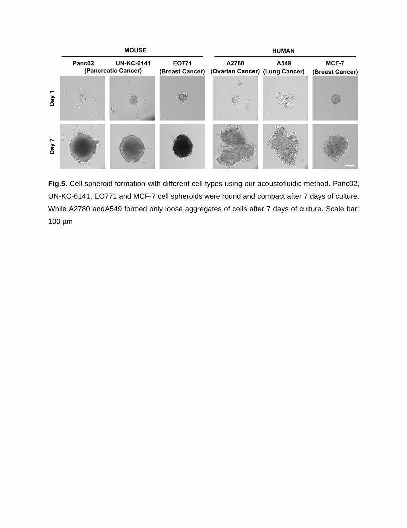
Fig.5. Cell spheroid formation with different cell types using our acoustofluidic method. Panc02,
UN-KC-6141, EO771 and MCF-7 cell spheroids were round and compact after 7 days of culture.
While A2780 andA549 formed only loose aggregates of cells after 7 days of culture. Scale bar:
100 µm
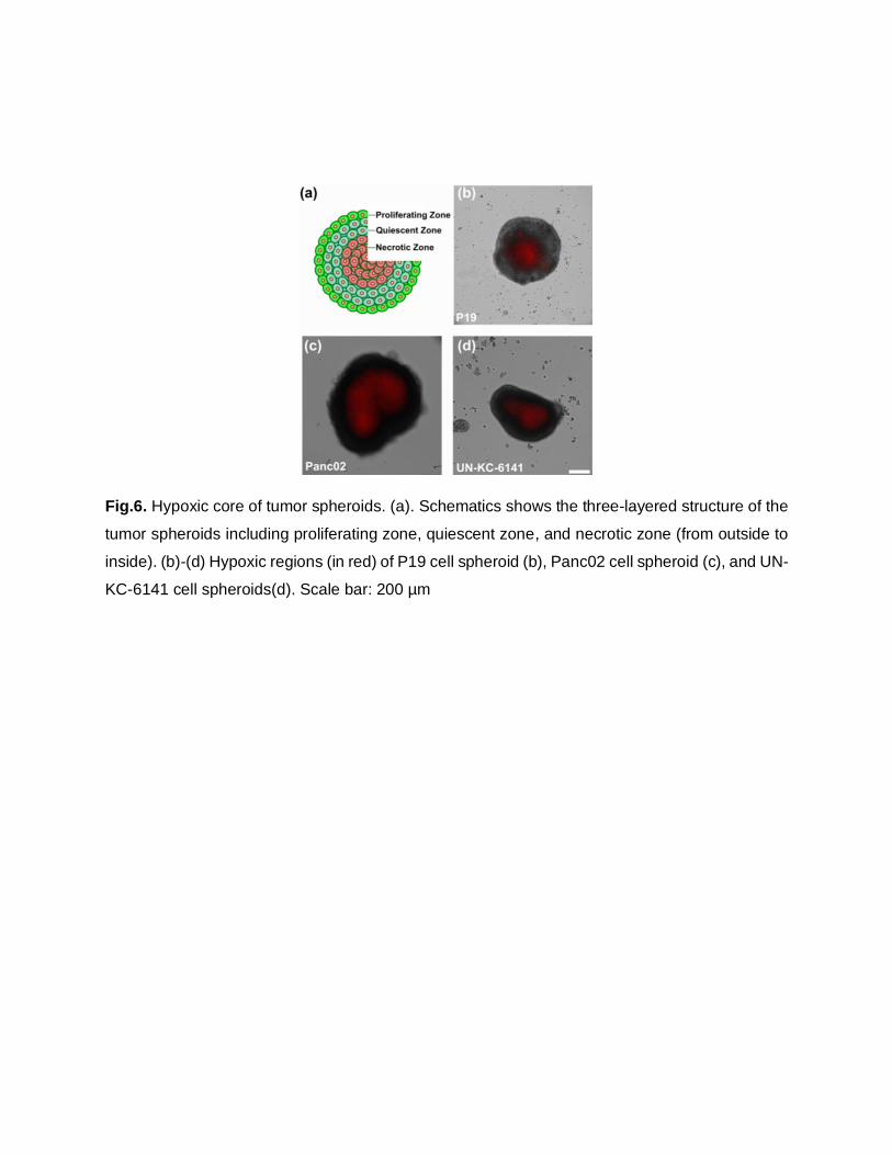
Fig.6. Hypoxic core of tumor spheroids. (a). Schematics shows the three-layered structure of the
tumor spheroids including proliferating zone, quiescent zone, and necrotic zone (from outside to
inside). (b)-(d) Hypoxic regions (in red) of P19 cell spheroid (b), Panc02 cell spheroid (c), and UN-
KC-6141 cell spheroids(d). Scale bar: 200 µm
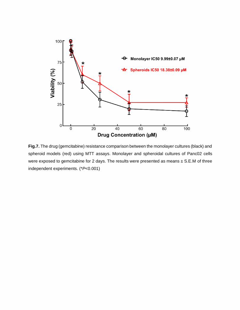
Fig.7. The drug (gemcitabine) resistance comparison between the monolayer cultures (black) and
spheroid models (red) using MTT assays. Monolayer and spheroidal cultures of Panc02 cells
were exposed to gemcitabine for 2 days. The results were presented as means ± S.E.M of three
independent experiments. (*P<0.001)
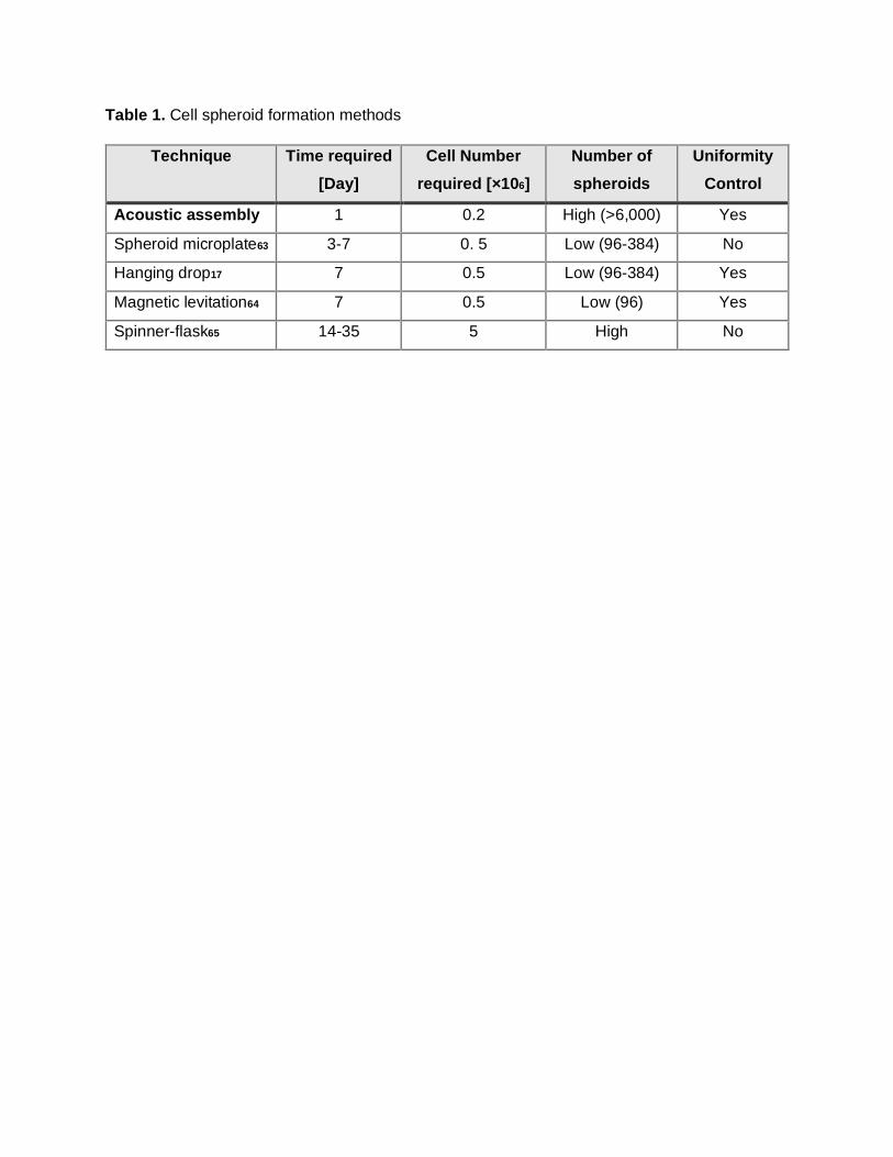
Table 1. Cell spheroid formation methods
Technique Time required
[Day]
Cell Number
required [×106]
Number of
spheroids
Uniformity
Control
Acoustic assembly 1 0.2 High (>6,000) Yes
Spheroid microplate63 3-7 0. 5 Low (96-384) No
Hanging drop17 7 0.5 Low (96-384) Yes
Magnetic levitation64 7 0.5 Low (96) Yes
Spinner-flask65 14-35 5 High No