Embed Size (px)
Citation preview
http://e-jbm.org/ 175
Copyright © 2015 The Korean Society for Bone and Mineral Research
This is an Open Access article distributed under the terms of the Creative Commons Attribution Non-Commercial Li-cense (http://creativecommons.org/licenses/by-nc/3.0/) which permits unrestricted non-commercial use, distribu-tion, and reproduction in any medium, provided the original work is properly cited.
J Bone Metab 2015;22:175-181http://dx.doi.org/10.11005/jbm.2015.22.4.175pISSN 2287-6375 eISSN 2287-7029
Osteoporotic Fracture: 2015 Position Statement of the Korean Society for Bone and Mineral ResearchJe-Hyun Yoo1, Seong-Hwan Moon2, Yong-Chan Ha3, Dong Yeon Lee4, Hyun Sik Gong4, Si Young Park5, Kyu Hyun Yang6
1Department of Orthopedic Surgery, Hallym University Sacred Heart Hospital, Anyang; 2Department of Orthopaedic Surgery, Yonsei University College of Medicine, Seoul; 3Department of Orthopaedic Surgery, Chung-Ang University College of Medicine, Seoul; 4Department of Orthopaedic Surgery, Seoul National University College of Medicine, Seoul; 5Department of Orthopaedic Surgery, Korea University College of Medicine, Seoul; 6Department of Orthopedic Surgery, Yonsei University College of Medicine, Gangnam Severance Hospital, Seoul, Korea
Osteoporotic fractures are one of the most common causes of disability and a major contributor to medical care costs worldwide. Prior osteoporotic fracture at any site is one of the strongest risk factors for a new fracture, which occurs very soon after the first frac-ture. Bone mineral density (BMD) scan, a conventional diagnostic tool for osteoporosis, has clear limitations in diagnosing osteoporotic fractures and identifying the risk of sub-sequent fractures. Therefore, early and accurate diagnosis of osteoporotic fractures us-ing the clinical definition which is applicable practically and independent of BMD, is es-sential for preventing subsequent fractures and reducing the socioeconomic burden of these fractures. Fractures caused by low-level trauma equivalent to a fall from a standing height or less at major (hip, spine, distal radius, and proximal humerus) or minor (pelvis, sacrum, ribs, distal femur and humerus, and ankle) sites in adults over age 50, should be first regarded as osteoporotic. In addition, if osteoporotic fractures are strongly suspect-ed on history and physical examination even though there are no positive findings on conventional X-rays, more advanced imaging techniques such as computed tomogra-phy, bone scan, and magnetic resonance imaging are necessary as soon as possible.
Key Words: Definition, Diagnosis, Osteoporosis, Osteoporotic fractures, Practice guide-lines as topic
INTRODUCTION
The costs and implications of osteoporotic fractures for national health care sys-tems are increasing rapidly, and as a result, intense efforts are being made to pre-vent second osteoporotic fractures in people who have already had first.[1] The World Health Organization (WHO) has defined osteoporosis as a metabolic bone disease characterized by low bone mass and microarchitectural deterioration of bone tissue leading to enhanced bone fragility and a consequent increase in frac-ture risk. The bone mineral density (BMD) scan is currently the gold standard as-sessment tool for diagnosing osteoporosis, which is measured at the lumbar spine and hip. However, diagnosing osteoporosis relying solely on BMD T-total scores
Corresponding authorSeong-Hwan MoonDepartment of Orthopaedic Surgery, Yonsei University College of Medicine, 50-1 Yonsei-ro, Seodaemun-gu, Seoul 03722, Korea Tel: +82-2-2228-2188Fax: +82-2-363-1139E-mail: [email protected]
Received: November 9, 2015Revised: November 24, 2015Accepted: November 24, 2015
No potential conflict of interest relevant to this article was reported.
Review Article

Je-Hyun Yoo, et al.
176 http://e-jbm.org/ http://dx.doi.org/10.11005/jbm.2015.22.4.175
identifies fewer than 50% of people who go on to have an osteoporotic fracture.[2] In addition, fractures at other sites such as the humerus or forearm contribute significantly to the burden of osteoporosis, particularly in younger indi-viduals in whom osteoporotic fractures at sites other than the hip and spine are much more common.[3]
As populations age, a number of studies have classified fractures of the vertebrae, proximal femur, and distal radi-us as the main osteoporotic fractures and have also includ-ed fractures of the pelvis, subtrochanter and diaphysis of the femur, ankle, and rib.[2-7] To date, the importance of fractures at sites other than the main fracture site has been emphasized as contributing to the numbers of fractures and increasing the socioeconomic burden.
As described above, osteoporotic fractures are defined as fractures at sites associated with low BMD, but low BMD alone might not fully detect the risk.[2,8] In addition, os-teoporotic fractures are not always associated with low BMD. Therefore, more accessible and effective tool for di-agnosing osteoporotic fractures is critical for reducing the risk and burden of subsequent fractures after the first one.
The objective of this review is to define osteoporotic fracture more practically and to present a more clinically applicable and useful tool for its diagnosis than the con-ventional method that depends only on areal BMD.
EPIDEMIOLOGY OF OSTEOPOROTIC FRACTURES IN KOREA
Since 1993, a number of studies regarding osteoporotic fracture have been conducted in Korea using cohort or na-tionwide medical claims database.[9-12] The first was by Rowe et al.,[11] who reported a hip fracture incidence of 33 per 100,000 adults (37/100,000 in men and 31/100,000 in women) using a cohort in Honam. These authors also performed a 10-year follow-up study in 2005 in Gwangju City and Chonnam Province and reported a hip fracture in-cidence of 133 per 100,000 adults (113/100,000 in men and 148/100,000 in women); the incidence increased four-fold over the 10-year study period.[10] Recently, a longitu-dinal cohort study of adults over age 50 on Jeju Island re-ported that the crude incidence of hip fractures had in-creased from 126.6 per 100,000 in 2002 (70.9/100,000 in men and 167.9/100,000 in women) to 183.7/100,000 in 2011 (89.4/100,000 in men and 261.9/100,000 in women).
[9] The annual increase in hip fractures was 4.3% (5.3% in women and 2.2% in men).[8]
Members of the Korean Society for Bone and Mineral Research and the Health Insurance Review and Assessment Service (HIRA), using HIRA data, reported that the incidence of osteoporotic fractures (hip, spine, distal radius, and hu-merus), in adults aged 50 or over between 2005 and 2008 had increased from 189,856 in 2005 to 210,592 in 2008. In 2008, the incidence of spine fractures was highest (969 per 100,000 persons), followed by the distal radius (422), hip (157), and humerus (81).[12] These findings reflect a trend of increasing numbers of osteoporotic fractures in Korea.
IMPLICATION OF CORTICAL THINNING AND POROSITY IN OSTEOPOROTIC FRACTURE
Cortical porosity is relatively more marked in men, where-as cortical thinning prevails more in women, especially in the early stages.[13] The cortical bone, as a source of fragil-ity, has more often been the focus for determining bone strength than the trabecular bone. Cortical bone loss oc-curs mainly at the endosteal surface and partly in the Ha-versian canals.[14] As endosteal resorption occurs, perios-teal apposition also progresses to compensate, which par-tially preserves bone strength. However, cortical thinning reduces the resistance to compressive and bending forces, and is prone to leading to osteoporotic fracture.[15] Many studies have reported that the cortical thickness of bones including the tibia, humerus, metacarpal bone, and man-dible can be used as an alternative for determining the risk of an osteoporotic fracture.[16-19] A recent review report-ed that the estimated cortical thickness of the mid-femoral neck might be of most importance in determining resis-tance to fracture.[20] Cortical porosity can be also used to identify the risk of osteoporotic fracture, but only the po-rosity of the outer compact‐appearing cortex, not that of the inner transitional zone.[21]
DEFINITION OF OSTEOPOROTIC FRACTURE
Osteoporotic (fragility) fractures are fractures that result from mechanical forces that would not ordinarily result in a fracture, known as low-level (or low-energy) trauma ac-

Osteoporotic Fracture: 2015 Position Statement
http://dx.doi.org/10.11005/jbm.2015.22.4.175 http://e-jbm.org/ 177
cording to National Institute for Health and Care Excellence (NICE) clinical guidelines. The WHO has quantified low-level trauma that causes osteoporotic fracture as force equiva-lent to a fall from a standing height or less. In addition, many clinicians consider the presence of an osteoporotic fracture (fracture caused by inadequate or mild trauma such as falling from standing height) as sufficient for a di-agnosis of osteoporosis regardless of the patient’s BMD.[22]
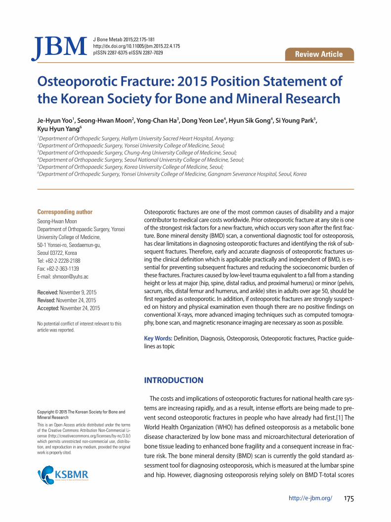
According to the conventional diagnosis based on the BMD T-score, osteoporosis is defined as T-score ≤-2.5 stan-dard deviation (SD) or the presence of a prevalent fragility fracture despite T-score >-2.5 SD.[23-25] Osteoporotic fractures are associated with low BMD measured at the fracture site. However, the occurrence of osteoporotic frac-ture is not always associated with low bone density equiv-alent to osteoporosis, and in most cases, central BMD mea-sures assessed mainly at the lumbar spine and the proxi-mal femur are used (Fig. 1).[26]
Based on the description above, clinical criteria are need-ed for defining osteoporotic fractures at sites other than
the lumbar spine and proximal femur, which are not com-monly used for measuring BMD, in order to recognize these fractures and initiate timely and appropriate therapy. The above-described definition of osteoporotic fracture can be easily obtained from the history of injury and radiographic findings. This clinically applicable definition of osteoporot-ic fracture regardless of BMD can determine the risk of sec-ond fractures and reduce the associated socioeconomic burdens.
ANATOMIC SITES OF OSTEOPOROTIC FRACTURES
Common osteoporotic fracture sites include bones that are under strain because they bear weight (such as the spine, hip, and pelvis) or that take the stress when a person catch-es him- or herself when falling from a standing height or less (such as the wrist, forearm, and upper arm).
Osteoporotic fractures occur mainly at sites that are as-sociated with low BMD and increase in incidence after the age of 50.[27] Conventionally, the spine, hip, and distal ra-dius have long been regarded as the quintessential osteo-porotic fracture sites. However, large studies have shown that nearly all types of fractures occur more often in pati-ents with low bone density irrespective of the site.[3,28,29]
Patients who have fractures at typical osteoporotic sites (spine, hip, wrist, and humerus) are most likely to have low BMD, but 74% of patients with fractures at less typical sites (ankle, hand, foot, other sites) also have low BMD at either the hip or the spine.[30] This finding reinforces the recom-mendation that history of any low-trauma fracture at any site should be an indication for osteoporosis evaluation.
According to NICE and National Osteoporosis Founda-tion (NOF) guidelines, osteoporotic fractures occur most commonly in the spine, hip, and distal radius but may also occur in the humerus, pelvis, ribs, and other bones. The WHO considers proximal humerus fractures to be one of the major osteoporotic fractures.
Recently, fractures of the pelvic ring in older populations have been classified as osteoporotic because this fracture type is caused by low-energy trauma or no trauma in pop-ulations with osteoporosis. Low-energy falls are responsi-ble for the majority of pelvic insufficiency fractures, and moreover, up to two-thirds of sacral insufficiency fractures have been noted to occur in the absence of trauma in old-
Fig. 1. A fracture of the right femoral neck in a 76-year-old male pa-tient is shown on a preoperative radiograph. The fracture was caused by a simple fall from a bed. There was no finding of osteoporosis on dual energy X-ray absorptiometry measured at the proximal femur and lumbar spine.

Je-Hyun Yoo, et al.
178 http://e-jbm.org/ http://dx.doi.org/10.11005/jbm.2015.22.4.175
er populations.[31] In addition, rib fractures from low-en-ergy trauma have been reported as common non-verte-bral fractures in the elderly.[32,33] These studies revealed an increasing pattern of fracture incidence, and a history of rib fracture carried at least a twofold increased risk of a sub-sequent osteoporotic fracture. In addition, ankle fractures have gained increasing attention as another type of fragili-ty fracture.[4,34] Low-energy ankle fractures can offer sig-nificant implications for identifying patients who need os-teoporosis treatment. One population-based study identi-fied radiologic findings and trauma history as valid tools for assessing osteoporotic ankle fractures.[35]
Following the description above, Table 1 lists what are considered to be the major and minor sites of osteoporotic fractures (Fig. 2, 3).
All of these fractures should be assessed based on the
clinical and research evidence and considering the benefits of osteoporosis management, including reducing the risk of osteoporotic fractures.
THE AGES OF POPULATIONS WITH OSTEOPOROTIC FRACTURES
Osteoporotic fractures and the associated costs are ex-pected to increase markedly as populations age.[36] These fractures are associated with low bone mass, and their in-cidence increases with age after the age of 50. The estimat-ed lifetime risk for an osteoporotic fracture among 50-year-old women in North America is approximately 18% for hip fracture, 16% for clinically diagnosed vertebral fracture, and 16% for distal radius fracture.[37] Overall, the NOF es-timates that 1 in 2 white women and 1 in 4 white men old-er than age 50 will sustain at least 1 osteoporosis-related fracture in their remaining lifetimes.[38] Prior osteoporotic fracture in this population is an important predictor of fu-ture fractures.[39] Therefore, fractures at the sites described above in adults over age 50 should be strongly suspected as osteoporotic. Their neglect ultimately can underestimate the burden of osteoporosis and lead to subsequent frac-tures, particularly in relatively younger individuals.
Finally, a recent fracture at any skeletal site described above in an adult older than 50 years of age should be con-sidered a significant event for the diagnosis of osteoporo-
Table 1. Sites of osteoporotic fractures
Major sites Hip Spine Distal radius Proximal Humerus
Minor sites Pelvis Sacrum Ribs Distal femur Distal humerus Ankle
A B C D
Fig. 2. Typical osteoporotic fractures at major sites. (A) Hip, (B) Spine, (C) Distal radius, (D) Proximal humerus.

Osteoporotic Fracture: 2015 Position Statement
http://dx.doi.org/10.11005/jbm.2015.22.4.175 http://e-jbm.org/ 179
sis and should trigger further assessment and treatment if necessary.
DIAGNOSIS OF OSTEOPOROTIC FRACTURE
Conventional X-rays are the first step of the diagnostic work-up to detect osteoporotic fracture. Simple X-rays can easily detect the presence of fractures, particularly in the upper and lower extremities, but occult fractures at any site are very difficult to diagnose on simple X-ray. If occult frac-ture is highly suspected on history and physical examina-tion, computed tomography (CT), bone scintigraphy (BS), or magnetic resonance imaging (MRI) are recommended as additional diagnostic tools to confirm osteoporotic fracture.
In general, osteoporotic fractures of the vertebrae and the pelvic ring and their severity are often underdiagnosed
or underestimated on conventional X-rays because of in-adequate film quality, lack of fracture recognition by radi-ologists, or use of ambiguous terminology in reports.[40-43]
In most fractures of the pelvic ring, conventional X-rays mainly detect ventral pelvic fractures; therefore, additional CT is necessary to evaluate the dorsal pelvis. Especially in cases in which osteoporotic fracture of the pelvic ring is strongly suspected without an obvious fracture line on conventional X-rays, MRI of the pelvis is of great help and may be a gold standard.[40,41]
Osteoporotic vertebral fractures are frequently undetect-ed, but their accurate and early diagnosis is of paramount importance because both symptomatic and asymptomatic vertebral fractures are associated with increased morbidity and mortality.[42] Preferentially, conventional X-rays are crucial for diagnosing and grading osteoporotic vertebral deformities and for differential diagnosis during assess-
A B
C D E F
Fig. 3. Typical osteoporotic fractures at minor sites. (A) Pelvis, (B) Sacrum, (C) Ribs, (D) Distal femur, (E) Distal humerus, (F) Ankle.
Je-Hyun Yoo, et al.
180 http://e-jbm.org/ http://dx.doi.org/10.11005/jbm.2015.22.4.175
ment for osteoporotic deformity. However, more advanced imaging techniques such as CT, BS, and MRI may be requir-ed to further investigate the etiology in some of these de-formities as well as to differentiate chronic from acute frac-tures, to assess compromise of the spinal canal, and for fol-low-up after specific treatments such as vertebroplasty.[43]
CONCLUSION
Osteoporotic fractures are a frequent and important cause of disability and medical costs worldwide. Moreover, a num-ber of osteoporotic fractures such as hip and vertebral frac-tures have very high morbidity and mortality.[28] Prior os-teoporotic fracture at any site is one of the strongest risk factors for a new fracture, partly independent of BMD [44]; The new fractures occur very soon after the first fracture. Therefore, early and accurate diagnosis of osteoporotic fractures using the clinical definition, which is applicable practically and independent of BMD, is crucial for prevent-ing subsequent fractures and reducing their associated so-cioeconomic burden. Fractures caused by low-level trauma equivalent to a fall from a standing height or less at sites described above in adults over age 50, should be first re-garded as osteoporotic. In addition, if osteoporotic frac-tures are strongly suspected on history and physical exam-ination even though there are no positive findings on con-ventional X-rays, more advanced imaging techniques such as CT, BS, and MRI are required. Subsequently, appropriate treatment should be administered as soon as possible af-ter the diagnosis of the first osteoporotic fracture in order to be most cost-effective.
REFERENCES
1. Yi H, Ha YC, Lee YK, et al. National healthcare budget im-pact analysis of the treatment for osteoporosis and frac-tures in Korea. J Bone Metab 2013;20:17-23.
2. Schuit SC, van der Klift M, Weel AE, et al. Fracture incidence and association with bone mineral density in elderly men and women: the Rotterdam Study. Bone 2004;34:195-202.
3. Johnell O, Kanis JA. An estimate of the worldwide preva-lence and disability associated with osteoporotic fractures. Osteoporos Int 2006;17:1726-33.
4. Lee KM, Chung CY, Kwon SS, et al. Ankle fractures have features of an osteoporotic fracture. Osteoporos Int 2013;
24:2819-25.5. Soles GL, Ferguson TA. Fragility fractures of the pelvis. Curr
Rev Musculoskelet Med 2012;5:222-8.6. Sajjan SG, Barrett-Connor E, McHorney CA, et al. Rib frac-
ture as a predictor of future fractures in young and older postmenopausal women: National Osteoporosis Risk As-sessment (NORA). Osteoporos Int 2012;23:821-8.
7. Ng AC, Drake MT, Clarke BL, et al. Trends in subtrochanter-ic, diaphyseal, and distal femur fractures, 1984-2007. Os-teoporos Int 2012;23:1721-6.
8. Marshall D, Johnell O, Wedel H. Meta-analysis of how well measures of bone mineral density predict occurrence of osteoporotic fractures. BMJ 1996;312:1254-9.
9. Ha YC, Park YG, Nam KW, et al. Trend in hip fracture inci-dence and mortality in Korea: a prospective cohort study from 2002 to 2011. J Korean Med Sci 2015;30:483-8.
10. Rowe SM, Song EK, Kim JS, et al. Rising incidence of hip fracture in Gwangju City and Chonnam Province, Korea. J Korean Med Sci 2005;20:655-8.
11. Rowe SM, Yoon TR, Ryang DH. An epidemiological study of hip fracture in Honam, Korea. Int Orthop 1993;17:139-43.
12. Park C, Ha YC, Jang S, et al. The incidence and residual life-time risk of osteoporosis-related fractures in Korea. J Bone Miner Metab 2011;29:744-51.
13. Meema HE, Meema S. Cortical bone mineral density versus cortical thickness in the diagnosis of osteoporosis: a roent-genologic-densitometric study. J Am Geriatr Soc 1969;17: 120-41.
14. Keshawarz NM, Recker RR. Expansion of the medullary cavity at the expense of cortex in postmenopausal osteo-porosis. Metab Bone Dis Relat Res 1984;5:223-8.
15. Ahlborg HG, Johnell O, Turner CH, et al. Bone loss and bone size after menopause. N Engl J Med 2003;349:327-34.
16. Haara M, Heliövaara M, Impivaara O, et al. Low metacarpal index predicts hip fracture: a prospective population study of 3,561 subjects with 15 years of follow-up. Acta Orthop 2006;77:9-14.
17. Mather J, MacDermid JC, Faber KJ, et al. Proximal humerus cortical bone thickness correlates with bone mineral den-sity and can clinically rule out osteoporosis. J Shoulder El-bow Surg 2013;22:732-8.
18. Roberts M, Yuan J, Graham J, et al. Changes in mandibular cortical width measurements with age in men and wom-en. Osteoporos Int 2011;22:1915-25.

Osteoporotic Fracture: 2015 Position Statement
http://dx.doi.org/10.11005/jbm.2015.22.4.175 http://e-jbm.org/ 181
19. Sadat-Ali M, Elshaboury E, Al-Omran AS, et al. Tibial corti-cal thickness: A dependable tool for assessing osteoporo-sis in the absence of dual energy X-ray absorptiopmetry. Int J Appl Basic Med Res 2015;5:21-4.
20. Johannesdottir F, Turmezei T, Poole KE. Cortical bone as-sessed with clinical computed tomography at the proxi-mal femur. J Bone Miner Res 2014;29:771-83.
21. Bala Y, Zebaze R, Ghasem-Zadeh A, et al. Cortical porosity identifies women with osteopenia at increased risk for forearm fractures. J Bone Miner Res 2014;29:1356-62.
22. Lenchik L, Rogers LF, Delmas PD, et al. Diagnosis of osteo-porotic vertebral fractures: importance of recognition and description by radiologists. AJR Am J Roentgenol 2004; 183:949-58.
23. WHO Study Group. Assessment of fracture risk and its ap-plication to screening for postmenopausal osteoporosis. Report of a WHO Study Group. World Health Organ Tech Rep Ser 1994;843:1-129.
24. Kanis JA, Melton LJ 3rd, Christiansen C, et al. The diagnosis of osteoporosis. J Bone Miner Res 1994;9:1137-41.
25. World Health Organization. Assessment of osteoporosis at the primary health care level. Summary Report of a WHO Scientific Group. 2007; www.who.int/chp/topics/rheuma-tic/en/index
26. Hey HW, Sng WJ, Lim JL, et al. Interpretation of hip frac-ture patterns using areal bone mineral density in the prox-imal femur. Arch Orthop Trauma Surg 2015;135:1647-53.
27. Kanis JA, Oden A, Johnell O, et al. The burden of osteopo-rotic fractures: a method for setting intervention thresh-olds. Osteoporos Int 2001;12:417-27.
28. Johnell O, Kanis J. Epidemiology of osteoporotic fractures. Osteoporos Int 2005;16 Suppl 2:S3-7.
29. Nguyen TV, Eisman JA, Kelly PJ, et al. Risk factors for osteo-porotic fractures in elderly men. Am J Epidemiol 1996;144: 255-63.
30. McLellan AR, Gallacher SJ, Fraser M, et al. The fracture liai-son service: success of a program for the evaluation and management of patients with osteoporotic fracture. Os-teoporos Int 2003;14:1028-34.
31. Tsiridis E, Upadhyay N, Giannoudis PV. Sacral insufficiency fractures: current concepts of management. Osteoporos Int 2006;17:1716-25.
32. Palvanen M, Kannus P, Niemi S, et al. Hospital-treated min-
imal-trauma rib fractures in elderly Finns: long-term trends and projections for the future. Osteoporos Int 2004;15:649-53.
33. Barrett-Connor E, Nielson CM, Orwoll E, et al. Epidemiolo-gy of rib fractures in older men: Osteoporotic Fractures in Men (MrOS) prospective cohort study. BMJ 2010;340:c1069.
34. Biver E, Durosier C, Chevalley T, et al. Prior ankle fractures in postmenopausal women are associated with low areal bone mineral density and bone microstructure alterations. Osteoporos Int 2015;26:2147-55.
35. Koski AM, Patala A, Patala E, et al. Incidence of osteopo-rotic fractures in elderly women and men in Finland dur-ing 2005-2006: a population-based study. Scand J Surg 2014;103:215-21.
36. Johnell O. The socioeconomic burden of fractures: today and in the 21st century. Am J Med 1997;103:20S-5S.
37. Melton LJ 3rd, Chrischilles EA, Cooper C, et al. Perspective. How many women have osteoporosis? J Bone Miner Res 1992;7:1005-10.
38. National Osteoporosis Foundation. Osteoporosis Disease Statistics. 2006 [cited by 2008 October 3]. Available from: http://www.nof.org/osteoporosis/stats.htm
39. Tromp AM, Ooms ME, Popp-Snijders C, et al. Predictors of fractures in elderly women. Osteoporos Int 2000;11:134-40.
40. Rommens PM, Wagner D, Hofmann A. Surgical manage-ment of osteoporotic pelvic fractures: a new challenge. Eur J Trauma Emerg Surg 2012;38:499-509.
41. Rommens PM, Ossendorf C, Pairon P, et al. Clinical path-ways for fragility fractures of the pelvic ring: personal ex-perience and review of the literature. J Orthop Sci 2015; 20:1-11.
42. Delmas PD, van de Langerijt L, Watts NB, et al. Underdiag-nosis of vertebral fractures is a worldwide problem: the IMPACT study. J Bone Miner Res 2005;20:557-63.
43. Link TM, Guglielmi G, van Kuijk C, et al. Radiologic assess-ment of osteoporotic vertebral fractures: diagnostic and prognostic implications. Eur Radiol 2005;15:1521-32.
44. Klotzbuecher CM, Ross PD, Landsman PB, et al. Patients with prior fractures have an increased risk of future frac-tures: a summary of the literature and statistical synthesis. J Bone Miner Res 2000;15:721-39.


![Major osteoporotic fragility fractures: Risk factor updates and ......aged 50 and older will have an osteoporotic fracture in the lifetime[6]. Although many national and international](https://img.pdfslide.net/doc/110x75/604cdf565cdcb6501161cb1b/major-osteoporotic-fragility-fractures-risk-factor-updates-and-aged-50.jpg)