Embed Size (px)
Citation preview
OXIDATIVE PHOSPHORYLATION
Is the process in which ATP is formed as a result of the transfer of electrons from
NADH or FADH2 to O2 by a series of electron carriers
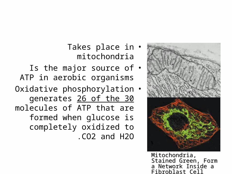
•Takes place in mitochondria•Is the major source of ATP in
aerobic organisms•Oxidative phosphorylation
generates 26 of the 30 molecules of ATP that are
formed when glucose is completely oxidized to CO2
and H2O .
Mitochondria, Stained Mitochondria, Stained Green, Form a Network Green, Form a Network Inside a Fibroblast CellInside a Fibroblast Cell
Cellular respiration
.1Carbon fuels are oxidized in the citric acid cycle to yield electrons with high
transfer potential.
.2This electron-motive force is converted into a proton-motive force.
.3The proton-motive force is converted into phosphoryl transfer potential .
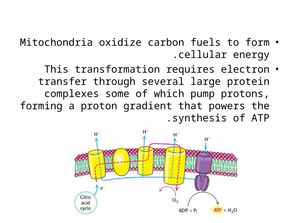
•Mitochondria oxidize carbon fuels to form cellular energy.
•This transformation requires electron transfer through several large protein complexes some of
which pump protons, forming a proton gradient that powers the synthesis of ATP .
conversion of electron-motive force into proton-motive force
•Carried out by three electron-driven proton pumps:.1NADH-Q oxidoreductase.2Q-cytochrome c oxidoreductase.3cytochrome c oxidase.
•These large transmembrane complexes contain multiple oxidation-reduction centers, including:
–Quinones (Q)–Flavins–Iron-sulfur clusters–Hemes–Copper ion
•The final phase of oxidative phosphorylation is carried out by ATP synthase, an ATP-synthesizing assembly driven by the flow of protons back into the mitochondrial matrix .
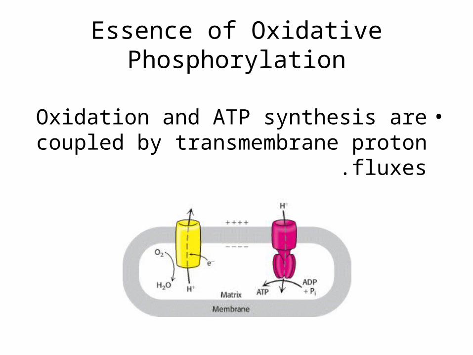
Essence of Oxidative Phosphorylation
•Oxidation and ATP synthesis are coupled by transmembrane proton fluxes .
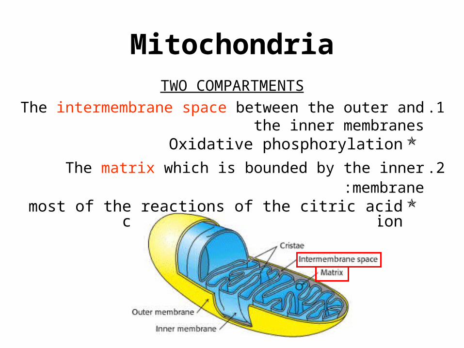
MitochondriaTWO COMPARTMENTS
.1The intermembrane space between the outer and the inner membranes
Oxidative phosphorylation
.2The matrix which is bounded by the inner membrane :most of the reactions of the citric acid cycle and fatty
acid oxidation
OUTER MEMBRANE
•Permeable to most small molecules and ions because it contains many copies of
mitochondrial porin:–VDAC is voltage-dependent anion channel.–VDAC plays a role in the regulated flux of
metabolites across the outer membrane: •Phosphate•Chloride•organic anions•adenine nucleotides
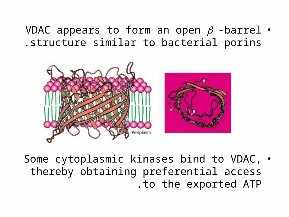
•VDAC appears to form an open -barrel structure similar to bacterial porins.
•Some cytoplasmic kinases bind to VDAC, thereby obtaining preferential access to the
exported ATP .
INNER MEMBRANE
•Impermeable to nearly all ions and polar molecules.•A large family of transporters shuttles metabolites across
the inner mitochondrial membrane like:–ATP–Pyruvate–citrate
•The two faces of this membrane will be referred to as:–matrix side also called the N side (the membrane
potential is negative)–cytosolic side (the latter because it is freely
accessible to most small molecules in the cytosol). also called the P side (the membrane potential is
positive)
High energy electrons: Redox potential & Free-energy change
•In oxidative phosphorylation, the electron transfer potential of NADH or FADH2 is
converted into the phosphoryl transfer potential of ATP.
•Recall that:G0’ of hydrolysis of ATP to ADP + Pi gives
idea about the phosphoryl transfer potential.–E’0 is the electron transfer potential: also
called the reduction potential, redox potential or oxidation-reduction potential
•A strong reducing agent (such as NADH) is poised to donate electrons and has a
negative reduction potential (-ve E’0).
•A strong oxidizing agent (such as O2) is ready to accept electrons and has a positive reduction potential (+ve E’0)..
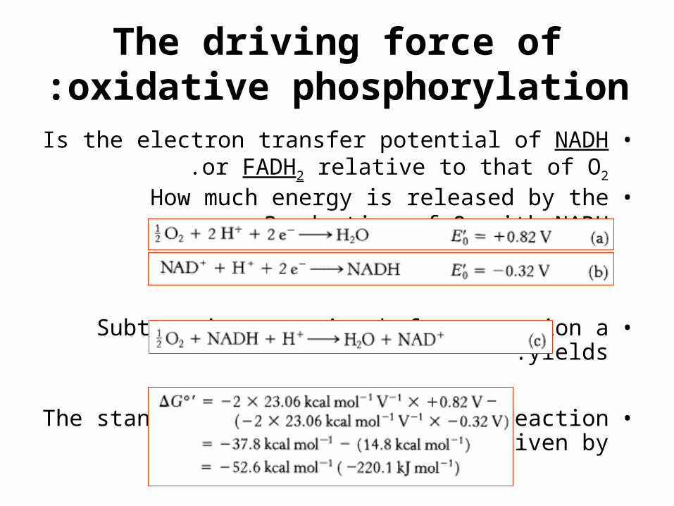
The driving force of oxidative phosphorylation:
•Is the electron transfer potential of NADH or FADH2 relative to that of O2.
•How much energy is released by the reduction of O2 with NADH?
•Subtracting reaction b from reaction a yields:
•The standard free energy for this reaction is then given by
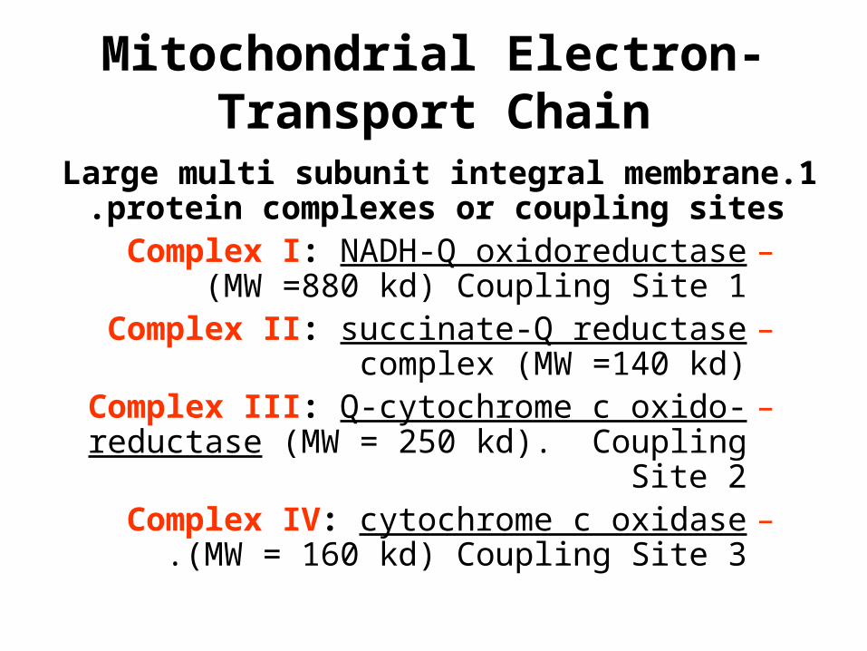
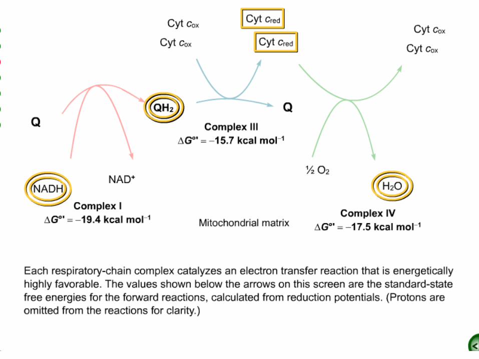
Mitochondrial Electron-Transport Chain
.1Large multi subunit integral membrane protein complexes or coupling sites.
–Complex I: NADH-Q oxidoreductase (MW =880 kd) Coupling Site 1
–Complex II: succinate-Q reductase complex (MW =140 kd)
–Complex III: Q-cytochrome c oxido-reductase (MW = 250 kd). Coupling Site 2
–Complex IV: cytochrome c oxidase (MW = 160 kd) Coupling Site 3.
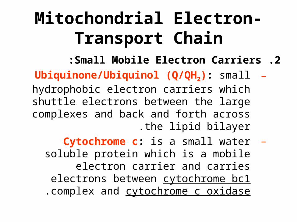
Mitochondrial Electron-Transport Chain
.2Small Mobile Electron Carriers:
–Ubiquinone/Ubiquinol (Q/QH2): small hydrophobic electron carriers which shuttle
electrons between the large complexes and back and forth across the lipid bilayer.
–Cytochrome c: is a small water soluble protein which is a mobile electron carrier
and carries electrons between cytochrome bc1 complex and cytochrome c oxidase.
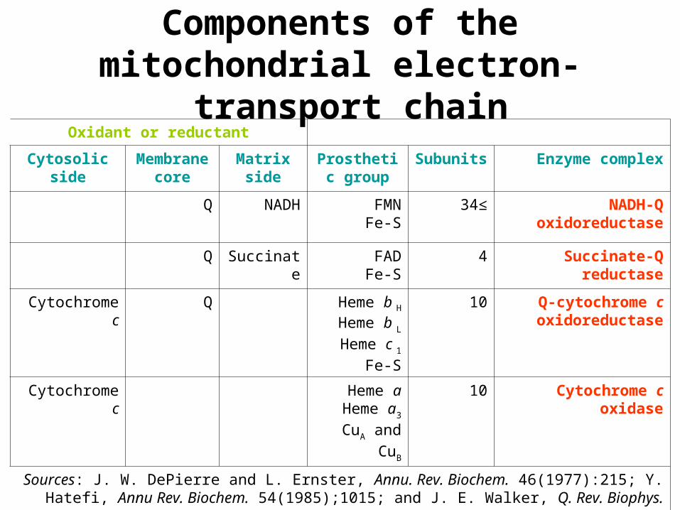
Oxidant or reductant
Enzyme complexSubunitsProsthetic group
Matrixside
Membrane core
Cytosolic side
NADH-Q oxidoreductase
≥34FMNFe-S
NADHQ
Succinate-Q reductase
4FADFe-S
SuccinateQ
Q-cytochrome c oxidoreductase
10Heme b H
Heme b L
Heme c 1
Fe-S
QCytochrome c
Cytochrome c oxidase10Heme aHeme a3
CuA and CuB
Cytochrome c
Sources: J. W. DePierre and L. Ernster, Annu. Rev. Biochem. 46(1977):215; Y. Hatefi, Annu Rev. Biochem. 54(1985);1015; and J. E. Walker, Q. Rev. Biophys. 25(1992):253.
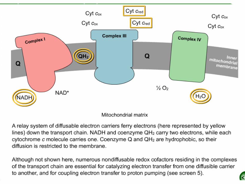
Components of the mitochondrial electron-transport chain
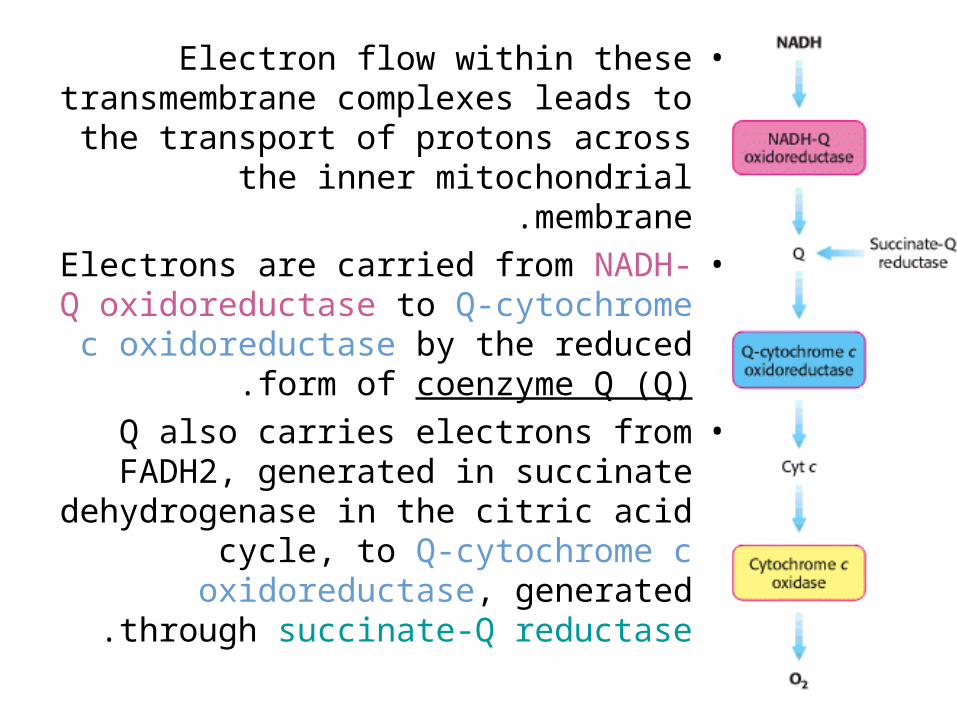
•Electron flow within these transmembrane complexes leads to
the transport of protons across the inner mitochondrial membrane.
•Electrons are carried from NADH-Q oxidoreductase to Q-cytochrome c
oxidoreductase by the reduced form of coenzyme Q (Q).
•Q also carries electrons from FADH2, generated in succinate
dehydrogenase in the citric acid cycle, to Q-cytochrome c oxidoreductase,
generated through succinate-Q reductase.
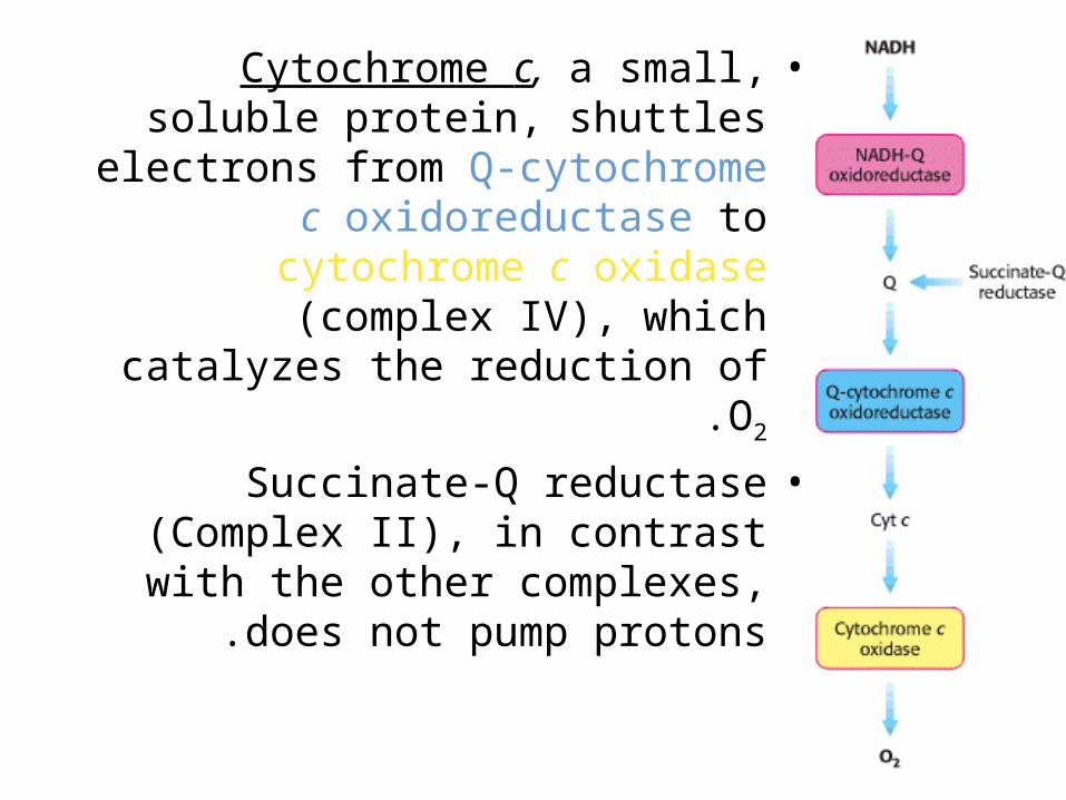
•Cytochrome c, a small, soluble protein, shuttles electrons from Q-
cytochrome c oxidoreductase to cytochrome c oxidase (complex IV), which catalyzes the reduction of O2.
•Succinate-Q reductase (Complex II), in contrast with the other complexes,
does not pump protons .

Coenzyme Q (Q)
•Q is a hydrophobic quinone that diffuses rapidly within the inner mitochondrial membrane.
•Q is a quinone derivative with a long isoprenoid tail.–The number of five-carbon isoprene units in
coenzyme Q depends on the species.–The most common form in mammals contains 10
isoprene units (coenzyme Q10).
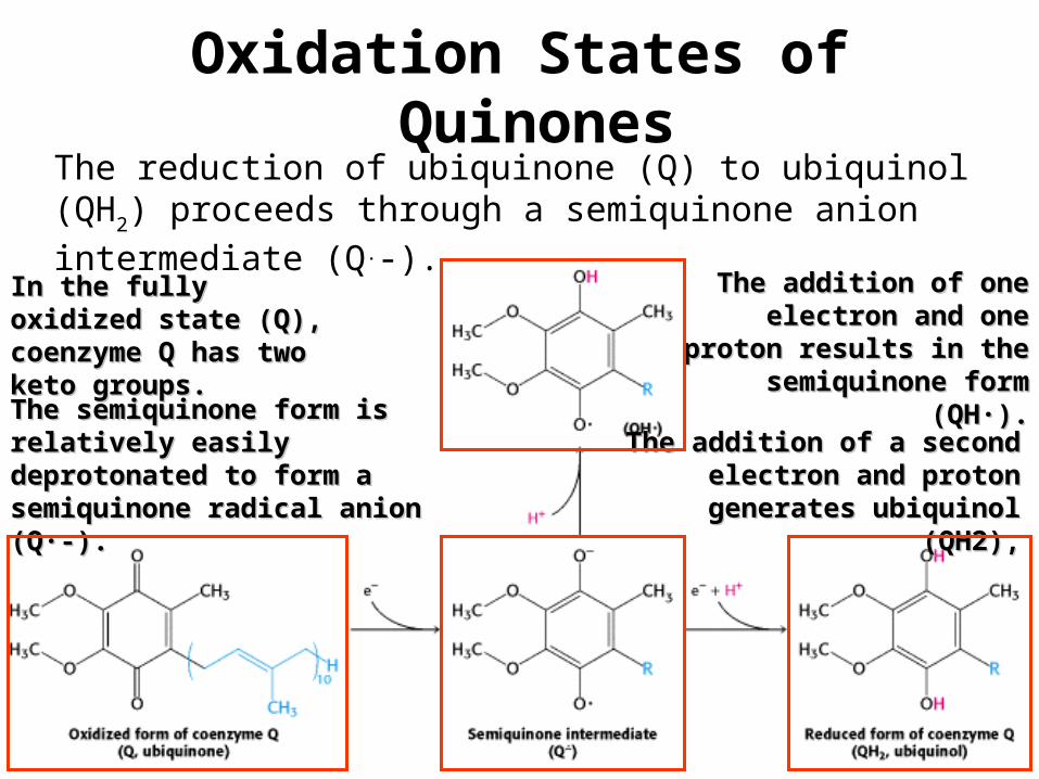
Oxidation States of Quinones
The reduction of ubiquinone (Q) to ubiquinol (QH2) proceeds
through a semiquinone anion intermediate (Q.-). In the fully oxidized state In the fully oxidized state (Q), coenzyme Q has two (Q), coenzyme Q has two keto groups.keto groups.
The addition of one The addition of one electron and one proton electron and one proton
results in the semiquinone results in the semiquinone form (QH·).form (QH·).
The semiquinone form is The semiquinone form is relatively easily deprotonated to relatively easily deprotonated to form a semiquinone radical form a semiquinone radical anion (Q·-).anion (Q·-).
The addition of a second The addition of a second electron and proton generates electron and proton generates
ubiquinol (QH2),ubiquinol (QH2),
•Thus, electron-transfer reactions of quinones are coupled to proton binding
and release, a property that is key to transmembrane proton transport .
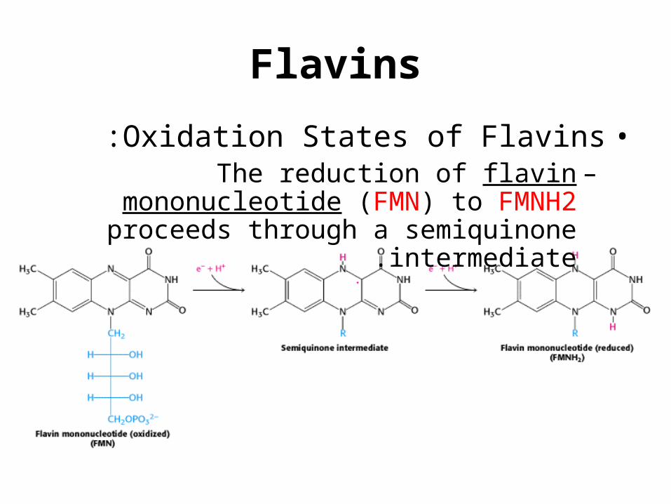
Flavins
•Oxidation States of Flavins:–The reduction of flavin mononucleotide (FMN)
to FMNH2 proceeds through a semiquinone intermediate.
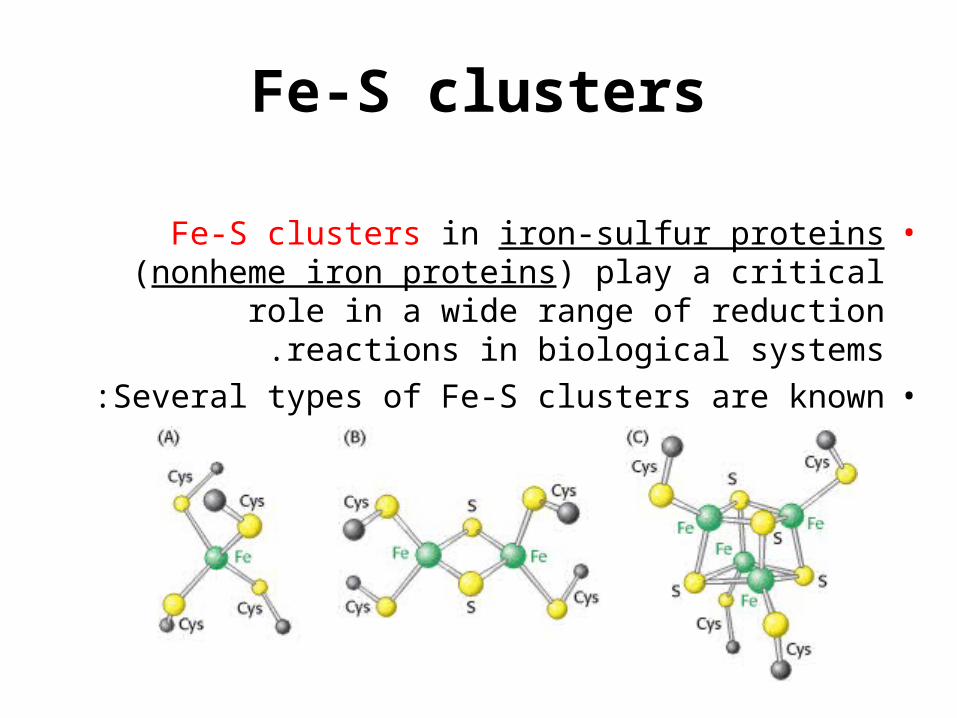
Fe-S clusters
•Fe-S clusters in iron-sulfur proteins (nonheme iron proteins) play a critical role in a wide range of
reduction reactions in biological systems.•Several types of Fe-S clusters are known :
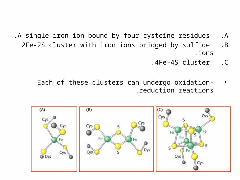
.A A single iron ion bound by four cysteine residues.
.B 2Fe-2S cluster with iron ions bridged by sulfide ions.
.C 4Fe-4S cluster.
•Each of these clusters can undergo oxidation-reduction reactions .
•Iron ions in these Fe-S complexes cycle between:
–Fe2+ (reduced state)–Fe3+ (oxidized state)
•Unlike quinones and flavins, iron-sulfur clusters generally undergo oxidation-
reduction reactions without releasing or binding protons .
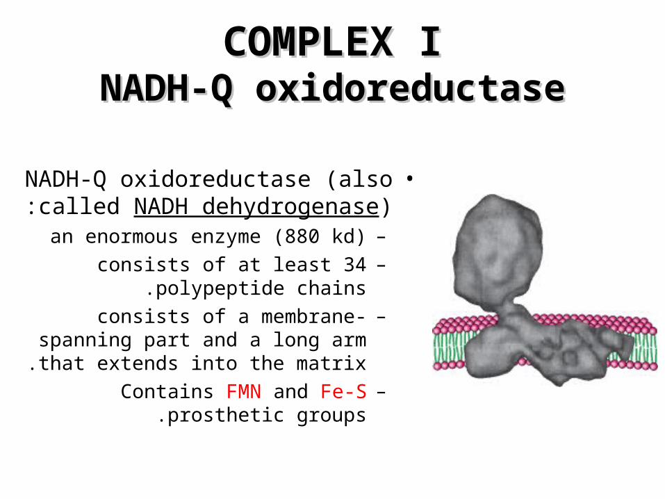
COMPLEX ICOMPLEX INADH-Q oxidoreductaseNADH-Q oxidoreductase
•NADH-Q oxidoreductase (also called NADH dehydrogenase):
–an enormous enzyme (880 kd)–consists of at least 34 polypeptide
chains.–consists of a membrane-spanning
part and a long arm that extends into the matrix.
–Contains FMN and Fe-S prosthetic groups.
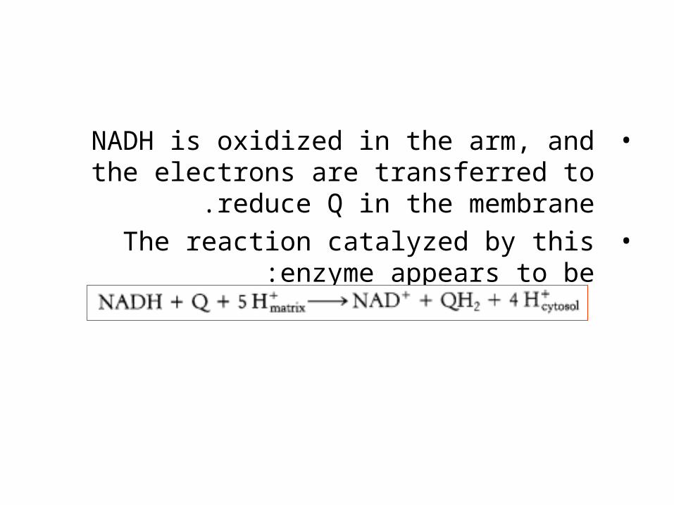
•NADH is oxidized in the arm, and the electrons are transferred to reduce Q in the membrane.
•The reaction catalyzed by this enzyme appears to be:
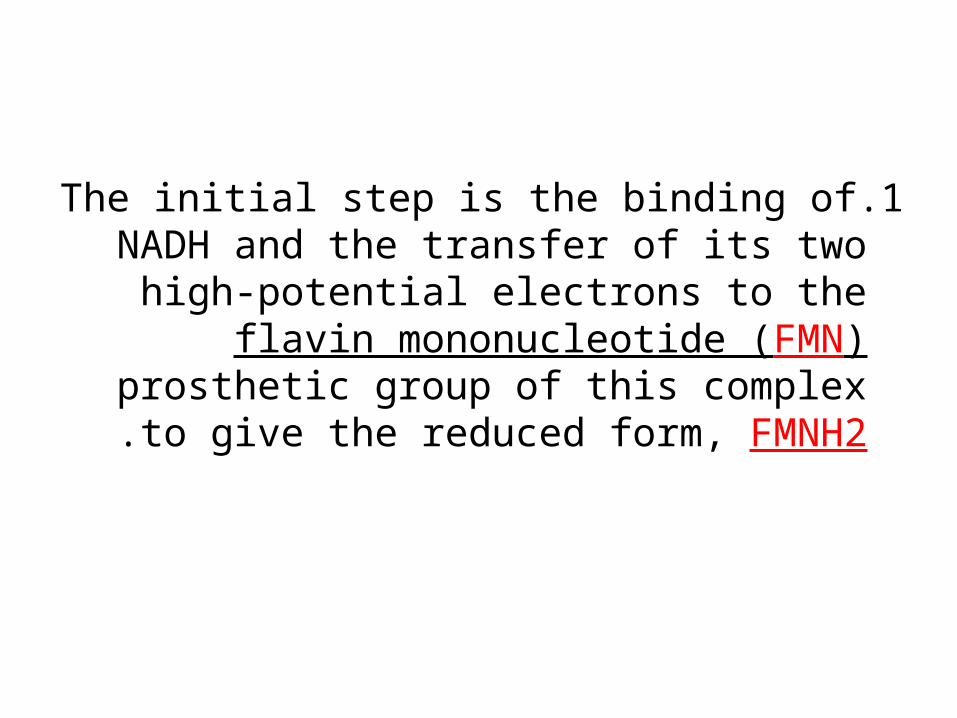
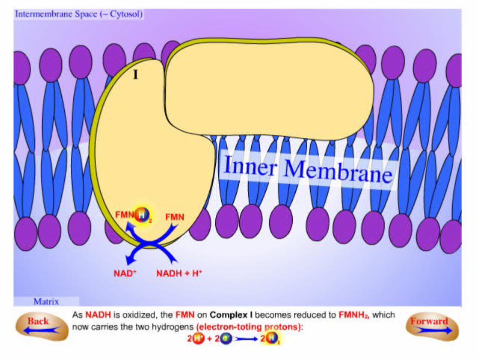
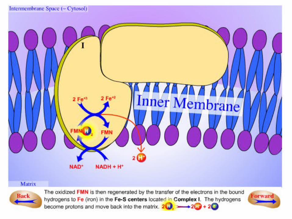
.1The initial step is the binding of NADH and the transfer of its two high-potential
electrons to the flavin mononucleotide (FMN) prosthetic group of this complex to
give the reduced form, FMNH2 .
.2Electrons are then transferred from FMNH2 to a series of 4Fe4S, the second
type of prosthetic group in NADH-Q oxidoreductase.
•NADH-Q oxidoreductase contains:–2Fe-2S cluster–4Fe-4S cluster
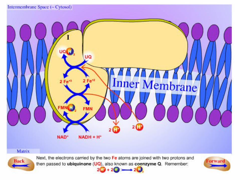
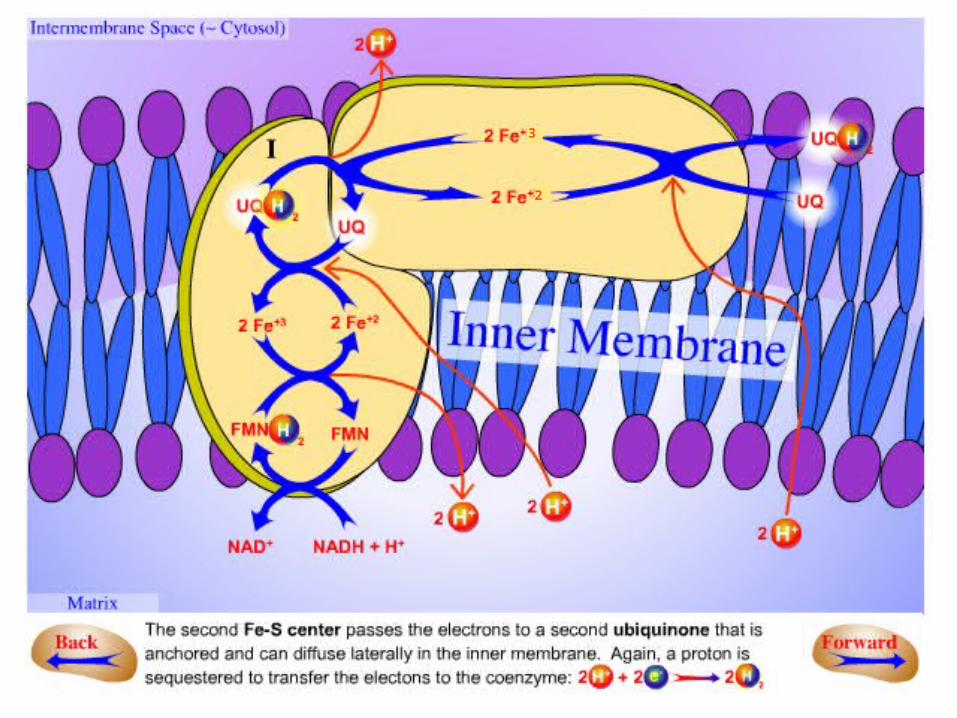
.3Electrons in the 4Fe4S of NADH-Q oxidoreductase are shuttled to coenzyme
Q.–The flow of two electrons from NADH to
coenzyme Q through NADH-Q oxidoreductase leads to the pumping of four
hydrogen ions out of the matrix of the mitochondrion.
–The coupled electron- proton transfer reactions of Q are crucial.

3
2
3
2
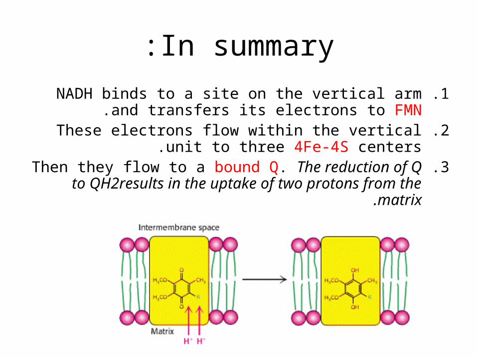
In summary:
.1NADH binds to a site on the vertical arm and transfers its electrons to FMN.
.2These electrons flow within the vertical unit to three 4Fe-4S centers.
.3Then they flow to a bound Q. The reduction of Q to QH2results in the uptake of two protons from the
matrix.
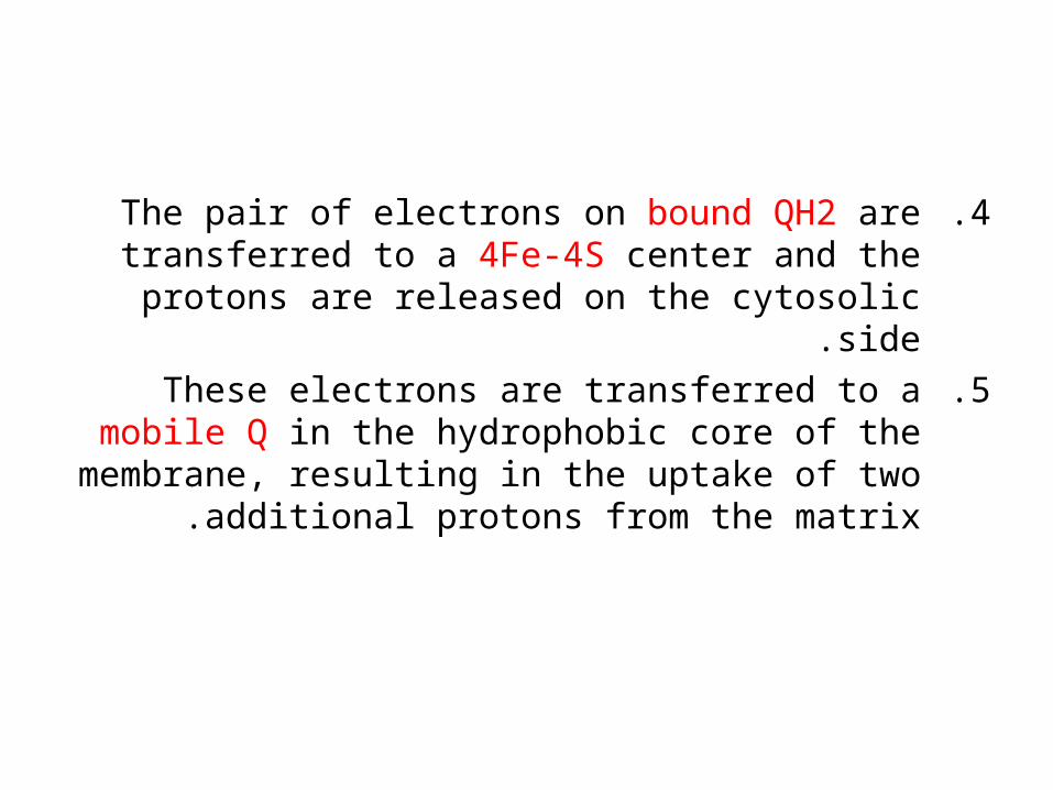
.4The pair of electrons on bound QH2 are transferred to a 4Fe-4S center and the protons are released on the
cytosolic side.
.5These electrons are transferred to a mobile Q in the hydrophobic core of the membrane, resulting in the
uptake of two additional protons from the matrix .
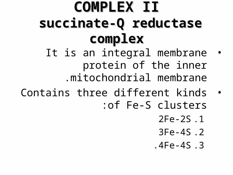
COMPLEX IICOMPLEX II succinate-Q reductase complex succinate-Q reductase complex
•It is an integral membrane protein of the inner mitochondrial membrane.
•Contains three different kinds of Fe-S clusters:
.12Fe-2S
.23Fe-4S
.34Fe-4S .
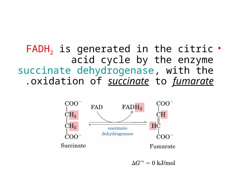
•FADH2 is generated in the citric acid cycle by the enzyme succinate dehydrogenase, with the oxidation of succinate to fumarate.
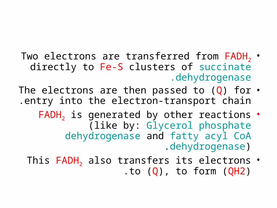
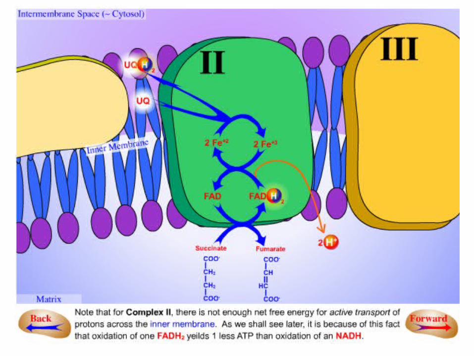
•Two electrons are transferred from FADH2 directly to Fe-S clusters of succinate
dehydrogenase.•The electrons are then passed to (Q) for entry
into the electron-transport chain.•FADH2 is generated by other reactions (like by:
Glycerol phosphate dehydrogenase and fatty acyl CoA dehydrogenase).
•This FADH2 also transfers its electrons to (Q), to form (QH2).
•The succinate-Q reductase complex and other enzymes that transfer electrons from FADH2 to
Q, in contrast with NADH-Q oxidoreductase, do not transport protons .
•Consequently, less ATP is formed from the oxidation of FADH2 than from NADH .
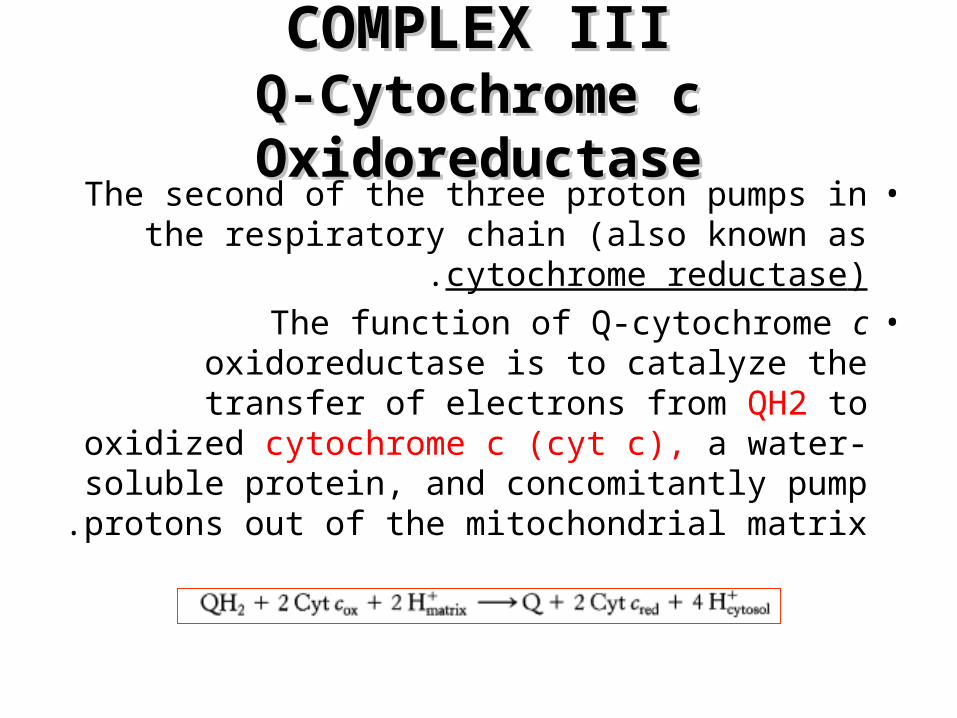
COMPLEX IIICOMPLEX IIIQ-Cytochrome c OxidoreductaseQ-Cytochrome c Oxidoreductase
•The second of the three proton pumps in the respiratory chain (also known as cytochrome
reductase).•The function of Q-cytochrome c oxidoreductase
is to catalyze the transfer of electrons from QH2 to oxidized cytochrome c (cyt c), a water-soluble protein, and concomitantly pump protons out of
the mitochondrial matrix .
•Q-cytochrome c oxidoreductase contains:–Cytochrome b562–Cytochrome b566–Cytochrome c1–An iron sulfur protein–At least six other subunits.
•A cytochrome is an electron-transferring protein that contains a heme prosthetic
group .
•The iron ion of a cytochrome alternates between a reduced ferrous (+2) state and
an oxidized ferric (+3) state during electron transport.
•The enzyme contains three heme prosthetic groups contained within two
cytochrome subunits:–Two b-type hemes within cytochrome b:
•Heme bL (L for low affinity)
•Heme bH (H for high affinity)
–One c-type heme within cytochrome c1 (Heme c1).
N
N
N
N
CH3 HC
CH3
S CH2
CH3
CH S CH2
CH3
CH2
CH2
COO
CH3
H3C
CH2CH2 OOC
protein
protein
Fe
Heme c
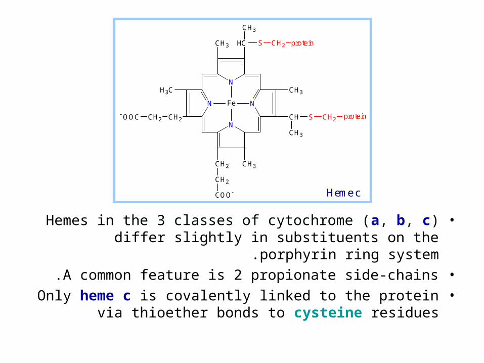
•Hemes in the 3 classes of cytochrome (a, b, c) differ slightly in substituents on the porphyrin ring system.
•A common feature is 2 propionate side-chains.•Only heme c is covalently linked to the protein via
thioether bonds to cysteine residues
•The enzyme also contains an iron-sulfur protein with an 2Fe-2S center (Rieske center).
•This center is unusual in that one of the iron ions is coordinated by two his residues rather than
two cysteine residues.•This coordination stabilizes the center in its
reduced form, raising its reduction potential.•Finally, Q-cytochrome c oxidoreductase contains
two distinct binding sites for ubiquinone termed (Qo) and (Qi), with the Qi site lying closer to the
inside of the matrix.
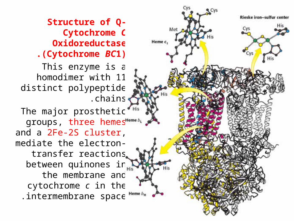
Structure of Q-Cytochrome C
Oxidoreductase (Cytochrome BC1).
This enzyme is a homodimer with 11 distinct
polypeptide chains.
The major prosthetic groups, three hemes and
a 2Fe-2S cluster, mediate the electron-transfer
reactions between quinones in the membrane
and cytochrome c in the intermembrane space .
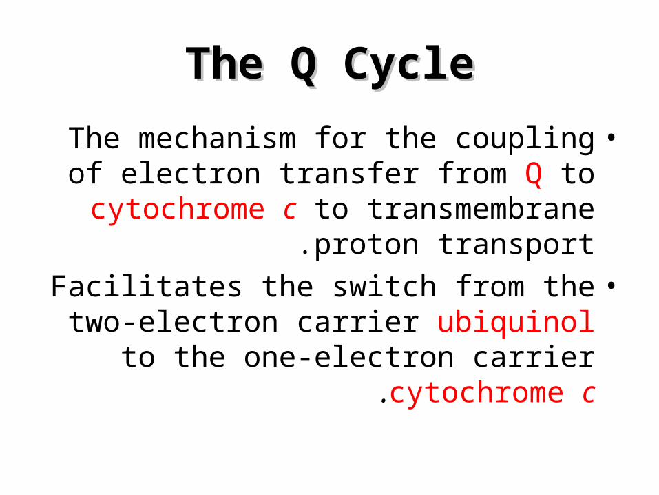
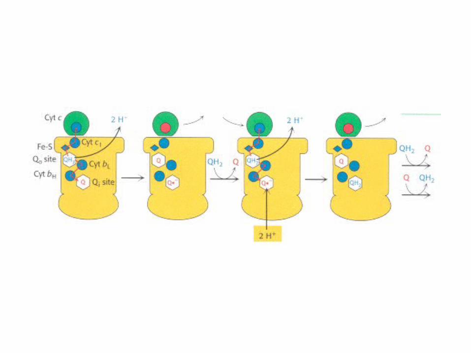
The Q CycleThe Q Cycle
•The mechanism for the coupling of electron transfer from Q to cytochrome c
to transmembrane proton transport.
•Facilitates the switch from the two-electron carrier ubiquinol to the one-electron carrier
cytochrome c.
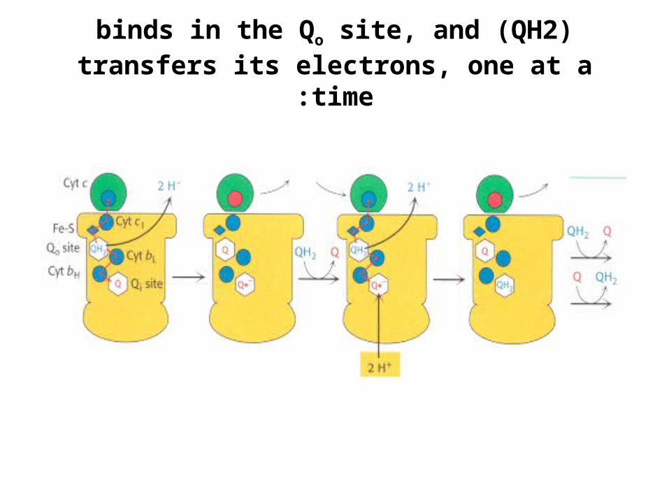
)QH2 (binds in the Qo site, and transfers its electrons, one at a time:
H+ H+Qo site
Cyt c
Fe-S
Cyt c1
Cyt bL
Cyt bH
Qi siteQ
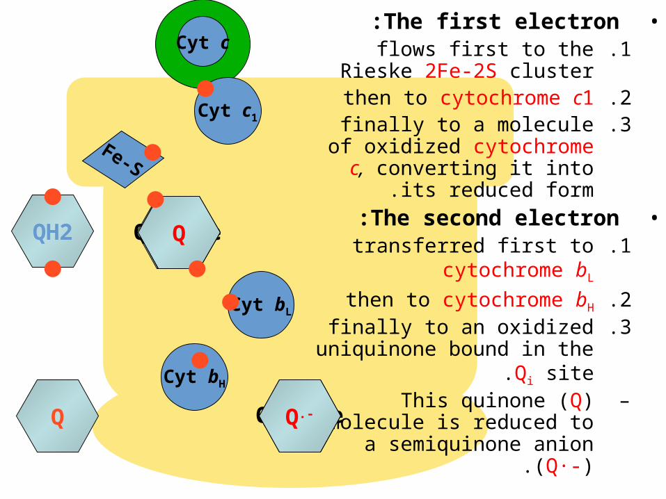
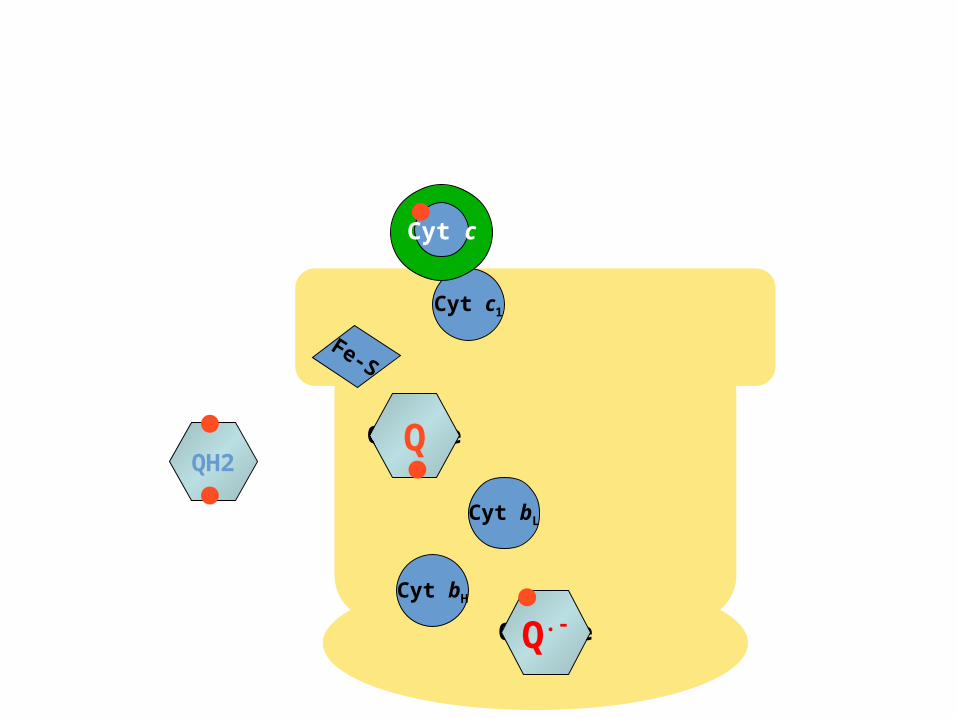
•The first electron:.1flows first to the Rieske
2Fe-2S cluster.2then to cytochrome c1.3finally to a molecule of
oxidized cytochrome c, converting it into its
reduced form.•The second electron:
.1transferred first to cytochrome bL
.2then to cytochrome bH
.3finally to an oxidized uniquinone bound in the Qi
site.–This quinone (Q) molecule
is reduced to a semiquinone anion (Q·-).
QH2 QHQ
Q.-
Fe-S
Cyt c1
Cyt bL
Cyt bH
Cyt c
Qo siteQH2
Qi siteQ.-
Q
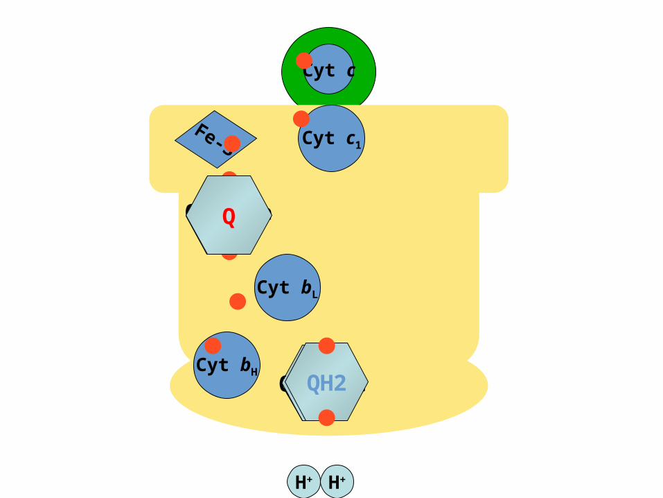
H+ H+
H+ H+
Cyt c
Fe-S Cyt c1
Cyt bL
Qo site
Cyt bH
QH2
Qi siteQ.-QH2
Q
COMPLEX IVCOMPLEX IVCytochrome c OxidaseCytochrome c Oxidase
•It catalyzes the coupled oxidation of the reduced cyt c generated by Complex III, and reduction of
O2 to two molecules of H2O.•The four-electron reduction of oxygen directly to
water without the release of intermediates is quite thermodynamically favorable.
DG°´ = -231.8 kJ mol-1•As much of this free energy as possible must be
captured in the form of a proton gradient for subsequent use in ATP synthesis .
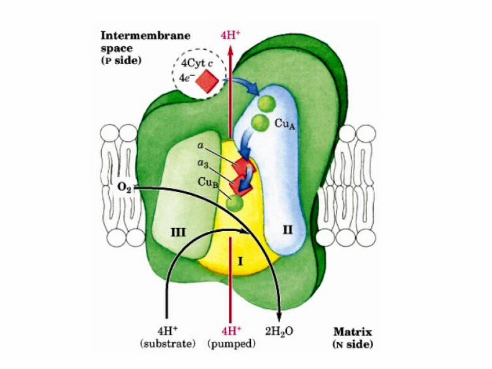
•It consists of 13 subunits, 3 of which (subunits I, II, and III) are encoded by the mitochondrial genome.
•Cytochrome c oxidase contains:–Three copper ions, arranged as two copper
centers, designated A and B:.1CuA/CuA: contains two copper ions linked by two bridging
cysteine residues. This center initially accepts electrons from reduced cytochrome c.
.2CuB: is coordinated by three histidine residues, one of which is modified by covalent linkage to a tyrosine residue.
–Two heme A groups: .1Heme a: functions to carry electrons from CuA/CuA
.2heme a3: passes electrons to CuB, to which it is directly adjacent.
•Together, heme a3 and CuB form the active center at which O2 is reduced to H2O.
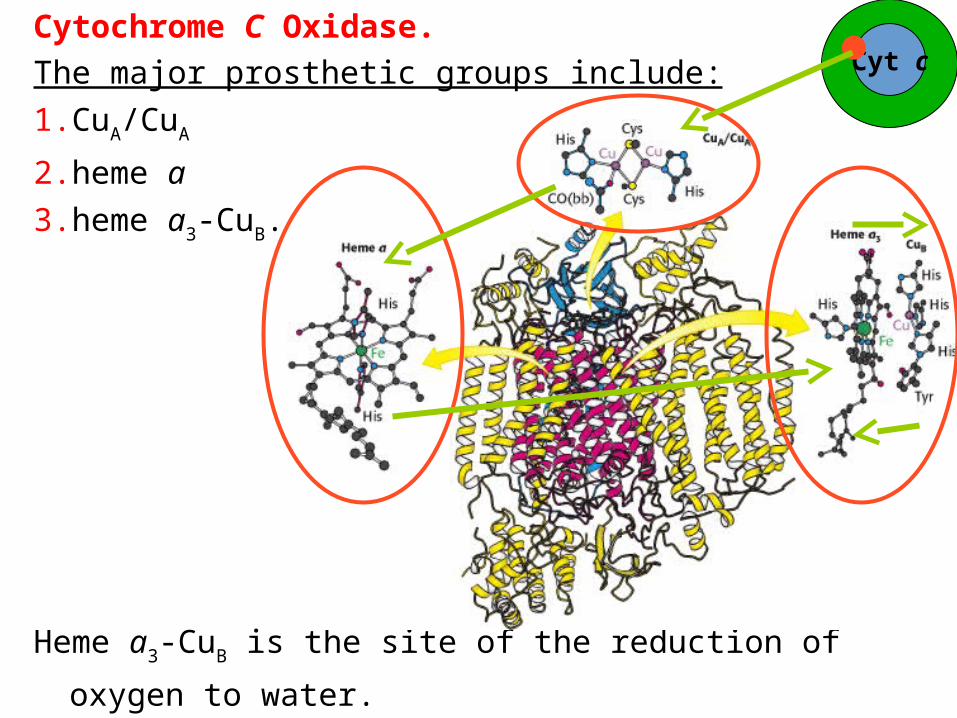
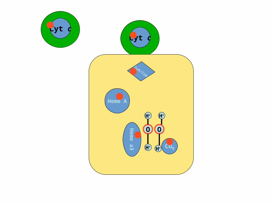
Cytochrome C Oxidase.
The major prosthetic groups include:
1. CuA/CuA
2. heme a
3. heme a3-CuB.
Heme a3-CuB is the site of the reduction of oxygen to water.
Cyt c
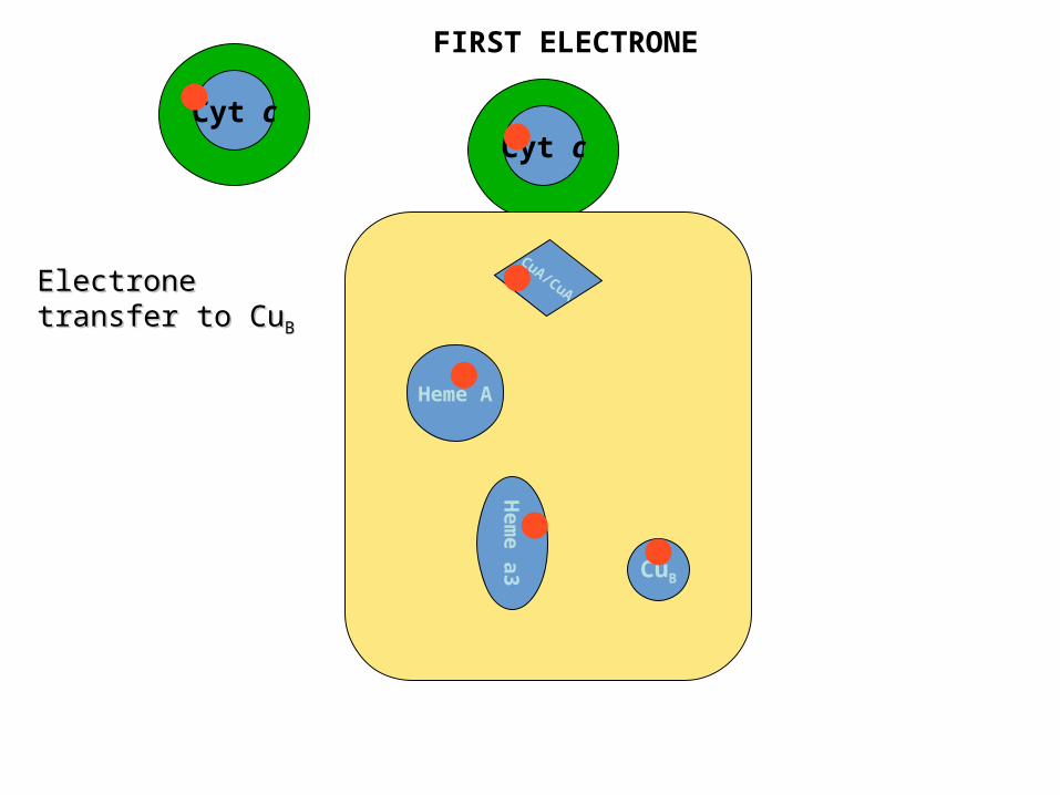
Cyt cCyt c
CuA/CuA
Heme A
Hem
e a
3 CuB
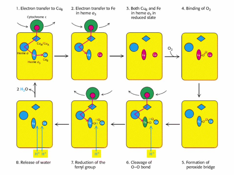
Electrone transfer Electrone transfer to Cuto CuBB
FIRST ELECTRONE

Cyt cCyt c
CuA/CuA
Heme A
Hem
e a
3 CuB
O O
O O
Electrone transfer Electrone transfer to to FeFe in in Heme aHeme a33
Both Both CuBCuB and and FeFe in in Heme aHeme a33 are in are in reduced formreduced form
Binding of Binding of O2O2
Formation of Formation of peroxide bridgeperoxide bridge
SECOND ELECTRONE
Cyt cCyt c
CuA/CuA
Heme A
Hem
e a
3 CuB
O O
H+
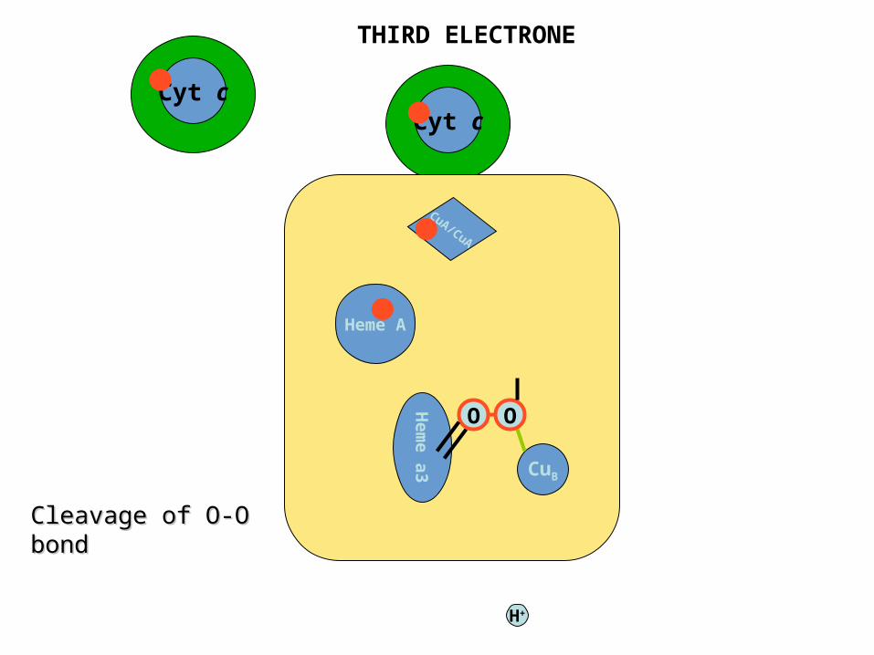
THIRD ELECTRONE
Cleavage of O-O Cleavage of O-O bondbond
Cyt cCyt c
CuA/CuA
Heme A
Hem
e a
3 CuB
O O
H+
H+
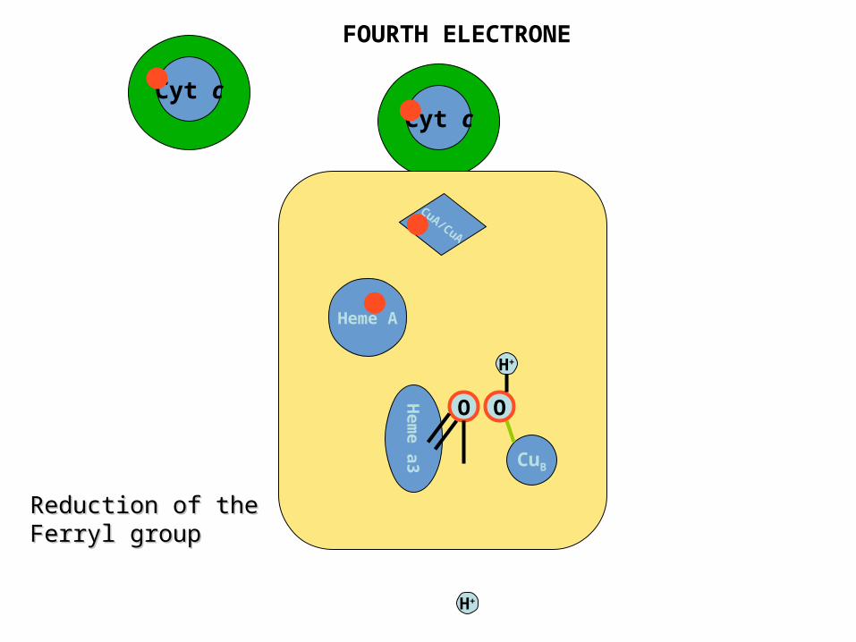
FOURTH ELECTRONE
Reduction of the Reduction of the Ferryl groupFerryl group
Cyt c
CuA/CuA
Heme A
Hem
e a
3 CuB
O O
H+
H+
H+ H+
H+
H+
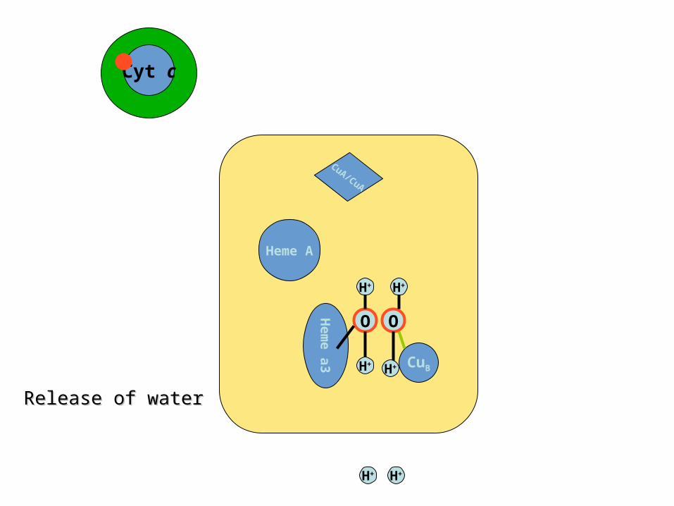
Release of waterRelease of water
Cyt cCyt c
CuA/CuA
Heme A
Hem
e a
3 CuB
O O
H+
H+
H+
H+

This reaction can be summarized as
•The four protons in this reaction come exclusively from the matrix.
•Thus, the consumption of these four protons contributes directly to the proton
gradient.•Each proton contributes (21.8 kJ mol-1) to
the free energy associated with the proton gradient .
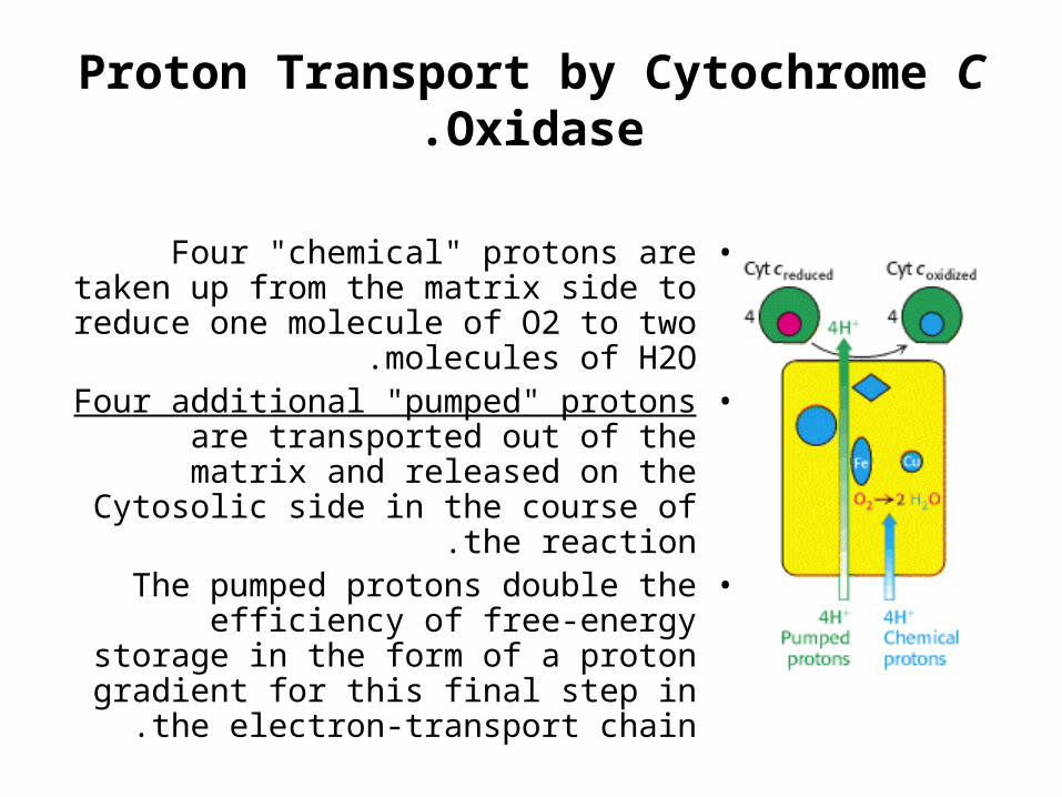
Proton Transport by Cytochrome C Oxidase.
•Four "chemical" protons are taken up from the matrix side to reduce one
molecule of O2 to two molecules of H2O.
•Four additional "pumped" protons are transported out of the matrix and
released on the Cytosolic side in the course of the reaction.
•The pumped protons double the efficiency of free-energy storage in the
form of a proton gradient for this final step in the electron-transport chain .
How these protons are transported through the protein?
•Still under study.•However, two effects contribute to the
mechanism:.1Charge neutrality tends to be maintained in the
interior of proteins.–Thus, the addition of an electron to a site inside a
protein tends to favor the binding of a proton to a nearby site.
.2Conformational changes take place, particularly around the heme a3-CuB center, in the course of
the reaction cycle.–These changes must be used to allow protons to
enter the protein exclusively from the matrix side and to exit exclusively to the cytosolic side .
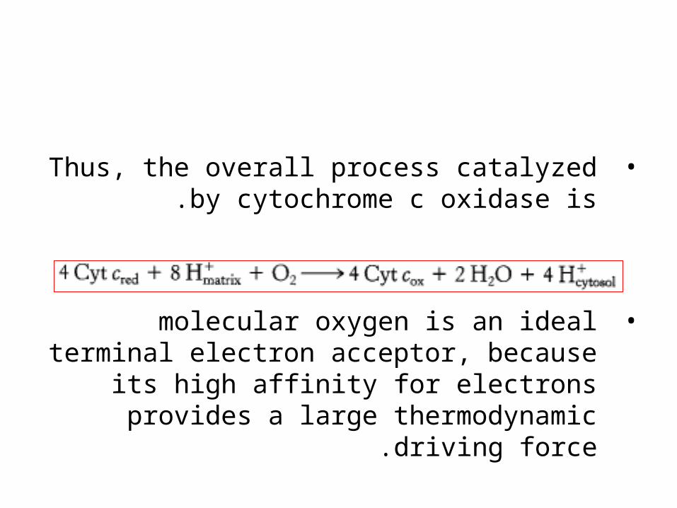
•Thus, the overall process catalyzed by cytochrome c oxidase is.
•molecular oxygen is an ideal terminal electron acceptor, because its high
affinity for electrons provides a large thermodynamic driving force.
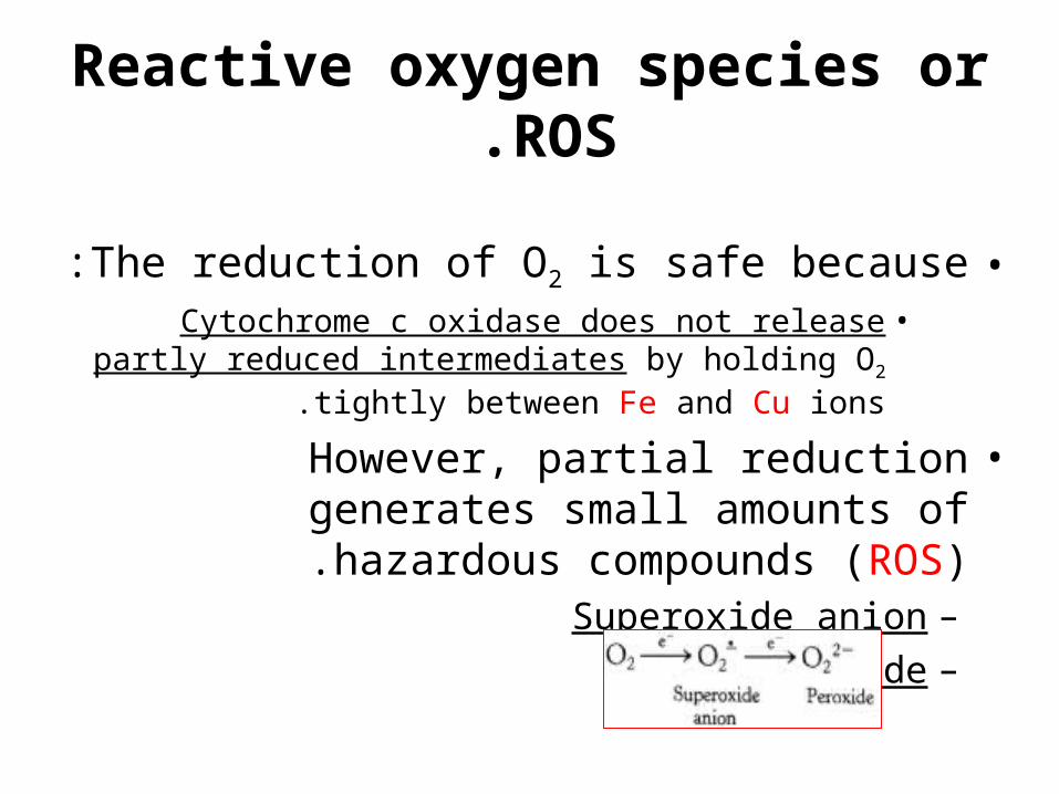
Reactive oxygen species or ROS.
•The reduction of O2 is safe because:•Cytochrome c oxidase does not release partly
reduced intermediates by holding O2 tightly between Fe and Cu ions.
•However, partial reduction generates small amounts of hazardous compounds (ROS).
–Superoxide anion–peroxide.
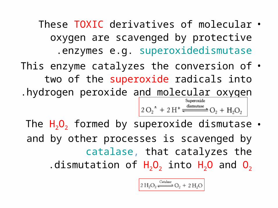
•These TOXIC derivatives of molecular oxygen are scavenged by protective enzymes e.g.
superoxidedismutase.
•This enzyme catalyzes the conversion of two of the superoxide radicals into hydrogen peroxide
and molecular oxygen.
•The H2O2 formed by superoxide dismutase and
by other processes is scavenged by catalase, that catalyzes the dismutation of H2O2 into H2O
and O2.
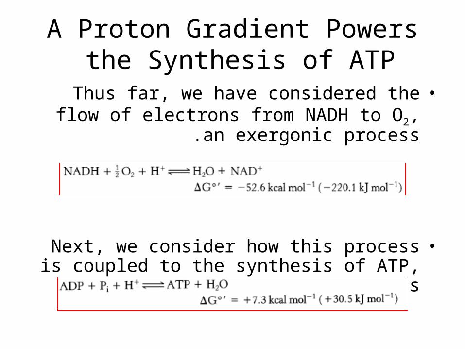
A Proton Gradient Powers the Synthesis of ATP
•Thus far, we have considered the flow of electrons from NADH to O2, an exergonic
process .
•Next, we consider how this process is coupled to the synthesis of ATP, an endergonic process ?

ATP synthase
•The synthesis of ATP is carried out by A molecular assembly in the inner mitochondrial membrane called:
–ATP synthase–mitochon-drial ATPase
–F1F0 ATPase
–Complex V
How is the oxidation of NADH coupled to the phosphorylationof ADP?
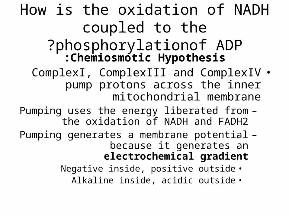
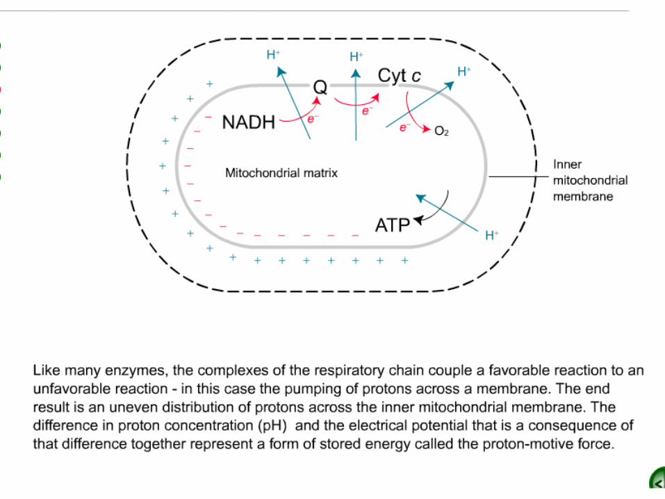
Chemiosmotic Hypothesis:•ComplexI, ComplexIII and ComplexIV pump
protons across the inner mitochondrial membrane
–Pumping uses the energy liberated from the oxidation of NADH and FADH2
–Pumping generates a membrane potential because it generates an electrochemical gradient
•Negative inside, positive outside•Alkaline inside, acidic outside
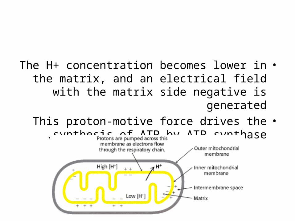
•The H+ concentration becomes lower in the matrix, and an electrical field with the matrix side
negative is generated•This proton-motive force drives the synthesis of
ATP by ATP synthase .
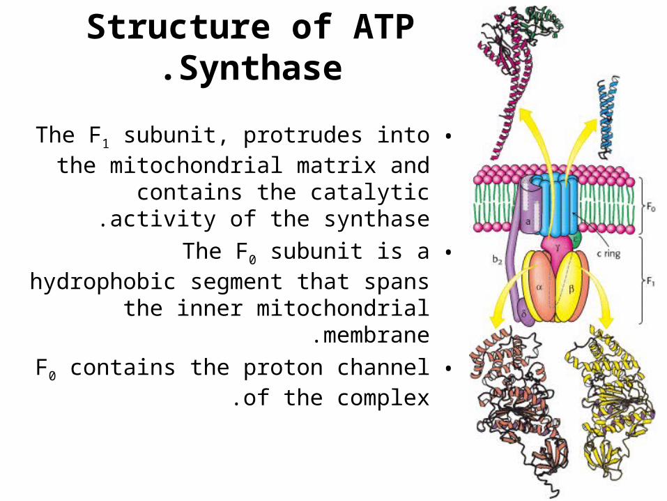
Structure of ATP Synthase.
•The F1 subunit, protrudes into the mitochondrial matrix and contains the
catalytic activity of the synthase.
•The F0 subunit is a hydrophobic segment that spans the inner mitochondrial
membrane.
•F0 contains the proton channel of the complex.
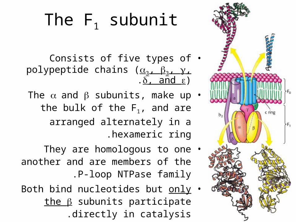
The F1 subunit
•Consists of five types of polypeptide chains (3, 3, , , and ).
•The and subunits, make up the bulk of the F1, and are arranged alternately in
a hexameric ring.
•They are homologous to one another and are members of the P-loop NTPase
family.
•Both bind nucleotides but only the subunits participate directly in catalysis.
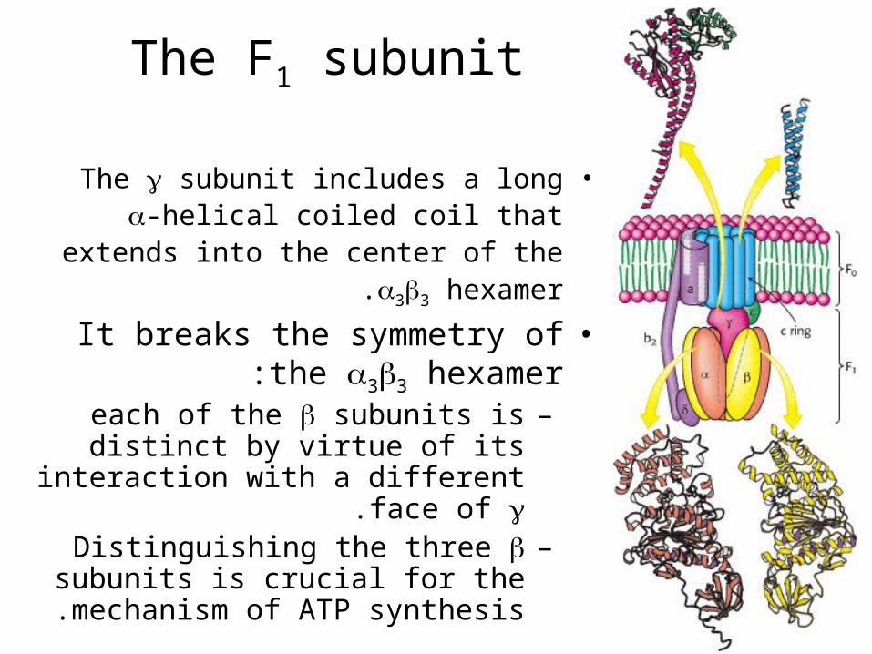
The F1 subunit
•The subunit includes a long -helical coiled coil that extends into the center of
the 33 hexamer.
•It breaks the symmetry of the 33 hexamer:
–each of the subunits is distinct by virtue of its interaction with a
different face of .–Distinguishing the three subunits
is crucial for the mechanism of ATP synthesis .
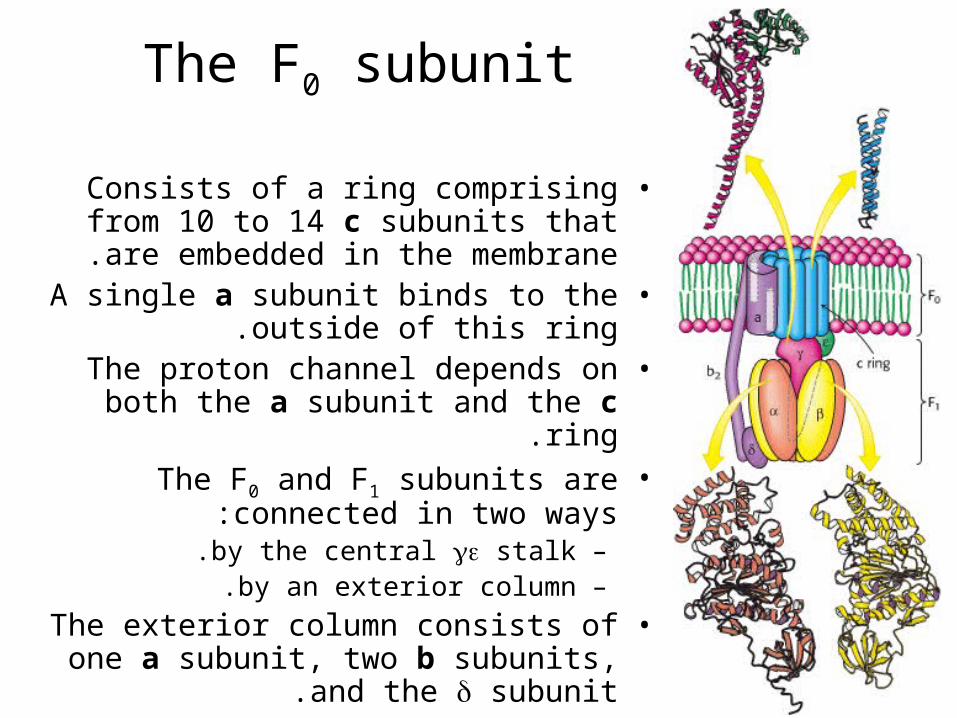
The F0 subunit
•Consists of a ring comprising from 10 to 14 c subunits that are embedded in the
membrane.•A single a subunit binds to the outside of
this ring.•The proton channel depends on both the
a subunit and the c ring.•The F0 and F1 subunits are connected in
two ways:–by the central stalk.–by an exterior column.
•The exterior column consists of one a subunit, two b subunits, and the
subunit.
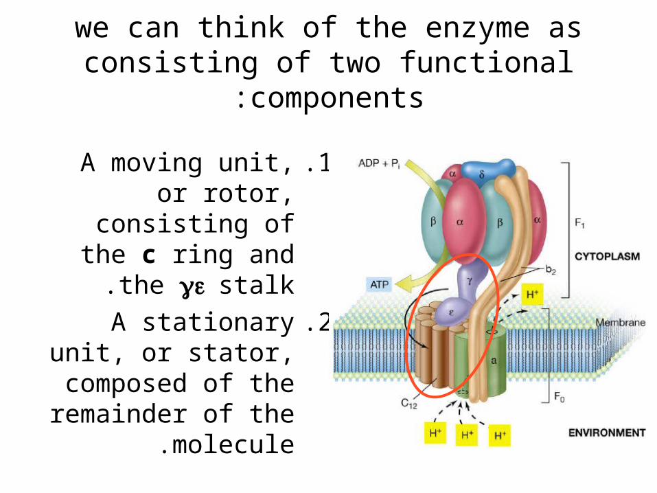
we can think of the enzyme as consisting of two functional components:
.1A moving unit, or rotor, consisting of the
c ring and the stalk.
.2A stationary unit, or stator, composed of the remainder of the
molecule.
The Binding-Change Mechanism