Embed Size (px)
Citation preview
4822 Biochemistry 1983, 22, 4822-4830
Schiff, P. B., & Horwitz, S . B. (1980) Proc. Natl. Acad. Sci.
Schiff, P. B., & Horwitz, S . B. (1981) Biochemistry 20,
Schiff, P. B., Fant, J., & Horwitz, S . B. (1979) Nature
Shelanski, M. L., Gaskin, F., & Cantor, C. R. (1973) Proc.
Thompson, W. C., Wilson, L., & Purich, D. L. (1981) Cell
U.S.A. 27, 1561-1565.
3247-3252.
(London) 277, 665-667.
Natl. Acad. Sci. U.S.A. 70, 765-768.
Muscle Motil. I , 445-454.
Wani, M. C., Taylor, H. L., Wall, M. E., Coggon, P., & Mac Phail, A. T. (1971) J . Am. Chem. SOC. 93, 2325-2327.
Weingarten, M. D., Lockwood, A. H., Hwo, S. Y., & Kir- schner, M. W. (1975) Proc. Natl . Acad. Sci. U.S.A. 72,
Weisenberg, R. C., Deery, W. J., & Dickinson, P. J. (1976)
Wilson, L., & Meza, I. (1973) J . Cell. Biol. 58, 709-719. Zeeberg, B., & Caplow, M. (1979) Biochemistry 18,
1858-1862.
Biochemistry 15, 4248-4254.
3 880-3 886.
Oxygen-Exchange Studies on the Pathways for Magnesium Adenosine 5’-Triphosphate Hydrolysis by Actomyosint
Kamal K. Shukla, Harvey M. Levy,* Fausto Ramirez, James F. Marecek, Brian McKeever, and Sarkis S. Margossian
ABSTRACT: At an intermediate stage in the hydrolysis of magnesium adenosine 5’-phosphate (MgATP) by myosin or actomyosin, there is an exchange of oxygen between water and the P, group of enzyme-bound nucleotide. Starting with [P,-’*O]ATP as substrate, the exchange is revealed in the [180]Pi species that are ultimately released as product into the reaction medium. An analysis of the distribution of these labeled Pi species, which contain 3, 2, 1, or none of the ‘*O atoms originally on the P, of ATP, is used to probe inter- mediate stages of the hydrolytic mechanism. In recent years, studies of this kind by several groups have shown that more than one pathway of hydrolysis operates. The work reported here demonstrates that two of these pathways are spurious; one is a “nonexchanging MgATPase” that is present in fresh myosin preparations; the other is an induced slow exchange
I n the hydrolysis of [P,-180]MgATP by myosin or acto- myosin, there is an exchange of oxygen between the P, group of enzyme-bound MgATP and water of the medium. This exchange is revealed by an examination of the distribution of l 8 0 in the inorganic Pi produced by the hydrolysis. Hydrolysis of MgATP by myosin follows a mechanism that is summarized in Scheme I according to Bagshaw & Trentham (1974). Scheme I
kl k2 M + ATP 4 M*ATP -
k , ( I50 s-’) k4 (0.03 s-’) ’ M* **ADP*Pi -
k-, ( 1 5 s-I) slow M*.ATP.
k5 k6 M*.ADP.P, - M**ADP + Pi -
ki M-ADP - M + ADP
’ From the Departments of Physiology and Biophysics, Health Sci- ences Center (K.K.S. and H.M.L.), and Chemistry (F.R., J.F.M., and B.M.), State University of New York at Stony Brook, Stony Brook, New York 11794, and the Department of Biochemistry and Medicine, Albert Einstein College of Medicine at Montefiore Hospital and Medical Center, Bronx, New York 10467 (S.S.M.). Received December 27, 1982; re- vised manuscript received June 8, 1983. This work was supported by Grant PCM 78-03259 from the National Science Foundation, Grant 1451 from the Muscular Dystrophy Association (to H.M.L.), Grant GM-20672 from the National Institutes of Health (to F.R.), and Grants MDA and NHLBI HL-26569 (to S.S.M.).
that develops in myosin during storage (-20 “C) and subse- quent aging (4 “C). However, after correction for these ar- tifacts, two normal pathways for actomyosin hydrolysis remain. These normal pathways differ in the mode of interaction be- tween actin and myosin in the course of hydrolysis; one is the Lymn-Taylor pathway where oxygen exchange occurs at a stage when actin and myosin are dissociated; the other is a pathway in which actin and myosin are associated during oxygen exchange. Each of these two pathways contributes an equal amount of Pi to the product pool. Thus, on average, each myosin head uses each of these pathways half the time. The findings suggest, e.g., that during contraction, myosin can dissociate from the actin filament only during every other cycle of MgATP hydrolysis or that only half the heads, at any one time, can exchange oxygen while free of the actin filament.
Oxygen exchange occurs at step 3, the hydrolytic cleavage step. Each time cleavage takes place, an oxygen of water is added to the P, of ATP, and each time this reaction reverses, an oxygen from the P, of ATP goes to water. It is assumed that the oxygens around the bound Pi of M**ADP.Pi can change position in space, e.g., by rotation; therefore, the oxygen lost during reverse cleavage can by chance be different from the one added during cleavage as indicated in Scheme 11.
The extent of exchange depends on the number of inter- conversions that can occur between M*ATP and M**ADP.Pi before the protein-nucleotide complex moves through the rate-limiting step 4 and ultimately releases Pi to the medium. Since the cleavage step is fast, the number of interconversions that actually take place is determined by the rate constant k-, for reverse cleavage and the rate constant k4 for the rate- limiting step. Actin activates the rate-limiting step. Thus the greater the concentration of actin, the shorter the lifetime of the exchanging intermediates and the lower the extent of exchange (Shukla & Levy, 1977a).
In the original Lymn-Taylor pathway (Lymn & Taylor, 197 l) , actomyosin hydrolysis follows the route shown in Scheme 111. In this scheme, exchange occurs when myosin is dissociated from actin. The distribution of labeled Pi species, containing 3, 2, 1 or none of the original l80 atoms on the P, of ATP, can be predicted theoretically for the Lymn-Taylor
0006-2960/83/0422-4822$01.50/0 0 1983 American Chemical Society

O X Y G E N E X C H A N G E B Y A C T O M Y O S I N V O L . 2 2 , N O . 2 0 , 1 9 8 3 4823
was dialyzed against the standard reaction solution before use. Actin was extracted from acetone-dried muscle powder by the method of Spudich & Watt (1971). Cardiac myosin was prepared from rabbit or dog heart muscle as described by Margossian & Lowey (1982). Cardiac or skeletal myosin depleted of the LC 2 light chain was made by digesting the protein with the neutral protease from myopathic hamster as described by Margossian et al. (1980). In some cases, the protein was recombined with LC 2 (Margossian et al., 1980).
Subfragment 1 (Sl) was made according to Weeds & Taylor (1975) with chymotrypsin but without chromatographic separation after proteolysis; thus, this preparation is a mixture of S1 A1 (alkali light chain 1) and S1 A2 (alkali light chain 2) with no LC 2 chain (the DTNB chain). Heavy meromyosin (HMM) with a full complement of LC 2 (often at a slightly lower than normal weight) was prepared with trypsin according to the method of Holt & Lowey (1 975). Gel electrophoresis was carried out as described by Weber & Osborn (1969).
MgATP hydrolysis by myosin and its subfragments was carried out in a reaction solution of the following composition: 50 mM KC1,25 mM Tris, pH 7.4, 1 mM DTT, 5 mM MgC12, and 2-5 mM [P,-180]ATP at 23 “C. In some runs, 250 gM bisadenosine pentaphosphate was added to inhibit any ade- nylate kinase activity. The reaction was quenched with tri- chloroacetic acid after 80-100% hydrolysis. The product Pi was isolated and converted to trimethyl phosphate and the fraction of each labeled Pi species determined by mass spec- trometry as described previously (Shukla et al., 1980, 1982). The labeled ATP contained 95-98 atom ’% excess l 8 0 in the P, position.
Mathematical Modeling of Oxygen Exchange. To interpret the oxygen exchange, we assume the sequence of steps shown in Scheme I. In this scheme, as outlined in the introduction, oxygen exchange occurs by a repeated interconversion of the two intermediates M*ATP and M**ADP-PI prior to the rate-limiting step, k4, and the ultimate release of products into the medium.
In the experiments described here, the P, oxygens of the substrate, ATP, are labeled with l 8 0 . Therefore, exchange is observed as the incorporation of unlabeled oxygen from water into the Pi that is released as product into the medium. Without any exchange, there is just one cleavage reaction putting one water oxygen into the bound Pi of M**ADP.Pi, and the species of Pi released into the medium contains this one unlabeled oxygen from water ( l 6 0 ) and three labeled oxygens originally on the P, of ATP. This species is designated n3 (three labeled oxygens retained in the product
With a sufficient number of interconversions between M*ATP and M**ADP.Pi, all of the l 8 0 in the original ATP may be exchanged for the I6O of water to produce the com- pletely exchanged species denoted as nO (none of the original l80 remaining); with partial exchange other species, n2 (two labeled oxygens retained) and nl (one of the original oxygens retained), are formed. Note that n4, with four labeled oxygens, is never formed even though the bridge oxygen between Ps and P, is labeled in the substrate we use; this is so because in this enzyme the water oxygen always adds to P, during cleavage while the labeled bridge oxygen remains on the Po of bound
Pi) *
Scheme IIa
0
HOH + M**ADP-O-i-O I k -1, MM*ADP-0*O--P-O I k,,t.t,on*
I 0
P 9 1 0
I k- 3 M***ADP-0.0-P-0 - HOH + M**ADP-O-P-O
0 is I6O; 0 is I8O.
model at different levels of actin, which set different turnover times for hydrolysis. Then, the fraction of each Pi species under different conditions can be measured by mass spec- trometry and compared to a theoretical distribution on the basis of the kinetic scheme. This kind of analysis proves to be a sensitive probe into the way that actin and myosin interact during the hydrolytic cleavage of bound MgATP. The physiological significance lies in the fact that such interactions in muscle are part of the contraction mechanism.
Scheme I11
AM + ATP + AMaATP + A + M**ATP e A 4- k-3
kdAM M**.ADP*Pi + AM***ADP.Pi - AM + ADP + Pi
The work along these lines reported here establishes that actomyosin can hydrolyze MgATP by two different routes under conditions where no artifactitious pathway is operating. The basic difference between these two normal actomyosin pathways is in the mode of interaction between myosin and actin in the course of hydrolysis. In one pathway, oxygen exchange occurs at the stage when myosin is dissociated from actin, in accordance with the Lymn-Taylor mechanism (Scheme 111); in the other pathway, however, actin and myosin are associated during the exchange reactions (Inoue et al., 1973; Stein et al., 1981; Midelfort, 1981).
These two pathways of actomyosin hydrolysis can now be distinguished clearly from other pathways of hydrolysis that are present in certain preparations of myosin and actin and that have complicated the analysis of actomyosin hydrolysis and oxygen exchange. These other activities, as we well de- scribe, are caused by changes in myosin that occur during storage and by a “nonexchanging MgATPase” that is present in fresh myosin preparations even after ammonium sulfate fractionation. We have now defined each of these activities and found ways to avoid or correct for them. Free of the complexities that these abnormal pathways introduce, the oxygen-exchange data now show that about half of the Pi from actomyosin hydrolysis comes from a Lymn-Taylor pathway, while the other half comes from a pathway where oxygen exchange occurs on undissociated actomyosin. The findings lead to a consideration of the possible role that two such pathways may play in the mechanism of muscle contraction.
Materials and Methods
Protein Preparations. Skeletal myosin was prepared from rabbit (or rat) back and leg muscle by the method of Mom- maerts & Parrish (1951) and fractionated with ammonium sulfate to remove actin. This purified myosin was used for the studies involving myosin itself and also to prepare myosin subfragments. The myosin was used fresh or after storage in 50% glycerol, 50 mM phosphate buffer (pH 6.5), 0.6 M KC1, and 1 mM dithiothreitol (DTT)’ at -20 OC. Stored myosin
’ Abbreviations: S1, subfragment 1; HMM, heavy meromyosin; LC 2, light chain 2 or DTNB chain; DTNB, 5,5’-dithiobis(2-nitrobenzoic acid); DTT, dithiothreitol or 1,4-dimercapto-2,3-butanediol; EGTA, ethylene glycol bis(0-aminoethyl ether)-N,N,N’,N’-tetraacetic acid; SDS, sodium dodecyl sulfate; NEM, N-ethylmaleirnide; TNBS, 2,4,6-tri- nitrobenzenesulfonate; Tris, tris(hydroxymethy1)aminomethane.
4824 B I 0 C H E M I S T R Y S H U K L A ET A L .
ADP. Also, note that the lSO label that goes into the water in the course of exchange can be neglected in subsequent interconversions, since it never rises to a significant level relative to the molarity of water.
The quantitative analysis of the distribution of the various species of Pi produced during hydrolysis stems basically from a consideration of the probability for an exchange to occur between I6O and lSO after one, two, or any number of se- quential interconversions between M*ATP and M**ADP-Pi prior to the release of the products. The analysis starts quite simply with the assumption that in a mechanism such as that outlined in Scheme 11, the chance is three out of four (3/4) that, in a given interconversion, the oxygen lost during reverse cleavage will be different from the one added during the previous cleavage reaction. This follows directly from the assumption that all four oxygens around the bound Pi of M**ADP.Pi are randomly scrambled by the effective rotation, shown in Scheme 11, and therefore, have an equal chance to go to water during reverse cleavage.
The distribution of labeled Pi species depends on the rate at which the exchanging intermediates interconvert and the time available for them to do this, i.e., the average lifetime of the exchanging intermediates. In the pathway of hydrolysis shown in Scheme I, these two determinants of oxygen exchange are given by the two rate constrants k-, and k4. The constant k-, limits the rate of the exchange cycle, Le., limits the rate of the interconversions. This is so because the cleavage re- action, k3, is known to be relatively fast (of the order of 150 s-l) and the rate of rotation is assumed to be fast, whereas k-, is of the order of 15 s-l (Webb & Trentham, 1981). The constant k4 determines the time available for exchange by determining the overall turnover number. Thus, the extent of exchange is a function of k-, (the rate constant of reverse cleavage) times l/k4 (the turnover time of hydrolysis); the ratio of these two constants (k-3/k4) is designated here as R. The greater the value of R, the greater is the extent of exchange.
In theoretically predicting the distribution of labeled Pi species, the exchange is mathematically determined for a series of interconversions where the chance for losing a number of the lSO is some function of the two rate constants k-, and k,; e.g., the probability that Pi will be released from M**ADP.Pi without exchange is given by k4/(k-3 + k4).
During every interconversion, the chance is three out of four for an exchange between phosphate and water oxygen. However, as unlabeled water oxygen is progressively incor- porated into the bound Pi through repeated interconversions, the chance progressively increases that the next unlabeled oxygen from water will exchange for an unlabeled oxygen already added to the phosphate during a previous cycle. In other words, there are diminishing returns for an exchange of I6O for I8O with each additional interconversion, and the mathematics take this into account. It turns out, finally, that the expected distribution for a given value of R is given by the following relatively simple set of equations that apply when the starting ATP is 100% labeled at P, or when the data are normalized to this condition as we have done.
n3 = [4/(3R + 4)] X 100
n2 = (100 - n3)[2/(R + 2)]
nl = (100 - n3 - n2)[4/(R + 4)]
nO = 100 - n3 - n2 - nl
In summary, the equations give the percent of each Pi species, n3, n2, n l , and nO (the number indicating how many
original P, oxygens have been retained), as a function of R (k-,/k4) under conditions where the probability for a water- phosphate oxygen exchange is three chances out of four for each interconversion, with diminishing returns on 160-180 exchange as the process repeats. A number of mathematical treatments leading to equivalent equations have been given (Hackney, 1980; Midelfort, 1981; Webb & Trentham, 1981).
It is important to make clear the effect of actin on the Lymn-Taylor pathway of Scheme I11 in terms of these dis- tribution studies. In the Lymn-Taylor pathway, actin de- creases the lifetime of the exchanging intermediates through an activation of the rate-limiting step k4. In doing so, actin decreases the extent of exchange; and the apparent value of R is decreased because the denominator k4 of this ratio is increased. Actin does not appear to have any effect on k-,, consistent with the assumption that in this scheme exchange occurs on myosin dissociated from actin. Thus, actin can be used to determine k-3 for myosin by measuring the distribution of labeled Pi species for different values of R set by the actin level; this is done by using the equation
R = k-,/k,
This analytical approach has been developed and used by a number of workers (Sleep et al., 1978; Midelfort, 1981; Webb & Trentham, 1981; Shukla et al., 1982). It is outlined here in general terms to give the reader a picture of the logic used in the analysis of the distribution data and to show the general relationship between the distributions of labeled Pi species and the rate constants k-, and k4 in the pathways of hydrolysis by myosin and actomyosin.
Under simple conditions, the experimental values for the distribution of Pi species fit the set of theoretical equations. Thus, with chymotryptic subfragment 1, where only the Lymn-Taylor pathway of hydrolysis appears to occur, the experimental distribution can be matched to a theoretical distribution for a single pathway with the value of R set by the actin concentration. From any value of R, the value of k-3 can be estimated from the equation k-3 = Rk,,, (which comes from substituting the measured turnover number k,,, for the value of the rate-limiting step k,). When this is done for a set of actin concentrations, the value for k-, is in the range of 10-15 s-l. This estimate agrees well with the value of 15 s-l recently determined from oxygen-exchange analysis of pre-steady-state intermediates (Webb & Trentham, 198 1) and with the value of 10-20 s-l determined by rapid-reaction techniques for studying the kinetics of the system (Bagshaw & Trentham, 1974).
Thus, the distribution studies on chymotryptic subfragment 1 with actin support the Lymn-Taylor pathway of hydrolysis and fit a scheme in which it is the only pathway operating. However, when the same kind of distribution studies have been made with myosin or heavy meromyosin (Shukla et al., 1980; Midelfort, 1981), the data have not fit a single set of exper- imental distributions for any value of R. Instead, with these proteins, the data fit a scheme in which two different pathways of hydrolysis operate with widely differing values of R and with each pathway contributing a substantial fractioqf, of the P, to the product pool.
The fitting of experimental values to a theoretical two- pathway system is a process of closer and closer approxima- tions. However, with four variables, two values for R and two forf, the ability to find fits that are meaningful depends on the fact that the pathways that appear in the actomyosin system have widely different values of R. Thus, there is typically one type of pathway with a value of R greater than 50, the high-exchange type, and another type of pathway with

O X Y G E N E X C H A N G E B Y A C T O M Y O S I N
an R of less than 2, the low-exchange type. As we will discuss, there are three different pathways of the low-exchange type.
Because of the large difference in R between the two types of pathways that operate, a good fit to the data can be made by assuming that virtually all of the Pi species at n3 and n2 come from the low-exchange pathway. Then from the ratio of n2 f n3, a good estimate of the R for low exchange comes by matching this ratio to the theoretical ratio for some value of R (a table of R vs. n2/n3 is easily generated by computer). Once having this value of R for the low-exchange pathway, the small contribution it would be expected to make to Pi species at nl and nO can be determined from the equations or from a computer generated table of distributions over a range of R values. After subtraction of the contribution of low exchange to the n l and nO values, the remaining values can then be used to obtain a ratio of nl/nO for the fast ex- change. With this nl/nO ratio for fast exchange, one obtains the R for fast exchange by matching it to a theoretical value of R. In a second approximation, the small contribution made by the high exchange to n3 and n2 can be subtracted away to give a new ratio of n2/n3 and a closer approximation to the R of low exchange. Usually, only one or two approxi- mations need to be made to obtain a reasonable fit. We do not attempt here to use this kind of data fitting to make fine distinctions although that should be possible with the high accuracy that can be obtained.
Simplifying our analysis considerably, there is not a con- tinuum of R values for low exchange. Instead, in the course of accumulating over 100 distributions, it has become evident that the distributions fall into three easily distinguishable categories. In the first category, the R values fall below 0.2 and indicate the presence of a nonexchanger; in the second category, the R values cluster around 0.4 and, as we will discuss, characterize what we call induced slow exchange; and then there is.a third category seen only in the presence of actin, termed actomyosin minimal exchange, where the R values are between 1 and 2.
When the nonexchanger is operating, this is directly evident by the exceedingly low fraction of Pi at n2. This column under these circumstances is virtually "empty" because the nonex- changer, we believe, does not produce any of this species and the high-exchange pathway produces less than 2%. When the slow exchanger is operating, there is a significantly higher level of Pi at n2, giving an n2/n3 ratio of about 0.3 or less; and this is distinguished from the presence of actomyosin minimal exchange, which has an n2/n3 ratio above 0.5. In general, it is directly evident from the n2/n3 ratio which of the low- exchange pathways is operating, and a fit is not difficult to obtain. Nevertheless, the difference in R values is not the primary criterion for distinguishing the three types of pathways to be defined in this paper. The distinctions depend primarily on major differences between them in their origin, lability, and response to actin and in the ability to selectively remove all but one by setting the appropriate experimental conditions.
Several identical Pi distribution experiments run on the same protein gave data that agree within 5%. The mass spectro- metric analyses are, of course, more accurate (<2%) (Mey- erson et al., 1982).
Results Nonexchange Actiuity. Table I shows typical [180]Pi dis-
tributions obtained from the hydrolysis of labeled ATP by fresh preparations of myosin from rabbit or rat skeletal muscle or from rabbit heart. The data show a relatively large percent of Pi with no exchange (n3). This kind of distribution shows an extremely small fraction of the Pi species with just one
VOL. 2 2 , N O . 2 0 , 1 9 8 3 4825
Table I: Preparations of Fresh Myosin Showing the Presence of Nonexchange Activitya
percent of [ l80,]Pi species
protein n 3 b n2 n l nO
skeletalmyosin 15.3 0.8 2.3 81.5 (rabbit) 14.7 1.1 2.5 81.8
13.0 0.9 2.4 83.8 skeletalmyosin 18.3 1.4 4.1 76.3
cardiac myosin 12.4 1.4 1.6 84.6 (rat)
(rabbit)
Reaction conditions: 50 mM KC1, 25 mM Tris, pH 7.4, 1 mM DTT, 5 mM MgCI,, and 2-5 mM [y"O]ATP at 23 "C. The presence of nonexchange activity is indicated by the relatively high percent of the Pi species at n3.
Table 11: Loss of Nonexchange Activity on Aging Myosin in Low-Salt Solutiona
percent of [180n]Pi species time of aging (days)b n3C n2 nl nO
1 13.0 0.9 2.4 83.8 2 10.4 1.1 2.5 85.9 3 8.3 1.0 2.4 88.2 9 3.5 1.1 2.3 93.0
15 2.7 1.6 2.8 92.9 a Reaction conditions: see Table I.
was dialyzed at 4 "C against 50 mM KC1, 25 mM Tris, pH 7.4, and 1 mM DTT. decrease of the Pi species at n3.
To age the protein, it
The loss of nonexchange activity is seen in the
Table 111: Retention of Nonexchange Activity on Aging Myosin in High-Salt Solutiona
percent of ['80,]Pi species time of aging (days)b n3C n2 nl nO
3 10.7 1.3 2.6 85.5 9 10.4 1.0 2.5 86.1
15 14.7 1 .1 2.5 81.8 a Reaction conditions: see Table I. To age the protein, it
was dialyzed against 0.6 M KC1, 25 mM Tris, pH 7.4, and 1 mM DTT at 4 "C. high percent of the Pi species at n3.
Nonexchange activity is indicated by the relatively
oxygen exchanged (n2). This leads to a very low n2/n3 ratio of less than 0.1, characteristic of the nonexchange activity. Normally, in order to remove traces of actin, myosin is frac- tionated with ammonium sulfate, and the portion that pre- cipitates out between 40 and 60% saturation is retained. The protein mixture is then dialyzed against the low-salt reaction solution to remove ammonium sulfate. The percent of non- exchange activity is highest just after this step. As shown in Table 11, this activity is lost when the protein is allowed to stand in a low-salt solution in a test tube or when it is dialyzed against such a solution. The nonexchange activity is not an effect of ammonium ion, since it is lost under conditions where any level of this ion that might be present would remain constant. Moreover, as shown in Table 111, the nonexchange activity is stable in high salt and remains at the original level after long-term dialysis against a number of large-volume changes of the dialyzate.
Table IV shows that this nonexchange activity of fresh myosin is lost in the preparation of heavy meromyosin (HMM) or of subfragment 1 (Sl). However, it is not inhibited by high salt and EGTA (to remove free CaZ+), conditions that would be expected to markedly inhibit any activation of myosin MgATPase by trace amounts of contaminating actin.
4826 El I O C HEM I S T R Y S H U K L A E T A L .
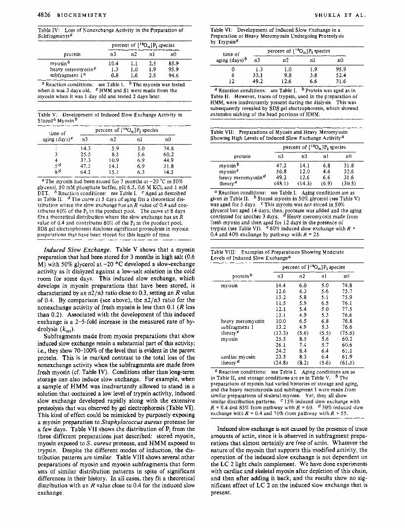
Table IV: Loss of Nonexchange Activity in the Preparation of Subfragmen t s a
percent of [180,]Pi species
protein n3 n2 n l nO
myosinb 10.4 1.1 2.5 85.9 heavy meromyosinC 1.3 1.0 1.9 95.9 subfragment l C 0.8 1.6 2.5 94.6
Reaction conditions: see Table I. The myosin was tested HMM and S1 were made from the when it was 3 days old.
myosin when it was 1 day old and tested 2 days later.
Table V: Development of Induced Slow Exchange Activity in Stored4 Myosinb
time of percent of [ '800,] Pi species
aging (days)c n3 n2 n l nO 1 14.3 5.9 5.0 74.8 3 25.5 8.5 5.6 60.2 4 37.3 10.9 6.9 44.9 5 d 47.3 14.1 6.9 31.8 8 d 64.2 15.1 6.3 14.5
a The myosin had been stored for 3 months at -20 "C in 50% glycerol, 5 0 mM phosphate buffer, pH 6.5, 0.6 M KCl, and 1 mM DTT. Reaction conditions: see Table I. Aged as described in Table 11. The curve at 5 days of aging fits a theoretical dis- tribution where the slow exchange has an R value of 0.4 and con- tributes 60% of the Pi in the product pool. The curve at 8 days fits a theoretical distribution where the slow exchange has an R value of 0.4 and contributes 80% of the Pi in the product pool. SDS gel electrophoresis discloses significant proteolysis in myosin preparations that have been stored for this length of time.
Induced Slow Exchange. Table V shows that a myosin preparation that had been stored for 3 months in high salt (0.6 M) with 50% glycerol at -20 OC developed a slow-exchange activity as it dialyzed against a low-salt solution in the cold room for some days. This induced slow exchange, which develops in myosin preparations that have been stored, is characterized by an n2/n3 ratio close to 0.3, setting an R value of 0.4. By comparison (see above), the n2/n3 ratio for the nonexchange activity of fresh myosin is less than 0.1 ( R less than 0.2). Associated with the development of this induced exchange is a 2-5-fold increase in the measured rate of hy- drolysis (k,,,).
Subfragments made from myosin preparations that show induced slow exchange retain a substantial part of this activity; i.e., they show 70-100% of the level that is evident in the parent protein. This is in marked contrast to the total loss of the nonexchange activity when the subfragments are made from fresh myosin (cf. Table IV). Conditions other than long-term storage can also induce slow exchange. For example, when a sample of HMM was inadvertantly allowed to stand in a solution that contained a low level of trypsin activity, induced slow exchange developed rapidly along with the extensive proteolysis that was observed by gel electrophoresis (Table VI). This kind of effect could be mimicked by purposely exposing a myosin preparation to Staphylococcus aureus protease for a few days. Table VI1 shows the distribution of Pi from the three different preparations just described: stored myosin, myosin exposed to S. aureus protease, and HMM exposed to trypsin. Despite the different modes of induction, the dis- tribution patterns are similar. Table VI11 shows several other preparations of myosin and myosin subfragments that form sets of similar distribution patterns in spite of significant differences in their history. In all cases, they fit a theoretical distribution with an R value close to 0.4 for the induced slow exchange.
Table VI: Development of Induced Slow Exchange in a Preparation of Heavy Meromyosin Undergoing Proteolysis by Trypsin"
percent of [180n]Pi species time of aging (days) n3 n2 n l nO
0 1.3 1.0 1.9 95.9 6 33.1 9.8 3.8 52.4
12 49.2 12.6 6.6 31.6
" Reaction conditions: see Table I. Protein was aged as in Table 11. However, traces of trypsin, used in the preparation of HMM, were inadvertantly present during the dialysis. This was subsequently revealed by SDS gel electrophoresis, which showed extensive nicking of the head portions of HMM.
Table VII: Preparations of Myosin and Heavy Meromyosin Showing High Levels of Induced Slow Exchange Activity"
percent of [ "On]Pi species
protein n3 n2 n l nO
myosin b 47.2 14.1 6.8 31.8 myosinC 50.8 12.0 4.6 32.6 heavy meromyosind 49.2 12.6 6.6 31.6 theorye (48.1) (14.3) (6.9) (30.5)
(I Reaction conditions: see Table I. Aging conditions are as given in Table 11. Stored myosin in 50% glycerol (see Table V) was aged for 5 days. glycerol but aged 14 days; then, rotease was added and the aging continued for another 3 days. BHeavy meromyosin made from fresh myosin and then aged for 12 days in the presence of trypsin (see Table VI). e 60% induced slow exchange with R = 0.4 and 40% exchange by pathway with R = 25.
This myosin was not stored in 50%
Table VIII: Examples of Preparations Showing Moderate Levels of Induced Slow Exchangea
percent of [ 180,]Pi species
proteinb n3 n2 n l nO
myosin 14.4 6.0 5.0 12.6 6.3 5.6 13.2 5.8 5.1 11.5 5.9 6.5 12.1 5.4 5.0 13.1 4.9 5.3
heavy meromyosin 10.0 6.5 6.8 subfragment 1 13.2 4.9 5.3 tHeoryC (13.3) (5.6) (5.5) myosin 25.5 8.5 5.6
26.1 7.4 5.7 24.2 8.4 6.4
cardiac myosin 23.5 8.3 6.4 theoryd (24.8) (8.2) (5.6)
74.8 75.7 75.9 76.1 77.5 76.6 76.8 76.6
(75.6) 60.2 60.6 61.1 61.9
(61.5)
a Reaction conditions: see Table I. Aging conditions are as in Table 11, and storage conditions are as in Table V. preparations of myosin had varied histories of storage and aging, and the heavy meromyosin and subfragment 1 were made from similar preparations of skeletal myosin. Yet, they all show similar distribution patterns. R = 0.4 and 85% from pathway with R = 60. exchange with R = 0.4 and 70% from pathway with R = 55.
The
15% induced slow exchange with 30% induced slow
Induced slow exchange is not caused by the presence of trace amounts of actin, since it is observed in subfragment prepa- rations that almost certainly are free of actin. Whatever the nature of the myosin that supports this modified activity, the operation of the induced slow exchange is not dependent on the LC 2 light chain complement. We have done experiments with cardiac and skeletal myosin after depletion of this chain, and then after adding it back, and the results show no sig- nificant effect of LC 2 on the induced slow exchange that is present.

O X Y G E N E X C H A N G E BY A C T O M Y O S I N V O L . 2 2 , N O . 2 0 , 1 9 8 3 4827
Table IX: Loss of Nonexchange Activity by Aging without Effect on Actomyosin Minimal Exchangea
protein concn percent of [ "On]Pi species time of myosin actin myosin
aging (days) (NM) ( w w i v ) kcat (s-') n3 n2 n l nO
0 none 17.0 0.038 8.2 1.0 2.7 88.0 0 1.8 4.2 0.18 24.9 11.9 1 1 . 7 51.7 3 none 17.0 0.037 4.9 1.3 2.4 91.3 3 1.8 4.2 0.21 22.1 13.9 12.5 51.6 6 none 17.0 0.028 3.8 1 .3 2.5 92.5 6 1.8 4.2 23.8 14.2 11.6 50.5 theoryd (23.4) (15.5) (10.8) (50.4)
Reaction conditions: see Table I. Aging conditions are as given in Table 11. Note in column n3 that the nonexchange activity (seen without actin) decreased with aging without any significant change in the actomyosin distribution. with R = 1.4 and 55% from pathway withR = 50.
45% actomyosin minimal exchange
Table X: Distribution of Labeled Pi Species from the Hydrolysis of "0-Labeled MgATP by Actomyosin: Evidence for Two Biochemical Pathways with One Supporting Actomyosin Minimal Exchange ~~~~~ ~ ~
protein concna
actin myosin O1M) (ccequiv) kcat ( s - ' ) ~ n3 n2 n l nO
none 18 0.04 2.4 1.5 2.1 94.1 0.2 4 0.1 20.5 10.5 5.9 63.2 1 4 0.2-0.3 25.3 16.0 10.4 48.1 2 4 0.2-0.3 22.4 16.0 10.4 51.8 2.5 4 0.2-0.3 23.4 15 .1 10.5 50.9 3.5 4 0.2-0.3 21.4 14.7 10.7 53.0 3.5 14 0.2-0.3 22.8 14.7 10.6 52.1 3.5 24 0.2-0.3 24.5 14.2 10.0 51.5 4.5 4 0.2-0.3 21.4 13.9 10.3 54.5 6 4 0.3-0.4 21.8 13 .3 10.6 54.3 12 4 0.3-0.4 20.6 13.4 11.0 54.9 18 4 0.3-0.4 21.5 14.4 11.4 52.7 23 4 0.5 21.9 14.8 10.9 52.6
theoryC (22) (16) (11) (51)
percent of ['aOn]Pi species
Reaction conditions: see Table I. 250 gM bisadenosine pentaphosphate was added to inhibit any possible adenylate kinase activity. The equivalent weight of myosin is taken as 240 000. The molecular weight of actin is taken as 43 000. * kcat is the turnover number per head (per active site) of myosin. tical distribution for a two-pathway system where 50% of the Pi comes from the Lymn-Taylor pathway (Scheme 111) and the other 50% comes from the actomyosin minimal exchange pathway (see text for discussion). The rate constant for exchange is assumed to be 15 s-' for both pathways. For actinomyosin minimal exchange, the operating k , is taken as 9 s- ' , equal to the V,,, for hydrolysis by actomyosin to give R = 15 s-'/9 s" = 1.7. For the Lymn-Taylor pathway, the operating kcat is taken as the measured value in the table. Between the measured kcat values of 0.3 and 0.5 s" , the distributions in the table do not change significantly because exchange by the Lymn-Taylor pathway is virtually complete over this range of turnover time (2-5 s). Under these conditions, over 90% of the Pi from the Lymn-Taylor pathway is at no. At the same time, the actomyosin minimal exchange pathway contributes virtually all of the species at n3 and n2, and the distribution from this pathway remains constant because the operation k, (=Vmax) is by definition independent of the actin concen- tration in solution. At the lowest level of actin with a turnover time of 10 s (kcat = 0.1 s - l ) , the percent of the Pi species at nO is relatively high because under these conditions about 10% of the total Pi (at no) comes from myosin that is not actin activated.
50% actomyosin minimal exchange with R = 1.7 and 50% exchange by pathway with R = 50. Theore-
Actomyosin Minimal Exchange. Table IX shows that there are two pathways for MgATP hydrolysis by actomyosin even after the nonexchange activity of fresh myosin is lost on standing for a few days. The actomyosin distribution, on the other hand, does not change significantly in this time, and no induced slow exchange appears. Thus the distributions for actomyosin shown in Tables IX and X are not caused by the two activities, nonexchange and induced slow exchange, that can complicate the analysis of actomyosin hydrolysis. With these activities under wntrpl, the distribution of Pi species from the hydrolysis of MgATP by actomyosin shows the operation of two different pathways, one with a large value of R in the range of 50, which we assume comes from the Lymn-Taylor pathway shown in Scheme 111, and one with a small value of R between 1 and 2, which is termed "actomyosin minimal exchange". From the distributions shown in Table X, it can be seen that the two pathways contribute about equally to the Pi product pool over a wide range of actin concentration and at different ratios of actin to myosin. The distribution pattern is similar to those previously reported (Shukla et al., 1980;
Midelfort, 1981). The interpretation and significance of ac- tomyosin minimal exchange will be considered under Dis- cussion.
Discussion The analysis described in this paper makes it clear that there
is not just one pathway for slow or nonexchange but, instead, a number of different pathways that can arise depending on the state of the myosin and the presence or absence of actin. The recognition of this complexity and the definition of each pathway now allows us to clarify a number of confusing issues that have arisen around the oxygen-exchange problem in recent years and to describe the two-pathway system that charac- terizes the normal hydrolysis of MgATP by actomyosin.
Three different general interpretations of the slow exchange seen in myosin and actomyosin systems have been made in the past. These may be summarized briefly as follows: (a) The protein preparations contain a contaminant ATPase that hy- drolyzes without exchange; evidence for the existence of such a contaminant in certain myosin preparations has been pres-
4828 B I O C H E M I S T R Y S H U K L A E T A L .
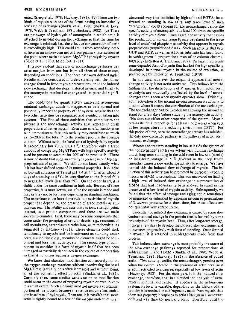
ented (Sleep et al., 1978; Hackney, 1981). (b) There are two kinds of myosin with one of the forms having an intrinsically low rate of exchange (Shukla et al., 1980; Shukla & Levy, 1978; Webb & Trentham, 1981; Hackney, 1982). (c) There are pathways of hydrolysis of actomyosin in which actin is attached to myosin during the exchange so that the time for exchange is minimal; Le., the effective concentration of actin is exceedingly high. This could result from secondary inter- actions in an actomyosin gel or from primary interactions at the point where actin activates MgATP hydrolysis by myosin (Sleep et al., 1980; Midelfort, 1981).
It is now evident that slow or nonexchange pathways can arise not just from one but from all three of these causes, depending on conditions. The three pathways defined under Results will be considered in order, starting with the nonex- changer found in fresh myosin preparations, on to the induced slow exchanger that develops in stored myosin, and finally to the actomyosin minimal exchange and its potential signifi- cance.
The conditions for quantitatively analyzing actomyosin minimal exchange, which now appears to be a normal and potentially important property of the system, require that the two other activities be recognized and avoided or taken into account. The first of these activities that complicates the picture is the nonexchange activity that appears in fresh preparations of native myosin. Even after careful fractionation with ammonium sulfate, this activity may contribute as much as 15-20% of the total Pi to the product pool, in the absence of actin. Without actin, the basal rate of hydrolysis by myosin is exceedingly low (0.02-0.04 s-’); therefore, only a trace amount of competing MgATPase with high specific activity need be present to account for 20% of the product Pi. There is now no doubt that such an activity is present in our freshest preparations of myosin. We still do not know exactly what it is but have defined some of its unusual properties. It is labile in low-salt solutions of Tris at pH 7.4 at 4 “ C ; after about 5 days of standing at 4 “C, its contribution to the Pi pool falls to negligible levels (less than 5%). On the other hand, it is stable under the same conditions in high salt. Because of these properties, it is most active just after the myosin is made and may or may not be lost later depending on standing conditions. The experiments we have done rule out activities of myosin proper that depend on the presence of trace metals or am- monium ion. The lability and sensitivity to ionic strength point, instead, to a protein component, and there are two main sources to consider. First, there may be some components that come under the grouping of cellular debris, e.g., remnants of cell membrane, sarcoplasmic reticulum, or mitochondria, as suggested by Hackney (1981). These elements could stick tenaciously to myosin and be inactivated on standing under certain conditions; e.g., membrane elements might be solu- bilized and lose their activity, etc. The second type of com- ponent to consider is a form of myosin itself that has been damaged or partially denatured in the course of preparation so that it no longer supports oxygen exchange.
We know that chemical modification can severely inhibit the oxygen-exchange reactions without inactivating the basal MgATPase (actually, this often increases) and without losing all of the activating effect of actin (Shukla et al., 1982). Certainly then, some similar denaturation or modification could occur in the course of preparing myosin or even in vitro to a small extent. Such a change need not involve a substantial portion of the protein because the native enzyme has such a low basel rate of hydrolysis. Then too, it is possible that some actin is tightly bound to a few of the myosin molecules in an
abnormal way (not inhibited by high salt and EGTA; inac- tivated on standing in low salt); any trace level of such “actomyosin” could account for the nonexchanger, since the specific activity of actomyosin is at least 100 times the specific activity of myosin alone. Then again, the activity that causes the production of nonexchange Pi may be related to the trace level of undefined phosphatase activity that appears in myosin preparations (unpublished data). Such an activity that uses GDP and ADP, as well as ATP, as substrate has been found in subfragment 1 preparations even after column chroma- tography (Eccleston & Trentham, 1979). Perhaps it represents some degraded form of myosin that has lost the high specificity developed in normal myosin in the course of evolution, as pointed out by Eccleston & Trentham (1979).
In any case, whatever the origin, it appears that nonex- change activity is not actin activated. This follows from the finding that the distributions of Pi species from actomyosin hydrolysis are practically unaffected by the level of nonex- changer that is seen when myosin operates alone. Evidently, actin activation of the normal myosin increases its activity to a point where it masks the contribution of the nonexchanger. The nonexchanger can be avoided by allowing the myosin to stand for a few days before studying the actomyosin activity. This does not affect other properties of the system. Myosin retains its initial properties for at least 1 or 2 weeks when kept at low temperature in a reducing environment (DTT). For this period of time, once the nonexchange activity has subsided, the only slow-exchange pathway is the pathway of actomyosin minimal exchange.
Whereas short-term standing in low salt rids the system of the nonexchanger and leaves actomyosin minimal exchange intact, long-term standing in the cold room in low salt (weeks) or long-term storage in 50% glycerol in the deep freeze (months) causes a slow-exchange activity to emerge. We have named this the induced slow exchange of myosin. The in- duction of this activity can be promoted by purposely exposing myosin or HMM to proteolysis. This was uncovered on finding a high level of induced slow exchange in a preparation of HMM that had inadvertantly been allowed to stand in the presence of a low level of trypsin activity. Subsequently, we found that the effect of storage and aging could sometimes be mimicked or enhanced by exposing myosin to preparations of S. aureus protease for a short time, but these effects are not quantitatively reproducible.
Evidently, the induced slow exchange is caused by some slow conformational change in the protein that is favored by some proteolysis of the myosin head. Thus, after long-term storage, it takes a few days to develop the induced slow exchange, and it increases progressively with time of standing. Once formed in myosin, it is retained in subfragments made from that protein.
This induced slow exchange is most probably the cause of the slow-exchange pathways reported for preparations of subfragment 1 and HMM (Shukla et al., 1980; Webb & Trentham, 1981; Hackney, 1982) in the absence of added actin. This activity, unlike the nonexchanger, persists even when the system is tested in the presence of actin because it is actin activated to a degree, especially at low levels of actin (Hackney, 1982). For the most part, it is the induced slow exchange, therefore, that has clouded the analysis of acto- myosin minimal exchange, It appears in the actomyosin system; its level is variable, depending on the history of the protein; it is retained in subfragments made from myosin that show this property; it responds to actin although in a somewhat different way than the normal protein. Therefore, until the
O X Y G E N E X C H A N G E B Y A C T O M Y O S I N
clear recognition of its existence and origin, it was difficult to study and characterize the true actomyosin minimal ex- change. For example, in our previous reported values for actomyosin minimal exchange in acto-HMM (Shukla et al., 1980), we also show a level of nonexchanger in HMM without actin, and this we now recognize as due to the induced slow exchanger. Also, in the studies of Midelfort (1981), his analysis of actomyosin minimal exchange, which we believe is essentially correct, may have been somewhat complicated by a low level of induced slow exchanger that makes its major contribution in the absence of actin or at low levels of actin and then is masked at higher actin levels. The reports of slow-exchange activity in S1 by other workers is also probably due in large part to the induced slow exchange, especially in the absence of actin (Webb & Trentham, 1981; Hackney, 1982).
The similar level of the n3 species of Pi produced by the nonexchanger, the induced slow exchange and actomyosin minimal exchange (all in the range of 15-25%) under the usual conditions of the experiments, has contributed to the potential for confusion. Thus, fortuitously, a preparation of fresh myosin tested without actin may typically show about 15% nonex- changed Pi at n3; then, after storage, myosin may typically develop an induced exchange activity that produces a com- parable level of n3; and this appears not only in myosin but also in subfragments made from that myosin. And then when such a myosin preparation is tested with actin, the normal pathway of actomyosin minimal exchange will appear with about 20-25% of the n3 species. Before recognizing and defining the properties of each pathway, it was this kind of situation, and the natural predisposition to simplify by as- suming one slow-exchange pathway, that stood in the way of resolving the complications of this system.
The properties of the induced slow exchange bear a resem- blance to the slow exchange induced by chemicals acting on myosin. In particular, we have shown that the binding of N-ethylmaleimide to the sulfhydryl 1-group of myosin or the binding of trinitrophenyl to the reactive lysine group markedly inhibits the rate of exchange, k,; these chemicals also decrease the extent of exchange by increasing the overall rate of hy- drolysis, k,, (Shukla et al., 1982). The induced slow exchange caused by proteolysis or storage and aging has an R value of about 0.4, and its development is usually associated with an increasing value for kat, from the normal level of about 0.03 s-' to as high as 0.1 s-l. Since R = k-3/kat (where k,, is taken to equal the rate-limiting step in the pathway of hydrolysis), an estimate of k-3 is obtained from the equation k-3 = Rk,,. If one takes R as 0.4 for induced slow exchange and a typical value for k,, of 0.07 s-l, the estimated value for k-3 by this activity is 0.03 s-1. This is about 10 times slower than the values for k-3 of the chemically induced slow exchange of myosin (which were 0.2-0.4 s-l). Nevertheless, the qualitative similarity is evidence that the changes occurring during storage and aging that produce the induced slow exchanger resemble those induced by chemical treatment.
Induced slow exchange is recognized by testing for it in the absence of actin and is avoided by using fresh preparations of myosin that show little or no proteolysis on gel electro- phoresis. The nonexchanger is avoided in myosin preparations by allowing it to subside during 2-3 days of standing or by working under conditions where the actomyosin activity is high enough to mask the nonexchanger.
With the complications of the nonexchanger and induced slow exchanger recognized and under control, the results of oxygen-exchange studies on actomyosin establish that under
VOL. 2 2 , N O . 2 0 , 1 9 8 3 4829
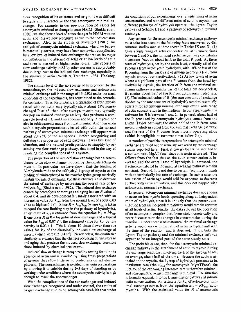
the conditions of our experiments, over a wide range of actin concentration, and with different ratios of actin to myosin, two different pathways of hydrolysis operate: the Lymn-Taylor pathway of Scheme I11 and a pathway of actomyosin minimal exchange.
Any scheme for the actomyosin minimal exchange pathway must take into account the following facts uncovered by dis- tribution studies such as those shown in Tables IX and X. (1) Over a wide range of actin concentration, at turnover times between 2 and 5 s, the minimal exchange pathway contributes a constant fraction, about half, to the total Pi pool. At these rates of hydrolysis, set by the actin level, virtually all of the Pi comes from actomyosin hydrolysis, with 10% or less of the Pi coming from the basal rate of myosin hydrolysis (Le., from myosin without actin activation). (2) At low levels of actin where a significant part of the Pi comes from the basal hy- drolysis by myosin, the fraction of Pi from the minimal ex- change pathway is a smaller part of the total, but nevertheless, it remains about half of the Pi from actomyosin hydrolysis. (3) The estimated value of R (the rate constant of exchange divided by the rate constant of hydrolysis) remains essentially constant for actomyosin minimal exchange over a wide range of actin concentration in the medium. In our experiments, the estimate for R is between 1 and 2. In general, about half of the Pi produced by actomyosin hydrolysis comes from the Lymn-Taylor pathway; the other half of the Pi from acto- myosin hydrolysis comes from the minimal exchange pathway; and the rest of the Pi comes from myosin operating alone (which is negligible at turnover times below 5 s).
A number of possible interpretations for actomyosin minimal exchange are ruled out or seriously weakened by the exchange studies reported here. First, it can no longer be ascribed to a contaminant MgATPase, since it is actin activated. This follows from the fact that as the actin concentration is in- creased and the overall rate of hydrolysis is increased, the fraction contributed by the minimal exchange pathway remains constant. Second, it is not due to certain free myosin heads with an intrinsically low rate of exchange. In such a case, the low extent of exchange would still be expected to decrease further with actin activation, and this does not happen with actomyosin minimal exchange.
In general actomyosin minimal exchange does not appear to occur on free myosin heads operating along an independent route of hydrolysis, since it is unlikely that the percent con- tribution from an independent pathway would remain constant at all levels of actin. Finally, the data rule out the operation of an actomyosin complex that forms stoichiometrically and never dissociates or that changes in concentration during the reaction. Under these circumstances, the minimal exchange activity would vary with the ratio of actin to myosin and with the time of the reaction, and it does not. Thus, both the Lymn-Taylor pathway and the minimal exchange pathway appear to be an integral part of the same steady state.
The probable cause, then, for the actomyosin minimal ex- change pathway is the attachment of actin to myosin during the exchange reactions, involving each of the myosin heads, on average, about half of the time. Because the actin is at- tached to the myosin, the k4 step of hydrolysis proceeds at its maximum rate (the V,,, for actomyosin MgATPase); the lifetime of the exchanging intermediates is therefore minimal, and consequently, oxygen exchange is minimal. The situation is formally equivalent to the Lymn-Taylor pathway at infinite actin concentration. An estimate for k-3 of actomyosin min- imal exchange comes from the equation k-3 = RVm',,,(acto- myosin). With the estimated value for R of actomyosin
4830 BIOCHEMISTRY S H U K L A ET A L .
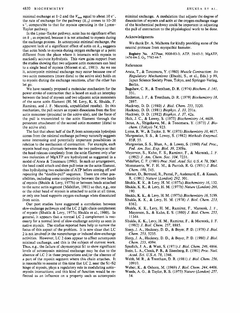
minimal exchange at 1-2 and the V, , , equal to about 10 s-l,
the rate of exchange for the pathway (k-J comes to 10-20 s-I, comparable to that for myosin operating in the Lymn- Taylor pathway.
In the Lymn-Taylor pathway, actin has no significant effect on k-3 as expected, because it is not attached to myosin during the exchange process. In actomyosin minimal exchange, the apparent lack of a significant effect of actin on k-3 suggests that actin binds to myosin during oxygen exchange at a point different from the place where it interacts with myosin to markedly activate hydrolysis. This view gains support from the studies showing that two adjacent actin monomers can bind to a single head of myosin (Mornet et al., 1981). As we see it, actomyosin minimal exchange may occur because one of two actin monomers (more distal to the active site) holds on to myosin during the exchange reactions, after the other has let go.
We have recently proposed a molecular mechanism for the power stroke of contraction that is based on such an interplay between the head of myosin and two adjacent actin monomers of the same actin filament (H. M. Levy, K. K. Shukla, F. Ramirez, and J. F. Marecek, unpublished results). In this mechanism, the pull occurs as myosin dissociates from the first actin monomer (proximal to the active site), and the force of the pull is transmitted to the actin filament through the persistent attachment of the second actin monomer (distal to the active site).
The fact that about half of the Pi from actomyosin hydrolysis comes from the minimal exchange pathway naturally suggests some interesting and potentially important possibilities in relation to the mechanism of contraction. For example, each myosin head may alternate between the two pathways so that the head releases completely from the actin filament only after two molecules of MgATP are hydrolyzed as suggested in a model of Arata & Tonomura (1980). In such an arrangement, the head could attach and then pull twice on the actin filament, thus hydrolyzing two molecules of ATP before coming off and repeating the “double-pull” sequence. There are other pos- sibilities, including some cooperativity between the two heads of myosin (Shukla & Levy, 1977b) or between heads attached to the same actin segment (Midelfort, 1981) so that, e.g., one or the other head of myosin is attached to actin at all times, or only one head supports oxygen exchange when dissociated from actin.
Our past studies have suggested a correlation between slow-exchange pathways and the LC 2 light chain complement of myosin (Shukla & Levy, 1977c; Shukla et al., 1980). In general, it appears that a normal LC 2 complement is nec- essary for a normal level of slow-exchange activity as seen in native myosin. The studies reported here help to narrow the focus of this aspect of the problem. It is now clear that LC 2 is not involved in the nonexchange or induced slow-exchange activities. However, LC 2 does appear to affect actomyosin minimal exchange, and this is the subject of current work. Thus, e.g., the failure of chymotryptic S1 to show significant levels of actomyosin minimal exchange may be due to the absence of LC 2 in these preparations and/or the absence of a part of the myosin segment where this chain attaches. It is reasonable to assume at this time that LC 2, near the Sl-S2 hinge of myosin, plays a regulatory role in modulating actin- myosin interactions; and this kind of function would be re- flected as an influence on a property such as actomyosin
miminal exchange. A modulation that adjusts the degree of dissociation of myosin and actin at the oxygen-exchange stage of the biochemical pathway could be important in adjusting the pull of contraction to the physiological work to be done.
Acknowledgments
neutral protease from myopathic hamster. We thank Dr. A. Malhotra for kindly providing some of the
Registry No. ATPase, 9000-83-3; ATP, 56-65-5; MgATP, 1476-84-2; 02, 7782-44-7.
References
Arata, T., & Tonomura, Y . (1980) Muscle Contraction: Its Regulatory Mechanisms (Ebashi, S . , et al., Eds.) p 99, Japan Science Society Press, Tokyo, and Springer-Verlag, Berlin.
Bagshaw, C. R., & Trentham, D. R. (1974) Biochem. J . 141, 331.
Eccleston, J. F., & Trentham, D. R. (1979) Biochemistry 18, 2897.
Hackney, D. D. (1980) J . Biol. Chem. 255, 5320. Hackney, D. D. (1981) Biophys. J . 33, 231a. Hackney, D. D. (1982) Biophys. J . 37, 42a. Holt, J. C., & Lowey, S. (1975) Biochemistry 14, 4609. Inoue, A., Shigekawa, M., & Tonomura, Y . (1973) J . Bio-
Lymn, R. W., & Taylor, E. W. (1971) Biochemistry 10,4617. Margossian, S. S . , & Lowey, S. (1982) Methods Enzymol.
Margossian, S . S . , Bhan, A., & Lowey, S . (1980) Fed. Proc.,
Meyerson, S . , Kuhn, E. S . , Ramirez, F., & Marecek, J. F.
Midelfort, C. F. (1981) Proc. Natl. Acad. Sci. U.S.A. 78,2067. Mommaerts, W. F. H. M., & Parrish, R. G. (1951) J . Biol.
Mornet, D., Bertrand, R., Pantel, P., Audemard, E., & Kassab,
Shukla, K. K., & Levy, H. M. (1977a) Biochemistry 16, 132. Shukla, K. K., & Levy, H. M. (1977b) Nature (London) 266,
Shukla, K. K., & Levy, H. M. (1977~) Biochemistry 16,5199. Shukla, K. K., & Levy, H. M. (1978) J . Biol. Chem. 253,
8362. Shukla, K. K., Levy, H. M., Ramirez, F., Marecek, J. F.,
Meyerson, S., & Kuhn, E. S. (1980) J . Biol. Chem. 255, 1 1344.
Shukla, K. K., Levy, H. M., Ramirez, F., & Marecek, J. F. (1982) J . Biol. Chem. 257, 8885.
Sleep, J. A,, Hackney, D. D., & Boyer, P. D. (1978) J . Biol. Chem. 253, 5235.
Sleep, J. A,, Hackney, D. D., & Boyer, P. D. (1980) J . Biol. Chem. 255, 4094.
Spudich, J. A., & Watt, S. (1971) J . Biol. Chem. 246, 4866. Stein, L. A., Chock, P. B., & Eisenberg, E. (1981) Proc. Natl.
Webb, M. R., & Trentham, D. R. (1981) J. Biol. Chem. 256,
Weber, K., & Osborn, M. (1969) J . Biol. Chem. 244,4406. Weeds, A. G., & Taylor, R. S. (1975) Nature (London) 257,
chem. (Tokyo) 74, 923.
85B, 55.
Fed. Am. SOC. Exp. Biol. 39, 2309a.
(1982) J . Am. Chem. SOC. 104, 7231.
Chem. 188, 545.
R. (1981) Nature (London) 292, 301.
190.
Acad. Sci. U.S.A. 78, 1346.
10910.
54.