Embed Size (px)
Citation preview
Annals of the Rheumatic Diseases, 1988; 47, 809-815
Palmoplantar pustulosis and sternocostoclaviculararthro-osteitisEVA EDLUND,' ULF JOHNSSON,2 LARS LIDGREN,4HOLGER PETTERSSON,3 GUNNAR STURFELT,' BJORN SVENSSON, 6JAN THEANDER,7 AND HELENA WILLEN5
From the 'Department of Rheumatology, 2the Blood Bank, and Departments of 3Diagnostic Radiology,4Orthopedic Surgery, and 5Pathology, University Hospital, Lund; the 6Department of Rheumatology,General Hospital, Helsingborg; and the 7Department of Rheumatology, Kristianstad General Hospital,Sweden
SUMMARY Seventeen patients with shoulder pain and radiographic involvement of thesternoclavicular or sternocostal joints, or both, are described. Eleven of these patients also hadpalmoplantar pustulosis. Histological examination of the joints showed chronic and subacuteinflammation, increased osteoblastic activity, and cartilage degeneration. Propionibacteriumacnes was cultured in tissue samples from seven of the 15 biopsied patients, a finding at variancewith those of previous reports. The possibility that sternoclavicular arthro-osteitis is of infectiousorigin should be the subject of further investigation. Non-steroidal anti-inflammatory drugs(NSAIDs) may provide pain relief, possibly owing to their inhibitory action on osteoblasts. Incases of severely restricted movement or severe pain resection of the medial clavicle may beconsidered.
Key words: arthritis, osteoblasts, Propionibacterium acnes.
Pustulosis palmoplantaris (PPP) is a chronic skindisease with intraepidermal, sterile pustules of thepalms and soles. There is no effective treatment.The prevalence of PPP in Sweden is about 0-05%,and most patients are women, 40-60 years of age. 1 2Sternocostoclavicular arthro-osteitis has been de-scribed as a painful condition of unknown aetiologybut with characteristic radiographic findings, usuallyaffecting the anterior chest wall and the sternocla-vicular, manubriosternal, and sternocostal joints.Although sternocostoclavicular arthro-osteitis, withor without PPP, is not infrequent in Japan,'7 it hasbeen considered rare among Caucasians, and, withfew exceptions,z10 published reports deal withindividual cases. 11-18 Recently a larger series ofpatients with the disease was reported. 9 The aim ofthis study was to investigate clinical features in anextended series of 17 patients and to draw attentionto the possibility that the condition may not be asrare in Caucasian populations as previously thought.
Accepted for publication 2 March 1988.Correspondence to Dr Gunnar Sturfelt, Department of Rheuma-tology, University Hospital, Lund, S-221 85 Sweden.
Patients and methods
From November 1982 all patients with anterior chestwall pain seen at the departments of rheumatologyand orthopaedics in Lund, Helsingborg and Kris-tianstad were investigated and included in thepresent study, provided that they presented with thefollowing criteria3: swelling and tenderness of thecostoclavicular or manubriosternal joints, or both,in conjunction with abnormal x ray or scintigraphyfindings, or both.
Patients with previously diagnosed ankylosingspondylitis, Reiter's syndrome, or psoriatic arthro-pathy were excluded. Seventeen consecutivepatients had been included by March 1986.The following investigations were performed: (a)
Conventional radiographic examination with a lat-eral view of the sternum, and conventional tomo-graphy in the posteroanterior view, including theupper sternum, the medial ends of the clavicles, thesternoclavicular joints, and proximal sternocostalsynchondroses. (b) Bone scintigraphy with 99mTcmethylene diphosphonate. In all the patients thechest, including the sternum, sternoclavicular joints,
809
copyright. on F
ebruary 9, 2022 by guest. Protected by
http://ard.bmj.com
/A
nn Rheum
Dis: first published as 10.1136/ard.47.10.809 on 1 O
ctober 1988. Dow
nloaded from
810 Edlund, Johnsson, Lidgren, Pettersson, Sturfelt, Svensson, Theander, Willen
and sternocostal synchondroses, was examined. (c)Open biopsy of affected joints and para-articularbone was carried out in 15 patients. From eachbiopsy specimen five tissue samples were taken foraerobic and anaerobic bacterial and fungalcultures.20 (d) Laboratory tests (erythrocycte sedi-
mentation rate (ESR), haemoglobin, white bloodcell counts, C reactive protein, antinuclear anti-bodies and rheumatoid factor (Waaler-Rose), serum
calcium and serum alkaline phosphatase) all byroutine procedures.HLA-A, B, and C antigens were typed with the
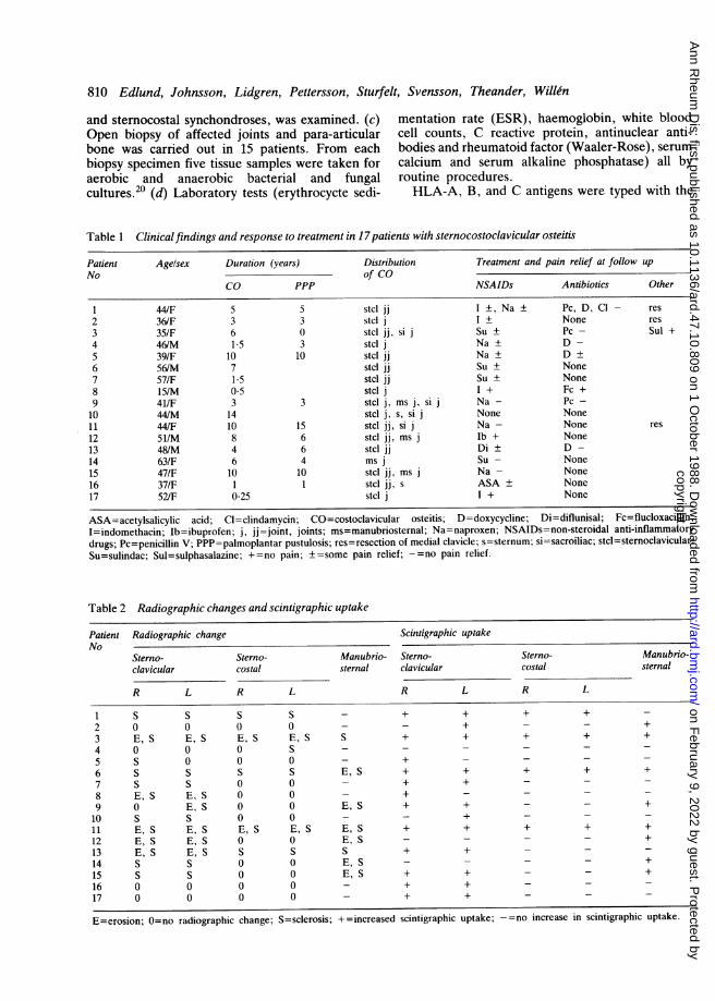
Table 1 Clinicalfindings and response to treatment in 17patients with sternocostoclavicular osteitis
Patient Agelsex Duration (years) Distribution Treatment and pain relief at follow upNo of CO
CO PPP NSAIDs Antibiotics Other
1 44/F 5 5 stcl jj I +,Na+ Pc, D, Cl res2 36/F 3 3 stcl j I ± None res3 35/F 6 0 stcl jj, si j Su+ Pc Sul +4 46/M 1-5 3 stcl j Na+ D5 39/F 10 10 stcl jj Na+ D±6 56/M 7 stcl jj Su + None7 57/F 1.5 stcl jj Su + None8 15/M 0-5 stcl j I + Fc +9 41/F 3 3 stcl j, ms j, si j Na- Pc -
10 44/M 14 stcl j, s, si j None None11 44/F 10 15 stcl jj, si j Na - None res12 51/M 8 6 stcl jj, ms j lb + None13 48/M 4 6 stcl jj Di + D -
14 63/F 6 4 ms j Su None15 47/F 10 10 stcl jj, ms j Na None16 37/F 1 1 stcl jj, s ASA ± None17 52/F 0-25 stcl j I + None
ASA=acetylsalicylic acid; Cl=clindamycin; CO=costoclavicular osteitis; D=doxycycline; Di=diflunisal; Fc=flucloxacillin;I=indomethacin; Ib=ibuprofen; j, jj=joint, joints; ms=manubriosternal; Na=naproxen; NSAIDs=non-steroidal anti-inflammatorydrugs; Pc=penicillin V; PPP=palmoplantar pustulosis; res=resection of medial clavicle; s=sternum; si=sacroiliac; stcl=sternoclavicular;Su=sulindac; Sul=sulphasalazine; +=no pain; ±=some pain relief; -=no pain relief.
Table 2 Radiographic changes and scintigraphic uptake
Patient Radiographic change Scintigraphic uptakeNo
Sterno- Sterno- Manubrio- Sterno- Sterno- Manubrio-clavicular costal sternal clavicular costal sternal
R L R L R L R L
1 S S S S - + + + +
2 0 0 0 0 + - +
3 E, S E, S E, S E, S S + + + + +
4 0 0 0 S5 S 0 0 0 - +
6 S S S S E, S + + + + +
7 S S 0 0 + +
8 E, S E, S 0 0 + - - -9 0 E, S 0 0 E, S + + - - +
10 S S 0 0 - - + - - -11 E, S E, S E, S E, S E, S + + + + +
12 E, S E, S 0 0 E, S - - +
13 E, S E, S S S S + +
14 S S 0 0 E, S - - - +
15 S S 0 0 E, S + + - +
16 0 0 0 0 - + + - -17 0 0 0 0 + + - - -
E=erosion; 0=no radiographic change; S=sclerosis; +=increased scintigraphic uptake; -no increase in scintigraphic uptake.
copyright. on F
ebruary 9, 2022 by guest. Protected by
http://ard.bmj.com
/A
nn Rheum
Dis: first published as 10.1136/ard.47.10.809 on 1 O
ctober 1988. Dow
nloaded from
Palmoplantar pustulosis and sternocostoclavicular arthro-osteitis 811
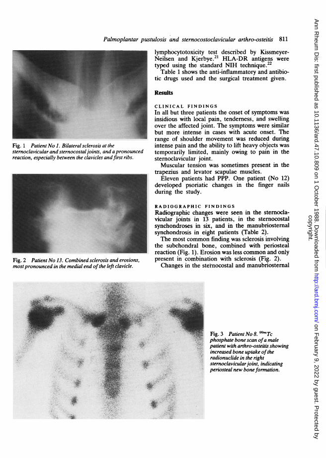
Fig. 1 Patient No 1. Bilateral sclerosis at thesternoclavicular and sternocostal joints, and a pronouncedreaction, especially between the clavicles andfirst ribs.
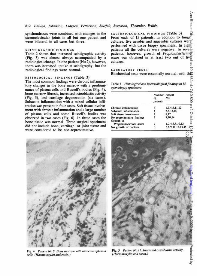
Fig. 2 Patient No 13. Combined sclerosis and erosions,mostpronounced in the medial end ofthe left clavicle.
lymphocytotoxicity test described by Kissmeyer-Neilsen and Kjerbye.21 HLA-DR antigens weretyped using the standard NIH technique.22Table 1 shows the anti-inflammatory and antibio-
tic drugs used and the surgical treatment given.
Results
CLINICAL FINDINGSIn all but three patients the onset of symptoms wasinsidious with local pain, tenderness, and swellingover the affected joint. The symptoms were similarbut more intense in cases with acute onset. Therange of shoulder movement was reduced duringintense pain and the ability to lift heavy objects wastemporarily limited, mainly owing to pain in thesternoclavicular joint.Muscular tension was sometimes present in the
trapezius and levator scapulae muscles.Eleven patients had PPP. One patient (No 12)
developed psoriatic changes in the finger nailsduring the study.
RADIOGRAPHIC FINDINGSRadiographic changes were seen in the sternocla-vicular joints in 13 patients, in the sternocostalsynchondroses in six, and in the manubriosternalsynchondrosis in eight patients (Table 2).The most common finding was sclerosis involving
the subchondral bone, combined with periostealreaction (Fig. 1). Erosion was less common and onlypresent in combination with sclerosis (Fig. 2).Changes in the sternocostal and manubriosternal
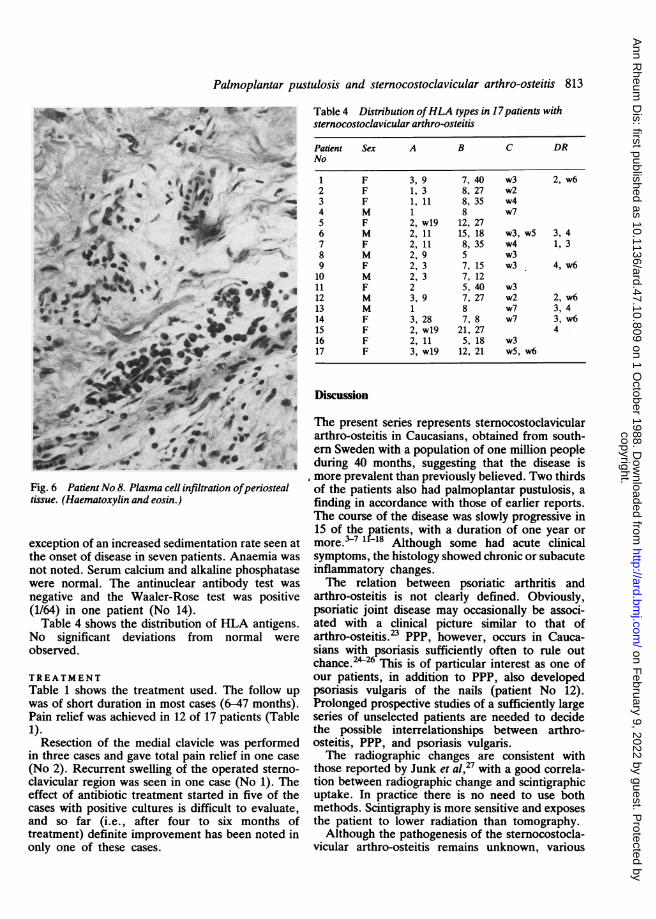
Fig. 3 Patient No 8. 9Tcphosphate bone scan ofa malepatient with arthro-osteitis showingincreased bone uptake oftheradionuclide in the rightsternoclavicular joint, indicatingperiosteal new boneformation.
4'.
copyright. on F
ebruary 9, 2022 by guest. Protected by
http://ard.bmj.com
/A
nn Rheum
Dis: first published as 10.1136/ard.47.10.809 on 1 O
ctober 1988. Dow
nloaded from
812 Edlund, Johnsson, Lidgren, Petuersson, Sturfelt, Svensson, Theander, Willen
synchondroses were combined with changes in thesternoclavicular joints in all but one patient andwere bilateral in all cases but three.
SCINTIGRAPHIC FINDINGSTable 2 shows that increased scintigraphic activity(Fig. 3) was almost always accompanied by aradiological change. In one patient (No 2), however,there was increased uptake at scintigraphy, but theradiological findings were normal.
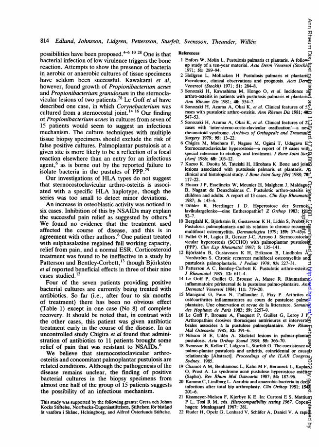
HISTOLOGICAL FINDINGS (Table 3)The most common findings were chronic inflamma-tory changes in the bone marrow with a predomi-nance of plasma cells and Russell's bodies (Fig. 4),bone marrow fibrosis, increased osteoblastic activity(Fig. 5), and cartilage degeneration (six cases).Subacute inflammation with a mixed cellular infil-tration was present in four cases. Soft tissue involve-ment with chronic inflammation and a large numberof plasma cells and some Russell's bodies wasobserved in two cases (Fig. 6). In three cases thebone tissue was normal. Three surgical specimensdid not include bone, cartilage, or joint tissue andwere considered to be non-representative.
BACTERIOLOGICAL FINDINGS (Table 3)From each of 15 patients, in addition to fungalcultures, five aerobic and anaerobic cultures wereperformed with tissue biopsy specimens. In eightpatients all the cultures were negative. In sevenpatients, however, growth of Propionibacteriumacnes was obtained in at least two out of fivecultures.
LABORATORY TESTSBiochemical tests were essentially normal, with the
Table 3 Histological and bacteriologicalfindings in 15open biopsy specimens
Number Patientof Nospatients
Chronic inflammation 6 1,3,4,5,11,12Subacute inflammation 4 2,6,13,15Soft tissue involvement 2 8,17No representative findings 3 9,10,14Growth of
Propionibacterium acnes 7 1,2,4,5,8,10,13No growth of bacteria 8 3,6,9,11,12,14,15,17
eff4' .A-
&A
Fig. 4 Patient No 8. Bone marrow with numerous plasmacells. (Haematoxylin and eosin.)
Fig. 5 Patient No 15. Increased osteoblastic activity.(Haematoxylin and eosin.)
ki
t4i.4
f
4ir
copyright. on F
ebruary 9, 2022 by guest. Protected by
http://ard.bmj.com
/A
nn Rheum
Dis: first published as 10.1136/ard.47.10.809 on 1 O
ctober 1988. Dow
nloaded from
Palmoplantar pustulosis and sternocostoclavicular arthro-osteitis 813
-at. . I'' ''4 _ii7_ s~~~~~~~, ISf
*b li*f *_ - V
Ylll_--V.
.-,.
0, Tr I
A#*' _ ,0 I.
Fig. 6 Patient No 8. Plasma cell infiltration ofperiostealtissue. (Haematoxylin and eosin.)
exception of an increased sedimentation rate seen atthe onset of disease in seven patients. Anaemia was
not noted. Serum calcium and alkaline phosphatasewere normal. The antinuclear antibody test wasnegative and the Waaler-Rose test was positive(1/64) in one patient (No 14).Table 4 shows the distribution of HLA antigens.
No significant deviations from normal wereobserved.
TREATMENTTable 1 shows the treatment used. The follow upwas of short duration in most cases (6-47 months).Pain relief was achieved in 12 of 17 patients (Table1).
Resection of the medial clavicle was performedin three cases and gave total pain relief in one case(No 2). Recurrent swelling of the operated sterno-clavicular region was seen in one case (No 1). Theeffect of antibiotic treatment started in five of thecases with positive cultures is difficult to evaluate,and so far (i.e., after four to six months oftreatment) definite improvement has been noted inonly one of these cases.
Table 4 Distribution ofHLA types in 17patients withsternocostoclavicular arthro-osteitis
Patient Sex A B C DRNo
1 F 3, 9 7, 40 w3 2, w62 F 1, 3 8, 27 w23 F 1, 11 8, 35 w44 M 1 8 w75 F 2, w19 12, 276 M 2, 11 15, 18 w3, w5 3, 47 F 2, 11 8, 35 w4 1, 38 M 2, 9 5 w39 F 2, 3 7, 15 w3 4, w610 M 2, 3 7, 1211 F 2 5, 40 w312 M 3, 9 7, 27 w2 2, w613 M 1 8 w7 3, 414 F 3, 28 7, 8 w7 3, w615 F 2, w19 21, 27 416 F 2, 11 5, 18 w317 F 3, w19 12, 21 w5, w6
Discussion
The present series represents sternocostoclaviculararthro-osteitis in Caucasians, obtained from south-ern Sweden with a population of one million peopleduring 40 months, suggesting that the disease ismore prevalent than previously believed. Two thirdsof the patients also had palmoplantar pustulosis, afinding in accordance with those of earlier reports.The course of the disease was slowly progressive in15 of the patients, with a duration of one year ormore.37 1 f-18 Although some had acute clinicalsymptoms, the histology showed chronic or subacuteinflammatory changes.The relation between psoriatic arthritis and
arthro-osteitis is not clearly defined. Obviously,psoriatic joint disease may occasionally be associ-ated with a clinical picture similar to that ofarthro-osteitis.23 PPP, however, occurs in Cauca-sians with psoriasis sufficiently often to rule outchance.2426 This is of particular interest as one ofour patients, in addition to PPP, also developedpsoriasis vulgaris of the nails (patient No 12).Prolonged prospective studies of a sufficiently largeseries of unselected patients are needed to decidethe possible interrelationships between arthro-osteitis, PPP, and psoriasis vulgaris.The radiographic changes are consistent with
those reported by Junk et al,27 with a good correla-tion between radiographic change and scintigraphicuptake. In practice there is no need to use bothmethods. Scintigraphy is more sensitive and exposesthe patient to lower radiation than tomography.Although the pathogenesis of the sternocostocla-
vicular arthro-osteitis remains unknown, various
copyright. on F
ebruary 9, 2022 by guest. Protected by
http://ard.bmj.com
/A
nn Rheum
Dis: first published as 10.1136/ard.47.10.809 on 1 O
ctober 1988. Dow
nloaded from
814 Edlund, Johnsson, Lidgren, Pettersson, Sturfelt, Svensson, Theander, Willen
possibilities have been proposed." 10 28 One is thatbacterial infection of low virulence triggers the bonereaction. Attempts to show the presence of bacteriain aerobic or anaerobic cultures of tissue specimenshave seldom been successful. Kawakami et al,however, found growth of Propionibacterium acnesand Propionibacterium granulosum in the sternocla-vicular lesions of two patients.28 Le Goff et al havedescribed one case, in which Corynebacterium wascultured from a sternocostal joint.'4 16 Our findingof Propionibacterium acnes in cultures from seven of15 patients would seem to suggest an infectiousmechanism. The culture techniques with multipletissue biopsy specimens should exclude the risk offalse positive cultures. Palmoplantar pustulosis at agiven site is more likely to be a reflection of a focalreaction elsewhere than an entry for an infectiousagent,6 as is borne out by the reported failure toisolate bacteria in the pustules of Ppp.29Our investigations of HLA types do not suggest
that sternocostoclavicular arthro-osteitis is associ-ated with a specific HLA haplotype, though theseries was too small to detect minor deviations.An increase in osteoblastic activity was noticed in
six cases. Inhibition of this by NSAIDs may explainthe successful pain relief as suggested by others.6We found no evidence that the treatment usedaffected the course of disease, and this is inagreement with other authors.6 One patient treatedwith sulphasalazine regained full working capacity,relief from pain, and a normal ESR. Corticosteroidtreatment was found to be ineffective in a study byPatterson and Bentley-Corbett,13 though Bjorkstenet al reported beneficial effects in three of their ninecases studied.12Four of the seven patients providing positive
bacterial cultures are currently being treated withantibiotics. So far (i.e., after four to six monthsof treatment) there has been no obvious effect(Table 1) except in one case (No 8) of completerecovery. It should be noted that, in contrast withthe other cases, this patient was given antibiotictreatment early in the course of the disease. In anuncontrolled study Chigira et al found that admini-stration of antibiotics to 11 patients brought somerelief of pain that was resistant to NSAIDs.6We believe that stemocostoclavicular arthro-
osteitis and concomitant palmoplantar pustulosis arerelated conditions. Although the pathogenesis of thedisease remains unclear, the finding of positivebacterial cultures in the biopsy specimens fromalmost one half of the group of 15 patients suggeststhe possibility of an infectious mechanism.
This study was supported by the following grants: Greta och JohanKocks Stiftelse, Norrbacka-Eugeniastiftelsen, Stiftelsen for bistandAt vanfora i Skane, Helsingborg, and Alfred Osterlunds Stiftelse.
References1 Enfors W, Molin L. Pustulosis palmaris et plantaris. A follow-up study of a ten-year material. Acta Derm Venereol (Stockh)1971; 51: 289-94.
2 Hellgren L, Mobacken H. Pustulosis palmaris et plantaris.Prevalence, clinical observations and prognosis. Acta DermVenereol (Stockh) 1971; 51: 284-8.
3 Sonozaki H, Kawashima M, Hongo 0, et al. Incidence ofarthro-osteitis in patients with pustulosis palmaris et plantaris.Ann Rheum Dis 1981; 40: 554-7.
4 Sonozaki H, Azuma A, Okai K, et al. Clinical features of 53cases with pustulotic arthro-osteitis. Ann Rheum Dis 1981; 40:547-53.
5 Sonozaki H, Azuma A, Okai K, et al. Clinical features of 22cases with 'inter-sterno-costo-clavicular ossification'-a newrheumatoid syndrome. Archives of Orthopedic and TraumaticSurgery 1979; 95: 13-22.
6 Chigira M, Maehara F, Nagase M, Ogimi T, Udagava E.Sternocostoclavicular hyperostosis-a report of 19 cases withspecial reference to etiology and treatment. J Bone Joint Surg[Am] 1986; 68: 103-12.
7 Kazuo K, Daoita M, Tateishi H, Hirohata K. Bone and jointlesions associated with pustulosis palmaris et plantaris. Aclinical and histological study. J Bone Joint Surg [Br] 1988; 70:117-22.
8 Huaux J P, Esselinckx W, Meunier H, Malghem J, MaldagueB, Nagant de Deuxchaisnes C. Pustulotic arthro-osteitis inchildren and adults. A report of 13 cases. Clin Exp Rheumatol1987; 5: 143-6.
9 Dohler R, Herrlinger J D. Hyperostose der Sternok-lavikulargelenke-eine Enthesopathie? Z Orthop 1983; 121:92-7.
10 Bergdahl K, Bjorksten B, Gustavsson K H, Liden S, Probst F.Pustulosis palmoplantaris and its relation to chronic recurrentmultifocal osteomyelitis. Dermatologica 1979; 159: 37-45.
11 Fallet G H, Lagier R, Gerster J-C, Arroyo J. Sternocostocla-vicular hyperostosis (SCCHO) with palmoplantar pustulosis(PPP). Clin Exp Rheumatol 1987; 5: 135-141.
12 Bjorksten B, Gustavsson K H, Eriksson B, Lindholm A,Nordstrom S. Chronic recurrent multifocal osteomyelitis andpustulosis palmoplantaris. J Pediatr 1978; 93: 227-31.
13 Patterson A C, Bentley-Corbett K. Pustulotic arthro-osteitis.J Rheumatol 1985; 12: 611-4.
14 Le Goff P, Guillet G, Brousse A, Masse R. Rhumatismeinflammatoire peristernal de la pustulose palmo-plantaire. AnnDermatol Venereol 1984; 111: 719-20.
15 Manigand G, Faux N, Taillandier J, Fixy P. Arthrites etosteoarthrites inflammatoires au cours de pustulose palmo-plantaire. Une observation et revue de la litterature. Semainedes Hopitaux de Paris 1983; 59: 2257-9.
16 Le Goff P, Brousse A, Fauquert P, Guillet G, Leroy J P.Arthropathies 6rosives thoraciques anterieures et intervert6-brales associ6es A la pustulose palmoplantaire. Rev RhumMal Osteoartic 1985; 52: 391-6.
17 Nilsson B E, Uden A. Skeletal lesions in palmar-plantarpustulosis. Acta Orthop Scand 1984; 55: 366-70.
18 Svensson B, Keller C, Lidgren L, Sturfelt G. The coexistence ofpalmo-plantar pustulosis and arthritis, coincidental or causalrelationship [Abstract]. Proceedings of the ILAR CongressSydney. 1985.
19 Chamot A M, Benhamouc L, Kahn M F, Beraneck L, Kaplan0, Prost A. Le syndrome acne pustulose hyperostose osteite(Sapho). Rev Rhum Mal Osteoartic 1987; 54: 187-96.
20 Kamme C, Lindberg L. Aerobic and anaerobic bacteria in deepinfections after total h,ip arthroplasty. Clin Orthop 1981; 154:201-6.
21 Kissmeyer-Nielsen F, Kjerbye K E. In: Curtoni E S, MattiuzP L, Tosi R M, eds. Histocompatibility testing 1967. Copen-hagen: Munksgaard 1967: 381.
22 Ruder H, Opelz G, Lenhard V, Schafer A, Daniel V. A rapid
copyright. on F
ebruary 9, 2022 by guest. Protected by
http://ard.bmj.com
/A
nn Rheum
Dis: first published as 10.1136/ard.47.10.809 on 1 O
ctober 1988. Dow
nloaded from
Palmoplantar pustulosis and sternocostoclavicular arthro-osteitis 815
screening technique for lymphocytotoxic antibodies using tray-frozen lymphocytes. Cryobiology 1984; 21: 480-5.
23 Becker N J, de Smet A, Cathcart-Raken W, Stechschulte D.Psoriatic arthritis affecting the manubrio sternal joint. ArthritisRheum 1986; 29: 1029-31.
24 Fransson J, Storgards K, Hammar H. Palmo-plantar lesions inpsoriatic patients and their relation to inverse psoriasis, tineainfection and contact allergy. Acta Derm Venereol (Stockh)1985; 65: 218-23.
25 Ward J M, Barnes R M. HLA antigens in persistent palmoplan-tar pustulosis and its relationship to psoriasis. Br J Dermatol1978; 99: 477-83.
26 Zachariae H, Obergaard Petersen H, Kissmeyer Nielsen F,Lamm L. HLA antigens in pustular psoriasis. Dermatologica1977; 154: 73-7.
27 Junk A G, Grandal H, de Carvalho A. Monarticular involve-ment of the manubriosternal joint. Skeletal Radiol 1985; 14:99-103.
28 Kawakami T, Toyoshima R, Furuse K, et al. So-calledsterno-costo-clavicular hyperostosis. Its etiology and manifesta-tions. Rinso Seikei Geka 1980; 15: 650-8.
29 Thormann J, Nyfors A. Pustulosis palmaris et plantaris. Areview. Ugeskr Laeger 1973; 135: 2098-101.
copyright. on F
ebruary 9, 2022 by guest. Protected by
http://ard.bmj.com
/A
nn Rheum
Dis: first published as 10.1136/ard.47.10.809 on 1 O
ctober 1988. Dow
nloaded from