Embed Size (px)
Citation preview
The Journal of Neuroscience December 1986, 6(12): 3682-3691
Perturbed Pattern of Catecholamine-Containing Neurons in Mutant Drosophila Deficient in the Enzyme Dopa Decarboxylase
Vivian Budnik,* Linda Martin-Morris,-f and Kalpana White-t *Biophysics Proaram, Brandeis Universitv, and i-Department of Biology, Brandeis University, Waltham, Maskabhusetts 02254
We have initiated a study of catecholamine-containing neurons in Drosophila melanogaster because of the potential, with this organism, to perturb catecholamine metabolism using genetic tools. The major objectives of this study were (1) to define the pattern of catecholamine-containing neurons and (2) to deter- mine the effect of the absence of dopa decarboxylase (DDC) enzyme activity on the catecholamine-containing neurons.
We chose to analyze the catecholamine-containing neurons in the ventral ganglion of the larval CNS. To define the catechol- amine-containing neurons, CNSs were dissected and reacted with glyoxylic acid. The catecholamine histofluorescence (CF) neu- ronal pattern (normal-CF neurons) in the wild-type ventral gan- glion is stereotypic. In the mutant ventral ganglia, in the ab- sence of DDC enzyme activity, most normal-CF neurons still exhibit CF, probably indicating the presence of accumulated L-dopa. Interestingly, in the mutant CNSs, additional novel neu- ronal subsets also exhibit CF. Analysis of CNSs from early developmental stages revealed that the novel-CF neurons be- come fluorogenic earlier than the normal-CF neurons in the mutant CNS.
To determine whether neuronal subsets, in addition to the normal-CF, neurons are able to sequester catecholamines, CNSs from wild-type larvae were incubated in exogenous catechol- amine (L-dopa or dopamine). Incubations in L-dopa or dopamine revealed normally nonfluorogenic neurons that are able to take up the amine and become fluorogenic. Among the neurons able to sequester L-dopa or dopamine are subsets that are similar to the novel-CF neurons in the mutant CNS. This similarity is best characterized by a major novel-CF neuronal cluster in the subesophageal-thoracic region. These results suggest that in the absence of DDC activity, subsets of normally nonfluorogenic neurons capable of sequestering Ldopa or dopamine accumulate the fluorogenic catecholamine. Hypotheses that might explain the mode of accumulation of the catecholamine within the novel- CF neurons are considered.
Within the last decade it has become increasingly possible to study the chemical anatomy of the nervous system, employing histochemical and immunohistochemical methods (e.g., Hiikfelt et al., 1984). Anatomical descriptions of the vertebrate and invertebrate systems focus on the localization of neurotrans- mitters, the enzymes utilized in the synthesis of neurotrans- mitters, and neuropeptides. A striking but not unexpected fea-
Received Mar. 11, 1986; revised June 23, 1986; accepted July 1, 1986. We appreciate the helpful comments of Eve Marder and Michael Nusbaum
during the preparation of this manuscript. We wish to thank Ana Maria Vall& for willingly providing invaluable help at many levels during the course of this work and for stimulating discussions. This work was supported by NIH Grant NS 235 10. V.B. is a predoctoral trainee, supported by a Brittner fellowship at Brandeis University.
Correspondence should be addressed to Kalpana White at the above address. Copyright 0 1986 Society for Neuroscience 0270-6474/86/123682-10$02.00/O
ture of the emerging picture is the stereotypic distribution within the CNS of a given chemical messenger. The advances in chem- ical neuroanatomy have contributed to the clarification of the chemical organization of the CNS. But at the same time, the discovery of a large number of molecules with putative neu- rotransmitter-like or neuromodulator-like functions has brought about a new awareness of the complex overall organization.
Presumably, for proper operation of the CNS, programmed differentiation of neurons and continuous overall regulation of the synthesis of a large array of molecules are essential. During development, different neurons develop the capacity to synthe- size and use 1 or more specific chemical signals, resulting in the final pattern. The mechanisms responsible for the exquisite con- trol of cellular differentiation and continued metabolic regula- tion operating in the nervous system are largely unknown.
It is indeed possible that some of the transmitter or modulator molecules might provide specific signals essential for the proper differentiation or maintenance of normal functions in some sub- sets of neurons. The recent work of Kater and co-workers on the regenerating neurons in cell cultures of the snail Helisoma implicate the biogenic amine transmitters, dopamine and sero- tonin, as regulators of neurite outgrowth (Haydon et al., 1984; McCobb et al., 1985; Mercier et al., 1985).
Genetically mutant animals incapable of synthesizing specific transmitters provide in vivo model systems in which to study the effect of the absence of the transmitter on the development and function of the nervous system (Greenspan, 1980; Green- span et al., 1980; VallCs and White, 1986). The study of neu- rochemical changes in neurons that are directly affected by the mutation, and of other associated chemical and morphological changes that might occur in the mutant CNSs may potentially provide insights when the primary biochemical lesion is well defined.
We have been studying the biogenic amine systems in the fruit fly, Drosophila melanogaster, as it is possible to generate animals that are genetically deficient in dopa decarboxylase (DDC) enzyme activity and thus are presumably deficient in serotonin and catecholamines (Livingstone and Tempel, 1983; Vall&s and White, 1986). In previous work, we studied the dif- ferentiation of serotonin-containing neurons in their transmit- terless state in Ddc mutant CNSs. Neurons normally committed to the serotonin differentiation pathway proceeded to express selective serotonin-uptake properties in the absence of DDC enzyme activity (VallCs and White, 1986). In this paper, we extend our investigation of CNSs deficient in DDC enzyme activity to catecholamine-containing neurons.
Materials and Methods
Fly cultures and strains All fly strains were raised at 25°C on standard medium. The mutant chromosomes used are listed below. For more complete descriptions of

The Journal of Neuroscience Catecholamines in Drosophila CNS 3683
B BRAIN
GANG LION
t dor
Figure 1. Normal-CF neurons in the larval wild-type ventral ganglion. A, Photomicrograph of a whole-mount preparation of a third instar wild- type CNS. For schematic representation, see B. In this plane, most of the Sb-Th neurons and all the AbU neurons can be resolved. Also some of the AbL neurons can be readily observed. Insert shows a first instar CNS dissected from a newly hatched larva. In this photographic plane, only some of the normal-CF neurons in the first instar larval CNS are observed. The neurons observed are from the Sb-Th region and some of the anterior AbU neurons. Bar, 30 pm. B, Schematic tracing of the normal-CF neurons adapted from whole-mount preparations of glyoxylic acid- reacted, wild-type larval CNSs. The histofluorescence in the neuropil and primary processes of the neurons are not drawn. ant, Anterior; AbL, abdominal lateral neurons; AbU, abdominal unpaired neurons: dor, dorsal; Sb, subesophageal paired and unpaired neurons; Th, thoracic paired and unpaired neurons; ThL, thoracic lateral neurons.
mutations, see VallCs and White (1986). The wild-type strain used was Canton S.
Df12LJ130. A cvtologically visible deletion, entirely removing the gene ijd, (Wright et al.,-1976).
Ddcn27. A 2 kb deletion removing the two 5’ exons of Ddc and 0.5 kb of 5’ flanking DNA (Gilbert etal., 1984).
CyO. A second chromosome balancer. SM6. A second chromosome balancer.
Generation of animals deficient in the gene Ddc The genetic scheme and specific crosses used to generate animals that were deficient in DDC activity (DfDdc) are detailed in VallCs and White (1986). The genetic crosses were as follows:
Df(zL)I3O/CyO x Ddc’27/Cy0 or DdP7/SM6 x Ddc+/SM6. Twenty- five percent of the progeny of these crosses were Ddc-deficient (ge- notype: Dff2L)l3O/Dd@ or Dd@/Ddcnz7).
Most Df Ddc mutant embryos die at the embryo larval boundary and do not hatch. However, they are fully developed and if liberated from the embryonic membranes about 40% are able to undergo larval de- velopment (VallCs and White, 1986). The mutant pharate larvae can be readily recognized in late embryogenesis (20-22 hr) because of their incompletely sclerotized mouth hooks. Naturally or mechanically hatched mutant animals were reared in a special medium. The details of this procedure are described elsewhere (Valles and White, 1986).
Glyoxylic acid-induced histofluorescence To visualize the catecholamine-containing neurons, the glyoxylic acid histofluorescence method, adapted for invertebrate tissue, was used (Flanagan, 1983). The larval CNSs were dissected in Mg2+-rich, ice-
cold Drosophila Ringer’s solution (NaCl, 110 mM; KCl, 4.7 mM; MgCl,, 20 mM; C&X,, 1.8 mM; KI-I,PO,, 0.74 mtw; Na,HPO,, 0.35 mM). The dissected CNSs were transferred to a glass microscope slide, gently blotted, and incubated in approximately 0.1 ml of the glyoxylic acid solution 15% alvoxvlic acid (Mallinckrodt). 0.2 M sucrose in Ca2+-free Drosophia R&e& solution’(NaC1, 110 t&; KCl, 4.7 mM; MgCl,, 1.80 mM; KI-I,PO,, 0.74 mM; Na,HPO,, 0.35 mM), adjusted to pH 7.01. The incubations were carried out in a moist, dark chamber for 1 hr at room temperature. The solution was then blotted away from the tissue and the samples were dried over Ca,SO, overnight in the dark. Thoroughly dried tissues were covered with a drop of heavy mineral oil and flash- heated for 2 min at 80°C to catalyze the second step of the glyoxylic acid reaction. The samples were viewed in epifluorescence in a Zeiss inverted microscope (Zeiss 487705) equipped with the appropriate ex- citation and barrier filters. Samples were stored in the dark at,4”C. Under these conditions, cells remained fluorescent for up to 3 months.
Preloading with catecholamines Larval CNSs were dissected in Drosophila Ringer’s (NaCl, 130 mM; KCl, 4.7 mM; CaCl,, 1.8 mM; KH,PO,, 0.74 mM; Na,HPO,, 0.35 mM)! A 1O-2 M stock solution of each, amine tested-L-dopa, dopamine, and norepinephrine-was made up into Ringer’s. Tyrosine was dissolved in EtOH at a final concentration of 10-l M. Dissected CNSs were incubated individually in concentrations ranging from lo-’ to lo-’ M amine in Ringer’s. Incubations were carried out for 30 min in the dark at room temperature, with 1 change of the amine solution after the first 15 min. Approximately 0.1 ml of the solution was used per CNS. After 30 min incubation time, the solution was removed and the tissue washed with Ringer’s 4 times over a period of 40 min at room temperature. The final wash solution was replaced with the glyoxylic acid solution and reacted as described above.

3684 Budnik et al. Vol. 6, No. 12, Dec. 1986
Figure 2. Normal-CF neuronal subsets in the ventral ganglion. A, Photomicrograph of a ventral plane showing the Sb-Th cluster (delineated by the 2 white arrows) and the AbU neurons located posterior to the second arrow. B, Same whole-mount as in A, but the plane of the photomicrograph represents some of the dorsally located lateral neurons. The white arrows point to the left AbL neurons; several of the right neurons are also observed. Bar, 20 pm. Axes, same as Figure 1.
Results
Catecholamine-containing neurons in Drosophila larval CNS Glyoxylic acid-induced histofluorescence. Glyoxylic acid con- verts certain monoamines to highly fluorescent derivatives (Lindvall and Bjorklund, 1974; Lindvall et al., 1974). The flu- orescent condensation products allow the histochemical local- ization of monoamine-containing neurons (Lindvall and Bjiirk- lund, 1974). Recently the glyoxylic acid method has been successfully employed to localize monoamine-containing neu- rons in invertebrate CNSs (Flanagan, 1983; Lent, 1982). There are 3 points regarding the specificity of the reaction that are pertinent to the interpretations of our results:
1. Both catecholamines and serotonin can react with glyoxylic acid to form fluorophores; the fluorescence specific to cate- cholamines is blue-white and is relatively stable, whereas the fluorescence specific to serotonin is yellowish and unstable (Lent, 1982; Lindvall et al., 1974). The pH optima for the catechol- amine and serotonin reactions are distinct (Lindvall et al., 1974). In this study, we used conditions that favored the catecholamine reaction (neutral pH), and only the blue-white fluorescence spe- cific to catecholamine-containing neurons was observed. These neurons will be referred to as “CF neurons.”
2. Glyoxylic acid reacts with several compounds in the cate- cholamine pathway. A major disadvantage of the method is that it is virtually impossible to distinguish between L-dopa, dopa- mine, and norepinephrine since the fluorophores they yield have
similar spectral characteristics (Lindvall et al., 1975). Therefore, positive identification of the catecholamine responsible for the fluorescence is not possible using this method alone.
3. In the histochemical application of this method for visu- alizing catecholamine-containing neurons, it is generally as- sumed that the amine responsible for the fluorescence is do- pamine or norepinephrine. L-Dopa, the precursor to dopamine, does not make a significant contribution to the fluorescence, as the intracellular pools of L-dopa are negligible in comparison to the dopamine or norepinephrine pools (Klemm, 1980). The fluorescence yield from octopamine is very low compared to that of dopamine or epinephrine; therefore, the method is not effective in detecting octopamine-containing neurons.
Larval CF pattern. A stereotypic pattern of CF neurons is observed after the glyoxylic acid treatment of intact CNSs dis- sected from the Drosophila larvae (Budnik et al., 1985). Figure 1A is a photomicrograph of a glyoxylic acid-treated whole-mount preparation. Figure 1B is a schematic representation of a third instar larval CNS onto which the somata of the CF neurons are drawn. The tracing is adapted from glyoxylic acid-treated CNS whole-mounts similar to the one shown in Figure 1A.
CF neurons were observed in the paired brain hemispheres and in the ventral ganglion. In the brain lobes, 3 bilaterally symmetrical clusters of 4-6 CF neurons each were routinely observed. In this paper we focus on the CF neurons observed in the ventral ganglion, as the regularity of the pattern allows direct comparison between different samples. The ventral gan- glion of the Drosophila larva is a compound ganglion composed
The Journal of Neuroscience Catecholamines in Drosophila CNS 3685
Table 1. Normal-CF neurons in the individual larval ventral ganglion tyrosine tryptophan
samples tyrosme tprosule
Sb-Th Ab Th Total eecarboxylase hydroaylase tryptopmn
(Maximum expected) hgaroaylase
Sample u P u L A
no. (4) (4) (8) (14) k, (36) tyramme I -dopa 5-hydroxytryptophan
1 4 4 7 14 5 34 2 4 4 7 14 4 33 tyramine bopa/5HTP bopa/5HTP 3 4 4 7 14 4 33 fl-hyaroqlase decarbo@ase decarboxylase
4 4 4 7 14 4 33 5 4 4 8 12 4 32 6 4 4 8 14 2 32 octopamme dopamme serotonin
7 4 4 7 13 4 32 8 4 4 7 14 2 31 9 4 4 6 13 3 30
10 4 4 I 14 0 29 11 4 4 7 14 0 29 12 4 4 7 11 0 28 13 4 4 5 10 0 27
of subesophageal (Sb), thoracic (Th) and abdominal (Ab) neu- romeres. The pattern of CF neurons observed in the wild-type ventral ganglion can be divided into 3 subgroups of neurons for clarity of presentation. These 3 neuronal subgroups will be re- ferred to as the normal-CF neuronal set in this paper. They are as follows:
1. Medial subesophageal-thoracic neurons. This region con- tains a total of 8 CF somata, located in the ventral cellular rind. There are 4 unpaired medial neurons (U) and 2 pairs of medially located neurons (P). At present, the boundary between the sub- esophageal and thoracic regions is not clear. Tentatively, the anterior pair of neurons and the 2 anterior unpaired medial neurons are localized to the subesophageal ganglion, and the posterior pair and the posterior unpaired medial neurons are localized to the thoracic neuromeres. This anatomical subdi- vision is supported by the observation that a medial pair and 2 unpaired neurons are observed in the subesophageal ganglion in the adult brain (V. Budnik, unpublished observations). Figure 2A depicts the 4 paired and 4 unpaired neurons in the medial Sb-Th region.
2. Abdominal unpaired medial row. A row of single unpaired CF neurons (AbU) was observed along the ventral midline in the abdominal region. Presumably, each of the anterior abdom- inal neuromeres contains 1 unpaired CF neuron (Fig. 2A). In most preparations, 7 neurons were observed, although, on oc- casion, 8 neurons were observed.
3. Paired lateral row. A single CF soma was observed in each of the 3 thoracic hemineuromeres (ThL) and in the anterior 7 abdominal hemineuromeres (AbL). The location of the lateral neuronal somata was dorsal to the medial unpaired neurons. The neuronal cell bodies along the lateral row appeared to be smaller than the medial neuronal cell bodies. This pattern was bilaterally symmetrical (Fig. 2B).
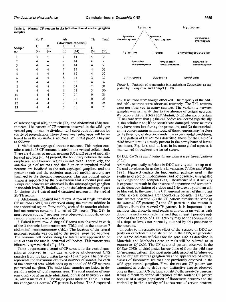
Table 1 represents a count of CF somata in the ventral gan- glion as observed in individual wild-type whole-mount CNS samples from the third instar larvae (13 samples). The first row represents the maximum observed number of somata for each of the neuronal sets, which adds up to a total of 36 CF neurons per ventral ganglion. The individual samples are listed in de- scending order of total neurons seen. The total number of neu- rons observed in an individual ganglion varied between 27 and 34, with a mean of 3 1. The major point made in Table 1 is that the endogenous normal-CF pattern is robust. The 8 expected
Figure 3. Pathway of monoamine biosynthesis in Drosophila, as sug- gested by Livingstone and Tempel (1983).
Sb-Th neurons were always observed. The majority of the AbU and AbL neurons were observed routinely. The ThL neurons were not observed in many samples. The variability between samples was primarily due to the absence of certain neurons. We believe that 2 factors contributing to the absence of certain CF neurons were that (1) the cell bodies are located superficially in the cellular rind; if the sheath was damaged, some neurons may have been lost during the procedure, and (2) the catechol- amine concentration within some of these neurons may be close to the threshold of detection under the experimental conditions.
The pattern of CF neurons described above for the CNS of a third instar larva is already present in the newly hatched larvae (see insert, Fig. lA), and, at least in its more global aspects, is maintained throughout the larval stages.
Df Ddc CNSs of third instar larvae exhibit a perturbed pattern of CF Animals genetically deficient in DDC activity can live up to 6- 7 d and develop as far as the late larval stages (Vallb and White, 1986). Figure 3 depicts the biochemical pathway used in the synthesis of serotonin, dopamine, and octopamine, as suggested by Livingstone and Tempel(1983). The absence of DDC activity is expected to result in the absence of dopamine and serotonin, as the decarboxylation of L-dopa and 5-hydroxytryptophan will be blocked. In the case of the CF neuronal pattern of the mutant CNSs, several scenarios are theoretically possible: (1) CF neu- rons are not observed; (2) the CF pattern remains the same as the normal-CF pattern; (3) the CF pattern in the mutant is different from the normal-CF pattern. It is important to re- member that glyoxylic acid reacts with L-dopa (as well as with dopamine and norepinephrine) and that at least 1 possible out- come of the absence of DDC activity may be the accumulation of L-dopa to levels not normally attained by this precursor in the neurons.
In order to investigate the effect of the absence of DDC ac- tivity on catecholamine distribution in the CNS, we generated and reared animals deficient for the gene Ddc, as described in Materials and Methods (these animals will be referred to as mutant or Df Ddc). The CF neuronal pattern observed in the Df Ddc CNSs of third instar larvae differed from the wild-type CF neuronal pattern. The most noticeable aspect of CF observed in the mutant ventral ganglion was the appearance of several classes of fluorescent neurons not previously observed in the wild-type ventral ganglion. First, the mutant CF pattern was examined in order to define any distinctive aspects observed only in the mutant CNSs; these constitute the novel-CFneurons. It was difficult to define all features of the mutant CF pattern because of a larger population of fluorescent neurons and the variability in the intensity of fluorescence of certain neurons.

3686 Budnik et al. Vol. 6, No. 12, Dec. 1986
Figure 4. CF neuronal pattern in the Sb-Th region of the Df Ddc and wild- type third instar larval CNSs. The prims are a negative image of a flu- orescence micrograph. A, Photomi- crograph showing the novel-ST clus- ter in the mutant Df Ddc CNS. White arrows point to the limits of the novel- STcluster. Black arrowheads point to 2 of the lateral neurons. B, Photomi- crograph showing the normal-CFpat- tern in the wild-type CNS. White ar- rows delineate the normal-CF neurons in the medial Sb-Th region. Compare the CF neurons in the region between the arrows in A and B. Bar, 20 pm. Axes, same as Figure 1.
Nevertheless, a novel-CFneuronal set not observed in wild-type was found in the Sb-Th region in all mutant CNSs; we will refer to this set as novel-St neurons. In addition, the mutant CNSs were also dotted with several other fluorescent neurons with low and variable intensities. We could not reliably define their pat- terns in the third instar CNSs.
Figure 4, A and B, shows the prominent CF neurons in the mutant and wild-type Sb-Th region. As shown in Figure 4A, in the mutant CNS the CF neuronal pattern in the Sb-Th region is dominated by the novel-ST cluster. The novel-ST cluster is composed of a dozen or more irregularly shaped, intensely llu- orescent cells. The somata of these neurons appear to be larger and more intensely fluorescent than any other CF neurons in the mutant CNS.
The increase in the total number of fluorescent neurons and a reduction in the resolution of individual fluorescent somata in the mutant CNSs made it difficult to ascertain whether the entire normal-CF neuronal complement was present in the mu- tant CNSs. These problems were most acute in the medial Sb- Th region, where the novel-ST neurons dominated the histoflu- orescence (Fig. 4A). Consequently, the presence or absence of the normal neurons could not be ascertained in the medial Sb- Th region. The AbU neurons were rarely observed in the mutant CNSs. Sometimes, especially in the early third instar mutant CNS, a set of diffusely fluorescent, irregularly shaped neurons was observed near the midline in the thoracic and abdominal regions. These neurons appeared to be unpaired, but were dis- tinct from the AbU normal-CF neurons and will be referred to as irregular medial (IM) neurons. Neurons in a location similar to the AbL row were routinely observed (Fig. 4A). The somata of the AbL neurons were small, as in the case of the wild-type.
The perturbed CF pattern is already present in the newly hatched Df Ddc larval CNS Since the dominant feature of the perturbation in the CF pattern (the novel-ST cluster) was routinely observed in the CNSs dis- sected from Df Ddc third instar larvae, we believe that the altered CF pattern is a specific result of the absence of endog- enous DDC activity. The novel-CF neurons observed in the mutant CNSs raise a number of questions regarding the origin of the novel-CFneurons and the identity of the catecholamine(s) responsible for the histofluorescence. When during development does the mutant pattern become evident? Are the novel-CF neurons born in response to the absence of DDC activity, or is some normally occurrfng subset of neurons showing a change in its qualitative or quantitative chemical constitution?
In order to determine when during development the differ- ences between the mutant and wild-type CF pattern first appear, CNSs from mutant and wild-type pharate larvae, mid-first instar larvae, and second instar larvae were examined.
Pharate larval CNS. The CNS at this early developmental stage is small and fragile (compare first and third instar CNSs in Fig. lA), and, in addition, Df Ddc neural tissue tends to be extremely sticky, making it difficult to handle and orient. Be- cause of these technical difficulties, the criteria used to interpret the mutant and the wild-type CF patterns in the individual ganglia were simplified. Presence of the novel-ST cluster was considered indicative of the mutant pattern; presence of at least 4 AbL and AbU neurons was considered indicative of the re- spective normal patterns. We felt justified in this simplification because, on occasion, when a sample was intact and properly oriented for observation, like the sample depicted in Figure 5, the full complement of neurons was evident.
In wild-type CNSs dissected from newly hatched larvae, all the normal-CF neuronal subsets could be observed (7/7 sam- ples), although the high level of background fluorescence re- sulted in a somewhat blurred image. The mutant CNSs were dissected from larvae that were still in the embryonic mem- branes at a time when the wild-type would normally hatch. The histofluorescence in the mutant samples was low, but when CF neurons could be observed, they were of the novel-CF type observed in the mutant CNSs (8112 samples).
First instar and second instar larval CNS. Figure 5 shows the fluorescence micrograph of the mutant CNS dissected from an early second instar larva. In all the mutant CNS whole-mounts examined at this stage, novel-ST neurons, as well as normal- CF neurons (AbL and AbU), were observed (6/6 samples). Sim- ilar results were obtained from the mid-first instar mutant CNSs (515 samples). In most samples a second set of novel-CF neu- rons, consisting of 4-6 pairs of paramedial neurons (novel-PM neurons) located close to the midline were observed. The novel- PM neurons are depicted in Figure 5A. Their location was pos- terior to the novel-ST cluster and their cell bodies smaller than those of the novel-ST cluster. Figure 5A also shows the AbU set of normal-CF neurons. Figure 5B shows the same whole- mount preparation, but the plane of focus is on the dorsally located AbL set of normal-CF neurons.
Studies of the mutant CNS at different developmental stages led to the ,following conclusions: (1) The perturbations observed in the mutant CNSs are already present at hatching. The con- centration of catecholamine(s) in the novel-CF neurons is close to the detection level at hatching and thereafter remain above

The Journal of Neuroscience Catecholamines in Drosophila CNS 3687
the detection level throughout the larval developmental period. (2) The catecholamine concentrations within the normal-CF neurons, in the absence of DDC activity, are low at the beginning of the first larval instar. (3) The catecholamine level in the normal-CF neurons in the mutant CNS increases during the first larval instar such that both the novel-CF and the normal- CF neuronal patterns are observed during the second half of the first instar and the second larval instar stages. (4) During the third larval instar, the catecholamine level in the normal-CF neurons appears to decrease in the mutant CNS.
Catecholamine-uptake studies The embryonic origins of the novel-CF neurons in the mutant CNSs strongly imply that a specific set of neurons is exhibiting a change in its catecholamine composition in the mutant CNS. Presumably, in the wild-type CNS, this set of neurons either does not contain fluorogenic catecholamine(s) or the fluorogenic catecholamine(s) within these neurons do not attain the con- centrations required for histofluorescent detection. But in the Df Ddc mutant CNSs, they acquire high levels of catechol- amine(s).
Although the cellular mechanism(s) by which the fluorogenic amine(s) attains high concentration in the mutant CNSs is not
Figure 5. CF neuronal pattern of a DfDdc second instar larval CNS. The prints are a negative image of a flu- orescence micrograph. A, Photomi- crograph showing the novel-PM neu- rons and the AbU subset of the normal-CF pattern. White arrows point to the left row of novel-PMneu- rons, which flank the AbU neurons. Two of the posterior AbU neurons are pointed out by the white arrowheads. B, Same whole-mount as in A, but the plane of focus is dorsal, showing some of the AbL neurons. The black arrow- heads point to the right row of the AbL neurons. Bar, 15 pm. Axes, same as Figure 1.
known, the neurons must have a set of properties that distin- guish them from other neurons. These neurons may be in some way related to or share common properties with the normal- CF neurons. An obvious specific property to test is the selective catecholamine-uptake capability of the monoamine-containing neurons (Axelrod, 1965). The selective ability to take up mono- amines has been used to preload cells with fluorogenic amines for better visualization (Flanagan, 1983; Kerkut et al., 1967). In fact, such preloading of neurons with dopamine in the ganglia of the hemipteran insect Rhodnius prolixus results in more re- liable detection of the CF neurons, and reveals additional sets of neurons that are normally nonfluorogenic, but can sequester exogenous dopamine (Flanagan, 1983, 1986). Interestingly, nonfluorogenic dopamine-sequestering cells in Rhodnius are also found in the subesophageal and prothoracic region (Flanagan, 1983, 1986).
Catecholamine preloading of third instar larval CNS. CNSs were dissected from third instar wild-type larvae and incubated in exogenous amine at concentrations ranging from lo--’ to 10-Y M, washed and then reacted with glyoxylic acid, as described in Materials and Methods. As controls, CNSs were incubated in Ringer’s solution alone, washed, and processed in parallel to the experimental samples. Table 2 presents the data on the mean
Table 2. Mean number of neurons observed in normal-CT neuronal subsets”
Larval starre No. of Sb-Th Ab Th samnles u+p U L L Total
Third instal” 13 8 I 13 3 31 Third instar
Ringer’s (70 min) 22 8 7 12 3 31 Second instar
Ringer’s (70 min) 12 6 5 9 3 23
0 The mean was rounded to the closest integer. b Data from Table 1.

3688 Budnik et al. Vol. 6, No. 12, Dec. 1986
Figure 6. CF neuronal pattern after preloading with L-dopa ( 1O-3 M). A, Glyoxylic acid-reacted third instar larval CNS whole-mount. Note the additional CF neurons that are normally nonfluorogenic (compare to Fig. 1A). The white box delineates the Sb-Th region. B, A higher-magnification photograph of the whole-mount shown in A, showing the ventral medial row of CF neurons in the abdominal neuromeres. Note the increased number of somata compared to the AbU subset of the normal-CF neurons shown in Figure 2A. Note also the large and irregular shape of the irregular medial (IM) neurons. C, The normally nonfluorogenic Sb-Th neurons revealed after preloading with L-dopa. White arrows delineate the Sb-Th region. This photograph is of a different whole-mount sample than that shown in A and B. D, Photograph of the same whole-mount as in C, showing the L-dopa-sequestering paramedial (PM) neurons revealed after preloading with L-dopa. The white arrows point to the PM neurons. Compare the PM neurons in this photograph to the novel-PMneurons shown in Figure 4A. Bar, 20 pm in A, 10 pm in B-D. Axes, as in Figure 1.
number of CF somata observed in control ganglia. The 22 sam- ples were from 5 different experimental series. The top line shows the mean number of somata in different neuronal groups for the individual ganglia from Table 1. The data from the samples incubated for 70 min at room temperature (Table 2, line 2) and from those reacted immediately (Table 2, line 1) are very similar, indicating that the normal-CF pattern persists
incubated with tyrosine or norepinephrine at concentrations ranging from lo-’ to 1O-3 M were indistinguishable from the control. The CF patterns after incubations with 1O-7-1O-4 M of L-dopa or dopamine were also indistinguishable from the con- trol pattern. But incubations with L-dopa or dopamine at a concentration of 1 O-’ M revealed additional neurons (Fig. 6A). In these CNSs, all the elements of the normal-CF pattern could
throughout the manipulation. The CF neuronal patterns in the ventral ganglion of CNSs
be identified and, additionally, sets of neurons that are normally nontluorogenic now fluoresced. Most of the additional fluoro-

The Journal of Neuroscience Catecholamines in Drosophila CNS 3689
Table 3. CF neuronal sets revealed after preloading wild-type CNS with Ldopa or dopamine
Larval instar Amine cont.
L-dopa-sequestering neurons Para-
Sb-Th medial Irregular Normal- CF neurons
Third 1 Om3 M L-dopa 13/14 10/14 14/14 1 l/14 10-j M dopamine 8/9 6/9 919 9/9
Second 10m5 M L-dopa 12/16 8/16 14/16 16/16 1 Om3 M dopamine 7/8 3/8 S/8 S/8
Data presented as nos. of samples in which the CF neuronal set was observed/total no. of samples.
genie neurons were highly reminiscent of the novel-CF neurons observed in the Df Ddc CNSs. The normally nonfluorogenic neurons able to sequester L-dopa or dopamine are described below and will be referred to as “L-dopa-sequestering neurons.” We have focused on those neuronal sets that were reliably ob- served and had readily recognizable patterns.
1. Medial subesophageal-thoracic neurons. These neurons were located in the Sb-Th region near the dorsal midline. Their lo- cation, shape, and overall appearance were similar to those of the novel-ST cluster in the mutant CNS. Similar to the mutant CNS, these neurons constitute the most conspicuous feature of the CF pattern after preloading with the amine (Fig. 6, A, c).
2. Paired paramedial neurons. Four to 6 pairs of neurons located close to the midline were observed (Fig. 60). These neuronal pairs were similar to the novel-PM neuronal pairs observed in mutant CNSs with respect to their location, overall shape, and number (compare Fig. 54 to Fig. 60).
3. Latent irregular medial neurons. A set of diffusely fluores- cent, irregularly shaped neurons was observed near the midline in the thoracic and abdominal regions (approximately 20-25). These neurons were similar to the previously mentioned novel- ZM neurons observed in the early third instar mutant CNS (Fig. 6, A, 9.
The pattern of CF in individual ventral ganglia that were incubated in exogenous amine was hard to define with the same rigor as the normal-CF pattern in the controls because of the increased number of fluorogenic neurons in the CNS (Fig. 6, A- D) and a more variable degree of fluorescence in both the in- dividual neurons and the background. Therefore, simpler qual- itative criteria were used in their analysis. The presence of some AbL and AbU neurons was considered indicative of normal- CF neurons. Each ganglion was also inspected for the presence of neurons corresponding to the 3 neuronal subgroups evidenced after preloading with the amine, as described above. Table 3 provides a summary of CF neurons observed in the wild-type third instar ventral ganglia after incubations in 1O-3 M L-dopa or dopamine. The major point that emerges from the data presented in Table 3 is that the normal-CF neurons, as well as the neurons specifically observed only after incubation in L-dopa or dopamine, were consistently observed.
The additional CF neurons observed after preloading with the amine in the third instar CNSs were revealed only when high concentrations of the exogeneous amines (1O-3 M) were used. The high concentrations were probably required because of the inability of the amines to pass freely through the sheath that envelops the CNS. Two observations strengthen this pos- sibility. In broken samples, some additional CF neurons were observed at lower amine concentrations, but only in the vicinity of the break, and we have previously shown that the older third instar CNSs are less permeable than younger CNSs to exoge- nously added serotonin (VallCs and White, 1986). Therefore, amine-preloading experiments were performed with second lar- val instar CNS to test whether additional CF neurons could be revealed after incubation at lower concentrations of the amines.
L-Dopa preloading of second instar larval CNS. CNSs were dissected from second instar larvae incubated in L-dopa (1 O-6- 1 Om3 M) or dopamine ( 1 O-3 M) and processed as described above. Control CNSs, incubated in Ringer’s alone, were processed in parallel. The results of the control samples are summarized in Table 2, and the results of the experimental sample are sum- marized in Table 3. The normal-CF pattern evidenced in the control samples in the second instar larval ganglia was somewhat more variable than the control third instar ganglia (Table 3). The mean number of neurons observed in 12 control samples was 24. The deviations from the expected CF pattern were always in absence of neurons.
Incubation in dopamine at a concentration of 1O-3 M gave a pattern similar to that seen with the third instar CNSs incubated with dopamine or L-dopa. Incubations in L-dopa, however, re- vealed the additional sets of CF neurons reproducibly, at a lower concentration (1O-5 M). At a higher level of L-dopa (10m3 M), nonspecific staining of the tissue was routinely observed in these second instar samples (8/ 10 samples). The nonspecific staining was characterized by the presence of some degree of fluores- cence, coming from all cells in the tissue. Even in these samples showing nonspecific fluorescence in all neurons, the neuronal cluster in the Sb-Th region that was revealed after preloading with the amine was intensely fluorescent, while the remaining CF neuronal sets were frequently obscured. These data suggest that the second instar sheath may be more permeable to L-dopa than dopamine, or that L-dopa uptake might be more efficient at this stage. Another possibility is that dopamine is more readily converted to a nonfluorogenic product than L-dopa.
In summary, the studies of wild-type CNSs incubated in ex- ogenous L-dopa or dopamine at different amine concentrations lead to the following conclusions: (1) In the control samples incubated in Ringer’s alone, the normal-CF pattern persists (Table 2). (2) Normal-CF neurons are still fluorogenic after in- cubations with L-dopa or dopamine. (3) Incubations with L- dopa or dopamine reveal a population of neurons that is nor- mally nonfluorogenic but is capable of sequestering L-dopa and dopamine. (4) The subpatterns observed within the population of the additional CF neurons revealed after preloading are sim- ilar, in localization and overall appearance, to the novel-CF neurons observed in the mutant Df Ddc CNSs, although all the subpatterns may not be discerned in an individual mutant CNS.
Discussion
CF neurons In this study, the in situ detection of catecholamines is based entirely on glyoxylic acid-induced histofluorescence and, as such, all its results are subject to the limitations imposed by this methodology. As was discussed at the beginning of Results, the fluorogenic catecholamine(s) in any given neuron cannot be spe- cifically identified. It is reasonable to assume that at least some and probably most of the normal-CF neuronal subsets in Dro-

3690 Budnik et al. Vol. 6, No. 12, Dec. 1986
sophila contain dopamine (Livingstone and Tempel, 1983). But it is possible that certain neurons may instead be norepineph- rine-containing, or may contain another, as-yet-unidentified, fluorogenic catecholamine. A second point is that only neurons with fluorogenic catecholamine concentrations above a certain threshold are revealed by this assay.
Similar to what has been observed in other invertebrates, the normal-CF neuronal pattern is stereotypic in Drosophila larval CNS (Flanagan, 1983; Lent, 1982). CF neurons were observed in the brain lobes, the subesophageal ganglion, the 3 thoracic neuromeres, and in most abdominal neuromeres. Comparison of the catecholamine-containing neurons to the previously char- acterized serotonin-containing neurons in the Drosophila ven- tral ganglia shows that the 2 sets are nonoverlapping (Valles and White, 1986). The current evidence suggests that octopamine is present in Drosophila CNS (Livingstone and Tempel, 1983), but as yet the octopamine-containing neurons have not been directly identified.
Comparison of the CF neuronal pattern in Drosophila ventral ganglion to the pattern described for the ventral cord of the hemipteran Rhodnius prolixus (Flanagan, 1983, 1986) reveals remarkable similarities. For example, in Rhodnius the sub- esophageal ganglion and the prothoracic ganglion each contains a cluster of 3 intensely fluorescent cells. The cluster is composed of an unpaired medial cell and a pair close to the midline- similar to what is observed in the homologous ganglia in Dro- sophila. One distinction between the patterns in Drosophila and Rhodnius is that these cells are located in the ventral cellular rind in the fly CNS, whereas the Rhodnius cells are reported as being dorsally located (Flanagan, 1983, 1986).
Catecholamine-containing neurons dtflerentiate in the absence of DDC activity In Drosophila, the gene Ddc encodes an aromatic amino acid decarboxylase present in the brain and required in the synthesis of serotonin and dopamine (Livingstone and Tempel, 1983). Furthermore, it appears to be the only decarboxylase used in the synthesis of serotonin, judging by the complete absence of serotonin-immunoreactivity in the Df Ddc mutant CNSs (Vallts and White, 1986). The methodology used in this study did not permit direct assertion of dopamine absence in the CF neurons in the mutant CNSs.
In the wild-type CNS, the normal-CF neurons are routinely observed in the newly hatched larval CNS. In the mutant CNSs, the normal-CF neurons are not observed at the newly hatched stage but are evident several hours later, by the mid-first instar stage. We suggest that the fluorogenic amine in the normal-CF neurons that is found in the mutant CNSs is likely to be L-dopa, which will accumulate when conversion to dopamine is blocked (Fig. 3). The normal-CF neurons in the mutant CNSs seem to persist throughout the larval stages. These observations imply that the catecholamine-containing neurons differentiate in the absence of DDC activity. The situation with the normal cate- cholamine-containing neurons appears to be analogous to that of the serotonin-containing neurons, which also persist and ex- press at least some cell-type-specific properties (Vallts and White, 1986).
Novel-CF neurons are observed in the Df Ddc CNSs A novel set of fluorogenic neurons is observed in the mutant CNSs. Interestingly, in the mutant CNS, the novel-CF neurons fluoresce before the normal-CF neurons. Since the novel-CF neurons are present in the newly hatched larvae, they have “cell birthdays” during the embryonic period. It is highly unlikely that the novel neurons are born in response to the DDC defi- ciency, since apparently the same neurons can be labeled in wild-type CNSs (see below) and the first expression of the zygotic Ddc gene activity occurs at around 16 hr of embryogenesis (Beall
and Hirsh, 1984; Kraminsky et al., 1980) which is well after the period of active cell division in the embryonic CNS (Poul- son, 1950). Although the cell-division pattern in the mutant embryo has not been studied, it seems reasonable to suggest that a set of neurons that is normally nonfluorogenic in the wild- type becomes fluorogenic in the mutant CNS.
Normally nonfuorogenic neurons can take up L-dopa or dopamine The studies of the wild-type CNSs that are preloaded with L-do- pa or dopamine demonstrate that a set of normally nonfluo- rogenic neurons can take up exogenous L-dopa and dopamine. Among the CF neurons observed after incubations in exogenous L-dopa or dopamine are neuronal subsets that resemble the normal-CF neurons as well as the novel-CF neurons.
We compared the CF neuronal patterns revealed after pre- loading Drosophila and Rhodnius with the amine in their sub- esophageal and prothoracic ganglia (Flanagan, 1986). Analogous to the case of the normal-CF neurons, where remarkable sim- ilarities are observed, the patterns after dopamine uptake also showed striking similarities. In Rhodnius CNS, as in Drosophila CNS, after incubation in exogenous dopamine normally non- fluorogenic neurons became fluorogenic. A major cluster of these normally nonfluorogenic neurons that became fluorogenic in the Rhodnius CNS was located near the dorsal midline in the sub- esophageal ganglion and in the prothoracic ganglion. Flanagan (1983) has suggested that these midline subesophageal-pro- thoracic neurons may be octopaminergic because of their sim- ilarity to the well-characterized midline octopaminergic neurons in the locust and because they also can be stained with the vital dye neutral red.
Accumulation of catecholamine in the novel-CF neurons in the mutant CNSs The circumstantial evidence presented here suggests that subsets of the normally nonfluorogenic neurons capable of sequestering L-dopa or dopamine in the mutant Df Ddc CNSs appear as the novel-CF neurons. At present, we can only speculate about the mode of accumulation of the fluorogenic amine in the novel- CF neurons.
It is possible that the nonfluorogenic neurons that are able to sequester L-dopa or dopamine normally synthesize a catechol- amine that is nonfluorogenic or that the levels of fluorogenic catecholamine are normally below the level of detection. In the mutant, the block in the pathway leads to accumulation of a fluorogenic amine, L-dopa. The implication would be that these neurons in the wild-type contain tyrosine hydroxylase and DDC (see Fig. 3). Immunological tools could potentially be used to test for the presence of these 2 synthetic enzymes.
An alternate possibility is that the tyrosine hydroxylase gene might be activated in the novel-CF neurons in the mutant, lead- ing to conversion of tyrosine to L-dopa. A third scenario depends on the assumption that the normally nonfluorogenic neurons that are able to sequester L-dopa or dopamine are octopamine- containing. The Ddc gene does not seem to be responsible for the conversion of tyrosine to tyramine in Drosophila (Living- stone and Tempel, 1983). It is possible that in the mutant CNSs, catecholamines may be synthesized, using tyramine as a pre- cursor in octopamine-containing neurons, by an alternate bio- chemical pathway.
A fourth plausible explanation is that the high levels of L-dopa are acquired in a nonautonomous fashion by the novel-CF neu- rons serving as a sink for the excess L-dopa secreted by other neurons or tissues in the mutant. In support of this hypothesis is the observation that in the wild-type CNS there appear to be neurons that can take up L-dopa in homologous positions to the novel-CF neurons in the mutant. However, since the novel-CF neurons already fluoresce in the newly hatched larval CNS, this
The Journal of Neuroscience Catecholamines in Drosophila CNS 3691
hypothesis would imply that high levels of L-dopa are accu- mulated within a few hours in the absence of DDC activity and that L-dopa is available to the neurons.
To conclude, we have demonstrated that the genetic defi- ciency of DDC enzyme activity leads to perturbation of the normal-CF neuronal pattern, and we have characterized the conspicuous aspects of these perturbations. We have also pro- vided evidence that normally nonfluorogenic neurons in the wild-type CNS in positions homologous to the novel-CF neu- rons in the mutant CNSs can sequester L-dopa or dopamine.
References Axelrod, J. (1965) The metabolism, storage and release of catechol-
amine. Recent Prog. Horm. Res. 21: 597-622. Beall, C. J., and J. Hirsh (1984) High levels of intron-containing RNAs
are associated with expression of the Drosophila dopa decarboxylase gene. Mol. Cell. Biol. 4: 1669-1674.
Budnik, V., A. M. Vallts, and K. White (1985) Histofluorescence of catecholamine-containing neurons in Drosophila. Sot. Neurosci. Abstr. 11: 944.
Flanagan, T. R. J. (1983) Monoaminergic innervation in a hemipteran nervous system: A whole-mount histofluorescence survey. In Func- tional Neuroanatomy, N. J. Strausfeld, ed., pp. 317-329, Springer, New York.
Flanagan, T. R. J. (1986) Serotonin-containing catecholamine-con- taining and dopamine-sequestering neurons in the ventral nerve cord of the hemipteran Rhodnius prolixus. J. Insect Physiol. 32: 17-26.
Gilbert, D., J. Hirsh, and T. R. F. Wright (1984) Molecular mapping of a gene cluster flanking the Drosophila dopa decarboxylase gene. Genetics 106: 679-694.
Greenspan, R. J. (1980) Mutations of choline acetyltransferase and associated neural defects in Drosophila melanogaster. J. Comp. Phys- iol. 137: 83-92.
Greenspan, R. J., J. A. Finn, and J. C. Hall (1980) Acetylcholinesterase mutants in Drosophila and their effects on the structure and function of the central nervous system. J. Comp. Neurol. 189: 741-774.
Haydon, P. G., D. P. McCobb, and S. B. Kater (1984) Serotonin selectively inhibits growth cone motility and synaptogenesis of specific identified neurons. Science 226: 56 l-564.
Hiikfelt, T., 0. Johansson, and M. Goldstein (1984) Chemical anat- omv of the brain. Science 225: 1326-I 334.
Kerkut, G. A., C. B. Sedden, and R. J. Walker (1967) Uptake of dopa and 5-hydroxytryptophan by monamine-forming neurons in the brain of Helix aspersa. Comp. Biochem. Physiol. 23: 159-162.
Klemm, N. (1980) Histochemical demonstration of biogenic mono- amines (Falck-Hillarp method) in the insect nervous system. In Neu- roanatomical Techniques-Znsect Nervous System, N. J. Strausfeld and T. A. Miller, eds., pp. 52-69, Springer, New York.
Kraminsky, G. P., W. C. Clark, M. A. Estelle, R. D. Gietz, B. A. Sage, J. D. O’Connor, and R. B. Hodgetts (1980) Induction of translatable mRNA for dopa decarboxylase in Drosophila: An early response to ecdvsterone. Proc. Natl. Acad. Sci. USA 77: 4 175-4 179.
Lent, -C. M. (1982) Fluorescent properties of monoamine neurons following glyoxylic acid treatment of intact leech ganglia. Histochem- istry 75: 77-89.
Lindvall, O., and A. Bjorklund (1974) The glyoxylic acid fluorescence histochemical method: A detailed account of the methodology for visualizing of catecholamine neurons. Histochemistry 39: 97-127.
Lindvall, O., A. Bjorklund, and L. A. Svensson (1974) Fluorophore formation from catecholamines and related compounds in the glyox- ylic acid fluorescence histochemical method. Histochemistry 39: 197- 227.
Lindvall, O., A. Bjijrklund, B. Falck, and L. A. Svensson (1975) Com- bined formaldehyde and glyoxylic acid reactions. I. New possibilities for microspectrofluorometric differentiation between phenylethyl- amines, indolylethylamines and their precursor amino acids. Histo- chemistry 46: 27-52.
Livingstone, M. S., and B. L. Tempel (1983) Genetic dissection of monoamine neurotransmitter synthesis in Drosophila. Nature 303: 67-70.
McCobb, D. P., P. G. Haydon, and S. B. Kater (1985) Dopamine: An additional regulator of neurite outgrowth in Helisoma. Sot. Neurosci. Abstr. II: 761.
Mercier, A. J., P. G. Haydon, and S. B. Kater (1985) A role for serotonergic neurons in the regulation of specific electrical synapses in Helisoma. Sot. Neurosci. Abstr. I I: 6 14.
Poulson, D. F. (1950) Histogenesis, organogenesis and differentiation in the embryo of Drosophila melanogaster. In Biology of Drosophila, M. Demerec, ed., pp. 168-174, Wiley, New York. -. - -
Valles. A. M.. and K. White (1986) Develonment of serotonin-con- taming neurons in Drosophili mutants unable to synthesize serotonin. J. Neurosci. 6: 1482-1491.
Wright, T. R. F., R. B. Hodgetts, and A. F. Sherald (1976) The genetics of dopa decarboxylase in Drosophila melanogaster. I. Isolation and characterization of deficiencies that delete the dopa-decarboxylase- dosage-sensitive region and the alpha-methyl-dopa-hypersensitive lo- cus. Genetics 84: 267-285.


![Asymptotic behavior of singularly perturbed control …€¦ · Asymptotic behavior of singularly perturbed control ... [Lions, Papanicolau, Varadhan 1986]; ... Asymptotic behavior](https://img.pdfslide.net/doc/110x75/5b7c19bc7f8b9a9d078b9b98/asymptotic-behavior-of-singularly-perturbed-control-asymptotic-behavior-of-singularly.jpg)