Embed Size (px)
Citation preview
1 | P a g e
PMB definition guideline for early stage gastric / gastroesophageal junction cancer
PMB definition guideline for early stage gastric/ gastro-oesophageal junction
cancer
2 | P a g e
PMB definition guideline for early stage gastric / gastroesophageal junction cancer
Disclaimer:
The early stage gastric / gastro-oesophageal junction (GEJ) cancer benefit definition has been developed
for the majority of standard patients. These benefits may not be sufficient for outlier patients. Therefore
Regulation 15(h) and 15(I) may be applied for patients who are inadequately managed by the stated
benefits. The benefit definition does not describe specific in-hospital management such as theatre,
anaesthetists, anaesthetist drugs and nursing care. However, these interventions form part of care and
are prescribed minimum benefits
3 | P a g e
PMB definition guideline for early stage gastric / gastroesophageal junction cancer
Table of Contents
1. Introduction ............................................................................................................................................... 5
2. Scope and Purpose .................................................................................................................................. 5
3. Epidemiology ............................................................................................................................................ 6
4. Diagnosis and Staging investigations ....................................................................................................... 6
5. Treatment options for early stage gastric / gastro-oesophageal junction cancer ...................................... 8
6. Follow Up Care ....................................................................................................................................... 11
7. References ……………………………………………………………………………………………………….13
4 | P a g e
PMB definition guideline for early stage gastric / gastroesophageal junction cancer
Abbreviations
5FU Fluorouracil
CMS Council for Medical Schemes
CT Computed tomographic
DTPs Diagnosis treatment pairs
EMR Endoscopic mucosal resection
EGC Early gastric carcinomas
ESD Endoscopic submucosal dissection
EUS Endoscopic ultrasound
FBC Full Blood Count
GEJ Gastro-oesophageal junction
PMB Prescribed minimum benefit
5 | P a g e
PMB definition guideline for early stage gastric / gastroesophageal junction cancer
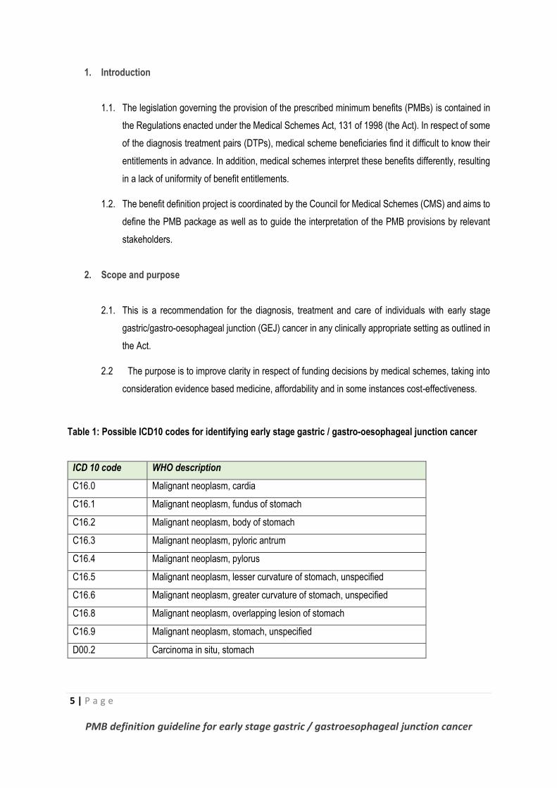
1. Introduction
1.1. The legislation governing the provision of the prescribed minimum benefits (PMBs) is contained in
the Regulations enacted under the Medical Schemes Act, 131 of 1998 (the Act). In respect of some
of the diagnosis treatment pairs (DTPs), medical scheme beneficiaries find it difficult to know their
entitlements in advance. In addition, medical schemes interpret these benefits differently, resulting
in a lack of uniformity of benefit entitlements.
1.2. The benefit definition project is coordinated by the Council for Medical Schemes (CMS) and aims to
define the PMB package as well as to guide the interpretation of the PMB provisions by relevant
stakeholders.
2. Scope and purpose
2.1. This is a recommendation for the diagnosis, treatment and care of individuals with early stage
gastric/gastro-oesophageal junction (GEJ) cancer in any clinically appropriate setting as outlined in
the Act.
2.2 The purpose is to improve clarity in respect of funding decisions by medical schemes, taking into
consideration evidence based medicine, affordability and in some instances cost-effectiveness.
Table 1: Possible ICD10 codes for identifying early stage gastric / gastro-oesophageal junction cancer
ICD 10 code WHO description
C16.0 Malignant neoplasm, cardia
C16.1 Malignant neoplasm, fundus of stomach
C16.2 Malignant neoplasm, body of stomach
C16.3 Malignant neoplasm, pyloric antrum
C16.4 Malignant neoplasm, pylorus
C16.5 Malignant neoplasm, lesser curvature of stomach, unspecified
C16.6 Malignant neoplasm, greater curvature of stomach, unspecified
C16.8 Malignant neoplasm, overlapping lesion of stomach
C16.9 Malignant neoplasm, stomach, unspecified
D00.2 Carcinoma in situ, stomach
6 | P a g e
PMB definition guideline for early stage gastric / gastroesophageal junction cancer
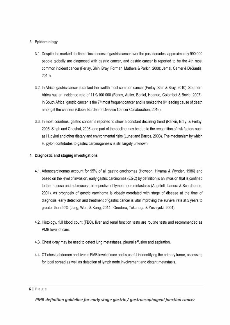
3. Epidemiology
3.1. Despite the marked decline of incidences of gastric cancer over the past decades, approximately 990 000
people globally are diagnosed with gastric cancer, and gastric cancer is reported to be the 4th most
common incident cancer (Ferlay, Shin, Bray, Forman, Mathers & Parkin, 2008; Jemal, Center & DeSantis,
2010).
3.2. In Africa, gastric cancer is ranked the twelfth most common cancer (Ferlay, Shin & Bray, 2010). Southern
Africa has an incidence rate of 11.9/100 000 (Ferlay, Autier, Boniol, Heanue, Colombet & Boyle, 2007).
In South Africa, gastric cancer is the 7th most frequent cancer and is ranked the 9th leading cause of death
amongst the cancers (Global Burden of Disease Cancer Collaboration, 2016).
3.3. In most countries, gastric cancer is reported to show a constant declining trend (Parkin, Bray, & Ferlay,
2005; Singh and Ghoshal, 2006) and part of the decline may be due to the recognition of risk factors such
as H. pylori and other dietary and environmental risks (Lunet and Barros, 2003). The mechanism by which
H. pylori contributes to gastric carcinogenesis is still largely unknown.
4. Diagnostic and staging investigations
4.1. Adenocarcinomas account for 95% of all gastric carcinomas (Howson, Hiyama & Wynder, 1986) and
based on the level of invasion, early gastric carcinomas (EGC) by definition is an invasion that is confined
to the mucosa and submucosa, irrespective of lymph node metastasis (Angelelli, Lanora & Scardapane,
2001). As prognosis of gastric carcinoma is closely correlated with stage of disease at the time of
diagnosis, early detection and treatment of gastric cancer is vital improving the survival rate at 5 years to
greater than 90% (Jung, Won, & Kong, 2014; Onodera, Tokunaga & Yoshiyuki, 2004).
4.2. Histology, full blood count (FBC), liver and renal function tests are routine tests and recommended as
PMB level of care.
4.3. Chest x-ray may be used to detect lung metastases, pleural effusion and aspiration.
4.4. CT chest, abdomen and liver is PMB level of care and is useful in identifying the primary tumor, assessing
for local spread as well as detection of lymph node involvement and distant metastasis.
7 | P a g e
PMB definition guideline for early stage gastric / gastroesophageal junction cancer
4.5. Upper gastrointestinal endoscopy remains the gold standard to detect and diagnose gastric cancer. It is
performed so as to assess for macroscopic appearance of the stomach as well as the morphology and
location of the lesion(s) in question (Russell, Hsu & Mansfield).
4.6. Endoscopic ultrasound (EUS) is a useful staging tool in gastric cancer, specifically to determine pre-
therapy T and N stages so as to guide the sequence of therapy as well as enhance the information on
the extent of disease (Fairweather, Jajoo, Sainani, Bertagnolli & Wang, 2015; Yoshinaga, Oda, Nonaka,
Kushima & Saito, 2012). It is also used preoperatively to assess the submucosal vasculature in order to
predict intraoperative bleeding during endoscopic therapy. Apart from the utility of EUS for diagnosing
invasion depth, EUS can be used preoperatively to assess the submucosal vasculature in order to predict
intraoperative bleeding during endoscopic therapy (Kikuchi, Lizuka & Hoteya, 2011).
4.7. The depth of mural invasion and the presence of extragastric lesions can be determined with endoscopic
ultrasonography (US) and computed tomography (CT).
4.8. To overcome the limitations of contrast-enhanced imaging, diagnostic laparoscopy is strongly
recommended as an additional staging tool to avoid nontherapeutic laparotomy. Staging laparoscopy can
detect radiographically occult peritoneal metastases and prevent futile laparotomy in patients with gastric
adenocarcinoma. This is evidenced by reports of up to 30% of patients with no preoperative evidence of
metastatic disease that harbor occult intra-abdominal metastases that cannot be detected
radiographically by modern imaging techniques (Kriplan and Kapur, 1991; Possik, Franco & Pires, 1986;
Sarela, Lefkowitz, Brennan & Karpeh, 2006).
4.9. Given the relatively low sensitivities for detection of gastric cancer, the value of PET/CT in diagnosis and
evaluation remains controversial (Dassen, Lips, Hoekstra & Pruijt, 2009; Filik, Kir & Aksel, 2015; Kim,
Kang & Lee, 2006). PET scan is not recommended as PMB level of care.
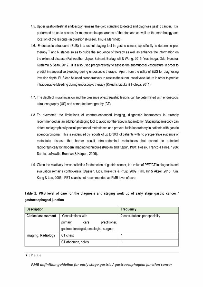
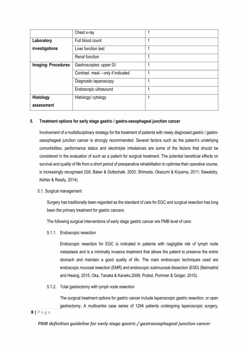
Table 2: PMB level of care for the diagnosis and staging work up of early stage gastric cancer /
gastroesophageal junction
Description Frequency
Clinical assessment Consultations with
primary care practitioner,
gastroenterologist, oncologist, surgeon
2 consultations per speciality
Imaging: Radiology CT chest 1
CT abdomen, pelvis 1
8 | P a g e
PMB definition guideline for early stage gastric / gastroesophageal junction cancer
Chest x-ray 1
Laboratory
investigations
Full blood count 1
Liver function test 1
Renal function 1
Imaging: Procedures Gastroscopies: upper GI 1
Contrast meal – only if indicated 1
Diagnostic laparoscopy 1
Endoscopic ultrasound 1
Histology
assessment
Histology/ cytology 1
5. Treatment options for early stage gastric / gastro-oesophageal junction cancer
Involvement of a multidisciplinary strategy for the treatment of patients with newly diagnosed gastric / gastro-
oesophageal junction cancer is strongly recommended. Several factors such as the patient’s underlying
comorbidities, performance status and electrolyte imbalances are some of the factors that should be
considered in the evaluation of such as a patient for surgical treatment. The potential beneficial effects on
survival and quality of life from a short period of preoperative rehabilitation to optimise their operative course,
is increasingly recognised (Gill, Baker & Gottschalk, 2003; Shimada, Okazumi & Koyama, 2011; Sawatzky,
Kehler & Ready, 2014).
5.1. Surgical management
Surgery has traditionally been regarded as the standard of care for EGC and surgical resection has long
been the primary treatment for gastric cancers.
The following surgical interventions of early stage gastric cancer are PMB level of care:
5.1.1. Endoscopic resection
Endoscopic resection for EGC is indicated in patients with negligible risk of lymph node
metastasis and is a minimally invasive treatment that allows the patient to preserve the entire
stomach and maintain a good quality of life. The main endoscopic techniques used are
endoscopic mucosal resection (EMR) and endoscopic submucosal dissection (ESD) (Balmadrid
and Hwang, 2015; Oka, Tanaka & Kaneko,2006; Probst, Pommer & Golger, 2010).
5.1.2. Total gastrectomy with lymph node resection
The surgical treatment options for gastric cancer include laparoscopic gastric resection, or open
gastrectomy. A multicentre case series of 1294 patients undergoing laparoscopic surgery,
9 | P a g e
PMB definition guideline for early stage gastric / gastroesophageal junction cancer
reported 5 year disease free survival to be 99.8% for stage IA disease, 98.7% for stage IB
disease, and 85.7% for stage II disease (Kitano, Shiraishi & Uyama, 2007).
5.1.3. Esophagogastrectomy with lymph node resection
Patients with proximal tumours usually require an esophagectomy or extended gastrectomy, with
the stomach or jejunum used for intestinal continuity.
5.1.4. Subtotal gastrectomy with lymph node resection
Comparisons disease-free survival between total and subtotal gastrectomy for distal gastric
cancer has shown no significant difference in overall or disease free survival. The 5-year overall
survival rate is 41% for total gastrectomy and 43% for subtotal gastrectomy is 41% and 43%
respectively (Brancato and Miner, 2008). Subtotal gastrectomy has been associated with better
nutritional outcomes and better quality of life when compared with total gastrectomy (Bozzetti,
Marubini & Bonfanti, 1999).
5.2. Chemotherapy
The following perioperative chemotherapy agents are used only for gastric carcinoma of the distal
esophagus or gastroesophageal junction (Cunningham, Allum & Stenning, 2006; Macdonald, Smalley &
Benedetti, 2001; Sumpter, Harper-Wynne & Cunningham, 2005).
Epirubicin,
5FU
Cisplatin
Capecitabine
5.2.1. The MAGIC trial involved random assignment of 503 patients with resectable stage IB-IV gastric
cancer to either perioperative chemotherapy of epirubicin cisplatin, 5-FU (ECF)] and surgery or
surgery alone. The 5-year survival rate, following a median follow up of years, was 36% in the
perioperative chemotherapy group vs 23% in the surgery alone group (HR = 0.75; 95%CI: 0.6-
0.93; P = 0.009) ( Cassidy, Saltz, Twelves, Van Cutsem, Hoff, Kang, Saini, Gilberg &
Cunningham, 2011).
5.2.2. Adjuvant therapy for gastric cancer has been investigated in a number of clinical trials to improve
outcomes for gastric cancer and to define the appropriate adjuvant regimen. A major survival
benefit from postoperative adjuvant chemoradiotherapy has been demonstrated by the INT0116
trial.
10 | P a g e
PMB definition guideline for early stage gastric / gastroesophageal junction cancer
5.2.3. Following surgery, 556 patients stage IB-IV gastric cancer were randomly assigned to either
observation or adjuvant therapy with 4 monthly cycles of bolus 5-fluorouracil (5-FU) and
leucovorin combined with radiation to 45 Gray in 25 fractions. The 3-year survival rate was 50%
in the CRT group vs 41% in the surgery alone group (P = 0.005) (Van Hagen, Hulshof & Van
Lanschot, 2012).
5.2.4. Capecitabine is recommended as an alternative to 5FU. Capecitabine is at least equivalent to 5-
FU in terms of overall survival in patients with gastrointestinal cancers (Cassidy et al, 2011).
5.2.5. The medicines listed below may be used in recognised combinations.
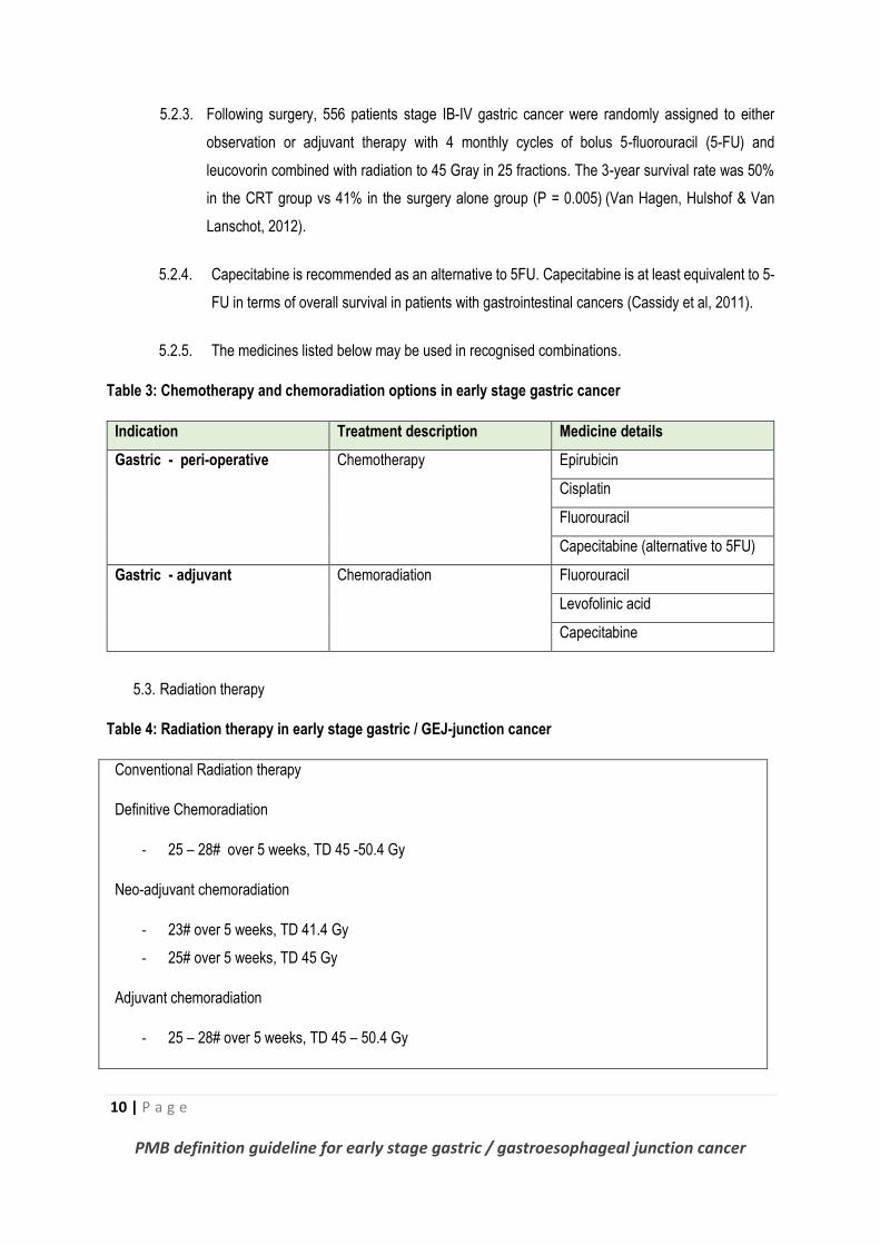
Table 3: Chemotherapy and chemoradiation options in early stage gastric cancer
Indication Treatment description Medicine details
Gastric - peri-operative Chemotherapy
Epirubicin
Cisplatin
Fluorouracil
Capecitabine (alternative to 5FU)
Gastric - adjuvant Chemoradiation Fluorouracil
Levofolinic acid
Capecitabine
5.3. Radiation therapy
Table 4: Radiation therapy in early stage gastric / GEJ-junction cancer
Conventional Radiation therapy
Definitive Chemoradiation
- 25 – 28# over 5 weeks, TD 45 -50.4 Gy
Neo-adjuvant chemoradiation
- 23# over 5 weeks, TD 41.4 Gy
- 25# over 5 weeks, TD 45 Gy
Adjuvant chemoradiation
- 25 – 28# over 5 weeks, TD 45 – 50.4 Gy
11 | P a g e
PMB definition guideline for early stage gastric / gastroesophageal junction cancer
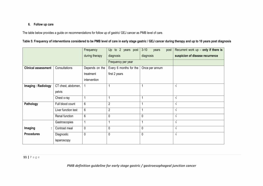
6. Follow up care
The table below provides a guide on recommendations for follow up of gastric/ GEJ cancer as PMB level of care.
Table 5: Frequency of interventions considered to be PMB level of care in early stage gastric / GEJ cancer during therapy and up to 10 years post diagnosis
Frequency
during therapy
Up to 2 years post
diagnosis
3-10 years post
diagnosis
Recurrent work up – only if there is
suspicion of disease recurrence
Frequency per year
Clinical assessment Consultations Depends on the
treatment
intervention
Every 6 months for the
first 2 years
Once per annum
Imaging : Radiology CT chest, abdomen,
pelvis
1 1 1 √
Chest x-ray 1 1 1 √
Pathology Full blood count 6 2 1 √
Liver function test 6 2 1 √
Renal function 6 0 0 √
Imaging :
Procedures
Gastroscopies 1 1 1 √
Contrast meal 0 0 0 √
Diagnostic
laparoscopy
0 0 0 √
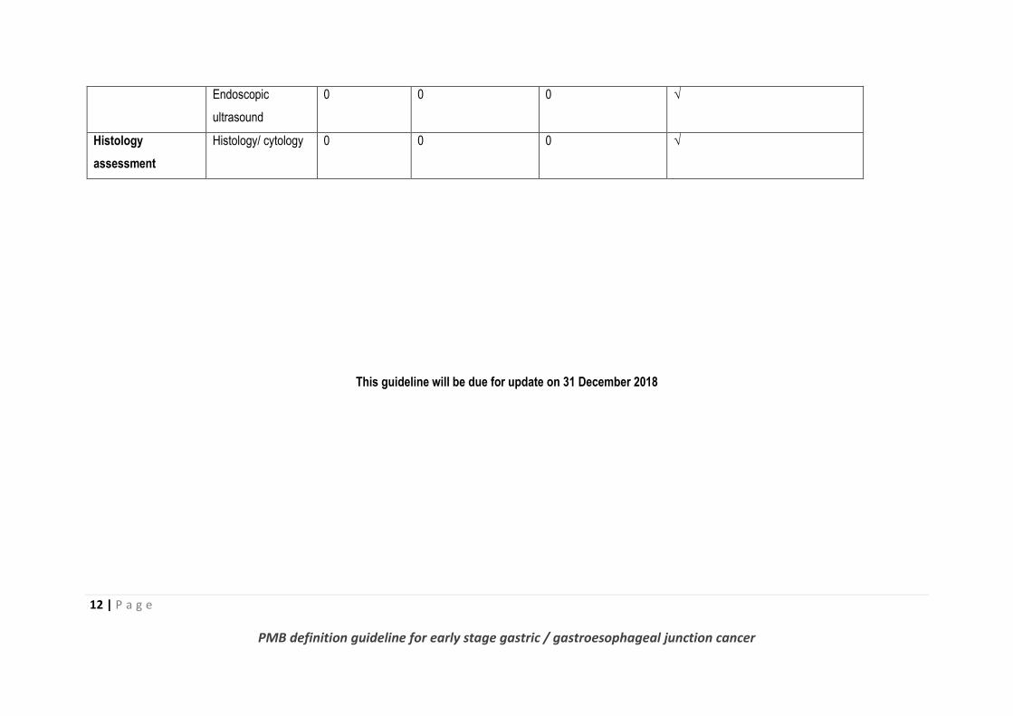
12 | P a g e
PMB definition guideline for early stage gastric / gastroesophageal junction cancer
Endoscopic
ultrasound
0 0 0 √
Histology
assessment
Histology/ cytology 0 0 0 √
This guideline will be due for update on 31 December 2018
13 | P a g e
PMB definition guideline for early stage gastric / gastroesophageal junction cancer
7. References
Angelelli, G., Ianora, A.A. & Scardapane, A. 2001. A. Role of computerized tomography in the staging of
gastrointestinal neoplasms. Seminars in Surgical Oncology, 20:109–121.
Balmadrid, B. and Hwang, J.H. 2015. Endoscopic resection of gastric and oesophageal cancer. Gastroenterology
Report, 3(4):330-338.
Bozzetti, F., Marubini, E. & Bonfanti, G. 1999. Italian Gastrointestinal Tumor Study Group. Subtotal versus total
gastrectomy for gastric cancer: five-year survival rates in a multicentre randomized Italian trial. Annals of Surgery,
230(2):170-178.
Brancato, S. and Miner, T.J. 2008. Surgical management of gastric cancer: review and consideration for total care
of the gastric cancer patient. Current Treatment Options in Gastroenterology, 11(2); 109-118.
Cassidy, J., Saltz, L., Twelves, C., Van, Cutsem. E., Hoff, P., Kang, Y., Saini, J.P., Gilberg, F. & Cunningham, D.
2011. Efficacy of capecitabine versus 5-fluorouracil in colorectal and gastric cancers: a meta-analysis of individual
data from 6171 patients. Annals of Oncology, (12):2604-9.
Cunningham, D., Allum, W.H. & Stenning, S.P. 2006. Perioperative chemotherapy versus surgery alone for
resectable gastroesophageal cancer. New England Journal of Medicine, 355:11–20.
Dassen, A.E., Lips, D.J., Hoekstra, C.J. & Pruijt, J.F. 2009. FDG-PET has no definite role in preoperative imaging
in gastric cancer. European Journal of Surgical Oncology, 35:449–455.
Fairweather, M., Jajoo, K., Sainani, N., Bertagnolli, M.M. & Wang, J. 2015. Accuracy of EUS and CT imaging in
preoperative gastric cancer staging. Journal of Surgical Oncology, 111(8):1016-1020.
Ferlay, J., Autier, P., Boniol, M., Heanue, M., Colombet, M. & Boyle, P. 2007. Estimates of the cancer incidence
and mortality in Europe in 2006. Annals of Oncology, 18: 581-592
Ferlay, J., Shin, H.R. & Bray, F. 2010. Estimates of worldwide burden of cancer in 2008: GLOBOCAN. 2008.
International Journal of Cancer, 127: 2893–917.
Ferlay, J., Shin, H.R., Bray, F., Forman, D., Mathers, C. & Parkin, D.M. GLOBOCAN. 2008 v2.0, Cancer Incidence
and Mortality Worldwide: IARC Cancer Base No. 10 [Internet] Lyon, France: International Agency for Research on
Cancer. Available from: http://globocan.iarc.fr [Accessed 12 January 2017]
Filik, M., Kir, K.M. & Aksel B. 2015. The Role of 18F-FDG PET/CT in the Primary Staging of Gastric Cancer.
Molecular Imaging and Radionuclide Therapy, 24(1):15-20.
Gill, T.M., Baker, D.I. & Gottschalk, M. 2003. A prehabilitation program for physically frail community-living older
persons. Archives of Physical Medicine and Rehabilitation, 84(3):394-404.
Global Burden of Disease Cancer Collaboration. 2016. Global, Regional, and National Cancer Incidence, Mortality,
Years of Life Lost, Years Lived With Disability, and Disability-Adjusted Life-years for 32 Cancer Groups, 1990 to
2015. A Systematic Analysis for the Global Burden of Disease Study. Journal of the American Medical Association-
Oncology, E1-E25.
Howson, C.P., Hiyama, T. & Wynder, E.L. 1986. The decline in gastric cancer: epidemiology of an unplanned
triumph. Epidemiologic Reviews: 8:1–27.
Jemal, A., Center, M.M., & DeSantis, C. 2010. Global patterns of cancer incidence and mortality rates and trends.
Cancer Epidemiology Biomarkers & Prevention, 19:1893–907.
14 | P a g e
PMB definition guideline for early stage gastric / gastroesophageal junction cancer
Jung, K.W., Won, Y.J. & Kong, H.J. 2014. Cancer statistics in Korea: incidence, mortality, survival, and prevalence
in 2011. Cancer Research and Treatment, 46(2); 109–123.
Kikuchi, D., Lizuka, T. & Hoteya, S. 2011. Usefulness of endoscopic ultrasound for the prediction of intraoperative
bleeding of endoscopic submucosal dissection for gastric neoplasms. Journal of Gastroenterology and
Hepatology: vol. 26, no. 1, pp. 68–72.
Kim, S.K., Kang, K.W. & Lee, J.S. 2006. Assessment of lymph node metastases using 18F-FDG PET in patients
with advanced gastric cancer. European Journal of Nuclear Medicine and Molecular Imaging, 33:148-55.
Kitano, S., Shiraishi, N. & Uyama, I. 2007. A multicenter study on oncologic outcome of laparoscopic gastrectomy
for early cancer in Japan. Annals of Surgery, 245; 68-72.
Kriplani, A.K. and Kapur, B.M. 1991. Laparoscopy for pre-operative staging and assessment of operability in gastric
carcinoma. Gastrointestinal Endoscopy; 37:441–443.
Lunet, N. & Barros, H. 2003. Helicobacter pylori infection and gastric cancer: facing the enigmas. International
Journal of Cancer, 106: 953-960.
Macdonald, J.S., Smalley, S.R. & Benedetti, J. 2001. Chemoradiotherapy after surgery compared with surgery
alone for adenocarcinoma of the stomach or gastroesophageal junction. New England Journal of Medicine,
345:725–730.
Oka, S., Tanaka, S. & Kaneko, I. 2006. Advantage of endoscopic submucosal dissection compared with EMR for
early gastric cancer. Gastrointestinal Endoscopy, 64: 877–883.
Onodera, H., Tokunaga, A. & Yoshiyuki, T. 2004. Surgical outcome of 483 patients with early gastric cancer:
prognosis, postoperative morbidity and mortality, and gastric remnant cancer. Hepatogastroenterology: 51; 82–85.
Parkin, D.M., Bray, F. & Ferlay, J. 2005. Global cancer statistics, 2002. CA cancer journal for clinicians, 55: 74-
108.
Possik, R.A., Franco, E.L. & Pires, D.R. 1986. Sensitivity, specificity, and predictive value of laparoscopy for the
staging of gastric cancer and for the detection of liver metastases. Cancer: 58:1–6.
Probst, A., Pommer, B. & Golger, D. 2010. Endoscopic submucosal dissection in gastric neoplasia - experience
from a European center. Endoscopy, 42:1037–1044.
Russell, M.C., Hsu, C. & Mansfield, P.F., Primary gastric malignancies. In: Feig BW, Ching CD (eds). MD Anderson
Surgical Oncology Handbook. 5th ed. New York, NY: Lippincott Williams & Wilkins; 270-315.
Sarela, A., Lefkowitz, R., Brennan, M. & Karpeh, M. 2006. Selection of patients with gastric adenocarcinoma for
laparoscopic staging. American Journal of Surgery, 191:134–138.
Sawatzky, J.A., Kehler, D.S. & Ready, A.E. 2014. Prehabilitation program for elective coronary artery bypass graft
surgery patients: a pilot randomized controlled study. Clinical Rehabilitation, 28(7):648-657.
Shimada, H., Okazumi, S. & Koyama, M. 2011. Japanese Gastric Cancer Association Task Force for Research
Promotion: clinical utility of 18F-fluoro-2-deoxyglucose positron emission tomography in gastric cancer. A
systematic review of the literature. Gastric Cancer, 14:13–21
Singh, K. & Ghoshal, U.C. 2006. Causal role of Helicobacter pylori infection in gastric cancer: an Asian enigma.
World Journal of Gastroenterology, 12: 1346-1351.
15 | P a g e
PMB definition guideline for early stage gastric / gastroesophageal junction cancer
Yoshinaga, S., Oda, I., Nonaka, S., Kushima, R. & Saito, Y. 2012. Endoscopic ultrasound using ultrasound probes
for the diagnosis of early oesophageal and gastric cancers. World Journal of Gastrointestinal Endoscopy: vol. 4,
no. 6, pp. 218–226.