Embed Size (px)
Citation preview
Hartmann 1
Postpartum Angiopathy with Cerebral Infarction, Subarachnoid Hemorrhage
and Intraparenchymal Hemorrhage: A Case Discussion
Alexander J.P.W. Hartmann, M.D.*, Edward Livingstone II M.D.**, and Timothy
Meadows M.D.**
Department of Neurology, University of Minnesota* and Hampton Roads Neurology**
Case:
A 25-year-old female two weeks postpartum presented to the emergency
department with the chief complaint of a headache. She had been evaluated five days
prior to admission at an outside hospital for sudden onset of severe right-sided
headache with no history of headaches. A head computed tomographic (CT) scan at
that location revealed subarachnoid hemorrhage, after which the patient was
transferred for further evaluation. A cerebral angiogram was performed which was
completely normal, including the venous phase. The patient's headache resolved and
she was discharged home. Five days later, she returned to the emergency
department with recurrence of her headache and new complaints of numbness and
weakness in her left hand.
Her medical history was remarkable only for being two weeks postpartum from
an uncomplicated pregnancy and vaginal delivery. She also reported a remote history
of Bell's palsy, but had otherwise been healthy and denied any substance use. Her
family history was remarkable for rheumatoid arthritis in her mother and scleroderma
in her maternal grandmother. She denied taking any medications or having allergies.
Her vital signs were stable upon admission. The exam showed shortened attention
span and reduced concentration, and a left homonymous hemianopia. Strength was
intact throughout with slowed alternate motion rate in her left upper extremity. Toes
were downgoing bilaterally.
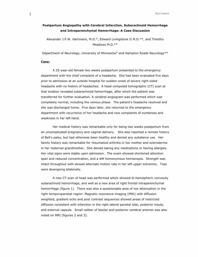
A new CT scan of head was performed which showed bi-hemispheric convexity
subarachnoid hemorrhage, and well as a new area of right frontal intraparenchymal
hemorrhage (figure 1). There was also a questionable area of low attenuation in the
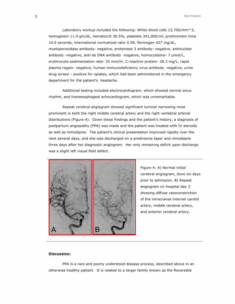
right temporoparietal region. Magnetic resonance imaging (MRI) with diffusion
weighted, gradient echo and post contrast sequences showed areas of restricted
diffusion consistent with infarction in the right lateral parietal lobe, posterior insula,
and external capsule. Small caliber of basilar and posterior cerebral arteries was also
noted on MRI (figures 2 and 3).
Hartmann 2
Figure 1: CT scan of head on hospital day
1 showing the presence of subarachnoid
hemorrhage that is more prominent on
the right side.
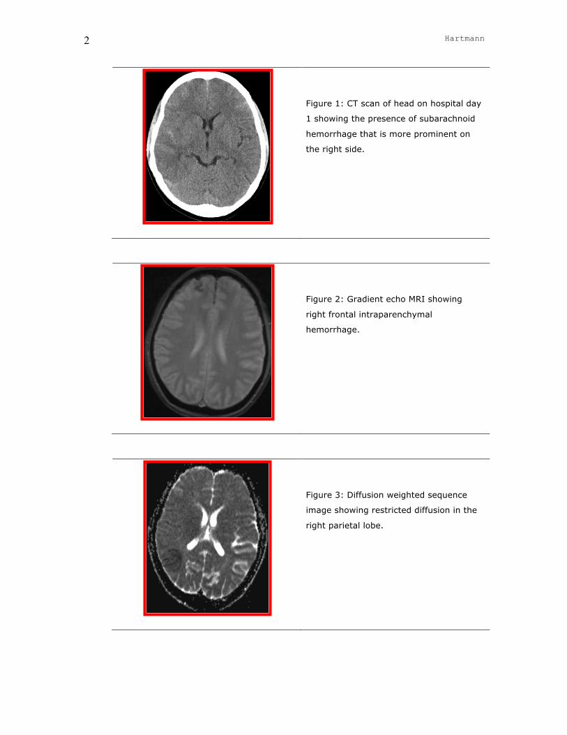
Figure 2: Gradient echo MRI showing
right frontal intraparenchymal
hemorrhage.
Figure 3: Diffusion weighted sequence
image showing restricted diffusion in the
right parietal lobe.
!
!
!
Hartmann 3
Laboratory workup included the following: White blood cells 12,700/mm^3,
hemoglobin 11.9 gm/dL, hematocrit 36.5%, platelets 341,000/ml, prothrombin time
10.6 seconds, international normalized ratio 0.99, fibrinogen 427 mg/dL,
myeloperoxidase antibody- negative, proteinase 3 antibody- negative, antinuclear
antibody -negative, anti-ds DNA antibody -negative, homocysteine- 7 µmol/L,
erythrocyte sedimentation rate- 35 mm/hr, C-reactive protein- 38.3 mg/L, rapid
plasma regain- negative, human immunodeficiency virus antibody- negative, urine
drug screen - positive for opiates, which had been administered in the emergency
department for the patient’s headache.
Additional testing included electrocardiogram, which showed normal sinus
rhythm, and transesophageal echocardiogram, which was unremarkable.
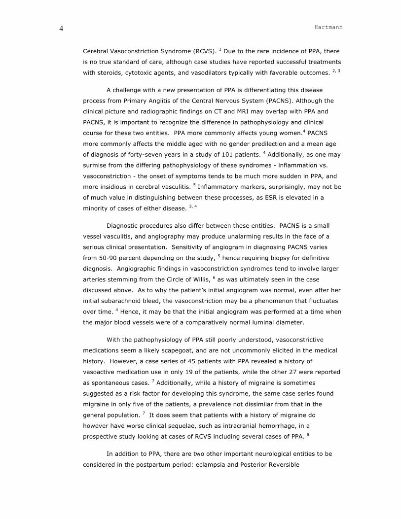
Repeat cerebral angiogram showed significant luminal narrowing most
prominent in both the right middle cerebral artery and the right vertebral arterial
distributions (Figure 4). Given these findings and the patient's history, a diagnosis of
postpartum angiopathy (PPA) was made and the patient was treated with IV steroids
as well as nimodipine. The patient’s clinical presentation improved rapidly over the
next several days, and she was discharged on a prednisone taper and nimodipine
three days after her diagnostic angiogram. Her only remaining deficit upon discharge
was a slight left visual field defect.
Figure 4: A) Normal initial
cerebral angiogram, done six days
prior to admission. B) Repeat
angiogram on hospital day 3
showing diffuse vasoconstriction
of the intracranial internal carotid
artery, middle cerebral artery,
and anterior cerebral artery.
Discussion:
PPA is a rare and poorly understood disease process, described above in an
otherwise healthy patient. It is related to a larger family known as the Reversible
Hartmann 4
Cerebral Vasoconstriction Syndrome (RCVS). 1 Due to the rare incidence of PPA, there
is no true standard of care, although case studies have reported successful treatments
with steroids, cytotoxic agents, and vasodilators typically with favorable outcomes. 2, 3
A challenge with a new presentation of PPA is differentiating this disease
process from Primary Angiitis of the Central Nervous System (PACNS). Although the
clinical picture and radiographic findings on CT and MRI may overlap with PPA and
PACNS, it is important to recognize the difference in pathophysiology and clinical
course for these two entities. PPA more commonly affects young women.4 PACNS
more commonly affects the middle aged with no gender predilection and a mean age
of diagnosis of forty-seven years in a study of 101 patients. 4 Additionally, as one may
surmise from the differing pathophysiology of these syndromes - inflammation vs.
vasoconstriction - the onset of symptoms tends to be much more sudden in PPA, and
more insidious in cerebral vasculitis. 5 Inflammatory markers, surprisingly, may not be
of much value in distinguishing between these processes, as ESR is elevated in a
minority of cases of either disease. 3, 4
Diagnostic procedures also differ between these entities. PACNS is a small
vessel vasculitis, and angiography may produce unalarming results in the face of a
serious clinical presentation. Sensitivity of angiogram in diagnosing PACNS varies
from 50-90 percent depending on the study, 5 hence requiring biopsy for definitive
diagnosis. Angiographic findings in vasoconstriction syndromes tend to involve larger
arteries stemming from the Circle of Willis, 6 as was ultimately seen in the case
discussed above. As to why the patient’s initial angiogram was normal, even after her
initial subarachnoid bleed, the vasoconstriction may be a phenomenon that fluctuates
over time. 4 Hence, it may be that the initial angiogram was performed at a time when
the major blood vessels were of a comparatively normal luminal diameter.
With the pathophysiology of PPA still poorly understood, vasoconstrictive
medications seem a likely scapegoat, and are not uncommonly elicited in the medical
history. However, a case series of 45 patients with PPA revealed a history of
vasoactive medication use in only 19 of the patients, while the other 27 were reported
as spontaneous cases. 7 Additionally, while a history of migraine is sometimes
suggested as a risk factor for developing this syndrome, the same case series found
migraine in only five of the patients, a prevalence not dissimilar from that in the
general population. 7 It does seem that patients with a history of migraine do
however have worse clinical sequelae, such as intracranial hemorrhage, in a
prospective study looking at cases of RCVS including several cases of PPA. 8
In addition to PPA, there are two other important neurological entities to be
considered in the postpartum period: eclampsia and Posterior Reversible
Hartmann 5
Encephalopathy Syndrome (PRES), which have been shown elsewhere to be closely
related. 9
Eclampsia is known to be associated at least with peripartum cerebrovascular
events. In one study of women in the peripartum period, the rate of comorbid
ischemic stroke was 27%, while that of intracerebral hemorrhage was 14%. 10
However the relationship between eclampsia with PPA specifically is less clear. In a
study comparing the radiographic findings in four patients with postpartum eclampsia
and four patients PPA, one of the eclampsia patients was incidentally found to have
angiopathy on angiogram, and one of the PPA patients was found to have
preeclampsia, although she did not meet the clinical requirements to make the
diagnosis of full eclampsia. 2 Another review of 4 cases of concomitant PPA and PRES,
discussed more below, found 3 of the 4 to be having seizures suggestive of possible
eclampsia. However none of these patients had the proteinuria encountered in
eclampsia. 11 It is unclear if the cerebral vasoconstriction that has been described with
eclampsia is a reaction to the hypertension associated with eclampsia, or if whether it
represents an independent, primary process. 12
Regarding PRES, there is a more firmly established relationship between this
entity and PPA. In a prospective series of 67 RCVS patients, including several patients
with PPA, two of the six cases that developed concomitant PRES syndrome were cases
of PPA. 13 A severe case of PPA with PRES was recently reported in Belgium. The
patient progressed to coma and required intubation, although was ultimately
discharged with a Modified Rankin Score of 0, suggesting that developing PRES in and
of itself does not necessarily reflect negatively on prognosis. 14
Conclusion:
PPA is an uncommon disease process, for which data remains limited. Its
presentation may mimic other disorders, and a thorough workup including CT, MRI,
and angiography is warranted. Although its presentation may be alarming, outcomes
are generally quite favorable in the literature reviewed as was the case here. There is
established comorbidity between PPA and PRES with a fairly low incidence. Although
vasoconstriction has been demonstrated in eclampsia, the exact relationship between
eclampsia and PPA as a distinct entity is not well known.
Teaching Points:
1) A postpartum woman with neurologic deficits should be taken seriously, with
imaging performed if necessary.
Hartmann 6
2) PPA may present as intraparenchymal hemorrhage, subarachnoid hemorrhage,
ischemic stroke, or more than one of these as was the case here.
2) If PPA is suspected, one negative angiogram does not rule it out.
4) Consider repeat angiogram before proceeding to brain biopsy.
5) Outcome is favorable with treatment of combination of steroids and calcium channel
blockers.
References:
1. Ducros A, Bousser MG. Reversible cerebral vasoconstriction syndrome. Pract Neurol.
2009;9:256-267
2. Fletcher JJ, Kramer AH, Bleck TP, Solenski NJ. Overlapping features of eclampsia and
postpartum angiopathy. Neurocrit Care. 2009;11:199-209
3. Geocadin RG, Razumovsky AY, Wityk RJ, Bhardwaj A, Ulatowski JA. Intracerebral
hemorrhage and postpartum cerebral vasculopathy. J Neurol Sci. 2002;205:29-34
4. Salvarani C, Brown RD, Jr., Calamia KT, Christianson TJ, Weigand SD, Miller DV,
Giannini C, Meschia JF, Huston J, 3rd, Hunder GG. Primary central nervous system
vasculitis: Analysis of 101 patients. Ann Neurol. 2007;62:442-451
5. Birnbaum J, Hellmann DB. Primary angiitis of the central nervous system. Arch Neurol.
2009;66:704-709
6. Singhal AB, Bernstein RA. Postpartum angiopathy and other cerebral vasoconstriction
syndromes. Neurocrit Care. 2005;3:91-97
7. Williams TL, Lukovits TG, Harris BT, Harker Rhodes C. A fatal case of postpartum
cerebral angiopathy with literature review. Arch Gynecol Obstet. 2007;275:67-77
8. Ducros A, Fiedler U, Porcher R, Boukobza M, Stapf C, Bousser MG. Hemorrhagic
manifestations of reversible cerebral vasoconstriction syndrome: Frequency, features,
and risk factors. Stroke. 2010;41:2505-2511
9. Wagner SJ, Acquah LA, Lindell EP, Craici IM, Wingo MT, Rose CH, White WM, August P,
Garovic VD. Posterior reversible encephalopathy syndrome and eclampsia: Pressing
the case for more aggressive blood pressure control. Mayo Clin Proc. 2011;86:851-
856
10. Kittner SJ, Stern BJ, Feeser BR, Hebel R, Nagey DA, Buchholz DW, Earley CJ, Johnson
CJ, Macko RF, Sloan MA, Wityk RJ, Wozniak MA. Pregnancy and the risk of stroke. N
Engl J Med. 1996;335:768-774
11. Singhal AB. Postpartum angiopathy with reversible posterior leukoencephalopathy.
Arch Neurol. 2004;61:411-416
Hartmann 7
12. Donaldson JO. Eclampsia and postpartum cerebral angiopathy. J Neurol Sci.
2000;178:1
13. Ducros A, Boukobza M, Porcher R, Sarov M, Valade D, Bousser MG. The clinical and
radiological spectrum of reversible cerebral vasoconstriction syndrome. A prospective
series of 67 patients. Brain. 2007;130:3091-3101
14. Lemmens R, Smet S, Wilms G, Demaerel P, Thijs V. Postpartum rcvs and pres with
normal initial imaging findings. Acta Neurol Belg. 2012
Corresponding Author:
Alexander J.P.W. Hartmann, M.D.
Department of Neurology, University of Minnesota
420 Delaware St SE MMC 295
Minneapolis, MN 55455
Tel 612-626-6519
Fax 612-625-7950
Email: [email protected]