Embed Size (px)
Citation preview
PALEOPATHOLOGY ASSOCIATION
ABSTRACTS
40th Annual North American Meeting KNOXVILLE, TENNESSEE
April, 2013
*** Entrant for the Cockburn student prize. Poster in the special session in honor of Dr. Donald J. Ortner.
PALEOPATHOLOGY ASSOCIATION
40th
Annual North American Meeting
KNOXVILLE, TENNESSEE
April 9 & 10, 2013
SCIENTIFIC PROGRAM
MONDAY, APRIL 8TH
REGISTRATION (6.00-9.00pm) Lobby, Hilton Knoxville
TUESDAY, APRIL 9TH
REGISTRATION (8.00am – 5.00pm) Outside Meeting Rooms 200 A-E, Knoxville Convention Centre
MORNING SESSION (9.00 – NOON): In recognition of the 21 workshop contributions of the late
Don Ortner to the PPA meetings, our 2013 meeting continues with:
9.00 – NOON. The "Donald J. Ortner PPA Workshop" RHEUMATIC DISEASES IN
ANTIQUITY AND CONTEMPORARY POPULATIONS. organized by Dawnie Steadman
and Heli Maijanen 200AB
9.00 – NOON. The "Donald J. Ortner PPA Workshop" THE BIOMECHANICS, BIOLOGY
AND INTERPRETATION OF BONE FRACTURE. organized by Dawnie Steadman and Heli
Maijanen 200DE
12.00 – 1.55 LUNCH
AFTERNOON SESSION I (2.00 – 3.20) Chair: Susan Pfeiffer
Podium Presentations 200DE
2.00 Announcements and Session Opening
2.05 SHOT THROUGH THE HEAD, AND WHO’S TO BLAME? FOURTEEN
HISTORIC PERIOD CASES FROM ALASKA. J. Christopher Dudar & Lars
Krutak
2.20 VIKINGS IN ORKNEY, GENOCIDE OR INTEGRATION?:
OSTEOLOGICAL ANALYSIS OF THE MEDIEVAL POPULATION OF
ORKNEY. Ceilidh Lerwick ***
2.35 QUANTITATIVE ANALYSIS OF PERIMORTEM TRAUMA IN A
FRAGMENTED, DISARTICULATED, AND COMMINGLED SKELETAL
SAMPLE: SUCCESS AND FAILURE AT SMITH’S KNOLL. Laura Lockau,
Ana-Maria Dragomir, & Megan Brickley. ***
*** Entrant for the Cockburn student prize. Poster in the special session in honor of Dr. Donald J. Ortner.
2.50 PALEOPATHOLOGICAL ENIGMAS OF MATRIX 101: AN
UNPRECEDENTED MASS HUMAN SACRIFICE FROM HUACA LAS
VENTANAS, PERU. Haagen Klaus, Steve Nau, Kevin Reed, Alexis Meeks, Angelina
DeMarco, Jenna Hurtubise, Jose Pinilla, Ana Alva, & Carlos Elera.
3.05 TESTING THE RELATIONSHIP BETWEEN SEXUAL DIMORPHISM
AND CHILDHOOD HEALTH IN PREHISTORIC THAILAND. Angela L.
Clark, Nancy Tayles, & Siân Halcrow.
3.20 – 3.50 BREAK
AFTERNOON SESSION II (3.50 – 5.15) Chair: Sandra Gravie Lok
Podium Presentations 200DE
3.50 BREAST CANCER IN ANTIQUITY? DIFFERENTIAL DIAGNOSIS OF
TWO CASES FROM ANCIENT EGYPT. Tosha L. Dupras, Lana J. Williams, Peter
S. Sheldrick, Brittany Walter, Bart VanThuyne, Sandra Wheeler, & Harco Willems.
4.05 THE ANTIQUITY OF CANCER: A SURVEY OF PALAEO-
ONCOLOGICAL “CASE STUDIES” FOR IDENTIFICATION AND
METHODOLOCICAL IMPROVEMENT. Kathryn J. Hunt.***
4.20 FUNCTIONAL ORTHO-PROSTHESIS IN A THIRD/SECOND CENTURY
BC GRAVE FROM TURFAN, CHINA. Julia Gresky, Mayke Wagner, Pavel Tarasov,
Xiao Li, Xiaohong Wu, Yongbin Zhang, Arno Schmidt, & Tomasz Goslar.
4.35 AN ICELANDIC SAGA OF A DIFFERENT SORT: HIGH FREQUENCIES
OF ANTEMORTEM TOOTH LOSS IN MEDIEVAL ICELANDERS
COMPARED TO GREENLANDERS, NORWEGIANS, AND DANISH
VIKINGS. Amanda R. Harvey, Roman Schomberg, Diana Malarchik, & G. Richard Scott. ***
4.50 STANDARDISING THE DIAGNOSIS OF DENTAL CARIES IN
ARCHAEOLOGICAL REMAINS – A STUDY FROM PREHISTORIC
SOUTHEAST ASIA. Stephanie A. Shkrum, Nancy Tayles, & Siân E. Halcrow.***
5.05 Announcements
Student Action Committee (5.15 – 6.15) 200DE
6.30 Cash Bar, followed by Association Business Meeting and Buffet Dinner 200ABC
WEDNESDAY, APRIL 10TH
REGISTRATION (8.00 – 12.00 noon) Outside Meeting Rooms 200 A-E, Knoxville Convention
Centre
MORNING SESSION I (8.00-9.20) Chair: Gretchen Dabbs
Podium Presentations 200DE
*** Entrant for the Cockburn student prize. Poster in the special session in honor of Dr. Donald J. Ortner.
8.00 Announcements
8.05 DELVING DEEP INTO THE ORIGINS OF TUBERCULOSIS WITH THE
AID OF ROBUST LIPID BIOMARKERS. David E. Minnikin, Oona Y-C. Lee,
Houdini H.T. Wu, Gurdyal S. Besra, Oussama Baker, Olivier Dutour, Bruce Rothschild, Richard
Laub, Mark Spigelman, & Helen D. Donoghue
8.20 PALEOEPIDEMIOLOGY OF A PREHISTORIC CENTRAL CALIFORNIA
SHELLMOUND IN RELATION TO THE RISE OF INFECTIOUS
MYCOBACTERIUM TUBERCULOSIS. Reshma E. Varghese, Elaine M. Burke, Emily
A. Bulger, Gabrielle E. Aldern, Gary D. Richards, & Rebecca S. Jabbour***
8.35 HETEROGENEITY IN LIPID BIOMARKER PROFILES FOR ANCIENT
LEPROSY. Oona Y-C Lee, Houdini H.T. Wu, Charlotte A. Roberts, Maria Giovanna
Belcastro, Valentina Mariotti, Mark Spigelman, Helen D. Donoghue, Gurdyal S. Besra, & David
E. Minnikin
8.50 AN ANALYSIS OF OSTEOARTHRITIS OF THE COSTOVERTEBRAL
AND COSTOTRANSVERSE JOINTS FROM 3 MEDIEVAL NUBIAN
SITES. Jennifer L. Willoughby.
9.05 CHRONIC COXOFEMORAL LUXATIONS IN 13TH
CENTURY VIRGINIA
DOGS: DIFFERENTIATION FROM HIP DYSPLASIA. Elizabeth W. Uhl, Shelby
F. Jarrett, Charles Kelderhouse, & Jeffrey P. Blick.
9.20 – 10.40 BREAK & POSTER SESSION I Clinch Concourse
Posters in place all day but authors of odd numbered posters will be present during this break.
Poster titles & authors listed below.
MORNING SESSION II (10.40 – 12.00) Chair: Daniel Temple
Podium Presentations 200DE
10.40 CONTRIBUTION OF OSTEOPOROSIS TO FRACTURE PATTERNS
AMONG PRECOLUMBIAN AMERINDIANS OF WEST-CENTRAL
ILLINOIS. Susan Dale Spencer, Polly Husmann, & Della Collins Cook.
10.55 URBAN AND RURAL HEALTH IN POST-MEDIEVAL LONDON: A
BIOARCHAEOLOGIAL ANALYSIS OF MIDDLE- AND OLD ADULT
WOMEN. Alexandra Perrone.
11.10 THE PERILS AND POSSIBILITIES OF INFERRING DISEASE
PRESENCE IN ANTIQUITY WITH LITERARY EVIDENCE. Stephanie
Marciniak.
11.25 INQUISITION OF ÉVORA (PORTUGAL): INVESTIGATION OF HUMAN
REMAINS FROM THE ‘JAIL CLEANING YARD’. Bruno Silva Magalhães,
Angela Araújo, & Ana Luísa Santos.
11.40 THE EARLIEST EVIDENCE FOR INTESTINAL PARASITES IN
GREECE: IMPLICATIONS FOR THE INTERPRETATION OF THE
*** Entrant for the Cockburn student prize. Poster in the special session in honor of Dr. Donald J. Ortner.
ANCIENT MEDICAL TEXTS OF HIPPOCRATES. Piers D. Mitchell & Evilena
Anastasiou.
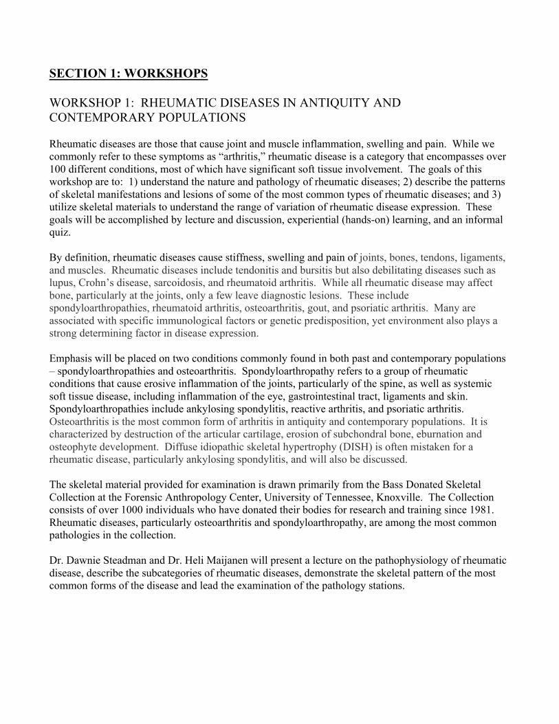
12.00 – 1.55 LUNCH – Let’s Do Lunch Chesapeake’s (See attached map)
AFTERNOON SESSIONS I & II. SPECIAL SESSION IN MEMORY OF DR. DONALD J.
ORTNER (2.00 – 3.05 AND 4.05-5.05)
Podium Presentations I 200DE Chair: Mary Lucas Powell
2.00 A TRIBUTE TO DON BY KEITH MANCHESTER. Charlotte Roberts, PPA
President.
2.05 IN MEMORIAM Jane E. Buikstra & Bruno Frohlich
2.25 A POSSIBLE TREPHINATION FROM THE NORTH COAST OF PERU
AND THE CHALLENGES OF DIFFERENTIAL DIAGNOSIS OF HEALED
CRANIAL DEFECTS. John W. Verano & Tania Delabarde
2.40 PALEOPATHOLOGICAL DESCRIPTION AND DIAGNOSIS OF
METASTATIC CARCINOMA IN A BRONZE AGE HUNTER-GATHERER
FROM CIS BAIKAL, SIBERIA. Daniel H. Temple, Angela R. Lieverse, Vladimir I.
Bazaliiski, & Andrzej W. Weber.
2.55 “THE LEGACY OF BONE PATHOLOGY REFERENCE SERIES: DON
ORTNER AND THE GALLER COLLECTION”. Frank Rühli & Thomas Böni.
3.10 – 4.20 BREAK & POSTER SESSION II Park Concourse
Posters in place all day but authors of even numbered posters will be present during this break.
Posters in the session in honor of Dr. Donald J. Ortner are located on even-numbered boards 64-
78 inclusive.
Poster titles & authors listed at the end of the program.
Podium Presentations II Chair: Jerry Rose
4.20 A CASE OF LEPROMATOUS LEPROSY FROM KODIAK ISLAND,
ALASKA. Cynthia A. Wilczak & Donald J. Ortner.
4.35 CRANIAL TRAUMA PATTERNS AMONG HOLOCENE FORAGERS –
INTENTIONAL AND ACCIDENTAL CAUSES. Susan Pfeiffer.
4.50 THE STUDY OF ANCIENT TUBERCULOSIS: AS DONALD J. ORTNER
ADVOCATED. Jane E. Buikstra & Charlotte A. Roberts.
5.05 Closing Remarks, and Announcements, Award of Cockburn Student Prize
CASH BAR ‘Highlights of PPA: 1973 – 2013’. Mary Lucas Powell.
______________________________________________________________________________
*** Entrant for the Cockburn student prize. Poster in the special session in honor of Dr. Donald J. Ortner.
POSTER PRESENTATIONS:
(Number refers to poster board number) An author of the poster should be present at the poster during
their assigned poster session. Authors of odd numbered posters should be present at the first poster
session (Wed., April 10th
, 9.20 – 10.40 am) and those of even numbers posters should be present at the
second session (Wed., April 10th
, 3.10 – 4.20 pm).
1. DEVELOPMENTAL DYSPLASIA OF THE HIP IN A CHILD FROM
MEDIEVAL POLAND
Amanda M. Agnew & Hedy M. Justus
2. HUMAN-INDUCED TRAUMATIC SCOLIOSIS IN A COMMERSON’S
DOLPHIN (Cephalorhyncus commersoni)
Kathleen B. Adia & Charles W. Potter
3. HEALTH AND MORBIDITY AT ANGEL MOUNDS (12VG1): THE
PALEOPATHOLOGY OF A GERMINATING TOWN
Erica L. Ausel***
4. LYTIC LESIONS ON THE EUBOEAN: A CASE STUDY FROM BRONZE AGE
MITROU
Amy Anderson & Nicholas P. Herrmann
5. DESERT REHYDRATION: TESTING PALEOHISTOLOGICAL FIELD
METHODOLOGY OF MUMMIFIED TISSUES FROM DAKHLEH OASIS,
EGYPT
J. Branson, L. Williams, T.L. Dupras, & S. Wheeler
6. CASES OF SYPHILIS IN THE ROBERT J. TERRY ANATOMICAL
SKELETAL COLLECTION
Daniel L. DiMichele & David Hunt
7. DENTAL DISEASE AT RAS AL-KHAIMAH, UAE: HEALTH INDICATORS
FROM COMINNGLED SKELETAL REMAINS
Alyson Caine
8. THE APPLICATION OF DIGITAL RADIOGRAPHIC ANALYSIS TO
SKELETAL ASSEMBLAGES
Jelena Bekvalac & Gaynor Western
9. SPECIAL CHILD, SPECIAL DEATH: CRANIOSYNOSTOSIS & MORTUARY
RITUAL IN PREHISPANIC NORTH MEXICO
John J. Crandall, Jennifer L. Thompson, & Della C. Cook***
*** Entrant for the Cockburn student prize. Poster in the special session in honor of Dr. Donald J. Ortner.
10. THE MISSHAPEN MAN: A DIFFERENTIAL DIAGNOSIS
Tracy K. Betsinger & Amy B. Scott
11. BIOARCHAEOLOGICAL ANALYSIS OF THE SHADY GROVE OSSUARY
(22QU525)
C. Brady Davis
12. THE PREVALENCE OF DENTAL CARIES IN NEOLITHIC ANATOLIA,
TURKEY
Başak Boz, Marin A. Pilloud, & Simon Hillson
13. LIVES OF STRESS? RECONSTRUCTING THE LIFE HISTORIES, HEALTH,
AND IDENTITY OF THE SACRIFICE VICTIMS AT MATRIX 101, SICÁN,
PERU
Angelina DeMarco, Haagen Klaus, Jenna Hurtibise, Alexis Meeks, Steve Nau, Kevin Reed, Jose Pinilla,
Ana Alva, & Carlos Elera
14. A HANGING-DECAPITATION FROM IRON AGE YORK
Jo Buckberry, Terry O’Connor, Andrew Wilson, & Sonia O’Connor
15. CUT MARKS: NEW EVIDENCE OF HUMAN SACRIFICE AT HUACA DE A
LUNA
Brittany Dement***
16. A PUZZLING PURPLE PIGMENT: PRESERVATION AND
PSEUDOPATHOLOGY PREDICAMENTS Julie A. Bukowski & Jennifer E. Mack
17. PALEOPATHOLOGICAL EVIDENCE OF SEXUAL DIVISON OF LABOR
AND DIET IN THE SAN FRANCISCO BAY AREA
Melanie M. Beasley & Eric J. Bartelink***
18. BONE MINERAL DENSITY ASSESSMENT IN THE DISTAL RADIUS: AN
INADEQUATE OPTION IN PALEOPATHOLOGY?
Francisco Curate, Anabela Albuquerque, Izilda Ferreira, & Tânia Ferreira
19. YOU ARE NOT WHAT YOU EAT DURING STRESS: ISOTOPIC
EVALUATON OF HUMAN HAIR FROM BELLEVILLE, ONTARIO Lori D’Ortenzio & Tracy Prowse***
*** Entrant for the Cockburn student prize. Poster in the special session in honor of Dr. Donald J. Ortner.
20. PATTERNS OF OSTEOPOROSIS SYMMETRICA AND CRIBRA
ORBITALIA AMONG PREHISTORIC SOCIETIES OF THE ARGENTINEAN
NORTHWEST
Hilton D. Drube
21. PATTERNS OF HEALTH AND CHILDHOOD STRESS SPANNING THE
LATE MEDIEVAL AGRARIAN CRISIS IN TWO DANISH CEMETERY
SAMPLES
Julia A. Gamble***
22. OSTEOLOGICAL EVIDENCE OF THE RELATIONSHIP BETWEEN THE
BODY AND THE ENVIRONMENT OF OSSONOBA: ATYPICAL DENTAL
WEAR AND AURICULAR EXOSTOSES IN INDIVIDUALS FROM 1-3RD
CENTURIES AD
Hélder Fernandes, Ana Luísa Santos, Ana Gonçalves, & Paula Tavares
23. INVESTIGATION OF THE EFFECT OF DIET, SEX, AND AGE ON DENTAL
HEALTH AMONG ANCIENT ASIAN POPULATIONS FROM CHINA AND
MONGOLIA
Jamie M. Gomez, Jacqueline T. Eng, & Erdene Myagmar
24. REVEALING CARSON: A HEALTH PROFILE OF CARSON MOUNDS
Falicia L. Gordon & Elizabeth Miller
25. ANALYSIS OF A PREHISTORIC ASIAN YOUTH FROM NON NOK THA,
THAILAND: PALEOPATHOLOGICAL EVIDENCE
Krystal M. Hammond & Jennifer L. Thompson***
26. A POSSIBLE CASE OF SCOLIOSIS IN A ROMAN-ERA WOMAN FROM
HELIKE, ACHAEA, GREECE
Sandra J. Garvie-Lok, Nicole Burt, & Dora Katsonopoulou
27. ANCIENT DNA EVIDENCE OF MYCOBACTERIUM TUBERCULOSIS
COMPLEX DNA AT THE GENTLEMAN FARM SITE, LASALLE COUNTY,
ILLINOIS
Jessica L. Harrison, Leslie Coss, Della Collins Cook, & Frederika A. Kaestle***
28. THE FIRST THREE VICTIMS EXAMINED FROM THE 1806 LANDSLIDE
DISASTER AT GOLDAU, SWITZERLAND
Martin Häusler, Thomas Reichlin, Frank Rühli, & Sabrina Meyer
*** Entrant for the Cockburn student prize. Poster in the special session in honor of Dr. Donald J. Ortner.
29. MANDIBULAR AND MAXILLARY FIRST MOLAR SIZE OF THE TIPU
POPULATION: CORRELATIONS BETWEEN CHILDHOOD DEATH, TOOTH
SIZE, AND THE PRESENCE OF DENTAL HYPOPLASIA
Michelle Haynes ***
30. HYPEROSTOSIS FRONTALIS INTERNA: A PALEOPATHOLOGY OF THE
FUTURE?
Lindsey L. Jenny, Michael G. Koot, & Wendy Lackey-Cornelison
31. FAMINE IN THE MIDST OF RETREAT: AN ISOTOPIC ANALYSIS OF
DIETARY VARIATION AND STARVATION IN NAPOLEON’S GRAND ARMY
Sammantha N. Holder, Tosha Dupras, Rimantas Jankauskas, Lana Williams, & John Schultz
32. A TALE OF TWO TRAUMAS: A OHIO HOPEWELL CASE STUDY
Michael G. Koot
33. AMBIGUOUS DEATHS? THROAT-SLITTING, STAB WOUNDS, AND
PALEOPATHOLOGICAL PERSPECTIVE ON THE ABSENCE OF EVIDENCE
OF VIOLENT DEATH AT MATRIX 101, HUACA LAS VENTANAS, PERU
Jenna Hurtubise, Haagen Klaus , Steve Nau, Kevin Reed, Alexis Meeks, &
Angelina DeMarco
34. INTERPERSONAL VIOLENCE IN PREHISTORIC WESTERN UKRAINE
Gwyn D. Madden & Jordan K. Karsten
35. IMPAIRED, NOT ‘DISABLED’: INDICATORS OF CEREBRAL PALSY ON
THE 16TH
CENTURY ENGLISH WARSHIP MARY ROSE
Rose Drew
36. POSSIBLE POT-POLISH IN HUMAN REMAINS FROM SIR JOHN
FRANKLIN’S LAST EXPEDITION TO THE ARCTIC, 1845
Simon Mays & Owen Beattie
37. LOCAL PREVALENCES OF CRANIAL SHARP FORCE TRAUMA IN THE
EARLY-MEDIEVAL GRAVEYARD OF LAUCHHEIM
Felix Engel***
38. TB OR NOT TB. POSSIBLE EVIDENCE FOR THE PRESENCE OF
TUBERCULOSIS AT 40RE12 IN EAST TENNESSEE
Donna M. McCarthy & Kevin B. Hufnagl
39. INJURY RECIDIVISM IN URBAN AND RURAL LATE-MEDIEVAL POLISH
POPULATIONS
Gabriela J. Jakubowska***
*** Entrant for the Cockburn student prize. Poster in the special session in honor of Dr. Donald J. Ortner.
40. A TREPANNED SKULL FROM THE MODERN AGE (19th
CENTURY)
FOUND IN STEINHAUSEN, CANTON OF ZUG, SWITZERLAND
Sabrina Meyer, Adriano Boschetti-Maradi, Rudolf Hauri, & Martin Häusler
41. PSEUDOPATHOLOGY AND ALTERATION OF PATHOLOGICAL
CONDITIONS: EFFECTS OF SOIL CHEMISTRY ON TOOTH AND BONE
G. Llew Kinison & Kerriann Marden***
42. THE PATHOLOGY AND TRAUMA OF A HISTORIC GALLOWS MOUND
IN DENMARK
Charlotte Primeau, Sara Andersson, & Lars Jørgensen
43. EPIDEMIOLOGICAL PROFILE OF NEOPLASMS ON FOUR PORTUGUESE
IDENTIFIED SKELETAL COLLECTIONS (19TH
- 20TH
CENTURIES)
Carina Marques, Eugénia Cunha, & Albert Zink***
44. ANEURYSMAL BONE CYST OF THE ORBIT FOLLOWING TRAUMA – A
CASE FROM THE MIDDLE BRONZE AGE OF SIDON, LEBANON
Holger Schutkowski, Michael Golloher, & Richard Mikulski
45. DENTAL DISEASE AND TOOTH WEAR IN HELIKE, GREECE FROM THE
HELLENISTIC TO THE BYZANTINE PERIODS
Courtney McConnan Borstad***
46. EVALUATING ALCOHOL RELATED BIRTH DEFECTS IN THE PAST:
SKELTAL AND BIOCHEMICAL EVIDENCE FROM A COLONIAL RUM
PRODUCTING COMMUNITY IN BARBADOS, WEST INDIES
Kristrina A. Shuler & Hannes Schroeder
47. DEVELOPING A SYSTEMATIC TOOL FOR DIFFERENTIAL
DIAGNOSIS OF JUVENILE SCURVY AND RICKETS IN ARCHAEOLOGICAL
COLLECTIONS A. O’Donnell, A.L.M. Rautman, & A.L.W. Stodder
48. AN ANALYSIS OF SKELETRAL TRAUMA IN TWO LATE WOODLAND
POPULATIONS FROM WEST CENTRAL ILLINOIS
Robert Taylor
49. A POSSIBLE CASE OF NON-VENEREAL TREPONEMATOSIS AT PETE
KLUNK MOUND GROUP
Olof D. Olafardottir***
*** Entrant for the Cockburn student prize. Poster in the special session in honor of Dr. Donald J. Ortner.
50. CHALLENGES OF TERRAIN AND HUMAN INTERACTION: FRACTURE
PATTERNS FROM A LATE INTERMEDIATE HIGHLAND SAMPLE FROM
MARCAJIRCA, DEPARTMENT OF ANCASH, PERU.
Anne R. Titelbaum, Bebel Ibarra, Stephan Naji, Óscar Loyola Azàldegui, Katya Valladares, & Madeline
Zhu
51. TUBERCULOSIS OSTEOMYELITIS: A CASE FROM THE GEORGE S.
HUNTINGTON COLLECTION
Kristen E. Pearlstein & David R. Hunt
52. MEDICINAL MERCURY AND A PROBABLE CASE OF ITS USE IN
COLONIAL ANTIGUA, W.I. Tamara Varney, Treena Swanston, Ian Coulthard, Reg Murphy, & David Cooper
53. SURVIVAL OF SEVERE TRAUMA IN A BIGHORN SHEEP (OVIS
CANADENSIS) Jenny M. Riley
54. HEALTH OF THE MEDIEVAL PEASANTS OF APIGLIANO, ITALY
Jennifer Vollner, Carolyn Hurst, & Todd Fenton
55. A PALEOPATHOLOGICAL ASSESSMENT OF OSTEOARTHRITIS IN THE
LOWER APPENDICULAR JOINTS OF INDIVIDUALS FROM THE KELLIS 2
CEMETERY IN THE DAKHLEH OASIS, EGYPT
Joshua B. Robin & Tosha Dupras
56. A PROBABLE CASE OF CONGENITAL SYPHILIS IN KOREA (19th
CENTURY A.D.) Eun Jin Woo, Hee-Jin Kim, & Sunyoung Pak
57. LOWER LEG FRACTURES IN AN ELDERLY WOMAN FROM THE
«ESPÍRITO SANTO» HERMITAGE – ALMADA, PORTUGAL
Fernando Robles, Telmo António, Sérgio Rosa, & Francisco Curate
58. SICKNESS OR STYLE? A PRELIMINARY INVESTIGATION OF A DENTAL
ANOMALY FROM PREHISTORIC MIDDLE TENNESSEE
Katherine Wright
59. ARCHAEOPARASITOLOGY IN THE HISTORICAL TOWN OF ARROW
ROCK, MISSOURI
Amanda Rollins, Frederika Kaestle, & Peter Warnock***
*** Entrant for the Cockburn student prize. Poster in the special session in honor of Dr. Donald J. Ortner.
60. DIFFERENTIAL DIAGNOSIS OF HEALED SCALPING OR A SEVERE
BURN AT THE MISSISSIPP MISSISSIPPIAN BANKS VILLAGE SITE IN
ARKANSAS
Katie Zejdlik & Della Collins Cook
61. THE USE OF DENTAL PATHOLOGY TO IDENTIFY A POSSIBLE
ARCHAIC COMPONENT AT KOSTER MOUNDS (GREENE COUNTY, IL)
Lita Sacks***
62. HEALTH OF THE ENSLAVED AND THE FREE IN THE DANISH WEST
INDIES: PRELIMINARY RESULTS FROM A PALEOPATHOLOGICAL
ANALYSIS of 18th
to 20th
CENTURY SKELETAL REMAINS FROM ST. CROIX,
USVI. Molly K. Zuckerman & Sarah A. Mathena
63. HIDDEN HEMATOMA: SUBADULT ENDOCRANIAL BLEEDING IN POST-
MEDIEVAL POLAND
Amy B. Scott & Tracy K. Betsinger
64. PALEOPATHOLOGICAL RESEARCH OF ANCIENT EGYPTIAN
MUMMIES FROM THE COLLECTION OF HRDLICKA MUSEUM OF MAN
USING COMPUTED TOMOGRAPHY
Lenka Cervenkova, Jan Cvrcek, Iva Grossova, Miroslav Kudela, Marco Stella, & Zuzana
Krupova
65. DETECTING HEALTH IN THE ABSENCE OF SKELETAL EVIDENCE:
THE HEALTH OF PEOPLE IN SHASTA COUNTY, CALIFORNIA DURING
THE GOLD RUSH (1848-1860) Heidi A. Shaw***
66. PLAGUES AND PEOPLE: SELECTIVE MORTALITY IN MEDIEVAL AND
POST-MEDIEVAL LONDON
Candice Chambers & Kenda Honeycutt
67. FETAL-PELVIC DISPROPORTION AND PELVIC ASYMMETRY AS A
POTENTIAL CAUSE FOR HIGH MATERNAL MORTALITY IN THE
DAKHLEH OASIS, EGYPT
S. Stansfield, T. Dupras, E. Maboudou, S. Wheeler, & L. Williams***
68. AN ASSESSMENT OF THE USE OF METAPHYSEAL LESIONS IN THE
DIAGNOSIS OF SCURVY IN JUVENILES
Danielle C. Hanson
*** Entrant for the Cockburn student prize. Poster in the special session in honor of Dr. Donald J. Ortner.
69. BUILDING A MODEL FOR TYPE 2 DIABETES IN PALEOPATHOLOGY
Charity F. Upson-Taboas***
70. RIDDLED WITH RIDDLES: A PUZZLING CASE OF DIFFUSE SKELETAL
LESIONS FROM SOUTHWESTERN ALASKA
Marilyn R. London, Morrie Kricun, Donald J. Ortner*, Dawn M. Mulhern, Claire O'Brien, Kathleen J.
Adia, J. Christopher Dudar, Janine Hinton, Erica B. Jones, & David R. Hunt
71. PATHOLOGY IN AN 18th
CENTURY DELAWARE SKELETAL
COLLECTION: IMPLICATIONS AND INTERPRETATIONS
Robyn Wakefield-Murphy
72. EXPANDING A BIT ON THE VERY LAST PAGE IN “IDENTIFICATION OF
PATHOLOGICAL CONDITIONS IN HUMAN SKELETAL REMAINS”: ON
THE HYPEROSTOSIS OF THE PALATE AND MANDIBLE IN THE
GREENLAND NORSE
Niels Lynnerup & Mathilde Baumann
73. HOLEY SCAPULAE: A CASE STUDY OF SCAPULAR ANOMALIES AT
KHIRBET QAZONE
Jessica L. Walker & Megan A. Perry***
74. DIAGNOSTIC MARKERS OF SCURVY IN CHORNANCAP
Alexis Meeks & Haagen D. Klaus, Angelina DeMarco, Jenna Hurtibise, Kevin Reed, and Steve
Nau
75. TOO MUCH MACRO, NOT ENOUGH MICRO, AND NO RADIOGRAPHY
AT ALL: AN OBJECT LESSON IN CHARACTERISTICS OF DIFFERENTIAL
DIAGNOSIS OF BREAST CANCER FROM ANCIENT EGYPT
B. Walter, L. Williams, T.L. Dupras, P. Sheldrick, B. VanThuyne, S. Wheeler, & H. Willems***
76. UNUSUAL PATHOLOGICAL REACTIONS IN AN INDIVIDUAL FROM
PREHISTORIC WEST-CENTRAL ILLINOIS
Jocelyn D. Minsky-Rowland & Dawnie Wolfe Steadman
77. ASSESSING THE OCCURENCE AND THE SEVERITY OF “LUMPY JAW”
IN WILD SHEEP (OVIS sp.). Amanda Williams, Alexis Berger, David Dyer, Emily Graslie, & Victoria Swenson***
78. CO-MORBIDITY INDEX ANALYSES OF SKELETONS AND SOFT TISSUE
AUTOPSY REPORTS IN AN EARLY 20TH
CENTURY SWISS BONE
REFERENCE SERIES
Katherine van Schaik & Frank Rühli
*** Entrant for the Cockburn student prize. Poster in the special session in honor of Dr. Donald J. Ortner.
79. OCCUPATIONAL HAZARDS AT TELL EL-AMARNA: A CASE FOR
REPEATED WORK-RELATED TRAUMATIC INJURY
Teresa V. Wilson & Robin Wineinger***
80. “The holy blissful martir for to seke …”- THE KILLING OF ARCHBISHOP
THOMAS BECKET (AND THE WHERABOUTS OF HIS REMAINS)
Christopher J. Knüsel
81. FIRST PREHISTORIC EVIDENCE OF TRIGONOCEPHALY: EVIDENCE
FROM SANTA ROSA ISLAND, CALIFORNIA (CA-SRI-24) Alexandra McGough, Mackenzie M. Alessi, Lauren C. Guthrie, Caitlin, L. Ibarra, & Gary D.
Richards***
82. SHIELDED IN DEATH, BUT NOT IN LIFE: TRAUMA AND FACETS OF
WARRIOR IDENTITY IN EARLY MEDIEVAL ALAMANNIA
Nivien Speith & Christopher J. Knüsel
83. EVALUATION OF DENTAL HEALTH IN A MID 18TH
CENTURY FRENCH COLONIAL
CEMETERY: FT. MICHILIMACKINAC (22EM52) Tyler Cargill***
Location of the Let’s Do Lunch Venue
Chesapeake's 500 Henley St
Knoxville, TN 37902
SECTION 1: WORKSHOPS WORKSHOP 1: RHEUMATIC DISEASES IN ANTIQUITY AND CONTEMPORARY POPULATIONS Rheumatic diseases are those that cause joint and muscle inflammation, swelling and pain. While we commonly refer to these symptoms as “arthritis,” rheumatic disease is a category that encompasses over 100 different conditions, most of which have significant soft tissue involvement. The goals of this workshop are to: 1) understand the nature and pathology of rheumatic diseases; 2) describe the patterns of skeletal manifestations and lesions of some of the most common types of rheumatic diseases; and 3) utilize skeletal materials to understand the range of variation of rheumatic disease expression. These goals will be accomplished by lecture and discussion, experiential (hands-on) learning, and an informal quiz. By definition, rheumatic diseases cause stiffness, swelling and pain of joints, bones, tendons, ligaments, and muscles. Rheumatic diseases include tendonitis and bursitis but also debilitating diseases such as lupus, Crohn’s disease, sarcoidosis, and rheumatoid arthritis. While all rheumatic disease may affect bone, particularly at the joints, only a few leave diagnostic lesions. These include spondyloarthropathies, rheumatoid arthritis, osteoarthritis, gout, and psoriatic arthritis. Many are associated with specific immunological factors or genetic predisposition, yet environment also plays a strong determining factor in disease expression. Emphasis will be placed on two conditions commonly found in both past and contemporary populations – spondyloarthropathies and osteoarthritis. Spondyloarthropathy refers to a group of rheumatic conditions that cause erosive inflammation of the joints, particularly of the spine, as well as systemic soft tissue disease, including inflammation of the eye, gastrointestinal tract, ligaments and skin. Spondyloarthropathies include ankylosing spondylitis, reactive arthritis, and psoriatic arthritis. Osteoarthritis is the most common form of arthritis in antiquity and contemporary populations. It is characterized by destruction of the articular cartilage, erosion of subchondral bone, eburnation and osteophyte development. Diffuse idiopathic skeletal hypertrophy (DISH) is often mistaken for a rheumatic disease, particularly ankylosing spondylitis, and will also be discussed. The skeletal material provided for examination is drawn primarily from the Bass Donated Skeletal Collection at the Forensic Anthropology Center, University of Tennessee, Knoxville. The Collection consists of over 1000 individuals who have donated their bodies for research and training since 1981. Rheumatic diseases, particularly osteoarthritis and spondyloarthropathy, are among the most common pathologies in the collection. Dr. Dawnie Steadman and Dr. Heli Maijanen will present a lecture on the pathophysiology of rheumatic disease, describe the subcategories of rheumatic diseases, demonstrate the skeletal pattern of the most common forms of the disease and lead the examination of the pathology stations.
WORKSHOP 2: THE BIOMECHANICS, BIOLOGY AND INTERPRETATION OF BONE FRACTURE Bone fracture is one of most common pathologies in both archaeological samples and forensic casework. Fractures may result from accidental injury, interpersonal violence or internal pathology and can occur in any bone. The purpose of this workshop is to 1) discuss the biomechanics of fracture in different types of bone, 2) describe the physiology of fracture repair, 3) evaluate the complications of fracture healing, and 4) interpret fracture etiology. The workshop format includes lecture, experiential (hands-on) learning with skeletal materials and an informal quiz. Skeletal fractures typically result from external forces that bend, twist or crush bone beyond its capacity to biomechanically respond. However, pre-existing pathological conditions can weaken bone, thereby decreasing the biomechanical threshold for failure. The visco-elastic properties of bone permit some deformation but as the force load or speed increases the bone will ultimately fail. Differentiating peri-mortem fractures from damage that occurs to bone at a later date is an important first step in both paleopathological and forensic investigations. For both peri- and ante-mortem fractures, examination of the type and location of fracture provides information concerning the direction of force. Fracture repair is a complex, multi-step process that is dependent upon early re-vascularization of the area. The composition of the subsequent soft callus reflects the relative stability of the fracture site, which then dictates if and when a bony callus will form. Fracture healing is a delicate process and subject to a number of complications, including infection, deformation, non-union, pseudo-arthrosis, necrosis, post-traumatic arthritis and myositis ossificans. Medical intervention, including surgically implanted rods, plates and pins, modifies the normal repair process and shortens the healing time. Case histories for individuals in the Bass Donated Skeletal Collection will allow the impact of different levels of medical intervention to be evaluated. The skeletal material provided for examination is drawn primarily from the Bass Donated Skeletal Collection at the Forensic Anthropology Center, University of Tennessee, Knoxville. The Collection consists of over 1000 individuals who have donated their bodies for research and training since 1981. The accumulation of many different types of skeletal pathologies in the Collection, including fractures, reflects the long life histories of the majority of the donors. Dr. Dawnie Steadman and Dr. Heli Maijanen will present a lecture on the biomechanics of fracture, the physiological process of bone healing and associated complications, and how the etiology of fractures can be interpreted in bioarchaeology and forensic anthropology. They will also discuss common fracture-related orthopedic devices seen in contemporary forensic cases.
SECTION 2: PODIUM PRESENTATIONS TESTING THE RELATIONSHIP BETWEEN SEXUAL DIMORPHISM AND CHILDHOOD HEALTH IN PREHISTORIC THAILAND Angela L. Clark1, Nancy Tayles1, & Siân Halcrow1
1Department of Anatomy, School of Medical Sciences, University of Otago, New Zealand [email protected]
The level of sexual dimorphism in skeletal dimensions varies between populations and is influenced by genetics and environmental factors. Population sexual dimorphism is often used as a bioarchaeological indicator of health. It is assumed that any change in sexual dimorphism reflects the greater sensitivity of males compared with females, who are commonly thought to be genetically buffered against fluctuations in environmental conditions. Consequently, where sexual dimorphism is low, the conditions for growth and development are interpreted to be adverse. This paper assessed how sexual dimorphism relates to childhood health in a prehistoric adult sample (n = 190) from Ban Non Wat, Thailand during the intensification of agriculture (1750 – 420 B.C.). Previous bioarchaeological investigations suggest that health in Southeast Asia did not deteriorate as severely compared with some Western populations during this transitional period. Therefore, it is hypothesised that sexual dimorphism will not increase over time. Sexual dimorphism is quantified using long bone lengths and health was assessed from the prevalence of antimeric linear enamel hypoplasia (LEH) as an indicator of non-specific systemic stress during the period of growth in childhood. The prevalence of LEH generally decreased over time at Ban Non Wat, suggesting that conditions for childhood health improved with the intensification of agriculture. However, sexual dimorphism also decreased over time. This diachronic change in sexual dimorphism was the consequence of greater female variation compared with males. Overall, this research provides evidence that assumptions regarding the relationship between health and sexual dimorphism are complex, and regionally variable. SHOT THROUGH THE HEAD, AND WHO’S TO BLAME? FOURTEEN HISTORIC PERIOD CASES FROM ALASKA J. Christopher Dudar1 & Lars Krutak1,
1National Museum of Natural History, Smithsonian Institution, Washington D.C. [email protected]
This paper presents fourteen cases of cranial perforations consistent with the passage of projectiles as determined by: osteological observation; small radiodense material fragments visible on x-ray (possible bullet wipe or bullet splatter); and/or x-ray fluorescence detection (XRF) revealing the presence of elemental lead. These historic-period remains are from a variety of Native Alaskan communities, and as such, likely represent individuals interred in subsurface burials just above the permafrost layer or in sheltered rock cairns, but subsequently show indication of some surface exposure as seen by variable sunbleaching, weather checking, and moss/lichen growth. These crania likely displayed variable organic flexibility when the projectiles passed through, since classic bullet entry and exit wounds are present; however, not all diagnostic criteria for bullet trauma were observed such as radiating fractures.
Oral history accounts are explored revealing possible perimortem and postmortem origins for this damage, such as senilicide, murder, and target practice involving old bones. BREAST CANCER IN ANTIQUITY? DIFFERENTIAL DIAGNOSIS OF TWO CASES FROM ANCIENT EGYPT Tosha L. Dupras1, Lana J. Williams1, Peter S. Sheldrick2, Brittany Walter3, Bart VanThuyne4, Sandra Wheeler1, & Harco Willems4
1Department of Anthropology, University of Central Florida, Orlando, FL 2Chatham, Ontario, Canada 3Department of Anthropology, University of South Carolina, Columbia, SC 4Near Eastern studies, Faculty of Arts, Katholieke Universiteit, Leuven, Belgium [email protected]
Although considered a disease of modern industrial societies, skeletal and soft tissue evidence of secondary malignant cancers is becoming more evident in the ancient world. Here we present two cases of advanced metastatic carcinoma from ancient Egypt, both most likely representative of breast cancer that metastasized throughout the skeleton. The first case, from the site of Dayr al Barsha, dates to the 3rd Dynasty period (circa 2660 BC) and may well represent the earliest skeletal evidence for this disease. This individual, an adult female approximately 45 years of age at death, displays multiple characteristic metastatic lesions throughout her skeleton. The second case, from the Kellis 2 cemetery in Dakhleh Oasis dating to the Romano-Christian period (circa 100 to 360 AD), is also an adult female approximately 40 years of age at death and displays similar characteristic metastatic lesions, however, in limited distribution. Given the advanced stage of cancer represented in both cases, we also explore possible pain management strategies used in ancient Egypt. FUNCTIONAL ORTHO-PROSTHESIS IN A THIRD/SECOND CENTURY BC GRAVE FROM TURFAN, CHINA Julia Gresky1 Mayke Wagner1, Pavel Tarasov2, Xiao Li3,4, Xiaohong Wu5, Yongbin Zhang4, Arno Schmidt6, & Tomasz Goslar7
1 German Archaeological Institute, Berlin, Germany 2 Institute of Geological Sciences, Free University, Berlin, Germany 3 School of Chinese Classics Renmin University of China, Beijing, China 4 Turfan Academy, Turfan, China 5 School of Archaeology and Museology, Beijing, China 6 Otto Bock HealthCare, Duderstadt, Germany 7 Faculty of Physics, Adam Mickiewicz University and Poznan Radiocarbon Laboratory, Poznan, Poland [email protected]
In a tomb of Shengjindian graveyard in Turfan, North-west China dating to about 300-200 BC the skeleton of a 50-65 year-old man and his wooden leg prosthesis were discovered. This ortho-prosthesis is the oldest functional leg prosthesis known to date. It consists of wooden thigh stabilizer, peg, leather straps, sheep/goat horn reinforcement of the peg tip, and horse/donkey hoof as sink resistance. The man suffered from ankylosis of the left knee joint in 135° flexion and 11° internal rotation, most probably due to tuberculosis infection. The lost mobility of the disabled shank was regained by using an externally fitted wooden prosthesis. It was fixed with leather stripes which started from the wooden lateral part and most probably reached an inner, perhaps leather part, which is not preserved. Most likely the prosthesis was fixed with a broader belt which was pulled obliquely over the right shoulder, similar to prosthesis used in the early 20th century. The distal part was useful for different undergrounds: the
horn at the top was used for the hard underground to reduce the wear due to rubbing while dragging it along, the hoof with its wider contact surface was situated above to prevent sinking in sand or wetlands. Heavy traces of wear and absence of muscle atrophy indicate long-term use of the prosthesis. The development of such a highly specialized prosthesis in those early times is really stupendous and shows that skillfulness is not bound to special periods of time. AN ICELANDIC SAGA OF A DIFFERENT SORT: HIGH FREQUENCIES OF ANTEMORTEM TOOTH LOSS IN MEDIEVAL ICELANDERS COMPARED TO GREENLANDERS, NORWEGIANS, AND DANISH VIKINGS Amanda R. Harvey1, Roman Schomberg1, Diana Malarchik1, & G. Richard Scott1
1Department of Anthropology, The University of Nevada, Reno [email protected]
An individual’s dental health reflects an interaction between diet, dietary behavior, and microorganisms in the oral cavity. An overall indicator of dental health in individuals and groups is antemortem tooth loss (AMTL) which can be caused by a number of factors, including caries, trauma, abscesses, and periodontal disease. An investigation of AMTL in medieval Icelanders, Greenlanders, medieval Norwegians, and Danish Vikings, was undertaken to examine variation in dental health among North Atlantic populations. AMTL frequencies, tabulated by number of abscessed teeth to number of observable sockets, are broken down by sex, age, jaw, and anterior versus posterior teeth. Results demonstrate more tooth loss in the maxilla than the mandible, while posterior and anterior teeth have similar frequencies. In all cultures, females had more AMTL than males. As expected, AMTL rates increased with age; adults 51+ exhibited the highest amount of AMTL, with all groups at or above 18%. Greenlanders and Norwegians, with a 6% frequency of AMTL, enjoyed better dental health than the Vikings and Icelanders. Despite similar diets, food preparation techniques, and a general lack of caries, Icelanders had a much higher frequency of AMTL than contemporary Greenlanders and Norwegians. The earlier Danish Viking sample had AMTL frequencies between Iceland and Greenland/Norway, but this is could be partially attributable to their much higher frequency of caries. Lacking caries entirely, the high frequency of tooth loss in medieval Icelanders reflects factors that have yet to be identified, but might include the impact of soil chemistry on local foodstuffs. THE ANTIQUITY OF CANCER: A SURVEY OF PALAEO-ONCOLOGICAL “CASE STUDIES” FOR IDENTIFICATION AND METHODOLOCICAL IMPROVEMENT Kathryn J. Hunt1
1University of Durham, Durham, UK [email protected]
Cancer causes 7.6 million human deaths worldwide each year. Research into its aetiology, prevention, and treatment continues to be a vital goal in improving the quality of life for thousands of people. However, there has been little synthesis of information on neoplastic diseases in palaeopathology, or in ancient documents and art (Spigelman and Bentley 1997). Our understanding is based mostly on literary evidence and archaeological case studies (e.g. Cattaneo et al. 1994) rather than populations. This is mainly due to the rarity of published studies and the limited analytical methods used in many investigations. The examination of ancient cancers in early societies could provide us with valuable
insights into how neoplastic diseases challenged the health of our ancestors, and reveal how its continual presence is influenced by human genetics and environmental factors. This study aims to present a deeper understanding of palaeo-oncology in the worldwide archaeological record by synthesizing published case studies from 1909 to current, proceeding research. This paper will examine 13 variables within 150 case studies of ancient neoplastic disease; age, sex, date, archaeological site location, preservation conditions, skeletal or soft tissue evidence, population, differential diagnoses, diagnostic and analytical methodology, type and location of disease, and source. The resulting data and methodological limitations could aid in initiating a collective dialogue between researchers from different disciplines, as well as lay the foundation for future palaeo-oncological studies. References: Spigelman, M. and Bentley, P. 1997 Cancer in Ancient Egypt. Journal of Paleopathology 9:2, 107-114. Cattaneo, C., Gelsthorpe, K., Phillips, P., Waldron T., Booth, J.R., and Sokol, R.J. 1994 Immunological diagnosis of multiple myeloma in a medeival bone. Int. J. Osteoarchaeology 4: 1-2. HETEROGENEITY IN LIPID BIOMARKER PROFILES FOR ANCIENT LEPROSY Oona Y-C Lee1, Houdini H.T. Wu1, Charlotte A. Roberts2, Maria Giovanna Belcastro3, Valentina Mariotti3, Mark Spigelman4, Helen D. Donoghue4, Gurdyal S. Besra1, & David E. Minnikin1
1School of Biosciences, University of Birmingham, Birmingham, UK 2Department of Archaeology, Durham University, Durham, UK 3Anthropology-Department of Biology E.S., University of Bologna, Italy 4Centre for Clinical Microbiology (M9), Royal Free Campus, University College London, London, UK [email protected]
The ancient epidemiology of tuberculosis and leprosy can be followed by analysis of key biomarkers. Where preservation allows, DNA analyses provide good signposts for diagnosis, but the study of characteristic robust lipid biomarkers offers an alternative approach. Quantitative profiles of particular long-chain biomarkers, such as the mycolic and mycocerosic acids, can even provide information about the relative bacterial load in the case of co-infections with leprosy and tuberculosis (Lee et al. 2012). The aim of this study was to survey the lipid biomarker composition of some representative skeletons, mainly suspected to have exhibited the symptoms of leprosy infection. The examples studied were from Casalecchio di Reno, Bologna, Italy (4th – 3rd century BC), Vicenne-Campochiaro, Molise, Italy (7th Century AD) and Chichester Leprosy Cemetery, UK (11th – 17th Century AD). Lipids were extracted, derivatised and analysed by standard protocols (Lee et al. 2012 PLoS ONE 7: e41923) to provide profiles of mycolic acids by fluorescence HPLC and mycocerosic acids by GCMS. Leprosy was confirmed in the majority of the samples analysed, but two Chichester cases showed only evidence for tuberculosis. The most diagnostic biomarker for leprosy was a very characteristic 34-carbon mycocerosate, which is not found in cases of tuberculosis. However, some of the mycolic acid profiles from these leprosy skeletons exhibited some interesting variations, in comparison with the patterns of those from standard Mycobacterium leprae. References: Lee OY-C, Wu HHT, Donoghue HD, Spigelman M, Greenblatt CL, Ian D. Bull, Bruce M. Rothschild, Larry D. Martin, David E. Minnikin, Gurdyal S. Besra (2012). Mycobacterium tuberculosis Complex Lipid Virulence Factors Preserved in the 17,000-Year-Old Skeleton of an Extinct Bison, Bison antiquus. PLoS ONE 7(7): e41923. doi:10.1371/journal.pone.0041923.
VIKINGS IN ORKNEY, GENOCIDE OR INTEGRATION?: OSTEOLOGICAL ANALYSIS OF THE MEDIEVAL POPULATION OF ORKNEY Ceilidh Lerwick1
1University of Bradford, Bradford, UK [email protected]
The Viking dominion over Orkney has long been the subject of debate. Historically, during the mid-9th century, Orkney was under the control of the Vikings. Was this a peaceful settlement, or a violent event that resulted in an ethnic cleansing of the earlier population? One hundred and nine individuals from Medieval Orkney (circa 7th to 13th centuries) underwent a full macroscopic osteological analysis. A few individuals exhibited peg teeth and supernumerary dentition. There were cases of TMJ, cribra orbitailia, and spina bifida occulta. Two individuals displayed tympanosclerosis or comparable condition. Along with a high rate of degenerative vertebral changes there were a few individuals with osteoporosis (or osteopenia). Additionally, nine cases of possible tuberculosis were present. However, other than one well healed broken rib, no evidence of trauma was found. These conditions were found in skeletons dating to all periods of the time frame in question and across the geography of the archipelago. The congruency in the data is consistent with a long term population affinity. The lack of trauma identified in the sample is suggestive of a peaceful settlement, rather than violent conquest of Orkney. QUANTITATIVE ANALYSIS OF PERIMORTEM TRAUMA IN A FRAGMENTED, DISARTICULATED, AND COMMINGLED SKELETAL SAMPLE: SUCCESS AND FAILURE AT SMITH’S KNOLL Laura Lockau1, Ana-Maria Dragomir1, & Megan Brickley1
1 Department of Anthropology, McMaster University, Canada [email protected]
Recent attention to perimortem trauma has increased its recognition and subsequent examination in archaeological human remains. Evaluation of potential perimortem lesions formed an important part of the analytical work completed for the Smith’s Knoll skeletal collection. Analysis of this War of 1812 assemblage from southern Ontario, Canada was particularly problematic as the collection was disarticulated and commingled, with the bone in a severely fragmented and damaged state. The postcranial elements of the collection were examined for evidence of perimortem traumatic injury, and fractures, sharp force, and musket lesions recorded were then both qualitatively and quantitatively analyzed. Prevalence values were calculated for each type of lesion by element and by individual when possible. These calculations utilized several different methods in order to attempt to account for variability in the completeness of fragments, both those that did and those that did not display lesions. The results of this analysis indicate that lesion prevalence in this sample does differ significantly if calculation methods are altered to account for potential variance in fragment completeness. Given this demonstrable effect of methodological choice and adjustment, specific discussion of the methods used in a given analysis is extremely valuable for allowing accurate and nuanced interpretation of the results obtained. Greater discussion of attempts to undertake difficult quantitative analyses on problematic skeletal samples, such as that completed for Smith’s Knoll, will greatly help in moving these developing areas in the study of perimortem trauma forward.
INQUISITION OF ÉVORA (PORTUGAL): INVESTIGATION OF HUMAN REMAINS FROM THE ‘JAIL CLEARING YARD’ Bruno Silva Magalhães1, Angela Araújo1, & Ana Luísa Santos1,2
1 Department of Life Sciences, University of Coimbra, Portugal 2 CIAS – Research Centre for Anthropology and Health, University of Coimbra, Portugal [email protected]
The religious court of the Évora Inquisition operated between 1536 and 1821. In 2007 and 2008 an area of 20.75 m2 was excavated and 12 articulated individuals (3 male and 9 female adults) and a minimum of 16 individuals represented by commingled bones were recovered. The absence of funerary structures, the unusual position of the skeletons (four in dorsal decubitus, four lateral, two ventral, one undeterminated) with various orientations suggests that the human remains represent dead prisoners discarded in an area referred to as the ‘jail cleaning yard’. The use of both documentary and osteological evidence assisted in framing and interpreting these individuals. Research on Inquisition court records housed in Torre do Tombo (Lisbon) allowed the identification of 265 prisoners. Of these, 87 (38 men and 47 women, aged 19-80 years old, mean=53.97 years) died during the period in which the dump has been in use (between 1568 and 1634). The charges that were investigated (mainly Judaism, heresy, apostasy), the jobs, the jail times (ranging from a few days up to 7 years), the tortures suffered (strappado and potro) and the causes of death detailed in the court records will be presented. The osteological analysis revealed pathological lesions mostly compatible with osteoarthritis, entheseal changes, new bone formations and oral pathology (ante mortem tooth loss, dental caries, periapical lesions or calculus). This information helped in the prisoner’s biographical characterization, as well as in the better understanding of the Portuguese Inquisition History. THE PERILS AND POSSIBILITIES OF INFERRING DISEASE PRESENCE IN ANTIQUITY WITH LITERARY EVIDENCE Stephanie Marciniak1
1Department of Anthropology, McMaster University, Canada [email protected]
Narratives of disease in antiquity are conveyed through surviving literary evidence, to be deciphered within the framework of contemporary investigations of health and illness. Accordingly, it is necessary to examine the challenges and possibilities of incorporating literary texts in the reconstructions of health and disease in ancient Rome. The retrospective diagnosis offers a means of deducing such an historical nosological reality; however, the conceptual gaps between ancient and modern knowledge systems, raises the question of whether “real” disease is identifiable within ancient literary records. The particular case of malaria is examined to highlight the risks of presuming a correlation between ancient and modern disease counterparts. The conceptualization of malaria from Hippocrates to 19th century physicians indicates that despite shifts in the understanding of the disease, interpretations convey a fixed rather than dynamic nature of this disease entity. Descriptions in ancient Greek and Roman texts do not permit unequivocal identification of “malaria”, only the identification of “fevers” with specific periodicities, seasonality and geography. Usage of the term “malaria” denotes a particular causative agent and symptomology in modern frameworks where its back-projection through retrospective diagnoses establishes a permanence of the disease concept not subject to proof or disproof. Ancient literary texts and contemporary inferences demonstrate the narrative of malaria in ancient Rome is representative of an accumulation of
disparate accounts. The immutability of this particular disease identity negates the conceptualization in antiquity to be a dynamic product of the social and cultural context. DELVING DEEP INTO THE ORIGINS OF TUBERCULOSIS WITH THE AID OF ROBUST LIPID BIOMARKERS David E. Minnikin1, Oona Y-C. Lee1, Houdini H.T. Wu1, Gurdyal S. Besra1, Oussama Baker2, Olivier Dutour2, Bruce Rothschild3, Richard Laub4, Mark Spigelman5 & Helen D. Donoghue5
1School of Biosciences, University of Birmingham, Birmingham, UK 2Laboratoire de Paléoanthropologie EPHE, UMR 5199 PACEA, Université Bordeaux 1, Talence, France 3Department of Medicine, Northeast Ohio Medical University, Rootstown, OH 44505, USA 4Buffalo Museum of Science, Buffalo, NY 14211, USA 5Centre for Clinical Microbiology (M9), Royal Free Campus, University College London, London, UK [email protected]
Lipid biomarkers, such as the mycolic and mycocerosic acids, are becoming established as alternatives or complements to DNA analyses in charting the evolution of Mycobacterium tuberculosis. A key event in tuberculosis evolution is the passage through a “bottleneck” around 30 – 20ka BP, which preceded a rapid evolutionary explosion resulting in the onset of modern human tuberculosis. Before the bottleneck, there is minimal evidence for the presence of tuberculosis in humans, but evidence for widespread dissemination of the disease in Pleistocene megafauna is accumulating. It appears that such animals may have endured long-term infection, resulting in typical undermining of the articular surfaces of metapodials. In one particular case of a 17ka BP extinct bison from Natural Trap Cave, Wyoming, the presence of tuberculosis was confirmed by the detection of DNA and pristine lipid biomarkers (Lee et al. 2012 PLoS ONE 7: e41923). On the proximal side of the evolutionary bottleneck, excellent lipid biomarker profiles supported DNA evidence, for a 9ka BP woman and child from Atlit Yam, Israel (Hershkovitz et al. 2008 PLoS ONE 3: e3426). This communication will focus on a comparison of human and animal tuberculosis before, after and through the evolutionary bottleneck. In particular, tuberculosis lipid biomarkers are being characterized in bones from the 10.5 – 10.2ka BP early Pre-Pottery Neolithic B (Early PPNB) Dja’de El Mughara (Syria) site. In parallel, promising investigations are underway on a diverse range of Pleistocene megafauna samples, including mastodons from Buffalo NY and bison from La Brea CA and the European North Sea. References: Lee OY-C, Wu HHT, Donoghue HD, Spigelman M, Greenblatt CL, Bull ID, Rothschild BM, Martin LD, Minnikin DE, Besra GS (2012) Mycobacterium tuberculosis Complex Lipid Virulence Factors Preserved in the 17,000-Year-Old Skeleton of an Extinct Bison, Bison antiquus. PLoS ONE 7: e41923. Hershkovitz I, Donoghue HD, Minnikin DE, Besra GS, Lee OY-C, Gernaey AM, Galili E, Eshed V, Greenblatt CL, Lemma, E, Bar-Gal, GK, Spigelman M (2008) Detection and Molecular Characterization of 9000-Year-Old Mycobacterium tuberculosis from a Neolithic Settlement in the Eastern Mediterranean. PLoS ONE 3: e3426. THE EARLIEST EVIDENCE FOR INTESTINAL PARASITES IN GREECE: IMPLICATIONS FOR THE INTERPRETATION OF THE ANCIENT MEDICAL TEXTS OF HIPPOCRATES Piers D. Mitchell1 & Evilena Anastasiou1
1Department of Archaeology and Anthropology, University of Cambridge, Cambridge, UK [email protected]
The aim of this paper is to present the parasite discoveries from prehistoric (Neolithic) and historic (Bronze Age and Roman) burials excavated from the Greek island of Kea. The paleoparasitological
analysis of the burials allows us to determine which parasitic worms were present in Greece by the time the key medical texts of the Hippocratic Corpus were being written in the 5th-4th century BC. Nine burials were excavated from the Neolithic cemetery of Kephala and 16 from the Chalcolithic-Byzantine occupation strata of Ayia Irini. Soil samples from the pelvic region of each burial, along with control samples from the area adjacent to the skull and feet were collected and analysed for parasitic worm eggs. The samples were processed with micro-sieves and chemicals in order to separate the parasite eggs from the soil components, while the identification of the extracted parasite species was achieved based on the morphological and morphometric characteristics of each species. Whipworm (Trichuris trichiura) was identified in the pelvic soil of one burial from Neolithic Kephala and one Roman period burial at Ayia Irini. Roundworm (Ascaris lumbricoides) was identified in one late Bronze age burial and one Roman burial at Ayia Irini. Control samples from head and feet were negative. This is the first time that parasitic worms have been identified from ancient Greece, and thus allows us to interpret the descriptions of intestinal worms in Hippocratic medical texts down to species level. PALEOPATHOLOGICAL ENIGMAS OF MATRIX 101: AN UNPRECEDENTED MASS HUMAN SACRIFICE FROM HUACA LAS VENTANAS, PERU Haagen Klaus1, Steve Nau1, Kevin Reed1, Alexis Meeks1, Angelina DeMarco1, Jenna Hurtubise2, Jose Pinilla3, Ana Alva3, & Carlos Elera3
1 Department of Behavioral Science, Utah Valley University 2University of Calgary, Canada 3Museo Nacional Sicán, Peru [email protected]
The Middle Sicán (or Classic Lambayeque culture; A.D. 900-1120) represented the zenith of pre-Hispanic economic, technological, and political developments in the Lambayeque valley – and almost as swiftly as they rose to power, they collapsed. Surprising new perspectives on this collapse are emerging from an enigmatic mass burial at the capital by the pyramid of Huaca Las Ventanas. This context, designated Matrix 101, contained the remains of 150-200 sacrificed individuals interred just before the capital’s abandonment. This paper presents an overview of the discovery and an integrated synthesis of the paleopathological observations among these skeletons to test three hypotheses: as with other late pre-Hispanic sacrifices, (1) a majority of victims were children; (2) victims were drawn from lower social substrata associated with strenuous lifestyles and poorer health, and (3) sacrifice was accomplished via throat slitting and chest opening. Our preliminary findings falsify each hypothesis, and lead to more questions than answers. Victims were almost exclusively middle- and old adult males. They possessed relatively excellent developmental and adult health aligning with the distinctive elite lifestyle and skeletal biology. Matrix 101 is clearly sacrificial in nature, but skeletal trauma was virtually absent; taphonomic data suggest live burial in some cases. This work demonstrates how a contextualized paleopathology can explore social identity and identify sacrifice where traumatic injury may be lacking. These observations also provide to new perspectives on the Middle Sicán collapse –when faced with impending calamity, the lords of Sicán and their affiliates may have sacrificed themselves in unparalleled scale and fashion. This work has been funded thanks to the generous support of the Unidad Ejecutora Naymlap-Lambayeque 005, Utah Valley University’s Office of Engaged Learning, and Utah Valley University’s Office of the Dean of the College of Humanities and Social Sciences.
URBAN AND RURAL HEALTH IN POST-MEDIEVAL LONDON: A BIOARCHAEOLOGIAL ANALYSIS OF MIDDLE- AND OLD ADULT WOMEN Alexandra Perrone1
1California State University, Chico [email protected]
The post-medieval period in London was characterized by population growth, industrialization and urban expansion. Studies of contemporary and past populations suggest that urbanization, industrialization, overcrowding and pollution result in health disparities between rural and urban groups. Several studies have shown a higher potential for disease in urban environments. As such, it is reasonable to expect that physiological stress was high in the urban center of post-medieval London, with a lower risk in rural locations. This study examines the health of middle- to older-aged women in upper-class post-medieval London, specifically addressing the impacts of urban versus rural living in the greater London area. Sex, age, and skeletal indicators of health were analyzed from a sample of sixty-five individuals curated by the Museum of London’s Centre for Bioarchaeology, thirty-three from the urban sample of St. Brides Church and thirty-two from the more rural sample of Chelsea Old Church. Contrary to the expectation, pathology was less prevalent in the urban sample, with several specific indicators of health showing significant differences between the samples. This type of paleopathological research highlights the potential to identify disease patterns and disease processes that occur in response to, or as a result of, changing environments. STANDARDISING THE DIAGNOSIS OF DENTAL CARIES IN ARCHAEOLOGICAL REMAINS – A STUDY FROM PREHISTORIC SOUTHEAST ASIA Stephanie A. Shkrum1, Nancy Tayles1, & Siân E. Halcrow1
1Department of Anatomy, University of Otago, New Zealand [email protected]
The accurate detection and measurement of dental caries is challenging. Recent advances in the bioarchaeological study of dental caries include the development of 'refined' methods for caries detection, which record dental caries at different stages of tissue destruction from precavitated lesions to 'frank' cavitation. However, these criteria have yet to be adopted widely, in part because the practicalities of applying the criteria have not been addressed. As a case study of the practicalities of caries diagnosis in bioarchaeology, this paper examines dental caries in a prehistoric sample (n=242) from the Thai site of Ban Non Wat that was occupied from the Neolithic period to the Iron Age (1700BC to 450AD). The detection of caries incorporated clinical methods where appropriate and was based on (1) clearly-defined stages of lesion progression; (2) specific diagnostic criteria for differentiating precavitated caries lesions from non-carious opacities and postmortem discolourations; and, (3) the use of digital radiography to improve the detection and measurement of 'hidden' caries. Results show that by quantifying a wider spectrum of the disease, it is possible to compare in detail different stages of disease progression over time in relation to subsistence change. Standardised criteria for caries diagnosis that are in line with clinical methods improve the reliability of disease detection and have the potential to enhance comparability among bioarchaeological studies.
CONTRIBUTION OF OSTEOPOROSIS TO FRACTURE PATTERNS AMONG PRECOLUMBIAN AMERINDIANS OF WEST-CENTRAL ILLINOIS Susan Dale Spencer1, Polly Husmann2, & Della Collins Cook3
1University of Southern Indiana, Evansville, Indiana 2Ashland University, Ashland, Ohio 3Indiana University, Bloomington, Indiana [email protected]
Schild Site, Greene County, Illinois, has a Late Woodland (AD 750-1000) component characterized by moderate maize consumption and a maize dependent Mississippian (AD 1000-1200) component. Bone density of the body of TV8 was measured using optical methods in 145 persons. Bone density was controlled by body size by dividing percent bone by the femoral length and expressed as thirds: low, medium and high. Bone fractures including vertebral wedging and spondylolysis were observed. Bone density and fracture frequency were age-dependent. Individuals with low bone density displayed more fractures and a higher frequency of multiple fractures than did persons with high or moderate density values. An elderly woman (SB257) survived a femoral neck fracture. Femoral neck fracture may be under-reported in paleopathology because signs of remodeling are subtle and the femoral neck is frequently damaged or poorly preserved. Decreased bone density in the maize-dependent Mississippian component at Schild contributes to increased risk of vertebral compression fracture. We explore implications for health and well-being at the agricultural transition. CHRONIC COXOFEMORAL LUXATIONS IN 13TH CENTURY VIRGINIA DOGS: DIFFERENTIATION FROM HIP DYSPLASIA Elizabeth W. Uhl1, Shelby F. Jarrett1, Charles Kelderhouse1, & Jeffrey P. Blick2
1Department of Pathology, College of Veterinary Medicine, University of Georgia, Athens, GA
2Department of Government and Sociology, Georgia College and State University, Milledgeville, GA [email protected]
Two dogs with coxofemoral lesions were found in a collection of over 120 canine skeletons excavated from Weyanoke Old Town, a Native American village site in what is now Virginia. Radiometric analysis of bone samples indicated these dogs lived in the 13th century. One was an older male as indicated by the prominence of tendon attachments and extent of dental wear, while the other had growth plates in its long bones. The right femoral head of the older dog was dorsally luxated and articulated with an acetabular-like structure composed of a rim of irregular osteophytes with eburnated bone at its center. The ventral aspect of the right femoral head was flattened and eburnated where it was in contact with the dorsal rim of the acetabulum. Mild dorsal flattening and wear along the cranial aspect of the right acetabulum was observed in the young dog. The lesions in the Pre-Columbian dogs were compared to those of canine hip dysplasia. Hip dysplasia is characterized by bilateral, gradual, post-natal craniodorsal subluxation of the femoral head. The lesions in the 13th century dogs were most likely traumatic, as they were not bilateral. In the older dog the dorsal acetabular rim was still intact, and there was ventral rather than cranial erosion of the femoral head. The lesions in the young dog were compatible with a mild subluxation. Comparisons of lesions found in ancient animal remains with those in modern animals can provide important information about animal health both in the past and the present.
PALEOEPIDEMIOLOGY OF A PREHISTORIC CENTRAL CALIFORNIA SHELLMOUND IN RELATION TO THE RISE OF INFECTIOUS MYCOBACTERIUM TUBERCULOSIS Reshma E. Varghese1, Elaine M. Burke2, Emily A. Bulger3, Gabrielle E. Aldern1, Gary D. Richards4, & Rebecca S. Jabbour5
1School of Public Health, University of California, Berkeley, CA 2Department of Molecular and Cell Biology, University of California, Berkeley, CA 3Department of Integrative Biology, University of California, Berkeley, CA 4Department of Biomedical Sciences, A.A. Dugoni School of Dentistry, University of the Pacific, San Francisco, CA 5Department of Biology, Saint Mary’s College of California, Moraga, CA [email protected]
Prehistoric evidence for Mycobacterium tuberculosis infection in the United States is patchily distributed and associated with large populations. The majority of cases derive from eastern states, while a few are known from southwestern states. Recently, we described a juvenile case of tuberculosis from California, confirming the presence of this disease on the West Coast. Population density at this locality (CA-CCO-138) was low and subsistence was non-agricultural. Delineating the history of tuberculosis at this locality requires a broader geographic assessment and understanding of the health status of the CCO-138 population over the duration of site occupation. To address the distribution of tuberculosis within California and establish a paleoepidemiological profile for CCO-138, we used two databases and examined specific remains. Pathology data are available for 2,570 of 7,250 individuals in the California collections, Phoebe Hearst Museum of Anthropology, UC Berkeley. Tuberculosis lesions were identified at this single California locality, in three individuals from the final two phases. A paleoepidemiological profile for CCO-138 (n=793) was compiled and subdivided into four time phases. Pathologies were identified in 52% of adults and 13% of subadults. Of adults assignable to a time phase, the frequency of an unusual arthritic condition ranges from 73-100%. Eleven major disease categories were established for CCO-138. The most prevalent diseases are arthritis, osteomyelitis, and periostitis. Joint destruction is extreme and the frequency of occurrence is unique to this locality. We discuss the ontogeny of tuberculosis and investigate potential links between the co-occurrence of tuberculosis and arthritis at this locality. AN ANALYSIS OF OSTEOARTHRITIS OF THE COSTOVERTEBRAL AND COSTOTRANSVERSE JOINTS FROM 3 MEDIEVAL NUBIAN SITES Jennifer L. Willoughby1
1University College London (UCL) [email protected]
Analysis of 78 skeletons from 3 medieval Nubian sites, stored at the British Museum, was conducted in order to determine the prevalence and distribution of osteoarthritis of the costovertebral and costotransverse joints throughout the thoracic spine. Osteoarthritis of these joints was determined according to an operational definition (Waldron 2009, 34), and prevalences were calculated for each joint with 95% confidence intervals.
The investigation revealed that 80% of the skeletons showed evidence of osteoarthritic changes at one or more joints. The overall distribution of disease showed a distinct pattern, with peaks in prevalence at T1, T11, and T12. Distinct differences were observed in frequency between the superior and inferior costovertebral facets, with the superior having statistically significant higher prevalences overall. Inferior costovertebral joints also showed a different distribution; the prevalences peaked at T8, with a curve from T6-T9. Distribution in the costotransverse facets showed a curve from T6-T8, and peaked at T8. Slight differences were noted between left and right sides, age and sex categories, and between sites. These differences were mostly in prevalence, while the distribution of osteoarthritis of the costovertebral and costotransverse joints remained similar. Results were similar to comparative material from medieval and post-medieval London, as well as 20th century data (Nathan et al. 1964). The striking similarities in the distribution of costovertebral and costotransverse osteoarthritis across space and time led to the conclusion that mechanical factors are more influential than environmental factors in the development of osteoarthritis of the costovertebral and costotransverse joints. References:Nathan,H.,Weinberg,H.,Robin,G.&Aviad,I.1964.TheCostovertebralJoints,Anatomical-ClinicalObservationsinArthritis.ArthritisandRheumatism,7,228-240.Waldron,T.2009.Palaeopathology,Cambridge,CambridgeUniversityPress.
SECTION 3: POSTER PRESENTATIONS HUMAN-INDUCED TRAUMATIC SCOLIOSIS IN A COMMERSON’S DOLPHIN (Cephalorhyncus commersoni) Kathleen B. Adia1 & Charles W. Potter2
1Department of Anthropology, Smithsonian Institution, Washington, DC 2 National Museum of Natural History, Smithsonian Institution, Washington, DC [email protected]
On December 15, 1978, four Commerson’s dolphins (Cephalorhynchus commersonii) were seized at JFK International Airport by the National Marine Fisheries Service under the Marine Mammals Protection Act while en route to a public aquarium in Japan from Argentina (Spotte et al. 1979). The dolphins were transferred into the care of the Mystic Marinelife Aquarium, and ultimately to the Smithsonian Institution National Museum of Natural History (NMNH). The Commerson’s dolphin is a small black and white delphinid indigenous to the waters of southern Chile, southern Argentina, the Falkland Islands, and Kerguelan Island (Aguayo 1975; Angot 1954; Brownell 1974). USNM 550155 is a female that exhibited lateral and ventral deviation of the vertebral column with a measured total length of 142 cm upon arrival in New York City. While at the aquarium, deviation of the vertebral column continued to worsen. When necropsied at the NMNH in 1982, its total length measured 117 cm. The skeletal remains exhibit severe scoliosis with fusion of several transverse processes on the left side and an acute angulation inferiorly of several transverse processes on the right side. The known information regarding the capture and transfer of this dolphin suggests that the scoliosis was due to trauma to the subdermal connective tissue sheath (SDS), a collagenous membrane anchored to the vertebral column and connected to the axial muscles which allow for dolphin movement (Pabst 1996). Unilateral damage to the SDS caused extreme and unchecked contraction to the muscles on the left side of the animal, resulting in scoliosis. Reference: Aguayo, L.A., 1975. Progress report on small cetation research in Chile. Journal of the Fisheries Research Board of Canada. 32, 1123-1143. Angot, M. 1954. Observations sur les mammifères marins de l’archipel de Kerguelen. Mammalia. 18, 1-111. Brownell, R.L., 1974. Small odontocetes of the Antarctic, in: Brown, S.G., Brownell, R.L., Erikson, A.W., Hoffman, R.J., Llano, G.A., Mackintosh, N.A. (Eds.), Antarctic Mammals. American Geographical Society., New York, pp. 13-19. Pabst, D.A., 1996. Morphology of the subdermal connective tissue sheath of dolphins: a new fibre-wound, thin-walled, pressurized cylinder model for swimming vertebrates. Journal of Zoology. 238, 35-52. Spotte, S., Radcliffe, C.W., Dunn, J.L., 1979. Notes on Commerson’s dolphin (Cephalorhynchus commersonii) in captivity. Cetology. 35, 1-9. DEVELOPMENTAL DYSPLASIA OF THE HIP IN A CHILD FROM MEDIEVAL POLAND Amanda M Agnew1,2 & Hedy M Justus2,3
1Division of Anatomy, The Ohio State University College of Medicine, Ohio 2Department of Anthropology, The Ohio State University 3Joint POW/MIA Accounting Command, Joint Base Pearl Harbor-Hickam, Hawaii [email protected]
Developmental dysplasia of the hip has an unknown etiology, but certainly genetic as well as environmental factors influence the progression of the malformation. Other variants of the condition are termed congenital dislocation of the hip or congenital acetabular dysplasia. Modern trends reveal a higher frequency of unilateral involvement, predominantly in the left hip, with females being affected more often than males. Polish Caucasian populations have been found to have one of the highest incidences of the condition. Grave 7/08 of the Giecz Collection (11-12th c., medieval Poland), a child of 8-10 years, exhibits evidence of unilateral (left) developmental dysplasia of the hip joint, including malformation of the ilium, ischium and proximal femur. The right hip appears normal. The entire left lower extremity is atrophied relative to the right with involvement of the femur, tibia, and fibula. In this case, it does not appear that the femoral head was dislocated from the joint itself as there is no evidence of a false acetabulum or secondary joint formed elsewhere on the pelvis. Instead, a deepening of the developing acetabulum is seen with sclerotic reaction surrounding the joint. While the absence of a femoral head/neck has been reported in other cases, both are present but deformed in this case and appear to fit within the similarly abnormal acetabulum. The lack of a defined secondary articular surface suggests the condition may be congenital and not traumatic in nature. LYTIC LESIONS ON THE EUBOEAN: A CASE STUDY FROM BRONZE AGE MITROU Amy Anderson1 & Nicholas P. Herrmann2
1Tennessee Valley Archaeological Research 2Mississippi State University [email protected]
The Greek site of Mitrou, located in East Lokris, is a small tidal islet in the Northern Euboean Gulf. Most material remains from the site date to the Bronze Age and subsequent Protogeometric period, when it occupied the main trade route between the northern and southern Greek mainland. Recent excavations have focused on elucidating the lives of its inhabitants during this dynamic time in Greek prehistory. A total of 77 graves have so far been recovered, dating from Early Helladic III to Late Protogeometric (2300-900 BCE). Of these, one individual, a middle-aged female (Grave 41, Burial 44) dating approximately from Middle Helladic to Late Helladic I (2000-1500 BCE), presents a particularly interesting palaeopathological case. Asymmetrically distributed peri-articular lytic lesions were observed on the postcranial skeleton, including appendicular and axial elements. All observations were macroscopic; radiography was unavailable. Various phases of lesion development are visible, from irregular depressions on the proximal right humerus to coalescing areas of resorption at the distal left humerus, proximal left ulna, and left ASIS. Most lesions, particularly on the vertebrae, metacarpals, and metatarsals, present an intermediate “space occupied” morphology of ellipsoid cavities between 1 and 7 mm in diameter. All three phases of lesion development can be seen on the distal right humerus. A differential diagnosis for the condition includes multiple myeloma, metastatic carcinoma, blastomycosis, brucellosis, leukemia, sarcoidosis, treponemal infection, tuberculosis, hyperparathyroidism, and smallpox; however, the lesion morphology and distribution does not conform to a textbook description of any of these conditions.

HEALTH AND MORBIDITY AT ANGEL MOUNDS (12VG1): THE PALEOPATHOLOGY OF A GERMINATING TOWN Erica L. Ausel1
1Indiana University Bloomington, Glenn A. Black Laboratory of Archaeology [email protected]
The methodological and analytical environment surrounding paleopathology continues to evolve while the acknowledgment of legacy collections as a key resource in the interpretation of pre-Columbian North American societies continues to increase. Analyses of these collections increasingly offer more in-depth understandings of sites, regions and continental-scale trends. This poster presents findings for ongoing paleopathological analysis of Angel Mounds (12Vg1), a prominent, fortified Mississippian period town located in southwestern Indiana. Excavated by Works Progress Administration between 1939 and 1942, this series remains one of the largest skeletal collections from the Lower Ohio River Valley. However, despite its size and regional importance, the collection has received relatively little attention over the past 70 years. The current research broadens our understanding of health and morbidity at Angel Mounds as site use and population dynamics dramatically changed during the mid-to-late Mississippian period. Recent chronological work has refined site chronology (Krus et al. 2011. Angel Mounds Chronology: A Bayesian Approach. SAA. Memphis, TN.), demonstrating that Angel Mounds existed as a sparsely inhabited mound center between AD 1050 and 1250. However, around AD 1300 population increased, likely through regional aggregation, and a series of palisades were built. The site was abandoned around AD 1450. Cross-cutting relationships demonstrate that the residential cemetery, which produced the majority of the skeletal collection, dates to this 150 year period. Macroscopic analyses of trauma, dental health, and infectious disease are presented. Preliminary analyses indicate the presence of violent trauma, including scalping, as well as poor dental health and treponemal infection. Reference: Krus, A., T. Schilling, G. W. Monaghan, T. Baumann and J. Wilson 2011. Angel Mounds Chronology: A Bayesian Approach. Paper presented at the Society for American Archaeology, Memphis, TN. PALEOPATHOLOGICAL EVIDENCE OF SEXUAL DIVISON OF LABOR AND DIET IN THE SAN FRANCISCO BAY AREA Melanie M. Beasley1 & Eric J. Bartelink2
1 Department of Anthropology, University of California, San Diego 2 Department of Anthropology, California State University, Chico [email protected]
Ethnohistoric and archaeological data suggest there was a marked sexual division in labor in prehistoric California, with males primarily exploiting protein-rich fauna and females focusing on carbohydrate-rich plant resources. The aim of this study is to evaluate sexual division of labor and access to food resources among an Ohlone Indian population from Ellis Landing (CA-CCO-295), a late Holocene shellmound in the San Francisco Bay Area (ca. 3740 B.P. to 760 B.P.).
Stable carbon and nitrogen isotope results indicate a mixed marine-terrestrial C3 diet (n=68; mean values δ13C = -14.3‰, δ15N = 14.7‰), with substantial variation among the population. Although males had significantly elevated δ15N values over females (p = .032), the small difference (0.5‰) may not be meaningful. We examined evidence of dental disease (dental caries, abscesses, and attrition) and
nutritional stress (cribra orbitalia and porotic hyperostosis) between males and females to evaluate differential access to food resources, which might provide support to the δ15N 0.5‰ sex difference. Comparisons of dental health and nutritional stress indicators suggest a similar pattern between the sexes. Auditory exostoses, which are often linked to the exploitation of marine resources in cold water, show a high occurrence among males (n=18/32;56.3%) and complete absence in females (n=0/32;0%). This suggests that males were habitually exposed to cold water at a higher frequency, and were likely the primary procurers of marine resources. At Ellis Landing, there is paleopathological evidence of a sexual division of labor with males procuring marine resources, but sharing foods equally with females. THE APPLICATION OF DIGITAL RADIOGRAPHIC ANALYSIS TO SKELETAL ASSEMBLAGES Jelena Bekvalac1 & Gaynor Western2
1Centre for Human Bioarchaeology, Museum of London, UK 2Ossafreelance, [email protected]
One of the most valued aspects of the analysis of human remains is the potential to gather pathological data. However, issues with diagnosing diseases in skeletal remains often leave questions in our knowledge of understanding patterns of disease in the past. Diagnosis of conditions is determined by the clinical base of orthopaedic knowledge and is recorded using radiological methodology in the clinical setting. Until recently, the application of radiography to skeletal assemblages has been limited due to time, financial constraints and wet film radiographs. The new development of digital radiography allows rapid and relatively inexpensive access to a new level of pathology data. The application of digital x-ray for St Bride’s, London crypt assemblage and the excavated remains from two pits at Worcester Royal Infirmary containing amputated limbs, preparations and dissected parts represent two quite different skeletal assemblages but with potential to provide further pathological insight and research value. All adult skulls and sub adults have been x-rayed at St Bride’s and at present a selected number of individuals with gross pathologies already provide a valuable resource of data. Comparatively from Worcester Royal Infirmary digital radiographic analysis was undertaken on 76 bisected and 59 pathological long bone fragments to help qualify their nature. Our radiographic investigation of the skeletal assemblages from St. Brides and Worcester Royal Infirmary clearly illustrate the need for a wider uptake of the application of this technique and for the development of a standardised approach in the methodology applied to digital radiographic analysis of skeletal assemblages. THE MISSHAPEN MAN: A DIFFERENTIAL DIAGNOSIS Tracy K. Betsinger1 & Amy B. Scott2
1 Department of Anthropology, SUNY College at Oneonta 2 Department of Anthropology, University of Manitoba [email protected]
Excavations of a post-medieval (17th-18th centuries) cemetery site in west-central Poland have uncovered over 200 inhumations of adults and subadults of both sexes. One burial, a 25-55 year-old male displayed a variety of pathological features. The individual has diffuse idiopathic skeletal hyperostosis (DISH), as evidenced by the fusion of five consecutive vertebral bodies (T6-T10) with the classic ‘dripping candle wax’ appearance of the right anterolateral aspect of the bodies. The spinous processes of the third and fourth thoracic vertebrae are elongated, possibly due to ossification of cartilage. In addition, the individual has well-healed punctures of the left ilium and a possible well-
healed fracture of the left clavicle. Finally, initial assessment of this male suggested the possibility that he suffered from acromegaly. However, despite an enlarged mandible and large body size for the population, as well as the association of acromegaly with DISH, several of the critical pathological features are not present, which prevents a differential diagnosis of acromegaly. HISTORIC CCR5D32 FREQUENCY IN CENTRAL EUROPE Abigail Bouwman1, Christina Warinner2, Natallia Shved1, Gülfirde Akgül1, Viera Trancik3, Frank Rühli1 1ZEM, Institute of Anatomy, University of Zürich, Switzerland
2Department of Anthropology, The University of Oklahoma, Dale Hall Tower 521, Norman, Ok 73019, USA 3 Archäoanthropologischer Dienst, Am Hübeli 12, CH-4147 Aesch, Switzerland [email protected]
CCR5 facilitates transport of chemokines across T-cell walls. A 32bp deletion CCR5 gene allele prevents expression and limits infection of certain pathogenic organisms, such as the HIV retrovirus. Resistance to HIV infection gives a large positive selection pressure and will increase frequency. However, the frequency in northern Europe is too high to have been caused by such a recent disease, and thus much research has been undertaken on CCR5D32 and the cause of the original selective pressure. Early dating of the mutation indicated that it first occurred during the Black Death and so it was linked to Yersina pestis epidemics. However, more recent data indicates that the allele is much older such as smallpox. There are three hypothesis as to the time it emerged, early (>500ybp), middle (1300-1100ybp) and late (<700ybp). Ancient DNA studies have already been shown that the CCR5D32 allele was present in similar frequency in the early and late 14th century, and was also present much earlier. But the sample sizes were too small for reliable frequency estimation. We are looking at archaeological central European populations to calculate the change in frequency of the allele over time, concentrating on Neolithic, pre-plague medieval and industrial populations. Using a real-time PCR system with two probes, one for wild type, one for deletion we have found that a rapid assessment of frequency is possible. Here we will present our data from a medieval Central European population and describe which emergence theory fits with the historic frequency. THE PREVALENCE OF DENTAL CARIES IN NEOLITHIC ANATOLIA, TURKEY Başak Boz1, Marin A. Pilloud2, & Simon Hillson3
1 Department of Archaeology, Trakya University, Edirne, Turkey 2 Joint POW/MIA Accounting Command, Central Identification Laboratory, Hawaii 3 Institute of Archaeology, University College London [email protected]
This study investigates the prevalence of dental caries in Neolithic Anatolia to address changes in diet, oral health, and social structure. Analyses were conducted on adult posterior dentition from Çatalhöyük (n=190; 1,694 teeth) and results were compared to other Neolithic sites in the region. The sample was divided by age, sex, lesion location, and site to identify patterns in the presence of dental caries.
In Çatalhöyük 43.2% of individuals and 13.8% of teeth have carious lesions. No differences between the sexes exist; 51.4% (38/74) of males and 53.8% (50/93) of females have dental caries. Significant positive associations with age and dental caries are present. The most common site of enamel destruction is the cervix (6% of observed teeth), followed by the occlusal surface (2.6% of teeth). The Çatalhöyük rates of carious lesions are much higher than those of other Neolithic sites; in Aşıklı Höyük 2.9% of all teeth show carious lesions (Özbek 1998), and in Çayönü 4.3% of teeth are affected (Özbek 1995). Aşıklı Höyük and Çayönü both show the occlusal surface as the most affected location of the tooth (Özbek 1995, 1998). These results suggest a carbohydrate rich diet for both sexes at Çatalhöyük and implies equality in diet and access to various foods between the sexes. The prevalence and location of carious lesions with Çatalhöyük is quite different from those at other Neolithic Anatolian sites. Çatalhöyük is the latest of these three sites, and these differences speak to the growing importance of grains and starches throughout the Neolithic. References: Ozbek, M. 1995. Dental Patholoy of the Prepottery Neolithic Residents of Cayonu, Turkey. Rivista Di Antropologia (Rome). Vol. 73. Pp.99-122. Ozbek, M. 1998 “Musular Neolitik Insani” XIII. Arkeometri Sonuclari Toplantisi. 161-173. Ankara DESERT REHYDRATION: TESTING PALEOHISTOLOGICAL FIELD METHODOLOGY OF MUMMIFIED TISSUES FROM DAKHLEH OASIS, EGYPT J. Branson1, L. Williams1, T.L. Dupras1, & S. Wheeler1
1Department of Anthropology, University of Central Florida [email protected]
Use of paleohistology to identify histological structures in mummified tissues can allow insight into diseases afflicting earlier populations such as parasites, cirrhosis and lung scaring. Increasing concerns in archaeological sciences include restrictions on removal of human remains from their site of origin for scientific study. In the case of mummified remains, the use of a field paleohistology kit may be deemed useful for ‘point-of-care’ pathological assessment of preserved tissues. This study evaluates field paleohistology protocols for mummified soft tissue based on techniques used in medical missions. The application of this technique alleviates the need for export or removal of remains from the site for analysis. Samples from the Kellis 1 Cemetery (c. 60BC – AD100) and Deir abu Metta Christian church (c. 4th century AD) in the Dakhleh Oasis, Egypt were processed using field paleohistology techniques in both a laboratory and field setting. Two processes of soft tissue preservation were identified; anthropogenic or ‘artificial’, and spontaneous or ‘natural’ mummification. The use of resin, both internally and externally, caused difficulty in the rehydration process and visualization of the cellular structures. In cases of natural mummification, the technique was more successful in rehydration, slide mounting, imaging, and detection of cellular structures. Results also showed some tissue samples to be unsuitable for this method due to variable preservation and loss of tissue integrity during processing (e.g., liver). However, consistent quality microscope slides and digital images were obtained from samples of skin, muscle, and lung, indicating this is a viable option for paleopathological field analyses of human remains.
A HANGING-DECAPITATION FROM IRON AGE YORK Jo Buckberry1, Terry O’Connor2, Andrew Wilson1, & Sonia O’Connor1
1Archaeological Sciences, University of Bradford, UK 2Department of Archaeology, University of York, UK [email protected]
In 2008, a human skull containing the remains of a brain was discovered in a waterlogged pit in Heslington East, York, UK. A radiocarbon date indicated this individual died between 673-482 BC cal 2 σ (O’Connor et al. 2011). The skull and first two cervical vertebrae were found alongside faunal remains and a single intermediate hand phalanx. The skull was assessed to be male and aged to approximately between 26 and 45 years. The second cervical vertebra displayed a peri-mortem fracture separating the neural arch from the vertebral body, consistent with traumatic spondylolysthesis of C2 – a so-called ‘hangman’s fracture’. In addition, the anterior surface of the vertebral body displayed a group of c. nine shallow V-shaped incisions, which were also peri-mortem. The fracture to C2 required considerable force. In modern populations this type of fracture is most commonly seen in road traffic accidents. Clearly, this clearly cannot have been the case with the Heslington specimen. Another cause of C2 fractures seen in modern cases is hanging, though not all hanging cases show evidence of fracture (James and Nasmyth-Jones 1992). The sharp force trauma to the anterior of the vertebral body suggests the initial trauma was followed by deliberate and careful dismemberment of the head between C2 and C3. This paper will discuss the osteological evidence and will place the remains within the context of Iron Age ‘trophy heads’. References: James, R. and R. Nasmyth-Jones (1992). The occurence of cervical fractures in victims of judicial hanging. Forensic Science International 54: 81-91. O’Connor, S., Ali, E., Al-Sabah, S., Anwar, D., Bergström, E., Brown, K.A., Buckberry, J., Buckley, S., Collins, M., Denton, J., Dorling, K.M., Dowle, A., Duffey, P., Edwards, H.G.M., Faria, E.C., Gardner, P., Gledhill, A., Heaton, K., Heron, C., Janaway, R., Keely, B.J., King, D., Masinton, A., Penkman, K., Petzold, A., Pickering, M.D., Rumsby, M., Schutkowski, H., Shackleton, K.A., Thomas, J., Thomas-Oates, J., Usai, M-R., Wilson, A.S., O’Connor, T. (2011). Exceptional preservation of a prehistoric human brain from Heslington, Yorkshire, UK. Journal of Archaeological Sciences 38 (7):1641–1654. A PUZZLING PURPLE PIGMENT: PRESERVATION AND PSEUDOPATHOLOGY PREDICAMENTS Julie A. Bukowski1 & Jennifer E. Mack2
1Illinois State Archaeological Survey, Prairie Research Institute, University of Illinois at Urbana-Champaign 2Office of the State Archaeologist of Iowa [email protected]
Staining of an unknown origin that ranges from a pink to purple hue has been identified on a number of human remains from three different contexts: a Middle Woodland mound and log crypt from the Dickison South Mounds in the Central Illinois River Valley; the Third Street historic cemetery in Dubuque, IA; and the University of Iowa-Stanford anatomical collection. Such staining is likely related to putrefaction and decomposition processes involving saprophytic and/or yeast-like fungus activity. Initial findings suggest that human remains with compromised mineral integrity, including pathology specimens, are more susceptible to this type of decomposition. Associated taphonomic changes such as warping and cortical destruction can mimic pathological processes and should be precluded during differential diagnosis. Findings also indicate these processes can occur during curation, and may
complicate skeletal analysis and pathology diagnosis in collections as well as recently excavated material. DENTAL DISEASE AT RAS AL-KHAIMAH, UAE: HEALTH INDICATORS FROM COMINNGLED SKELETAL REMAINS Alyson Caine1
1Durham University, Durham, UK [email protected]
Analysis of human remains is rare from the Wadi Suq period (2000 - 1600 B.C.) in the Oman Peninsula. Previous archaeological research in the region has focused predominantly on material culture and architecture, while ignoring human skeletal remains. Available collections from this region are often commingled and therefore difficult to analyze. Due to the state of preservation and context of burial, paleopathological studies are often considered too problematic, thus frequently absent from the literature. Further analysis of skeletal remains in this region will contribute to the understanding of pathology and health in this area, especially the role of dentition in commingled burials (Littleton and Frohlich 1993). Due to the relative resistance to taphonomic processes, teeth are more commonly found in the archaeological record. Analysis of dental health from four Wadi Suq (2000 B.C.- 1600 B.C.) tombs at Ras-al–Khaimah in the United Arab Emirates (UAE) will be used to indicate stress prevalence in the population. The aim of this study is to explore the dietary habits of, and environmental factors impacting the population represented by the remains at Ras al-Khaimah, focusing on the frequency of linear enamel hypoplasia and caries. Other studies in surrounding regions have shown a high frequency of linear enamel hypoplasia (89.7%) and dental caries (21.6- 49%) (Littleton 1998). These studies will be compared to findings from the Ras-al-Khaimah collection to explore health in the population and UAE. The primary research will concentrate on health profiles of Wadi Suq populations, depicting health in the Oman Peninsula. Reference: Littleton, J. (1998). Skeletons and social composition: Bahrain 300 BC-AD 250, Archaeopress. Littleton, J. and B. Frohlich (1993). "Fish-eaters and farmers: Dental pathology in the Arabian Gulf." American Journal of Physical Anthropology 92(4): 427-447. EVALUATION OF DENTAL HEALTH IN A MID 18TH CENTURY FRENCH COLONIAL CEMETERY: FT. MICHILIMACKINAC (22EM52) Tyler Cargill1 1Department of Archaeology, University of Exeter, UK [email protected] The influence of French colonialism in North America has been revealed through bioarchaeological analysis at only a limited number of sites. One of the largest skeletal series was recovered from Ft. Michilimackinac in northern Michigan. Fur traders, soldiers, Native Americans, and occasional families occupied the site from 1715 to 1763 C.E. In order to explore how subsistence patterns at this frontier site compared with those seen in more metropolitan areas of New France, such as Quebec and New Orleans, dental pathologies were assessed in the 29 individuals (19 adults and 10 juveniles) with dentition that could be evaluated
Overall, 85% of individuals exhibited at least one caries or tooth lost antemortem with a mean of 5.95 defects per individual. Abscesses were also commonly seen. Females generally had fewer lesions than did males, and lesions became more frequent with age.
These data are surprisingly in light of those seen at more urban centers. For example, St. Peters Cemetery in New Orleans exhibited a caries frequency rate of 22.3% in adults, and prisoners in Quebec City had a similar frequency of 27.1%. It was anticipated that the high protein diet accessible to the residents of Ft Michilimackinac would have resulted in comparatively better dental health. This minimally suggests that the fort was extensively provisioned either with maize grown by local Native Americans populations or with wheat brought in from Quebec or Illinois. Use of teeth as tools also undoubtedly increased antemortem loss. Future isotope studies should help explain these findings. SPECIAL CHILD, SPECIAL DEATH: CRANIOSYNOSTOSIS MORTUARY RITUAL IN PREHISPANIC NORTH MEXICO John J. Crandall1, Jennifer L. Thompson1, & Della C. Cook2
1Department of Anthropology, University of Nevada, Las Vegas 2Department of Anthropology, Indiana University [email protected]
Studies of disease and mortality in traditional societies show persons with developmental and behavioral defects sometimes survive into older childhood. Paleopathology can heighten our understandings of the social identities of children, in particular those with potential disabilities, through the study of burials exhibiting developmental defects (Cook et al. forthcoming). This poster presents an osteobiography of a 6 year old individual buried at La Cueva de Los Muertos Chiquitos (~660 AD) exhibiting early cranial suture closure to illustrate the merits of this approach. Of 31 individuals interred at this Tepehuan ceremonial site, this individual is the only burial exhibiting a developmental defect and evidence of dental pathology. Beyond exhibiting premature cranial suture closure, evidence of mostly healed scurvy and extensive dental caries is observed. These data are presented as part of a biological profile. Paleopathological examination of the early fusion of the sagittal suture is discussed using clinical references. Broader paleoepidemiological data situate this osteobiography. A 42% prevalence of subadult scurvy in the infant and child remains is discussed alongside extensive ethnographic evidence of disease ideologies found in the region. Taken in concert, these archaeological and pathological data in the context of child sacrifice links health and disease at the site to larger cultural and ideological practices. Such research illustrates how the paleopathological of developmental defects can enrich understandings of social identity, particularly those of children, in the past. References: Cook DC, Thompson AR, Rollins AA. Forthcoming Death and the Special Child: Three Examples from the Ancient Midwest. In Thompson JL, Alfonso-Durruty MP, Crandall JJ, editors. Tracing Childhood: Bioarchaeological Investigations of Early Lives in Antiquity. Gainesville: University Press of Florida. BONE MINERAL DENSITY ASSESSMENT IN THE DISTAL RADIUS: AN INADEQUATE OPTION IN PALEOPATHOLOGY? Francisco Curate1, Anabela Albuquerque2, Izilda Ferreira2, & Tânia Ferreira3
1 Research Centre for Anthropology and Health, University of Coimbra, Portugal
2 Nuclear Medicine Service, University of Coimbra Hospitals, Portugal 3 Department of Life Sciences, University of Coimbra, Portugal [email protected]
The diagnosis of osteoporosis is mainly substantiated in bone mineral density (BMD) mensuration by dual X-ray bone absorptiometry (DXA). The proximal femur has been favored in densitometric analyses focused in skeletal samples from archaeological contexts. Likewise, the distal radius may be a good skeletal site to evaluate bone loss in past populations. In this study we aimed to compare the densitometric results obtained in the proximal femur and the distal radius and to assess the value of the radius in the study of BMD in archaeological skeletal samples. 51 individuals (24 females; 27 males) from four Medieval Portuguese samples were scanned in the proximal femur (Regions of interest [ROI]: «neck» and «total hip») and the distal radius (ROI: «1/3 distal radius»). There is a strong linear relation between the BMD measured at the two ROI of the proximal femur (r = 0.892) and a moderate correlation between the ROI at the proximal femur and distal the radius (rneck vs. 1/3
radius= 0.334 / rtotal hip vs. 1/3 radius = 0.345). Although results could hint a theoretically expected skeletal heterogeneity in bone loss, they are probably an outcome of low precision in BMD assessment at the distal radius – in 39.2% (20/51) of the radii the densitometer was not able to fully detect BMD in the region of interest. BMD assessment at the distal radius in archeological samples is questionable and should not be used as a primary research technique in paleopathology. BIOARCHAEOLOGICAL ANALYSIS OF THE SHADY GROVE OSSUARY (22QU525) C. Brady Davis1
1 The University of Southern Mississippi [email protected]
Recent research of the Shady Grove (22QU525) population, a middle Mississippian mound center located in Quitman Co. Mississippi, helps to shed light on late prehistoric lifeways in the upper Yazoo Basin. Extensive sorting and inventorying of the highly commingled remains has taken place, and 20 individual skeletons have been partially reconstructed. The current minimum number of individuals is over 100. An assessment of the health patterns of adults has been completed, and has shown relatively high rates of enamel hypoplasias, but most have been of slight severity. Stature estimates reveal a mean height of 165.6 cm for males and 153.2 cm for females, which is comparable to other prehistoric series from north Mississippi. In agricultural populations, porotic hyperostosis and periosteal reactions are fairly frequent, and Shady Grove is no exception with over 40% exhibiting each condition. Compared to other regional contemporaneous populations, the frequency Shady Grove’s lesions falls in the middle, less than Upper Nodena (3MS4) where 86% of individuals are affected, but greater than Chucalissa (40SY1) where 36% show signs of periosteal reactions. Specific infectious diseases were low in frequency, but could be attributed to comingling; at least one individual displays evidence of tuberculosis and one possible case of treponemal disease. Presented here is the analysis and comparison of both the reconstructed individuals and isolated remains of the Shady Grove ossuary to other
populations from the surrounding areas. The findings from this study will provide valuable comparative data for future bioarchaeological research in North Mississippi. LIVES OF STRESS? RECONSTRUCTING THE LIFE HISTORIES, HEALTH, AND IDENTITY OF THE SACRIFICE VICTIMS AT MATRIX 101, SICÁN, PERU Angelina DeMarco1, Haagen Klaus1,3, Jenna Hurtibise2, Alexis Meeks1, Steve Nau1, Kevin Reed1, Jose Pinilla3, Ana Alva3, & Carlos Elera3
1 Behavioral Science Department, Utah Valley University 2University of Calgary, Canada 3Museo Nacional Sicán, Peru [email protected]
Studies of human sacrificial contexts from the late pre-Hispanic Lambayeque valley (north coast Peru) from sites such as Cerro Cerrillos, Chornancap, and Chotuna demonstrate ritual diversity though victims typically display very high prevalence of subadult skeletal stress and oral health consistent with low-status diets; our past studies suggest this was a reflection of victims being selected from the lower socioeconomic strata. Paleopathological examination of the 150-200 people interred in the mass burial of Matrix 101 at the Huaca Las Ventanas (Middle Sicán/Classic Lambayeque Culture, A.D. 900-1120) provides new windows on sacrifice victim selection and the lives they led before death. This poster presents the details of the quantitative analysis of pathological conditions among the Matrix 101 skeletons involving subadult biological stress (enamel hypoplasias, porotic hyperostosis, achieved adult stature), adult stress (non-specific infectious disease), physical activity (degenerative joint disease in load-bearing joint systems; accidental trauma), and oral health (dental caries, antemortem tooth loss, abscesses). We conducted a three-way examination of prevalence via calculation of frequency data, odds ratios, and G-tests between the skeletons of the Sicán elite, the individuals from Matrix 101, and the commoner population. We found multiple, statistically significant similarities between the victims and the elite, and furthermore, is strongly discontinuous from commoner patterns. The victims experienced excellent subadult health, taller than average stature, limited osteoarthritis, and good oral health. This work points to the possibility of a unique pattern of elite sacrifice victim selection associated with an intense El Niño event and the Middle Sicán collapse. CUT MARKS: NEW EVIDENCE OF HUMAN SACRIFICE AT HUACA DE A LUNA Brittany Dement1
1 Tulane University, New Orleans, LA [email protected]
Platform III, otherwise known as the New Temple, is part of the Huaca de la Luna complex within the Pyramids of Moche Archaeological Site, Trujillo, Peru. The Moche practiced human sacrifice at this site for over 600 years, from the time of the earliest constructions in AD 200 to the time of abandonment of Platform III around AD 850. During the 2012 field season cut marks were found on 12 sets of associated remains excavated at Platform III. Locations of cut marks are consistent with depictions of human sacrifice in Moche iconography, as well as, previous bioarcheaological findings from plaza 3A and plaza 3C at Huaca de la Luna. The evidence of cut marks at Platform III suggests the continuation of Moche Human sacrifice well into the Middle Horizon, much later than previously suspected.
This poster will utilize photographs and drawings to highlight a left os coxae on which 27 cut marks were found and a set of cervical vertebrae on which 56 cut marks were found, and provide an explanation for the location and frequency of these cut marks in the context of Moche sacrificial practices. CASES OF SYPHILIS IN THE ROBERT J. TERRY ANATOMICAL SKELETAL COLLECTION Daniel L. DiMichele1 & David Hunt1
1Department of Anthropology, National Museum of Natural History, Smithsonian Institution [email protected]
The distribution of lesions and morphological changes in the elements of the human skeleton have long been the basis for diagnosing trephonemal diseases in the human subject. Features including radial scars on the cranial vault, hypoplastic enamel defects, periostosis and periostitis on long bones and flat bones, and dense sclerotic bone and their distribution on the skeletal elements have been the hallmarks of diagnosing syphilis. To document these lesions and their association to syphilitic disease, and to help understand the dynamics of the disease expression, the current study presents several individuals of documented death from syphilis, describing the extent and severity and distribution of lesions identified to associate with syphilis. Thirty four individuals in the Terry Collection have recorded deaths from complications due to syphilis or lues. Forty seven percent of these individuals display the skeletal evidence attributed to this disease. The frequency of the lesions consistent with syphilis are presented – from the results, the elements most commonly involved are the tibia (23%), fibula (13%), and flat bones, which consisted of the parietals (13%) and frontals (10%). Individual 1268 in the Terry Collection shows signs of advanced periosteal apposition on the lower limbs creating a thickened and rugose appearance while individual 1312 exhibits shallow depressions characteristic of the healed stage of syphilitic lesions on the cranium, the radial scar. Several additional individuals which exhibit the most significant expression of the disease will be further described and the skeletal distribution of the disease on these remains will be discussed and illustrated. YOU ARE NOT WHAT YOU EAT DURING STRESS: ISOTOPIC EVALUATON OF HUMAN HAIR FROM BELLEVILLE, ONTARIO Lori D’Ortenzio1 & Tracy Prowse1
1Deptartment of Anthropology, McMaster University, Canada [email protected]
Carbon and nitrogen isotope ratios in sequential segments of human hair keratin provide an archive of temporal fluctuations in isotopic composition close to the time of an individual’s death. By combining stable isotope analysis with a microscopic examination of hair, this research explores health status prior to the death of early settlers from St. Thomas’ Anglican Church cemetery in Belleville, Ontario (1821-1874). The purpose of this study is to determine if there is a consistent difference in carbon and nitrogen isotopic signatures along sequentially segmented hair in individuals who have observable pathological conditions versus individuals who display no osteological evidence of pathology. Elevated nitrogen values can be associated with physiological stressors such as chronic illness, infection, or injury that affect an individual’s metabolism. Higher nitrogen values may represent a recycling of nitrogen derived from the breakdown of existing proteins in the body and subsequent tissue repair. Results on ten
individuals indicate that δ15N values are approximately 1‰ higher if an individual was suffering from a pathological condition (e.g. bone infection or fracture). The variability in nitrogen values of 1‰ over the expected trophic level shift may be indicative of factors such as nutritional or physiological stress. These results suggest that δ15N values are not only useful for studying diet, but may also be used as indicators of physiological stress. IMPAIRED, NOT ‘DISABLED’: INDICATORS OF CEREBRAL PALSY ON THE 16TH CENTURY ENGLISH WARSHIP MARY ROSE RoseDrew1 1DepartmentofArchaeology,UniversityofWinchesterUK [email protected],reportedanomaliessuggestiveofCerebralPalsy(CP)includehipdysplasia;Talipesequinus;andflexioncontractures.Basedonclinicalstudies,lowerlimbtraitsindicativeofCPrelatetoretentionofinfantilebonemorphologyduetoabnormalmuscleloading.Inthefemur,thiscanpresentasmaintenanceofinfantileanteversionandcoxavalgus.Whilsttheseconditionssuggestthepotentialfordifficultyinwalking,theydonotprecludeit,withambulationdependentuponthedegreeofanteversionandvalgus.
TheMaryRoseisanEnglishwarshipthatsanknearPortsmouthUKin1545duringanengagementwiththeFrench.Ninety-eightfemorafromtheshipwreckareexaminedforanteversionandcoxavalgusoutsidetherangeofnormaladultvalues.Reportedvaluesfortypicaladultnon-hunter-gathererfemoralnecktorsionandneck-shaftanglesare8-19degreesforanteversion,andapproximately125degreesforneck-shaftangles.Tenfemora(10.2%)haveoneorbothtraitssuggestiveofCP:6femoraareanteverted30-45degrees,withonealso140degreevalgus.Fourhave27-28degreesanteversion,onealso144degreevalgus.Thetwofemorawithbothtraitsarenotfromthesameindividual.Alternativediagnosesincludeidiopathicanteversion;congenitalhipdysplasia;andbonechangesduetoextremeactivityinyouth.IndividualsfromtheMaryRosewithpossibleCPwereemployedattimeofdeath,andthussupportrecentdisability/impairmentliteraturethatsuggestsinmedievalEurope,productivemembershipinsocietywastiedtothetasksonecoulddo,ratherthantasksonedidnot.
LOCAL PREVALENCES OF CRANIAL SHARP FORCE TRAUMA IN THE EARLY-MEDIEVAL GRAVEYARD OF LAUCHHEIM Felix Engel1
1Biological Anthropology, Freiburg University, Germany [email protected]
In the Early-medieval linear graveyard of Lauchheim (Germany), more than 1,300 burials accumulated over a period of about 200 years. Following contemporary burial customs, graves were placed in sequence, to the effect that spatial patterns reflect temporal developments. In a preliminary examination, only the skulls of the 996 adult skeletons were analysed for slashing wounds, and the resulting data used as a proxy for effects of sword fighting. Previously, local prevalence rates of cranial trauma had been
calculated, showing that later burials were more affected than earlier ones. This study evaluated the influence of preservation bias on these results. Prevalence rates were calculated for individual bone segments in sub-samples of 50 individuals, representing the consecutive deaths occurring in about five to ten years. These groups were weighed according to the mean numbers of preserved bone segments and appraised stepwise, gradually including lesser preserved material. Groups with more than half of the bone segments preserved on average, produced highly plausible spatial patterns within the later cemetery phases. Where less material was observable, rates were most likely under-estimated, as the probability of lesions being missed increased. This partly contributes to the lower estimates among the earlier burials. Groups with less than 15 observable segments on average (n = 184) had to be excluded, as the small denominators led to highly exaggerated prevalence rates. While these findings quantify the quality of the local prevalence rates at Lauchheim, they also illustrate the influence of preservation bias in general, that also affects smaller skeletal samples. OSTEOLOGICAL EVIDENCE OF THE RELATIONSHIP BETWEEN THE BODY AND THE ENVIRONMENT OF OSSONOBA: ATYPICAL DENTAL WEAR AND AURICULAR EXOSTOSES IN INDIVIDUALS FROM 1-3RD CENTURIES AD Hélder Fernandes1, Ana Luísa Santos1,2, Ana Gonçalves3, & Paula Tavares
1 Department of Life Sciences, Faculty of Sciences and Technology, University of Coimbra 2 CIAS – Research Centre for Anthropology and Health 3 ARKHAIOS – Archaeology and Landscape Professionals Ltd., Évora
[email protected] The necropolis of Ossonoba, located in the center of the current city of Faro (Algarve, Portugal), has been excavated by ARKHAIOS in 2004. The artifacts found indicate its use between the 1st and 3rd centuries AD. Of the 82 skeletons exhumed, 34 adult individuals have been macroscopically analyzed. The aim of this work is to reveal cases of atypical dental wear and auricular exostoses, conditions referred in the literature as markers of occupational activity. The observation of the dentition revealed striae and other characteristic signs of atypical wear in 12 (35%) individuals, with stage of wear of 6.45 in males (n = 9) and 6.14 in women (n = 3), according to Smith’s (1984) classification. These high stages of wear are supposed to be related to the extramastigatory uses of the teeth. In the remaining 22 individuals, the mean value was 2.6 in both sexes (females: n = 11, males: n = 10, indeterminate sex: n = 1), which is a common stage of wear in several studies. From these individuals 17 tympanic portions of the temporal bone were preserved, and three of it, from 2 males, shows exuberant exostoses of the external auditory canal. This study revealed the possible response of the body to the environmental stress caused by the extramastigatory use of the teeth, in activities such as the production of artifacts, eventually related to fishing, and by long exposures to cold water, possibly during diving or in the frigidarium cold bath, both practices documented in the region in Roman times. Reference: Smith, B. H. 1984. Patterns of molar wear in hunter-gathereres and agriculturalists. American Journal of Physical Anthropology, 63: 39 – 56.
PATTERNS OF HEALTH AND CHILDHOOD STRESS SPANNING THE LATE MEDIEVAL AGRARIAN CRISIS IN TWO DANISH CEMETERY SAMPLES Julia A. Gamble1
1Department of Anthropology, University of Manitoba, Canada [email protected]
The period known as the Late Medieval Agrarian Crisis saw episodes of population pressure and economic stress, famine, and disease across Europe, but a great deal of what we know comes from the historical literature. The bioarchaeological study of human cemetery populations is improving our understanding of the impact of these circumstances on population health. Denmark has a strong history of research into human skeletal remains from the Medieval period through the work of Møller-Christensen (Møller-Christensen and Faber 1952; Møller-Christensen 1953, 1965). More recent work on the period has also been conducted (Yoder 2006; Boldsen 1996). The current research investigates patterns of health in Medieval Denmark using two cemeteries (the rural site of Sejet and the urban site of Ole Wormsgade) which have not been the subject of extensive research prior to this point due to their relatively recent excavation. This research incorporates standard osteological evidence from 167 individuals as well as information on enamel microdefects to look at patterns of health between the two populations and over time. It further uses these data to investigate the relationship between childhood stress events and adult health in these populations. Preliminary results show significant differences between the two populations in mean age at death, and across time in stature. Different patterns of infectious disease are also suggested both between sites and from the earlier to the later periods. Finally, interesting differences in the impact of stress early in life on adult health between the two sexes are suggested for these populations. References: Boldsen, J. L. 1996. “Patterns of Childhood Mortality in Medieval Scandinavia.” Rivista Di Antropologia (Roma) 74: 147–159. Møller-Christensen, V. 1953. Ten Lepers from Næstved in Denmark: A Study of Skeletons from a Medieval Danish Leper Hospital. Copenhagen: Danish Science Press. ———. 1965. “New Knowledge of Leprosy Through Paleopathology.” International Journal of Leprosy 33 (3): S603–S610. Møller-Christensen, V., and B. Faber. 1952. “Leprous Changes in a Material of Medieval Skeletons from the St. Georges Court, Næstved.” Acta Radiologica [Old Series] 37 (3): 308–317. Yoder, C. J. 2006. “The Late Medieval Agrarian Crisis and Black Death Plague Epidemic in Medieval Denmark: A Paleopathological and Paleodietary Perspective”. Texas A&M University. INVESTIGATION OF THE EFFECT OF DIET, SEX, AND AGE ON DENTAL HEALTH AMONG ANCIENT ASIAN POPULATIONS FROM CHINA AND MONGOLIA Jamie M Gomez1, Jacqueline T. Eng1, & Erdene Myagmar2
1Department of Anthropology, Western Michigan University
2Deparment of Archaeology and Anthropology, National University of Mongolia [email protected]
While the effects of age, sex, gender, and diet on dental health has been widely studied within ancient hunter gatherers and agricultural populations, little attention has been paid to pastoral populations. Rates
of carious lesions, antemortem toothloss (AMTL), and abscesses are investigated among adults across age and sex groups between and within four ancient Asian groups that represent three subsistence patterns. Samples are from one agricultural population from Henan, China dating to 475-221 BCE (N=59), one agropastoral group from northeastern China dating to 337-410 AD (N=112), one pastoral sample from the Mongol Empire ca. 13th-14th c. AD (N=70), and another pastoral sample from the Xiongnu period 300 BCE–300 AD (N=55). It was observed that oral disease increased in frequency with age except for carious lesions. In both the agricultural and agropastoral samples, the females suffered worse overall dental health than the males. Within the pastoral samples, males and females both experienced high rates of oral disease with males having higher rates of AMTL and abscesses and females having higher rates of carious lesions. Interestingly, the agropastoral sample had the highest rates of AMTL (50%) and carious lesions (59%), followed by the agricultural population 39% and 58%, and then the pastoral samples with average AMTL and carious lesion rates of 36.5% and 30.5% respectively. For abscesses, the highest frequency was found in the pastoral samples (42% average), followed by the agropastoral sample (38%), and then the agricultural sample with 32%. Factors influencing dental health among these samples are investigated and discussed. REVEALING CARSON: A HEALTH PROFILE OF CARSON MOUNDS Falicia L. Gordon1 & Elizabeth Miller1,
1Califronia State University Los Angeles, Los Angeles, CA [email protected]
Carson Mounds is one of the largest prehistoric ceremonial sites in the Mississippi alluvial valley and is presumed to be an agricultural society relying on maize. It is located in Coahoma County Mississippi in the Yazoo basin. Using the Carson Mounds collection housed at the University of Alabama in Tuscaloosa, Alabama, the researcher has determined which pathological conditions are present in the Carson Mounds sample, and if these conditions vary between age and sex groups or across Mississippian. The researcher found that the adult male sample has higher occurrences of trauma, infections, congenital and dentals diseases than the female sample. This includes fractures, dental caries, osteoarthritis and porotic hyperostosis. The adult female sample of Carson Mounds has higher occurrence of parallelo-fronto-occipital cranial modifications and osteoporosis, while the occurrence of osteoarthrisits and caries were similar between males and females. Carson Mounds is a secondary burial location, and the researcher found similar conditions within single burial pits, indicating possible outbreaks during certain time periods. A POSSIBLE CASE OF SCOLIOSIS IN A ROMAN-ERA WOMAN FROM HELIKE, ACHAEA, GREECE Sandra J. Garvie-Lok1, Nicole Burt1, & Dora Katsonopoulou2
1Dept of Anthropology, University of Alberta, Canada 2Katsonopoulou – Helike Society, Athens, Greece [email protected]
In 373/372 BC an earthquake and tidal wave destroyed the classical city of Helike, founded in prehistoric times on the southwest shore of the Gulf of Corinth. Since 2000, the Helike Project, under the direction of Dr. Dora Katsonopoulou, has been conducting systematic excavations to locate the ruins of the city and document the long occupation history of the surrounding region. To date, sites dating from the Early Helladic (Bronze Age) to the Byzantine period have been discovered. These include a number of burial plots dating to the late Classical through Byzantine eras.
The skeleton discussed here was recovered from one of Helike’s Roman-era cemeteries and is likely that of a female aged 40 to 45 years at death. The fifth lumbar vertebra is partially sacralized on the right side and the sacrum shows a pronounced corresponding asymmetry. Sacralization of the fifth lumbar vertebra is common, and typically has minimal health effects. However, here the condition appears to have had a significant effect on quality of life. The fifth lumbar vertebra rests at an angle on the sacrum, and in situ photographs of the remains show an unusual curvature of the lower spine. These observations, along with severe arthritic degeneration of the spine, support a differential diagnosis of scoliosis. We discuss this diagnosis and review Roman-era writings on the treatment and perception of scoliosis that provide insights into the effects of the condition on this woman’s life experience. ANALYSIS OF A PREHISTORIC ASIAN YOUTH FROM NON NOK THA, THAILAND: PALEOPATHOLOGICAL EVIDENCE Krystal M. Hammond1 & Jennifer L. Thompson1
1University of Nevada, Las Vegas [email protected]
Prehistoric studies from the Southeast Asian region have revealed much on group health at the transition to agriculture. However, information focusing on individuals can be informative as well. This work will discuss data from the cemetery site of Non Nok Tha in Northeast Thailand (3000BC-200BC). An analysis of the remains housed at the University of Las Vegas, Nevada revealed a 14-16 year old (based on dental development) individual with a number of characteristics relevant to paleopathologists. The bones of the cranial vault exhibit pitting and reactive bone formation and the vertebrae contain at least one Schmorl’s node and osteophyte. The tibiae both show varying degrees of periostosis and a possible well-healed fracture of the radius is also present. Finally, bilateral lytic lesions can be observed on major joint surfaces as well as porosity on several unfused metaphyses. A differential diagnosis will be presented that considers several possible causes including rickets and treponematoses. Other subadults from this site do exhibit enamel hypoplasias and minor trauma but none present with multiple and marked pathologies as does this youth. The uniqueness of this individual adds to our understanding of the site and health conditions during the transition to agriculture. ANCIENT DNA EVIDENCE OF MYCOBACTERIUM TUBERCULOSIS COMPLEX DNA AT THE GENTLEMAN FARM SITE, LASALLE COUNTY, ILLINOIS Jessica L. Harrison1, Leslie Coss1, Della Collins Cook1, & Frederika A. Kaestle1
1Department of Anthropology, Indiana University, Bloomington, IN [email protected]
The Gentleman Farm Site is an Upper Mississippian Langford tradition site consisting of a low, broad mound constructed on top of an existing cemetery (Brown et al. 1967). Ancient DNA was extracted from rib fragments from all available individuals to determine if M. tuberculosis complex DNA was present at the site and, if present, what relationship these sequences would have to other reported cases. DNA extracts were tested for the presence of mitochondrial DNA and tuberculosis DNA using the Polymerase Chain Reaction (PCR) and subsequent sequencing of amplicons. M. tuberculosis complex DNA from the Gyrase B gene was detected in two individuals. The sequences were not identical; however, phylogenetic analysis shows that both sequences are basal to the M. tuberculosis complex,
consistent with previously reported pre-Columbian M. tuberculosis complex DNA. We present further characterization of these pre-contact M. tuberculosis complex genomes and potential implications of the relationship of these sequences to others from contemporaneous sites located in the Lower Illinois River Valley. This study was funded by NSF0925111. Reference: Brown, J. A., R. W. Willis, M. A. Barth, and G. K. Neumann. 1967. The Gentleman Farm site. Reports of Investigations, No.12. Illinois State Museum, Springfield. THE FIRST THREE VICTIMS EXAMINED FROM THE 1806 LANDSLIDE DISASTER AT GOLDAU, SWITZERLAND Martin Häusler1, Thomas Reichlin2, Frank Rühli1, & Sabrina Meyer1
1 Centre for Evolutionary Medicine, Institute of Anatomy, University Zürich, Switzerland 2 Arth-Goldau, Switzerland [email protected]
On September 2nd, 1806, some 36 million m3 of rock broke off from the Rossberg Mountain and buried the village of Goldau. The Goldau landslide is still Switzerland’s largest natural disaster. Hitting a valley already afflicted by the Napoleon wars, it released an international wave of solidarity. In his 1807 book, the local doctor, Karl Zay, extensively documented the catastrophe and recorded name, age, profession and the exact location of the houses of all 457 victims. Here we analyse three adult skeletons discovered 2012 during construction works at Harmettlen. The site is situated at the border of the area covered by sediments of the Goldau landslide. The geologic setting, 14C-dating and the heavy fragmentation of the skeletons with multiple peri-mortem juxtaarticular crush fractures indicate that they belong to the landslide fatalities. To our knowledge, they represent the first skeletons recovered worldwide showing the massive impact of a landslide. All three skeletons were found at the bottom of a 0.7 m wide trough formed by the squashed wooden floor of a house ruin that has been displaced several hundred meters by the debris avalanche. Associated ceramic fragments might suggest that the victims sought protection behind a tiled stove. Age and sex determination combined with planned aDNA analysis allows assignation to a narrow circle of persons of Karl Zay’s list and tentative identification of the victims as well as reconstruction of the original location of the house. This provides valuable insights into the paleopathology, dynamics and destructive forces of landslide disasters.
MANDIBULAR AND MAXILLARY FIRST MOLAR SIZE OF THE TIPU POPULATION: CORRELATIONS BETWEEN CHILDHOOD DEATH, TOOTH SIZE, AND THE PRESENCE OF DENTAL HYPOPLASIA Michelle Haynes1 1 Anthropology Department and Honor’s Center, SUNY Plattsburgh [email protected]
Tipu, Belize, was a sixteenth and seventeenth century Spanish/Maya visita mission. Excavations recovered ca 588 colonial individuals. Approximately half were children. The size of molar teeth is known to reflect health during their development (Harris et al 2001; Lukacs 2007). The hypothesis is that poor developmental conditions affect the probability of death in childhood; so six-year molars should be smaller in individuals dying in childhood than those surviving to age 15 (as independently determined by standard means). Tooth size was measured with digital calipers as maximum buccal-
lingual and anterior-posterior measurements in a sample of 100 individuals of which 55 were adult and 45 were children. The measurements were taken twice on different occasions. Intra-observer error was 0.028 mm. Sexual dimorphism in tooth size of known adults was accounted and used as a tentative basis for sexing juveniles. Molar sizes in juvenile and adult dentitions were not significantly different, indicating either that earlier developmental conditions did not affect age at death or did not affect molar size. However, six-year molars, as “polar” teeth, are better buffered than subsequent molars and may be less sensitive to developmental disturbance than twelve-year molars. The test should be repeated with available twelve-year molars from the entire sample of 588 dentitions. Data for hypoplasia are being integrated, which suggest short periods of stress in childhood in this population, with slightly higher incidences in males (Harvey 2011). References: Harris, E. F., R. H. Potter, and J. Lin 2001. Secular Trend in Tooth Size in Urban Chinese Assessed from Two-generational Family Data. American Journal of Physical Anthropology 5:312-18. Harvey, A. 2011. Consequences of Contact: An Evaluation of Childhood Health Patterns Using Enamel Hypoplasias Among the Colonial Maya of Tipu (Masters Thesis). University of Southern Mississippi, Hattiesburg, MS. Lukacs, J.R. 2007. Climate, Subsistence and Health in Prehistoric India: the Biological Impact of short-term subsistence shift. In Ancient Health eds. M. N. Cohen and G. M. M Crane Kramer,237-249. Gainesville, Fla.: University of Florida Press. FAMINE IN THE MIDST OF RETREAT: AN ISOTOPIC ANALYSIS OF DIETARY VARIATION AND STARVATION IN NAPOLEON’S GRAND ARMY Sammantha N. Holder1, Tosha Dupras1, Rimantas Jankauskas2, Lana Williams1, & John Schultz1
1Department of Anthropology, University of Central Florida, Orlando, FL 2Department of Anatomy, Histology and Anthropology, Vilnius University, Lithuania [email protected]
A mass grave containing the remains of at least 3269 Napoleonic soldiers was discovered in Vilnius, Lithuania in 2001. Historical evidence indicates a majority of these soldiers died of starvation, hypothermia and typhus during their march to France, following the failed invasion of Moscow in 1812 (Lobell 2002). Stable carbon and nitrogen isotopic analyses were performed on 78 femoral samples from this mass grave in order to examine dietary variation within the sample. Starvation and nutritional distress were also evaluated through the examination of nitrogen enrichment in bone collagen. Enriched nitrogen isotope values have been used in clinical studies to indicate periods of nutritional stress and starvation; however, this is the first study to examine its use as a diagnostic indicator of starvation in a historical population using bone tissue. The results of this study demonstrate the applicability of this method on archaeological samples as a line of evidence in the investigation of starvation and famine. Reference: Lobell JA. 2002. Digging Napoleon’s Dead. Archaeology 55(5):40-43. AMBIGUOUS DEATHS? THROAT-SLITTING, STAB WOUNDS, AND PALEOPATHOLOGICAL PERSPECTIVE ON THE ABSENCE OF EVIDENCE OF VIOLENT DEATH AT MATRIX 101, HUACA LAS VENTANAS, PERU. Jenna Hurtubise1, Haagen Klaus2 , Steve Nau2, Kevin Reed2, Alexis Meeks2, & Angelina DeMarco2
1UniversityofCalgary,Canada2Behavioral Science Department,[email protected]
Previous work by scholars working throughout the north coast of Peru established a basic understanding of the forms and history of human sacrifice rituals from the first millennium Moche culture to the time of European conquest (A.D. 250-1532). Evidence of perimortem trauma shows north coast traditions of sacrifice often involved variations on throat slitting, chest opening, decapitation, blunt force cranial trauma, defleshing, and dismemberment. The recently discovered mass burial at Matrix 101 at the Huaca Las Ventanas pyramid (Middle Sicán/Classic Lambayeque culture, ca. A.D. 1100-1120) does not fit this picture. Here, the remains of nearly 175 people have been documented, and multiple lines of archaeological evidence indicate these were almost certainly human sacrifices. However, physical evidence of violent death was surprisingly lacking. In this poster, we present a detailed analysis of the four individuals who displayed sharp force trauma on anterior cervical vertebrae. Comparison of wound size and morphology to other contemporaneous cases suggest common tools (bronze tumi knife) and victim manipulation (neck hyperextension) were used. Perimortem puncture wounds to the manubrium of another individual indicate he was stabbed in the upper chest: a highly unusual form of sacrificial trauma. The remaining ~ 170 individuals are trauma-free. We address this vexing conundrum involving such an “absence of evidence” at Matrix 101 in a paleopathological approach, and consider forms of ritual violence that did not affect bone – including that some victims may have been buried alive – in this unprecedented and unique context. This research was supported by grants to HDK from Utah Valley University’s Office of Engaged Learning and the College of Humanities and Social Sciences. INJURY RECIDIVISM IN URBAN AND RURAL LATE-MEDIEVAL POLISH POPULATIONS Gabriela J. Jakubowska1
1Department of Anthropology, The Ohio State University, Columbus, OH [email protected]
Analysis of traumatic injuries in archaeological populations provides insight into past lifestyles and hazards faced by those populations. Injury recidivism is an assessment of accumulation of trauma over the course of an individual’s life (Judd 2002). The goals of this study were 1) to assess the rates of injury among adults from urban and rural populations, (Toruń, 12-16th century; Pień, 13-16th century, respectively), and 2) to evaluate the rates of injury recidivism in these populations and determine whether these rates were related to different social environments. 114 individuals (86 urban and 28 rural) were analyzed for trauma, with 21 individuals (18.42%) exhibiting signs of injury. Rate of trauma was three times higher in the urban population than the rural population (22.09% vs. 7.14%). Among the urbanites with injuries, slightly more individuals had signs of one injury versus having multiple injuries (11.63 % vs. 10.47%). Only two rural individuals (7.14%) showed signs of repeat trauma. In both populations, ribs were the most frequently affected elements, and males were almost twice as likely as females to have multiple injuries. Because the total rates of traumatic injuries and rates of repeat trauma were higher in the urban population, the author suggests that urbanites, rather than their rural counterparts, were subjected to more risks, such as interpersonal aggression and/or accidents. Exposure to trauma was likely not limited to a specific time in an individual’s life but rather, continued as long as that individual remained in his/her social environment, thus often leading to injury recidivism.
Reference: Judd, M., & Roberts, C. 1999. Fracture trauma in a medieval British farming village. Am J Phys Anthropol 109:229-243. HYPEROSTOSIS FRONTALIS INTERNA: A PALEOPATHOLOGY OF THE FUTURE? Lindsey L. Jenny1, Michael G. Koot1, & Wendy Lackey-Cornelison1
1Division of Human Anatomy, Department of Radiology, Michigan State University [email protected]
Recent research suggests that Hyperostosis Frontalis Interna (HFI) is increasing in prevalence compared with historic skeletal samples. HFI is generally more prevalent in females but does occur in males as well. This pilot study explores the frequency and degree of expression of Hyperostosis Frontalis Interna (HFI) and Hyperostosis Cranii Interna (HCI) in a modern cadaver sample. A total of 96 cadavers were available at the initiation of this project, although due to dissection protocols, only 39 were observable for HFI and HCI. Of these, 20 (51%) were affected by HFI and 10 (26%) were affected by HCI. The individuals in this sample ranged in age at death from 63 years to 96 years. Preliminary results indicate that 13/21 females and 7/18 males were affected by HFI. Using morphologic classifications developed by Hershkovitz et al. 1999, the 20 affected individuals can be broken down into 7 individuals with Type A, 6 with Type B, 2 with Type C and 5 with Type D. Mays et al. (2011) have shown that HFI is increasing in prevalence and severity of expression in the last century. Modern cadaveric samples may provide a means to develop a better understanding of this bone pathology as these are documented samples reflecting current life trends. References: May, H., N, Peled, G. Dar, J. Abbas and I. Hershkovitz. (2011) "Hyperostosis Frontalis Interna: What Does It Tell Us About our Health?". American Journal of Human Biology. 23:392-397. Hershkovitz, I. C. Greenwald, B. Rothschild, B. Latimer, O. Dutour, L. Jellema, and S. Wish-Baratz. (1999). "Hyperostosis Frontalis Interna: An Anthropological Perspective." American Journal of Physical Anthropology. 109:303-325. PSEUDOPATHOLOGY AND ALTERATION OF PATHOLOGICAL CONDITIONS: EFFECTS OF SOIL CHEMISTRY ON TOOTH AND BONE G. Llew Kinison1 & Kerriann Marden2
1University of West Georgia 2 Biological and Forensic Anthropology Laboratory, Anthropology Department, University of West Georgia [email protected]
It can often be difficult to differentiate between taphonomic damage and pathological conditions in ancient skeletal remains. The caustic effects of soil chemistry can be particularly challenging to interpret. This study quantifies the taphonomic effects of naturally-occurring soil acidity, and compares these effects to known pathological conditions such as periostosis, periostitis, osteoporosis, thin dental enamel, and dental caries. Experiments were conducted using dental remains of human and pig (Sus scrofa domestica), and pig bone was used as an analogue for human skeletal remains. Specimens were immersed in a solution of distilled water and carbonic acid (H2CO3) to mimic acid levels occurring naturally in soil. Exposure time ranged from one to twelve weeks, and specimens were examined both microscopically and macroscopically at regular intervals. Taphonomic changes were recorded and compared to reference specimens bearing pathological conditions. Finally, specimens of pathological
teeth and pig bone were subjected to the same acidity to determine whether taphonomic changes would exacerbate and/or eradicate signs of disease in bones and teeth. The results of this study will provide archaeologists with experimentally-produced reference samples of the effects of soil acidity on osseous and dental remains for use in comparison to pathological conditions. This research also provides a controlled study of the effects of soil acidity on pathological conditions, to help to understand the interaction between taphonomic and pathological changes. “The holy blissful martir for to seke …”- THE KILLING OF ARCHBISHOP THOMAS BECKET (AND THE WHERABOUTS OF HIS REMAINS) Christopher J. Knüsel1
1Department of Archaeology, University of Exeter, United Kingdom [email protected]
On 29 December 1170, four knights, supporters of the English King Henry II, murdered Archbishop Thomas Becket within the walls of Canterbury Cathedral, the seat of the spiritual head of the Church in England. This act of desecration reverberated throughout Christendom, leading to the far-flung cult based on the martyrdom of the rapidly canonized saint. Although a number of detailed eyewitness and near contemporary accounts were written immediately afterwards, details of this event have been much contested, especially with regard to the whereabouts of the Archbishop’s remains after interment in the Cathedral. One version indicates that King Henry VIII destroyed them in the cleansing of Catholic icons that accompanied the establishment of the Church of England in the early 16th century. Another has them secreted in the Cathedral in order to escape destruction. Remains attributed to the Archbishop have been examined on at least three occasions since the time Stephan Langton, one of Thomas’ successors to the See, translated them in the 13th century to a splendid shrine within the Cathedral. Their identification hinges on the effects of sword injuries (sharp and blunt force trauma) to the Archbishop’s head. In the context of modern understandings of cranial fractures, this contribution considers photographs of the remains published in 1889 by the surgeon W. Pugin Thornton. It argues that these remains are those of an individual whose mechanism of death was by repeated weapon-related trauma, a finding remarked upon by the surgeon, but the significance of which has eluded more recent scholars. Reference: Pugin Thornton, W. 1889. Surgical report on a skeleton found in the crypt of Canterbury Cathedral. Archaeologia Cantiana XVIII: 257-260. A TALE OF TWO TRAUMAS: A OHIO HOPEWELL CASE STUDY Michael G. Koot1
1Division of Human Anatomy, Department of Radiology, Michigan State University [email protected]
This paper will examine the role of differential preservation in museum skeletal collections and its impact on the interpretation of skeletal trauma. The sample consists of 131 adults from six different Ohio Hopewell sites (Middle Woodland, 100 BC - 400 AD). Analysis focused on the presence of antemortem fractures to the major long bones of the upper and lower limbs, and the bones of the calvaria.
Overall, 14 of 131 (10.7%) individuals exhibit a fracture. Interestingly, 12 of those 14 (86%) have a fracture of the calvaria. The calvaria fractures are generally depressed, oval or round fractures of the outer table, showed signs of healing, and all fractures are either located on the frontal or parietal bones. At first glance, the data in this study may suggest that the Ohio Hopewell represent a violent society. However, only 43 individuals had six or more long bones that were at least two-thirds complete and observable for fractures, whereas 112 individuals were observable for calvarial fractures. Once the differential preservation of calvaria versus long bones in this sample is considered, the higher frequency of individuals with calvarial trauma suggests an alternate explanation is plausible. This higher frequency may simply be the result of the nature of the sample. This explanation is consistent with previous research that violence was not a common occurrence in Hopewell society. This study indicates it is imperative that bioarcheologists place the frequencies of trauma into the context of the skeletal sample they are analyzing. Interpersonal Violence in Prehistoric Western Ukraine Gwyn D. Madden1 & Jordan K. Karsten2
1Department of Anthropology, Grand Valley State University 2 Department of Anthropology, College of Arts & Sciences, State University of New York [email protected]
Cranial depression fractures dominate (72%) the peri-mortem trauma found at Verteba Cave, Western Ukraine. Forty seven percent of the 23 crania have one fracture, while 21% of the individuals display two or more. Human remains in the cave represent all age groups, although only adults present cranial fractures with no apparent preference for sex. Carnivore puncture marks on long bone epiphyses and sun bleaching on one cranium indicate bodies were left exposed. Of the nearly 1100 skeletal elements, these secondary burials are almost totally commingled with the exception of one partial vertebral column, two arms, one leg, and one additional hand and foot found in articulation. Human activity at the site dates between 3951-2620 cal B.C. (Kadrow et al. 2003), with a peak in activity around 3500 (Nikitin et al. 2010). It has been suggested that this peak was associated with a beginning in the decline of the local population. Researchers theorize that the cave was used as a refuge during inter- or intra-group violence over agricultural resources, with the remains collected from the battlefield and later buried in the cave. Only Verteba Cave and one additional site are known during this period with multiple inhumations; the development of a communal burial ritual may suggest that the inter- or intra-group violence drew the local population together reformulating the population’s identity. The burials at Verteba Cave then represent a mechanism to create group cohesion and a physical rooting in the land. References: KADROW, S., SOKHACKIY, M., TKACHUK, T., TRELA, E. 2003:Sprawozdanie ze studiów I wyniki analiz materiałów zabytkowych kultury trypolskiej z Bilcza Złotego znajdujacych się w zbiorach museum archeologicznego w Krakowie. Materiały Archeologiczne XXXIV, 53–143. NIKITIN, S., SOKHATSKY, M., KOVALIUKH, M, VIDEIKO, M. 2010: Comprehensive Site Chronology and Ancient Mitochondrial DNA Analysis from Verteba Cave – a Trypillian Culture Site of Eneolithic Ukraine. IANSA I1/2, 9-18.
EPIDEMIOLOGICAL PROFILE OF NEOPLASMS ON FOUR PORTUGUESE IDENTIFIED SKELETAL COLLECTIONS (19TH - 20TH CENTURIES) Carina Marques1, Eugénia Cunha2, & Albert Zink3
1Research Centre for Anthropology and Health (CIAS), Department of Life Sciences, Faculty of Sciences and Technology, University of Coimbra, Portugal. 2Department of Life Sciences, Faculty of Sciences and Technology, University of Coimbra and Centro de Ciências Forenses, Portugal. 3Institute for Mummies and the Iceman. Drususallee 1/Viale Druso 1- 39100 Bozen-Bolzano, Italy. [email protected]
Along time, the neoplasms, particularly malignant ones, become one of the leading causes of mortality. In modern populations, tumor-related mortality profiles displays sex and age-specific differentials. The present study addresses the question whether disparities in sex and age structures or chronology, among four Portuguese reference collections (19th-20th centuries), impacts on the neoplasms epidemiological profile. The research was undertaken by analyzing the documental records from the Museu Bocage Identified Skeletal Collection (MBISC, Lisbon), the Coimbra Identified Skeletal Collection (CISC), the Medical School (MSC, Coimbra) and International Exchange (IEC, Coimbra) Identified Skull Collections. From 2990 individuals scrutinized, 8.8% (n=263) had registers of tumours as cause of death on the biographic files. The crude prevalence of neoplasm was higher for the MBISC (11.8%, n=91) when compared with the remaining collections [CISC= 8.9% (n=45), IEC= 8.1% (n=96) and MSC= 5.8% (n=31)], a superiority that was maintained when age standardization procedures were applied. The MBISC have the latest chronology of death (extending until 1970s) and an older age structure. The results also indicate that age was an influential variable for the increasing prevalence of tumours, whilst no differences were detected for the sex cohort. Idiosyncrasies on age structure can hamper comparative analysis between samples for disease prevalence, thus age standardization procedures are essential tools for paleopathology. The influence of age at death on the frequency of the oncological diseases parallels with the data on the Portuguese mortality statistics for this period, nonetheless, the reported higher female mortality was not observed on the herein analyzed samples. POSSIBLE POT-POLISH IN HUMAN REMAINS FROM SIR JOHN FRANKLIN’S LAST EXPEDITION TO THE ARCTIC, 1845 Simon Mays1 & Owen Beattie2
1Analysis and Investigation Division, English Heritage, Portsmouth 2Department of Anthropology, University of Alberta, Canada [email protected]
The 1845 British naval expedition commanded by Sir John Franklin to map the Northwest Passage ended in disaster, and none returned alive from the Canadian Arctic. Nineteenth century Inuit testimony described cannibalism among Franklin’s men in the final throes of the expedition. Such claims were highly controversial at the time, but were supported in the 1980s and 90s when cut-marks were identified on human remains recovered from expedition sites on King William Island.
Survival cannibalism generally follows a sequence in which meat is initially cut from an intact corpse, but if further calories are required then successively greater effort is put into corpse processing. End-stage cannibalism is characterised by breakage and boiling of bones to extract marrow fat from medullary cavities and cancellous bone. The current work involves re-examination of human remains from the Franklin expedition. It describes for the first time, evidence for polishing of broken edges of parts of two human long-bones from the Franklin expedition. These alterations are tentatively ascribed to pot-polish – polishing arising due to contact between bone ends and the walls of a cooking vessel during boiling of the bones in water. If this interpretation is correct, then it constitutes the first osteological evidence of end-stage cannibalism among crew members on the expedition. Comparison of these osteological findings with 19th century Inuit reports provides further evidence supporting the veracity of Inuit descriptions of cannibalistic practices by expedition members. TB OR NOT TB. POSSIBLE EVIDENCE FOR THE PRESENCE OF TUBERCULOSIS AT 40RE12 IN EAST TENNESSEE. Donna M. McCarthy1 & Kevin B. Hufnagl1
1University of Tennessee, Knoxville [email protected]
Documentation of diseases in prehistory is important in the understanding of the antiquity and geographical extent of infectious agents. This is especially true for New World populations as such evidence refutes previous assumptions that these conditions were absent before European contact. Evidence for one of these diseases, tuberculosis, has been reported in numerous sites in North America, dating back as far as ca. 1000 years ago in Illinois and Arizona (Roberts and Buikstra, 2003). In Tennessee, prehistoric cases of tuberculosis have also been reported at the Arnold Site (~1200 AD) in Cumberland, and the Averbuch site (1275-1400 AD) in Nashville. The goal of this study is to contribute to the understanding of the health of prehistoric populations by presenting evidence for possible TB in a Mississippian age site, DeArmond (1200 -1450 AD), in the Watts Bar Basin of East Tennessee. Skeletons from the site were examined for the presence of infectious disease, with observed pathologies being recorded for type and location in the skeleton. Based on the combination of specific skeletal indicators, several individuals were flagged for the possible presence of TB. Descriptions and illustrations of the skeletal evidence are presented here. For a multitude of reasons, tuberculosis is often very difficult to document; however, continued reporting of possible evidence for this disease in archaeological sites in North America helps fortify the conclusion that these diseases were indeed a fact of life for individuals in the New World before contact. Reference: Roberts CA and Buikstra JE (2003) The Bioarchaeology of Tuberculosis: A Global View on a Reemerging Disease. Gainesville: The University Press of Florida.
DENTAL DISEASE AND TOOTH WEAR IN HELIKE, GREECE FROM THE HELLENISTIC TO THE BYZANTINE PERIODS Courtney McConnan Borstad1
1Department of Anthropology, University of Alberta, Canada [email protected]
In 373 BC, a major earthquake shook the northern coast of the Peloponnese and destroyed the city of ancient Helike. Recent archaeological excavations have uncovered human remains which indicate that this area was resettled after the earthquake and was continuously inhabited from the early Hellenistic to the late Byzantine periods (ca. 323 BC to 15th century AD). An examination of these skeletal remains shows that the frequency and prevalence of dental disease, including caries, abscesses, and calculus, is similar to those of other contemporary Greek populations. Tooth wear patterns and severity suggest that wear occurred not only from food consumption, but also from the use of teeth in crafting activities. FIRST PREHISTORIC EVIDENCE OF TRIGONOCEPHALY: EVIDENCE FROM SANTA ROSA ISLAND, CALIFORNIA (CA-SRI-24) Alexandra M. McGough1, Mackenzie M. Alessi1, Lauren C. Guthrie1, Caitlin, L. Ibarra2, & Gary D. Richards3.
1University of California, Berkeley and Institute for Dental History and Craniofacial Study, A.A. Dugoni School of Dentistry, University of the Pacific, San Francisco, CA, USA. 2Department of Anthropology, California State University East Bay, Hayward, CA, USA. 3Department of Biomedical Sciences, A.A. Dugoni School of Dentistry, University of the Pacific, San Francisco, CA, USA. [email protected]
Congenital malformations are rare in prehistoric contexts and diagnosed individuals span a relatively broad range of developmental abnormalities. Although etiologically complex, one relatively well represented set of abnormalities are the craniosynostoses, inclusive of cases of sutural agenesis. Within the craniosynostoses those related to the implied premature fusion of the sutura frontalis (trigoncephalics) are particularly rare in archaeological contexts, with no reported cases from prehistoric and only two potential historic cases. Herein we describe the first prehistoric evidence of trigonocephaly and provide new data on the relationship of the sutura frontalis to this cranial malformation. We describe a juvenile skull (8.0 y ±24 m) that derives from excavations on Santa Rosa Island (CA-SRI-24). The remains are housed in the Phoebe Hearst Museum of Anthropology, UC Berkeley. To assess the degree of shape change in the cranial vault and face and explore the endocranium for malformed neural units, we CT scanned the individual. Our comparative sample comprises infant crania (n=60) developmentally aged at 5.0-9.0 y. All isosurfaces and volumes were reconstructed with Amira v.5.3. Cranial shape/size characteristics indicate a severe developmental disorder. The skull is microcephalic (sensu lato) with an endocranial volume of ~700 cc. The reduced brain volume (microencephaly) mainly results from reduced frontal lobe size. Premature sutura frontalis fusion is indicated by hypotelersim, ovate orbits (superoinferiorly directed long axis), thickening/ridging of the former suture site, short anterior cranial fossa, flattened frontal eminences, and a ‘heart-shaped’ transverse cranial outline. This individual possesses all the features traditionally assigned to trigonocephaly.
A TREPANNED SKULL FROM THE MODERN AGE (19th CENTURY) FOUND IN STEINHAUSEN, CANTON OF ZUG, SWITZERLAND Sabrina Meyer1,4, Adriano Boschetti-Maradi1,2, Rudolf Hauri3, & Martin Häusler4
1Bureau for the Preservation of Historical Monuments and Archaeology, Canton Zug, Switzerland 2Institute of Art History, University of Zurich, Switzerland 3Health Department of the Canton of Zug, Switzerland 4Centre for Evolutionary Medicine, Institute of Anatomy, University of Zurich, Switzerland [email protected]
Cranial trepanation is one of the oldest documented surgical procedures in what is today Switzerland, it has been practiced since the Neolithic. Here we describe a trepanned specimen from an archaeological graveyard in Steinhausen, Canton of Zug, Switzerland, dated from 1810 - 1890 CE. Considering the history of trepanation in Switzerland, this calvarium represents a very late example. An anthropological examination, including conventional radiography and CT scans, showed that the calvarium belonged to an adult female over 35 years of age. Tuberculosis had also pathologically altered the skull. In total, six trepanations measuring 20-48.5 mm in length could be distinguished. Five show evidence of osseous regeneration, which implies survival after the surgery. The different stages of regeneration allowed us to arrange them chronologically. From the orientation of one of the trepanations, we could reconstruct the position of the patient during the surgery. The evidence and recent age of the trepanations are remarkable for Switzerland. DEVELOPING A SYSTEMATIC TOOL FOR DIFFERENTIAL DIAGNOSIS OF JUVENILE SCURVY AND RICKETS IN ARCHAEOLOGICAL COLLECTIONS A. O’Donnell1, A.L.M. Rautman1, & A.L.W. Stodder1
1Department of Anthropology, University of New Mexico [email protected]
Differential diagnosis of scurvy and rickets is a multi-faceted process, with no concrete pathognomonic traits for either disease. The aim of this project is to create a tool to facilitate the process of differential diagnosis of these conditions in juveniles. Diagnostic traits of rickets and scurvy in juveniles were culled from the literature and used to create a checklist-style form and Microsoft Access database. The checklist data are used to identify a diagnostic threshold for the presence of these diseases. The database can query for traits that are strongly diagnostic, as well as for those traits that, while not diagnostic themselves, tend to occur in concert with either scurvy or rickets. While the database and checklist can be used either during regular inventory and analysis of skeletal remains or as part of focused reanalysis of collections, the intended advantage of both is to expedite the process of differential diagnosis of two vitamin deficiency disorders in juveniles. To test the usefulness of the form and database, a sample of 20 juvenile individuals from the Pottery Mound site in New Mexico were inventoried and evaluated for the presence of both diseases. During the pilot study we evaluated the applicability of all of the diagnostic traits in the current literature, and found that several traits, such as delayed dental eruption, were difficult to use in studying archaeological remains because of taphonomically obscured features and lack of documented age.
A POSSIBLE CASE OF NON-VENEREAL TREPONEMATOSIS AT PETE KLUNK MOUND GROUP Olof D. Olafardottir1
1 Anthropology Department, Indiana University [email protected]
Treponematosis is a chronic or subacute infection caused by microorganisms called spirochetes of the genus Treoponema. On the basis of clinical and geographical variation, the infection is divided into four types: pinta, yaws, bejel (also known as endemic syphilis or treponarid) and veneral syphilis (which can be aqcuired or congenital). Of all four, only venereal syphilis, bejel, and yaws affect the skeleton (Aufderheide and Rodriguez-Martin 1998; Ortner 2003). One male individual from the Late Woodland Pete Klunk Mound Group is described. Paleopathological examination revealed bone lesions compatiple with non-venereal treponemal disease on both femura, right fibula, and right radius, where post mortem breaks exposing the medullary cavities show abnormal trabecular bone. The frontal bone also exhibits evidence for a possible case of healed treponematosis. The lack of periosteal involvement of the longbones, with minimal hypertrophic expansions, is troubling and a possible ground for differential diagnosis. However, given the history and the overall prevalence of treponemal disease in the area of Woodland and Mississippian remains from the Lower Illinois River Valley, and the pattern of the pathologies present; one can conclude that an infectious disease was indeed present and that it was likely of treponemal origin. References: Aufderheide AC, and Rogriguez-Martin C. 1998. The Cambridge Encyclopedia of Human Paleopathology. Cambridge University Press. Ortner DJ. 2003. Identification of Pathological Conditions in Human Skeletal Remains. San Diego: Academic Press. TUBERCULOSIS OSTEOMYELITIS: A CASE FROM THE GEORGE S. HUNTINGTON COLLECTION Kristen E. Pearlstein1 & David R. Hunt1
1Department of Anthropology, National Museum of Natural History, Smithsonian Institution [email protected]
This case study examines an individual from the George S. Huntington anatomical skeletal collection who exhibits prolific reactive bone formation and osteomyelitis as the result of a chronic tuberculous infection and the migration of tuberculous abcesses. Bony changes are observed at eleven different sites including the atlas and axis, thoracic and lumbar vertebrae, acromion and glenoid fossa, clavicle, ilium, ischium, femur, and calcaneous. These changes mostly consist of extensive bone formation, particularly on the dorsal surface of the acromion and posterior femur, but also include osteomyelitis of the ilium and clavicle. The combination of prolific bone formation and osteomyelitis is not typically attributed to tuberculosis, but in this study the pattern of skeletal changes is consistent with the migration of tuberculous abcesses from the paravertebral musculature following the muscle fascia to the tendinous insertions of various skeletal elements. Since lytic destruction of the spine or synovial joints is more frequently used as a diagnostic feature for tuberculosis, this study presents an unusual and alternative example of the manifestation of disease. The skeletal remains were examined using gross observation
and radiography, and the differential diagnosis of tuberculosis is supported by the recorded cause of death, although additional diagnoses are addressed and explored. THE PATHOLOGY AND TRAUMA OF A HISTORIC GALLOWS MOUND IN DENMARK Charlotte Primeau1, Sara Andersson2, & Lars Jørgensen3
1 Department of Forensic Medicine, University of Copenhagen, Denmark 2 The Department of Arts and Cutural Sciences, University of Lund, Sweden 3 National Museum of Denmark, Copenhagen, Denmark [email protected]
This poster presents the re-examination of human remains excavated more than 60 years ago at the gallows mound outside Slagelse in Denmark. In 1946 the remains were excavated and later analyzed in the 1970’s. The original black and white photographs and archaeological drawings show evidence of quartering and execution methods such as decapitation. The excavation revealed several virtually complete skeletons found in situ. A total of 30 skeletons where identified from 24 graves. 10 individuals were clearly decapitated with skulls either absent from burial or placed between their legs. 9 individuals had the head still in correct anatomical location, suggesting these individuals may have been hanged. Recently the material was re-examined for pathology and trauma. The results show a minimum number of 7 individuals present based on skulls (7) and left femora (7). There is evidence of both male and females being present, all adults. Only 1 individual showed osteological evidence of decapitation with a clear cut through the cervical vertebrae. The individuals present a variation of pathology and trauma such as long bone fractures, osteomyelitis, cribra orbitalia, spina bifida, and arthritis. This recent examination established that large portions of material have been lost from the archives and the remainder significantly degraded in the intervening 40 years between examinations. The results show the importance of correct curation as well as coherent record keeping of human remains. SURVIVAL OF SEVERE TRAUMA IN A BIGHORN SHEEP (OVIS CANADENSIS) Jenny M. Riley1
1Anthropology Department, Indiana University [email protected]
Modern comparative studies are important in the diagnosis of pathological conditions in animal remains from the past. A known life history coupled with veterinary science can help determine diagnoses that are more accurate. This case study focuses on a fused knee in an adult female bighorn sheep (Ovis canadensis), BICA 06, collected from Bighorn Canyon National Recreation Area, Montana in 2006. The pathology consists of a large calcified formation surrounding the radio-ulnar joint, carpal joint, and carpo-metacarpal joint of the right front leg. Lesions were viewed macroscopically and radiographed. Trauma and neoplasia are considered as differential diagnoses. This study will contribute to diagnoses of similar lesions in paleopathological studies, which often lack complete skeletons and modern comparative specimens. Lesions similar to these in ancient specimens should be reevaluated in light of new comparative studies.
A PALEOPATHOLOGICAL ASSESSMENT OF OSTEOARTHRITIS IN THE LOWER APPENDICULAR JOINTS OF INDIVIDUALS FROM THE KELLIS 2 CEMETERY IN THE DAKHLEH OASIS, EGYPT Joshua B. Robin1 & Tosha Dupras1
1Department of Anthropology, University of Central Florida [email protected]
Osteoarthritis (OA) is a degenerative pathological condition of the appendicular joints affecting the cartilage and underlying bone. Current clinical and bioarchaeological research indicates age and activity to be a significant factor regarding the prevalence and severity of OA. The purpose of this study is to assess the incidence, prevalence, and general severity of hip and knee OA in a Roman-Christian period (50 A.D – 450) Egyptian population sample (N=135; 83 females and 51 males). Findings from this sample indicate that age is highly correlated with OA prevalence for both males and females. Male incidence of the disease is significantly more than females in the hips (F: [L] 3.6%, [R] 5.9% and M: [L] 13.7%,[R] 13.7%) and also in the knees (F: [L] 17.5%,[R] 18.3% and M: [L] 22.9%,[R]21.3%). The joint surface observed with the highest OA prevalence is the femoral surface of the patella (F: [L] 17.5%,[R] 15.9% and M: [L] 21.3%,[R]. Isotopic and archaeological evidence indicates that the individuals from this sample maintained an agricultural subsistence regime, which would have been highly physically demanding, which is concordant with the incidence and prevalence of OA observed. Isotopic evidence also suggests that males may have been migrating to and from this region which would indicate a significant degree of terrestrial mobility. This is also concordant with the prevalence and sexual dimorphism of OA in the hips. While these finding are very general they have shed some light on the possible activity levels and patterns of this bioarchaeological sample. LOWER LEG FRACTURES IN AN ELDERLY WOMAN FROM THE «ESPÍRITO SANTO» HERMITAGE – ALMADA, PORTUGAL Fernando Robles 1, Telmo António 1, Sérgio Rosa 1, & Francisco Curate 2
1Municipality of Almada, Portugal 2Research Centre for Anthropology and Health – University of Coimbra, Portugal, [email protected]
An old adult (50+ years) female, exhumed in the «Espírito Santo» Hermitage (Almada, Portugal, 16th-19th centuries) displayed multiple fractures in both tibiae and fibulae, without involvement of other bones. Bilateral fractures of the lower legs are unusual, being associated with high-energy trauma or biomechanical stress. The non-symmetrical nature of the fractures and the severity of the lesions argue against a biomechanical stress etiology. A high-energy traumatic event stands as the most probable cause of these fractures, which certainly compromised this individual’s locomotor capacity. KEY WORDS multiple fractures, tibia, fibula, Portugal ARCHAEOPARASITOLOGY IN THE HISTORICAL TOWN OF ARROW ROCK, MISSOURI Amanda Rollins1, Frederika Kaestle1 & Peter Warnock2
1Department of Anthropology, Indiana University, Bloomington 2 Department of Anthropology, Missouri Valley College [email protected]
The aim of this study was to determine whether intestinal parasites have been preserved in the organic layers of night soil excavated from a late 19th to early 20th century latrine at an African-American meeting house located in Arrow Rock, Missouri. This is a preliminary feasibility study of a broader project intended to reconstruct the relationships between socio-economic status, diet, and parasitic disease of the inhabitants of this historic segregated village on the eastern edge of westward expansion. This particular study focused on the identification of internal parasites preserved both morphologically, as evidenced in microscopic identification of intestinal helminths, and genetically, as evidenced through ancient DNA analysis of latrine soils for parasite presence. A combination of these methods is crucial to fully evaluate the range of parasitic diseases due to the differential preservation of species in archaeological contexts. For example, only parasites with durable morphological features, such as thick-walled eggs or exoskeletons, preserve well enough for visual identification, whereas soft-bodied larval and adult helminths and protozoan parasites are usually degraded beyond all recognition. When parasites are preserved well enough for morphological identification, quantification of parasitic loads can also be ascertained. On the other hand, genetic analysis is vital for species confirmation when morphological preservation is less than ideal, as well as to detect soft-bodied parasitic species not expected to be otherwise preserved under archaeological conditions. A comparison of both methods for detecting intestinal parasites in the latrine soils is hereby presented, as well as the implications of such parasitic diseases for the inhabitants of the historical village of Arrow Rock. THE USE OF DENTAL PATHOLOGY TO IDENTIFY A POSSIBLE ARCHAIC COMPONENT AT KOSTER MOUNDS (GREENE COUNTY, IL) Lita Sacks1
1Department of Anthropology, Indiana University [email protected]
It is well established that Archaic hunter-gatherers had more severe occlusal wear than age-matched agriculturalists from the Woodland period onward. This discrepancy is due in part to their highly abrasive diet resulting from the presence of grit in food prior to the adoption of pottery in food preparation in the Woodland period (Deter 2009; Smith 1984). This poster presents a case study for the use of dental wear to distinguish Late Woodland individuals from a possible Archaic component at Koster Mounds, a North American Midwest burial site of questionable temporal occupation. Based on the presence of several Woodland artifacts, mound construction techniques, and radiocarbon dating, the occupation of Koster Mounds was identified as discontinuous but limited to the Late Woodland period (Perino 1973). Although there is undoubtedly a Late Woodland burial component at Koster Mounds, the presence of multiple Archaic artifacts in mound-fill and the later discovery of the adjacent Archaic Koster Site strongly suggest occupation of Koster Mounds began earlier than the Late Woodland period. Dental wear in adults from Koster Mounds was evaluated according to the Smith method (1984) within age groups. Individuals with severe occlusal wear given overall trends in their respective age groups were identified as possible Archaic individuals. Results of dental wear analysis support the presence of an Archaic component to Koster Mounds, most strongly associated with Mound °1, Mound °4, and Knoll 8. Fluoride dating was performed to assess the validity of using dental pathology as an indicator of archaeological temporal association. References: Deter, CA. 2009. Gradients of Occlusal Wear in Hunter-Gatherers and Agriculturalists. American Journal of Physical Anthropology 138:247-254.
Perino G. 1973. The Koster Mounds, Greene County, Illinois. Illinois Archaeological Survey Bulletin 9:141-210. Smith HB. 1984. Patterns of Molar Wear in Hunter-Gatherers and Agriculturalists. American Journal of Physical Anthropology 63:39-56. ANEURYSMAL BONE CYST OF THE ORBIT FOLLOWING TRAUMA – A CASE FROM THE MIDDLE BRONZE AGE OF SIDON, LEBANON Holger Schutkowski 1, Michael Golloher 1, & Richard Mikulski 2
1 School of Applied Sciences, Bournemouth University, UK 2 Department of Archaeology, University of Exeter, UK [email protected]
To date, excavations at the College Site of Sidon, Lebanon, have unearthed some 150 individuals from a stratigraphically well-defined Middle Bronze Age horizon, which makes it one of the most significant Levantine sites of that time period. Anthropological assessment of Burial 108, an old adult male, revealed a discrete spherical lesion located antero-laterally in the roof of the left orbit. The lesion presents as a rounded, depressed cavity of 8-10 mm diameter, smooth-edged with a slightly sclerotic border and a fine- granular surface, as identified through macroscopic and radiographic inspection. The orbital rim displays a microporotic surface and two fine fracture lines that merge just below the rim to run as a hairline through the lesion and the roof. The appearance of the lesion is consistent with descriptions of an aneurysmal bone cyst (ABC) of the orbit in the clinical literature. Usually located in the postcranial skeleton, there is cranial involvement but conditions of the orbit are rare (0.25%) and only 21 cases of this benign lesion have been presented so far. There is preponderance for post-traumatic origin of the condition as a result of sub-acute haemorrhage and/or distension following blood accumulation in the bone marrow. To our knowledge, this is the first observation of ABC from an archaeological context. The skeleton of Burial 108 reflects an active individual with multiple healed post-cranial fractures, which may be taken in support of the the aetiology. The diagnosis will be differentiated against leptomeningeal, hydatid and epidermoid cysts and orbital tuberculosis. HIDDEN HEMATOMA: SUBADULT ENDOCRANIAL BLEEDING IN POST-MEDIEVAL POLAND Amy B. Scott1 & Tracy K. Betsinger2
1Department of Anthropology, University of Manitoba, Winnipeg 2Department of Anthropology, SUNY College at Oneonta [email protected]
Over 200 individuals have been recovered from the Drawsko 1 site, a 17th-18th century cemetery located in the west-central region of Poland. Approximately 150 subadult burials have been identified, including an 8-11 year old of undetermined sex, which had evidence of substantial endocranial bleeding. Staining on the endocranial surface of the frontal, ethmoid, sphenoid, and parietals, indicated a subdural hematoma. Accessory vascularization is evident on the endocranial surface of the parietals, likely due to swollen meninges. Porosity was noted on the perpendicular plate of the vomer and the hard palate of the maxillae. Some hematoma staining was also evident on the inferior aspect of the mandibular body near
midline. Hematomas can result from a number of conditions, including meningitis, scurvy, osteogenesis imperfecta, and trauma. Osteogenesis imperfecta is unlikely, as there is no evidence of the disorder. The lack of traumatic injuries to the cranium or elsewhere on the body makes a traumatic origin less likely for this case; however, traumatic origin is possible without evidence of fracture. Scurvy and meningitis are both likely sources of the subdural hematoma. There is no porosity on the cranial vault, only minor porosity in the eye orbits, and no porosity on the greater wings of the sphenoid and the posterior aspect of the maxillae, which may suggest scurvy is less likely; however, scurvy has been diagnosed in other subadults from this site so the condition is known to exist for this population. DETECTING HEALTH IN THE ABSENCE OF SKELETAL EVIDENCE: THE HEALTH OF PEOPLE IN SHASTA COUNTY, CALIFORNIA DURING THE GOLD RUSH (1848-1860) Heidi A. Shaw1
1Durham University, UK [email protected]
The Gold Rush brought many things to California, including statehood, wealth, and prominence, but most noticeably it brought people. Before 1848, California only boasted a population of 162,000 people, but by the end there were more than 380,000 inhabitants. The well-documented California Gold Rush in Shasta County presents a unique opportunity to explore health through the application of the census records for 1850-1860 and county cemetery data. There is little evidence in published literature or from the archaeological record of the health of people who worked in the area during the gold rush, and how health may have been compromised. Diseases such as scurvy, dysentery, and cholera are known to have been present in Gold Rush communities, but their significance has not been investigated (Rawls et al. 1999). In correlation with cemetery records, a population of 8,306 (males= 7046, females=1260; age ranging from 1-85) individuals from the 1850, 1852, and 1860 censuses will be examined to reconstruct the health profile for Shasta County in the Californian Gold Rush. Variables such as name, age, sex, ethnicity, occupation, and residency were collected for each individual and compared to data collected from tombstones of known cemeteries active during this period. The findings of this study will be used to demonstrate the applicability of secondary sources in the examination of health, particularly in situations where the corresponding skeletal evidence is absent. Reference: Rawls, J.; Orsi, R.; Smith-Baranzini, M. 1999. A Golden State: Mining and Economic Development in Gold Rush California. University of California Press: Berkley. EVALUATING ALCOHOL RELATED BIRTH DEFECTS IN THE PAST: SKELTAL AND BIOCHEMICAL EVIDENCE FROM A COLONIAL RUM PRODUCTING COMMUNITY IN BARBADOS, WEST INDIES Kristrina A. Shuler1 & Hannes Schroeder2
1 Department of Sociology, Anthropology, and Social Work, Auburn University 2 Center for GeoGenetics, Natural History Museum of Denmark, University of Copenhagen [email protected]
Few historical studies have explored alcohol related birth defects (ARBD) in past communities. We evaluate possible osteological and biochemical evidence of alcohol exposures from a colonial rum producing plantation in the Caribbean. Earlier studies at Newton Plantation, Barbados demonstrated
significant lifetime lead exposure in adults of both sexes, which with archival data strongly point to ubiquitous consumption of lead-tainted rum. We hypothesize that this would have resulted in high rates of birth defects and elevated perinatal lead exposures in children who were exposed during gestation and/or lactation. To test this, we assessed 45 skeletons for evidence of clinically/experimentally reported ARBD followed by testing of enamel lead in 26 first molars using a quadrupole ICP-MS. As predicted, congenital defects (4%) and mean enamel lead levels (0.2-47.3 µg/g; mean=10.85 µg/g) were high, and only in Barbadian (n=18; x ̅=14.9278 µg/g) but not African-born (n=7; x ̅= 0.4714 µg/g) individuals. Co-occurring vertebral synostosis, micrognathism, and significant lead exposure in one individual best fit predictions for exposure to lead-tainted rum, but complex patterns are suggested. Diagnostic constraints in FASD are not limited to those working with past communities. Multidisciplinary approaches can contribute to better understandings of the developmental consequences of alcohol consumption at the population level. SHIELDED IN DEATH, BUT NOT IN LIFE: TRAUMA AND FACETS OF WARRIOR IDENTITY IN EARLY MEDIEVAL ALAMANNIA Nivien Speith1,2 & Christopher J. Knüsel1
1Department of Archaeology, University of Exeter, UK 2Division of Archaeological, Geographical and Environmental Sciences, University of Bradford, UK [email protected]
It is a commonly accepted assumption that warriorhood was a lived reality for men in early medieval Europe. The idea of the peasant-warrior lifestyle, attested by funerary evidence in the form of weaponry interred with males, shapes the interpretation of Alamannic “weapon burials”: life-long peasant-hood interspersed with warfare-active periods in an area and period that is marked by social and political instability. While the burials of male individuals found in the cemetery of Pleidelsheim (near Stuttgart), Baden-Württemberg, southwest Germany, appear to attest to the idea of Alamannic “warriorhood”, bioarchaeological analysis discloses the varied histories of such men.
This study presents the results of an integrated bioarchaeological analysis that highlights facets of warrior identity, with the analysis of traumatic injuries providing key evidence for revealing the identities of males in “weapon burials”. It focuses around the osteobiography of a young male, the victim of multiple peri-mortem post-cranial sharp-force injuries, who was buried with only his shield. Being one of only six individuals (9.7% of the total male population) in which ante- or peri-mortem sharp-force traumatic injuries could be observed, this individual’s injuries, their characterisation, locations, and patterning contribute to a reconstruction of his life and the circumstances of his death, which present pivotal defining features for a bioarchaeological approach to warrior identity. Skeletal evidence of patterns of health and activity, combined with funerary evidence, reveals the multi-faceted illustration of early medieval warrior identity.
FETAL-PELVIC DISPROPORTION AND PELVIC ASYMMETRY AS A POTENTIAL CAUSE FOR HIGH MATERNAL MORTALITY IN THE DAKHLEH OASIS, EGYPT S. Stansfield1, T. Dupras1, E. Maboudou2, S. Wheeler1, & L. Williams1
1Department of Anthropology, University Central Florida, Orlando 2Department of Statistics, University of Central Florida, Orlando [email protected]
Females of childbearing age are overrepresented in the population of the Kellis 2 cemetery (100-450 AD) in the Dakhleh Oasis, Egypt (Wheeler 2009). This demographic overrepresentation may be the result of complications related to childbirth. Clinical literature demonstrates that fetal size is rarely an explanation for failed labor (Cunningham et al. 2001), thus directing the focus to dimensions of the maternal pelvis for evidence of obstetrical issues, such as abnormally compressed pelvises. In order to test this explanation a total of 236 adults, 137 of which are female, were examined for this study. Pelvic dimension and asymmetry was determined through nine measurements of the pelvis and sacrum, and one measurement of femur length. Five of the measurements were used to calculate contraction of the pelvic inlet, midpelvis, and pelvic outlet (adapted from Williams’ Obstetrics 21st edition), while the other four measured pelvic symmetry (adapted from Tague 2009). In this population, it is expected that women on average would have experienced around six live births during their lifetime. Older females that demonstrate the pelvic measurements necessary to survive childbirth are therefore assumed to be multi-parous. Males were also measured to assess sexually dimorphic variation in this population. Principle components analysis was used to examine pelvic shape and a two-sample test was used to analyze variance and assess whether the younger females may have been at a higher risk of death during childbirth due to fetal-pelvic disproportion or asymmetry. Results indicate significant differences between age and sex groups. References: Cunningham FG, Gant NF, Gilstrap LC, Hauth JC, Wenstrom KD. 2001. Williams Obstetrics 21st edition. New York:
McGraw-Hill. Tague RG. 2009. High assimilation of the sacrum in a sample of American skeletons: Prevalence, pelvic size, and obstetrical
and evolutionary implications. Am J Phys Anthropol 138: 429-438. Wheeler SM. 2009. Bioarchaeology of Infancy and Childhood at Kellis 2 Cemetery, Dakhleh Oasis, Egypt. Ph.D.
Dissertation. London, Ontario, Canada: The University of Western Ontario. AN ANALYSIS OF SKELETRAL TRAUMA IN TWO LATE WOODLAND POPULATIONS FROM WEST CENTRAL ILLINOIS Robert Taylor1
1Department of Anthropology, Indiana University [email protected]
Skeletal trauma is a commonly observed and reported condition in paleopathological research. Previous studies have emphasized population based research that integrates environmental, behavioral, cultural and clinical information in order to better understand trauma etiology in past populations. This approach is used in the current study, which analyzes the rate of skeletal trauma in two Late Woodland (250 – 1000 A.D.) populations from West Central Illinois. A total of 85 individuals (38 males, 42 females and 5
of indeterminate sex) from the Late Woodland component of the Pete Klunk and Koster burial mounds were examined for signs of skeletal trauma. Macroscopic observations were recorded according to the specific bone involved, which side was affected, and the specific location of the injury. Trauma classifications that were considered during analysis included fractures, dislocations, scalping, as well as those resulting from stress and other pathologies. Of the 85 individuals examined, 12 exhibited evidence of traumatic injury (4 males and 8 females). Compression fractures of the lower vertebral column were the most common type of trauma observed in this collection, affecting 2 males and 3 females. Other injuries that were observed include rib and femur fractures, as well as trauma to the skull. These traumatic injuries are discussed within the wider social context of Late Woodland populations from the Midwest. CHALLENGES OF TERRAIN AND HUMAN INTERACTION: FRACTURE PATTERNS FROM A LATE INTERMEDIATE HIGHLAND SAMPLE FROM MARCAJIRCA, DEPARTMENT OF ANCASH, PERU Anne R. Titelbaum1, Bebel Ibarra2, Stephan Naji3, Óscar Loyola Azàldegui4, Katya Valladares4, & Madeline Zhu5 1University of Arizona College of Medicine, Phoenix 2Instituto de Estudios Huarinos, Peru
3CNRS-UMR 5199 PACEA, Bordeaux, France 4Equipo Peruano de Antropología Forense, Peru 5Harvard University [email protected] The distribution and pattern of fractures was examined among the disarticulated human skeletal remains of at least 90 adult individuals excavated from 4 chullpas (tombs) at the Late Intermediate (AD 1040-1640) site of Marcajirca, Department of Ancash, Peru. At an elevation of 3800 m, the site is situated on the top of a steep-sided, rocky mountain slope, and it was predicted that fractures would be typical of falling injuries. Results show that long bone fracture frequency was low (1.4%), with injuries being slightly more common among upper limb bones (1.8%) than the lower limb (1.2%) and shoulder girdle (1.3%). The radius was the most commonly fractured long bone (3%) followed by the tibia (2.5%). In contrast, of 24 adult crania, 11 demonstrated fractures (45.8%), with the frontal bone being most frequently affected, followed by the nasal and parietal bones. While the majority of healed postcranial trauma does appear to be related to the challenging physical environment, both healed and unhealed craniofacial trauma suggests the presence of interpersonal violence. The possibility for aggression may explain the precarious location of Marcajirca and the positioning of walls on the lateral borders of the site. BUILDING A MODEL FOR TYPE 2 DIABETES IN PALEOPATHOLOGY Charity F. Upson-Taboas1
1Department of Anthropology, Indiana University, Bloomington, IN [email protected]
Symptoms of type 2 diabetes are well documented in the medical literature, but have been minimally reported in the paleopathological record. The pathophysiology of type 2 diabetes is complicated and the effects on bone are indirect, making them easy to overlook or misdiagnose. There is a considerable suite of musculoskeletal changes that may occur with type 2 diabetes. Some of these include peripheral
neuropathy, diffuse idiopathic skeletal hyperostosis (DISH), shoulder adhesive capsulitis, and many more. An extensive literature review of occurrences of each of these changes with type 2 diabetes and with each other in modern first world medicine can be used in a predictive model to determine how likely type 2 diabetes may be present in a prehistoric skeletal population. Consideration of several other factors such as diet, activity, and status, must also be incorporated into the model for a more accurate interpretation. This model will influence evaluations of prehistoric health and may become invaluable for the identification of type 2 diabetes in the paleopathological record. MEDICINAL MERCURY AND A PROBABLE CASE OF ITS USE IN COLONIAL ANTIGUA, W.I. Tamara Varney1, Treena Swanston2, Ian Coulthard3, Reg Murphy5, & David Cooper2
1Department of Anthropology, Lakehead University, Thunder Bay, ON 2Department of Anatomy and Cell Biology, University of Saskatchewan 3Canadian Light Source, Saskatoon, SK 5National Parks Authority, Antigua and Barbuda, West Indies [email protected]
For centuries, a variety of elements toxic to human physiology were common components in medicinal treatments. A sample of individuals excavated from a cemetery associated with a Royal Naval Hospital (A.D. 1793-1822) in Antigua, WI was tested for toxic element content. Trace element analysis via inductively coupled plasma mass spectrometry (ICP-MS) revealed high levels of mercury (Hg) in one of the 14 individuals tested. The biogenic nature of Hg was confirmed using synchrotron x-ray fluorescence (SR-XRF) which revealed that the Hg was incorporated into the osteonal structure of the bone. On a gross morphological level the skeleton was devoid of any evidence of pathological conditions with the exception of excessive porosity of the alveolar sockets. This would be consistent with the known clinical signs of mercury poisoning. In the colonial era medicinal remedies containing mercurial compounds were commonly administered for a variety of ailments ranging from non-specific fevers to syphilis. The use of orally administered quicksilver (mercury containing) pills is documented to have been used for management of syphilis and yellow fever in early 19th century Antigua. Thus, all evidence combined supports the suggestion that this individual suffered from mercury poisoning stemming from treatment for an unknown condition. HEALTH OF THE MEDIEVAL PEASANTS OF APIGLIANO, ITALY Jennifer Vollner1, Carolyn Hurst1, & Todd Fenton1
1Michigan State University, Michigan [email protected]
Apigliano was a medieval peasant village located in the Puglia region of southern Italy. The peasants that inhabited this village were buried in a small cemetery of stone lined tombs with associated charnel pits both surrounding and within a modest church. Previous work completed by the authors revealed that subadults, especially neonates and infants, were buried preferentially in these sacred spaces. Pathological data, including linear enamel hypoplasias (LEHs), porotic hyperostosis, cribra orbitalia, and tibial periostitis was collected to assess the overall health of the individuals of Apigliano. The prevalence of such diseases can serve as an indicator of the degree of nutritional and pathological stress
that a population was experiencing, and will provide valuable information on the living conditions at Apigliano. In addition, a regional perspective was achieved by comparing pathological data to Quattro Macine, a contemporary medieval peasant village in the same region. Unlike locally owned Apigliano, Quattro Macine belonged to the Archibishop of Otranto and had land that was more conducive to agriculture. It is therefore hypothesized that inhabitants of Quattro Macine would be healthier than those of Apigliano. To test this hypothesis, linear enamel hypoplasias (LEHs) of the anterior dentition were compared by tooth between Apigliano and Quattro Macine data collected by the late Trevor Anderson. Chi-square tests indicate a significantly higher rate of linear enamel hypoplasias at Apigliano. This provides preliminary support for our hypothesis, but future work will compare rates of porotic hyperostosis, cribra orbitalia, and tibial periostitis between these two populations for further confirmation. PATHOLOGY IN AN 18th CENTURY DELAWARE SKELETAL COLLECTION: IMPLICATIONS AND INTERPRETATIONS Robyn Wakefield-Murphy1
1Department of Anthropology, University of Pittsburgh, PA [email protected]
A skeletal collection of Delaware Native American remains from the Chambers Site in Lawrence County PA (36LR11) was inventoried and evaluated for dental and bone pathologies prior to repatriation in accordance with NAGPRA regulations. This collection of 58 individuals was curated at the Carnegie Museum of Natural History in Pittsburgh, PA. Historical sources and radiocarbon dates indicate that this group of individuals occupied towns, known as the Kuskuskies Towns, in Lawrence County during the mid-18th century (McConnell, 1992; Sciulli, 1993). A preliminary analysis of the skeletal material from the site, performed by Sciulli (1993), was primarily focused on genetic affinities and metric analyses. The skeletal analysis performed by the author from 2010-2011 revealed a number of pathologies. Dental pathologies observed were a moderate number of caries, enamel hypoplasias, and dental calculus. Tooth wear was not extensive. Additionally, three cases of cribra orbitalia were observed along with four individuals with spinal joint disease. There was no evidence of chronic infectious disease or skeletal trauma, however, this does not rule out any conditions that may not leave skeletal markers. Preliminary interpretations of the prevalence of dental caries indicate a heavy reliance upon maize in the diet, though periods of malnutrition and stress were experienced as evidenced by the presence of cribra orbitalia and enamel hypoplasias in several individuals. These findings are consistent with other analyses of dental pathologies in post-European contact groups (Reeves, 2000). References: McConnell, M.N. (1992). A Country Between: The Upper Ohio Valley and Its Peoples 1724 – 1774. Lincoln, NE: University of Nebraska Press. Reeves, M. (2000). Dental Health at Early Historic Fusihatchee Town: Biocultural Implications of Contact in Alabama. In Lambert, P.M. (ed.) Bioarchaeological Studies of Life in the Age of Agriculture: A View from the Southeast. Tuscaloosa: University of Alabama Press pp. 78-95. Sciulli, P.W. (1993). Preliminary Analysis of the Chambers Site (36LR11) Skeletal Sample. Eighth Contribution to: Analysis of Skeletal Remains from the Carnegie Museum of Natural History OSU Project #768623. Unpublished.
HOLEY SCAPULAE: A CASE STUDY OF SCAPULAR ANOMALIES AT KHIRBET QAZONE Jessica L. Walker1 & Megan A. Perry2
1University of Pittsburgh, PA 2East Carolina University, NC [email protected]
Analysis of the human skeletons, excavated by the Hellenic Society for Near East Studies in 1996, 1997, and 2004 from the 1st – 3rd century A.D. cemetery of Khirbet Qazone (n=30), revealed a number of congenital conditions among the sample, including several scapular anomalies underreported in the bioarchaeological literature such as multiple foramina in the scapular body and suprascapular foramina. The etiology of suprascapular foramina is still poorly documented but their occurrence has been associated with nerve entrapment in a clinical context. Scapular foramina both in the scapular body and the suprascapular region are rare within archaeological contexts and the high prevalence of this condition at Khirbet Qazone allows for a more detailed examination of these traits and how they might have affected the lives of individuals in the past. TOO MUCH MACRO, NOT ENOUGH MICRO, AND NO RADIOGRAPHY AT ALL: AN OBJECT LESSON IN CHARACTERISTICS OF DIFFERENTIAL DIAGNOSIS OF BREAST CANCER FROM ANCIENT EGYPT B. Walter1, L. Williams2, T.L. Dupras2,3, P. Sheldrick4, B. VanThuyne5, S. Wheeler2, & H. Willems5
1Department of Anthropology, University of South Carolina, Columbia, SC 2Department of Anthropology, University of Central Florida, Orlando, FL 3Institute for the Study of the Ancient World, New York University, New York City, NY 4Chatham, Ontario, Canada 5Near Eastern studies, Faculty of Arts, Katholieke Universiteit, Leuven, Belgium [email protected]
Evidence of metastatic carcinoma is believed to be a rare occurrence in the archaeological record and can be overlooked without thorough contextual analysis of remains. When lesions characteristic of metastatic carcinoma are discovered, a differential diagnosis should be conducted to eliminate possible taphonomic effects or other diseases. It is crucial to employ all possible analytical methods, so that an accurate diagnosis may be achieved. However in many instances, analysis is limited to only macro- and/or microscopic methods, making it difficult to determine the type of disease or, more specifically, the type of cancer present. Here we present a comprehensive characteristic assessment with the aim of reaching the most accurate differential diagnosis of cancer possible without the aid of destructive analysis or radiographs. The remains of an adult female from Dayr al-Barshā, Egypt (circa 2660 BC) with extensive lytic lesions were evaluated to identify specific diagnostic traits and develop a stepped methodology for differential diagnosis of metastatic carcinoma in skeletal remains. Variable size and appearance of lesions, prevalence of lesions to appear on certain skeletal elements, and many additional taphonomic and physical factors were considered during analysis. It was determined through these methods that the individual most likely suffered from an advanced stage of breast cancer. Thus, the differentiation of metastatic carcinoma from taphonomic effects and other diseases is possible when the age and sex of the individual are considered in conjunction with a thorough macroscopic and microscopic analysis of the location and appearance of the lesions on the skeleton.
ASSESSING THE OCCURENCE AND THE SEVERITY OF “LUMPY JAW” IN WILD SHEEP (OVIS sp.). Amanda Williams1, Alexis Berger1, David Dyer2, Emily Graslie2, & Victoria Swenson1
1Dept. of Anthropology, The University of Montana 2Philip L. Wright Zoological Museum, Div. of Biological Sciences, The University of Montana [email protected]
Actinomycosis, or “lumpy jaw”, is an infectious disease caused by bacteria in the genus Actinomyces. It is a chronic infection that often becomes established in the bones of the oral cavity (Hoefs and Bunch, 2001). It is found frequently in domestic cattle, and is also found in wild ungulates. Using Ovis canadensis and Ovis dalli specimens from the Philip L. Wright Zoological Museum at The University of Montana, this study aims to examine the occurrence and create a scale that measures the severity of “lumpy jaw.” Previous research has suggested that there is a higher rate in Ovis canadesnsis than in Ovis dalli; however the severity of “lumpy jaw” is greater in Ovis dalli. We assessed 94 Ovis dalli and 154 Ovis canadensis from the Philip L. Wright Zoological Museum. Only the skull and mandible were examined, and elements were assessed for: presence of lumpy jaw, dental abnormalities, tooth wear, and sex and age of the specimens. A scale was created from zero to five to assess the severity. Zero represents lumpy jaw was not present while a level of five is described as having at least four fistulous tracts present on the mandible. This study found that of the specimens examined lumpy jaw occurred in 12.5 percent of Ovis dalli and 12.3 percent in Ovis canadensis. “Lumpy jaw” could be explained by the breaks in the oral mucosa, from either coarseness in diet or tooth eruption. Reference: Hoefs, M. and Bunch, T.D. 2001. Lumpy Jaw in Wild Sheep and Its Evolutionary Implications. Journal of Wildlife Diseases. 37(1). 39-48. OCCUPATIONAL HAZARDS AT TELL EL-AMARNA: A CASE FOR REPEATED WORK-RELATED TRAUMATIC INJURY Teresa V. Wilson1 & Robin Wineinger2,
1University of Arkansas, Fayetteville, AR 2New Mexico State University, Las Cruces, NM [email protected]
The rich written and illustrative record of the lives of ancient Egyptians makes the skeletal sample from the South Tombs Cemetery at Tell el-Amarna a unique opportunity to look at the occupations of the commoners from New Kingdom Egypt. Through the study of skeletal markers like degenerative joint disease (DJD) and antemortem trauma, it is possible to determine the possible occupations of the commoners of Amarna. A high percentage of individuals from the cemetery exhibit pathological trauma, but one skeleton excavated during the 2011 excavation season, Individual 230, exhibited an abnormally high number of traumatic injuries. While this individual had DJD in locations and degrees indicative of occupational labor and consistent with others from the cemetery, this individual has an abnormally high number of antemortem fractures in various stages of healing. The most serious and oldest of these fractures was a severe subtrochanteric fracture of the right femur that forced the shaft to curve laterally and would have resulted in a noticeable limp. Newer antemortem fractures were also present in the right ulna, left radius (Colles’ fracture), several right and left ribs, the right foot, the right transverse processes of the lower thoracic vertebrae, and severe compression fractures of vertebrae C4 and L5. The quantity and types of injuries seen on Individual 230 are consistent with falls from great
heights and are indicative of traumatic events that may be related to repeated occupational accidents including falls from ladders, cliffs, and rooftops. A PROBABLE CASE OF CONGENITAL SYPHILIS IN KOREA (19th CENTURY A.D.) Eun Jin Woo1, Hee-Jin Kim2, & Sunyoung Pak1
1Anthropology, College of Social Sciences, Seoul National University 2Oral Biology, College of Dentistry, Yonsei University [email protected]
Although the origin of syphilis remains a controversial issue, it is probable that syphilis was spread into a wide area from Europe to East Asia through India. According to historical records, syphilis was prevalent in China and Korea around 1510. However, specimens proving the existence of treponemal disease in archaeological series from Asia are scarce. The aim of this research is to examine the skeletal lesions related to a congenital syphilitic infection in Korea (19th century A.D.) and to compare the lesions to the stigmatic manifestations of congenital syphilis found in the West. Our specimen displays proliferative and destructive lesions of multiple bones commonly found in the majority of cases with venereal syphilis. Because of the sub-periosteal new bone formation, the medullary spaces in some long bones are narrowed. Also, there is destruction on the alveoli of the incisors and a periosteal reaction on the anterior portions of the maxilla. The size of the lower incisors is smaller; also, the lower incisors appear to be rounded when viewed from above, but bear no notches. However, linear enamel hypoplasia is displayed on the lower incisors and upper and lower canines. Meanwhile, no the caries sicca was found and dental stigmata such as Hutchinson’s incisor could not be examined because of post-mortem loss. The upper first molar does not resemble the typical form of the mulberry molar. Our specimen bears the characteristics of late congenital syphilis, and presents important new evidence for the state of treponemal diseases in East Asia. SICKNESS OR STYLE? A PRELIMINARY INVESTIGATION OF A DENTAL ANOMALY FROM PREHISTORIC MIDDLE TENNESSEE Katherine Wright1 1Durham University, Madison AL [email protected] While examining the skeletal remains of Middle to Late Mississippian (c.1100-1650 A.D) Native Americans in Middle Tennessee, it is not uncommon to encounter intentional cranial modification. The appearance of intentional dental modification, however, is more rare. This poster discusses the appearance of horizontal grooves in the central incisors of two Middle to Late Mississippian Native American adults from the Nashville-area Arnold site and presents the hypothesis that these horizontal grooves are most likely the result of intentional cultural modification. The course of this study macroscopically examined a total of 1,832 teeth from the Arnold site and the nearby Ganier site and revealed a 20-30 year old probable female and a 40-50+ year old male, both from the Arnold site, who exhibited the unusual grooves on the maxillary incisors. There is evidence to support a hypothesis of intentional modification. The Mississippian Culture is believed to originate in Mexico, where mound-building Mayans are known for their intricate dental modification techniques. Furthermore, these grooves in Tennessee are found only on the anterior teeth, where they would be visible. However, the
edges of these grooves do not appear exactly like known examples of dental grooving, and the grooves seem to continue onto the lingual surface of the tooth, which would not have been intentionally modified. Therefore, other hypotheses, such as severe linear enamel hypoplasia and trauma, are briefly discussed. This poster discusses the exciting possibility of intentional dental modification in Middle Tennessee, and highlights the importance of further study on these two individuals. DIFFERENTIAL DIAGNOSIS OF HEALED SCALPING OR A SEVERE BURN AT THE MISSISSIPPIAN BANKS VILLAGE SITE IN ARKANSAS Katie Zejdlik1 & Della Collins Cook1
1Department of Anthropology, Indiana University, Bloomington [email protected]
A skull with unusual traumatic and pathological modification was recovered from the Banks Village, Mississippian archaeological site in northeast Arkansas. Gregory Perino’s 1966 report of the excavation provides little information about the nearly 400 burials excavated from the site. Human remains were left in the ground with the exception of three skulls and a single individual represented by a pair of tibiae and a femur. An adult male, 35 to 50 years old, exhibits consistently dispersed nodular bone over the external surface of the right parietal. This modification crosses fused sutures and extends over the entire temporal fossa. Pathological modification is also present on the left supraorbital area and on the left and right zygomatics. The right temporomandibular joint is obliterated and the left shows severe modification. A focal point on the posterior, superior parietal shows a smooth area of necrotic bone. The endocranial surface directly internal to this point appears to have been undergoing active alteration of vascular features. Interpretation of the modification is challenging because it mildly represents a multitude of pathological conditions but has details that preclude many diagnoses. Examination of the modified area has eliminated treponematosis, ulcer, malignancy or otitis media as the cause of the modification. Differential diagnosis includes a healed scalp wound or severe burn. Mississippian ritualized violence portrayed through warrior figurines and iconography on ceramics, shell, and copper sheets further supports this interpretation. HEALTH OF THE ENSLAVED AND THE FREE IN THE DANISH WEST INDIES: PRELIMINARY RESULTS FROM A PALEOPATHOLOGICAL ANALYSIS of 18th to 20th CENTURY SKELETAL REMAINS FROM ST. CROIX, USVI. Molly K. Zuckerman1 & Sarah A. Mathena1,
1Department of Anthropology and Middle Eastern Cultures, Mississippi State University [email protected]
This paper presents results from a preliminary analysis of a small, commingled subsample of skeletal remains (N=10) from an 18th to early 20th century Lutheran cemetery in Frederiksted, St. Croix, U.S.V.I., which was excavated in the early 21st century. Records suggest that the remains, the remaining portion of a larger sample that was removed to Denmark for analysis, are those of enslaved Africans, free blacks, and free individuals of European descent. Osteological inventory reveals a low frequency of most skeletal stress indicators (c. 20%) but an overall early age at death, and a moderately high frequency (c. 40%) of a great range of activity markers on many of the individuals. These are consistent with the suspected origins and identity of many of those in the sample: an overall high quality of early life in indigenous communities in West Africa, capture and exportation to the Dutch West Indies,

followed by grueling labor, poor nutrition, and high levels of infectious disease during adulthood in the gang system of slave labor involved in sugar cane farming and related activities. Data from this sample is evaluated within biocultural and political economic perspectives to shed light on the population history and health costs of slavery on St. Croix, which has been the focus of relatively little bioarchaeological and paleopathological research, as part of the initial stages of a larger, multidisciplinary project examining this issue.
SECTION 4 SPECIAL SESSION IN MEMORY OF DR. DONALD J. ORTNER PODIUM PRESENTATIONS
THE STUDY OF ANCIENT TUBERCULOSIS: AS DONALD J. ORTNER ADVOCATED Jane E. Buikstra1 & Charlotte A. Roberts2
1School of Human Evolution and Social Change, Arizona State University 2Department of Archaeology, Durham University, UK [email protected]
Donald J. Ortner persistently emphasized thorough descriptions of skeletal lesions, the use of standard terminology, rigorous differential diagnosis, and the judicious application of new biomedical technologies. In this paper, we critically review the use of contemporary and recent clinical models for disseminated tuberculosis in the diagnosis of ancient tuberculosis. In so doing, we emphasize a rigorous approach to differential diagnosis, comparing clinical and populational methodologies. We close with our preliminary results from a recent collaboration between Arizona State University, Durham University, Manchester University, and the University of Tubingen. We have collected an unparalleled global sample of materials useful for discussion of phylogenetic relationships between ancient and recent tuberculosis strains, targeting relationships between Old and New World forms. The impact of newly developed next generation sequencing technologies upon our ability to characterize the co-evolution of humans and the Mycobacterium tuberculosis complex is considered in the course of reviewing our most recent research results. CRANIAL TRAUMA PATTERNS AMONG HOLOCENE FORAGERS – INTENTIONAL AND ACCIDENTAL CAUSES. Susan Pfeiffer1,2
1Department of Anthropology, University of Toronto, Canada 2Department of Archaeology, University of Cape Town, South Africa Susan.pfeiffer@utoronto
There is a growing literature on interpersonal violence during the Later Stone Age of southern Africa. Instances of perimortem trauma come from a limited geographic region and time period, while healed or healing lesions are seen more broadly. The Holocene actors are ancestral to KhoeSan (Bushmen) people, so interpretations are relevant to Pinker’s assertions (The Better Angels of our Nature: Why Violence has Declined) and the ensuing debate about his reading of the past. Two cases from disparate locales (east coast and south coast) demonstrate cranial trauma linked to foraging lifeways but not linked to purposeful interpersonal violence. From Ballito Bay, Natal, an adult male skeleton who died about 3000 years ago shows a healed lesion around a small piece of stone, still embedded in the outer table of the left parietal. A CT scan confirms that the object did not fully penetrate the skull bone, but a tissue reaction initially occurred. From Plettenberg Bay, Western Cape, an unprovenienced female skull dated to about 1400 years ago shows recovery from extensive bilateral penetrating wounds, possibly from an animal attack. These cases support a conclusion that uncritically associating cranial trauma with interpersonal violence can distort our reconstructions of past behaviors, and may lead to an over-emphasis on violence.
“THE LEGACY OF BONE PATHOLOGY REFERENCE SERIES: DON ORTNER AND THE GALLER COLLECTION” Frank Rühli1 & Thomas Böni 1,2
1Centre for Evolutionary Medicine, Institute of Anatomy, University of Zurich, Zurich Switzerland 2 Orthopedic University Hospital Balgrist, Zurich, Switzerland [email protected]
In 1981, Don Ortner (1938-2012) and Walter Putschar (1904-1987) published their groundbreaking treatise Identification of Pathological Conditions in Human Skeletal Remains. This work heavily relied on illustrative cases found in the pathology reference series nowadays known as “The Galler Collection”. This collection, belonging to the University of Zurich (and currently stored in Zurich and Basle), consists of bone and some soft tissue samples of several thousand patients and their corresponding autopsy reports. It has been used as a research and teaching tool repetitively and, eventually, will be available online. The initiator of this outstanding collection was Erwin Uehlinger (1899-1980) to whom Don Ortner and Walter Putschar dedicated the book. Don Ortner’s connection to the Galler collection endured until his final years; in fact he had intended a 2012 trip to Zurich to study the collection. Unfortunately, his tragic death prevented this plan’s completion. The aim of this presentation is to highlight the enormous impact Ortner’s and Putschar’s 1981 work had as well as the specific contributions of the Galler Collection as a research and teaching source. Particularly in the last few years, the value of human bone collections as a prime research and teaching tool have become more and more recognized, particularly since acquiring similar modern samples is de facto no longer possible. Thus, part of the presentation will also address the growing impact of such reference series to the progress of the field of paleopathology. PALEOPATHOLOGICAL DESCRIPTION AND DIAGNOSIS OF METASTATIC CARCINOMA IN A BRONZE AGE HUNTER-GATHERER FROM CIS BAIKAL, SIBERIA Daniel H. Temple1, Angela R. Lieverse2, Vladimir I. Bazaliiski3, & Andrzej W. Weber4
1Department of Anthropology, University of North Carolina Wilmington 2Department of Archaeology and Anthropology, University of Saskatchewan 3Department of Archaeology and Ethnography, Irkutsk State University 4Department of Anthropology, University of Alberta [email protected]
Lytic and blastic lesions were observed in an individual radiocarbon dated to 4556 + 32 BP, the site of Gorodishche II, located in the Cis Baikal region of Siberia (Russian Federation). The sex of the individual in question was determined to be male, while age was estimated between 45 and 55 years. Lytic lesions ranged in size from approximately 3.0 to 12.0 millimeters and had irregular, moth-eaten borders. Many of these lesions destroyed cancellous bone, though hollowed shells of cortical bone surrounding the lesions often remained observable. Radiographic analysis revealed numerous lesions within cancellous bone that had not yet involved cortical bone. Blastic lesions were identified as spiculated lines or bands of cortical bone formed at irregular intervals near the borders of destructive foci. Anatomical elements with the greatest involvement included the skull, vertebral bodies and neural arches, ribs, sternum, ilia and ischia, proximal femora, and proximal humeri. Osteocoalescence of destructive foci were observed on the left ilium and frontal bone, with the largest lesion found on the ilium. Differential diagnoses include metastatic carcinoma, mycotic infections, tuberculosis, Paget’s
disease, and multiple myeloma. Based on lesion appearance and distribution, age and sex of the individual, as well as pathogen endemism, the most likely diagnostic option for these lesions is metastatic carcinoma. The age, sex, and appearance of the lesions may reflect carcinoma of the lung, or possibly, prostate. This represents one of the earlier cases of metastatic carcinoma among hunter-gatherers and earliest case identified in Northeast Asia. A POSSIBLE TREPHINATION FROM THE NORTH COAST OF PERU AND THE CHALLENGES OF DIFFERENTIAL DIAGNOSIS OF HEALED CRANIAL DEFECTS John W. Verano1 & Tania Delabarde2 1Department of Anthropology, Tulane University, New Orleans
2Faculté de Médecine, Institut de Médecine légale, Strasbourg France [email protected]
In a discussion of the difficulties in evaluating “possible” trephinations in archaeological material, Donald Ortner noted, “In such situations the demonstration of unambiguous trephination in the same geographical area would certainly be significant in interpreting the equivocal cases.” (Ortner and Putschar 1981: 96). Indeed, it was the lack of such unambiguous evidence, along with a careful differential diagnosis, that led T. Dale Stewart to question the existence of “supra-inion” trephinations in coastal Peru (Stewart 1976). Occasional discoveries of skulls with healed defects continue to present interpretative challenges, particularly when they are found in a geographical area or time period from which no other trephined skulls have been reported (Alt et al. 1997; Lillie 1998). This paper presents one such case from the Pyramids at Moche in northern coastal Peru: a cranium with a healed 3.5 x 3.0 cm oval defect on the left parietal bone. Typically, such a defect would be classified without issue as a healed trephination if it were found in the Peruvian highlands, where there is abundant evidence of the practice. But its discovery in a Moche context in northern coastal Peru is anomalous, and thus calls for a careful differential diagnosis. References: Alt, K. W., C. 1997. Jeunesse, C. H. Buitrago-Tellez, R. Wachter, E. Boes and S. L. Pichler Evidence for stone age cranial surgery. Nature 387(6631):360. Lillie, M. C. 1998. Cranial surgery dates back to Mesolithic. Nature 391(6670):854. Ortner, D. J. and W. G. J. Putschar 1981. Identification of Pathological Conditions in Human Skeletal Remains. Smithsonian Contributions to Anthropology, No. 28. Smithsonian Institution Press, Washington, D.C. Stewart, T. Dale 1976. Are supra-inion depressions evidence of prophylactic trephination? Bulletin of the History of Medicine 50:414-434. A CASE OF LEPROMATOUS LEPROSY FROM KODIAK ISLAND, ALASKA Cynthia A. Wilczak1 & Donald J. Ortner2
1Department of Anthropology, San Francisco State University 2 National Museum of Natural History, Smithsonian Institution, Washington D.C. [email protected]
Kodiak Island is the largest island of an archipelago in the Gulf of Alaska. Aleš Hrdlička conducted archeological work on Kodiak for four field seasons (1931-1936) and during this time he accessioned a cranium, mandible, clavicles and right scapula (female, 25-35 years) to the Smithsonian with a note
indicating they were “badly damaged in life”. Subsequent evaluation of the remains by the authors found significant pathological changes consistent with lepromatous leprosy including: 1) resorption and rounding of the piriform aperture margins; 2) destruction of the anterior nasal spine, inferior nasal conchae and most of the nasal septum; 3) loss of maxillary incisors, canines, and the left first premolar with resorption of the alveoli; and 4) a perforating lesion through the palatine process of the right maxilla. In addition to the pathological changes associated with leprosy, there are healed fractures of the left zygomatic arch and mandibular ramus with significant deformity of the mandible. While we found the diagnosis of leprosy to be particularly strong, the dating of the sample is more problematic. Hrdlička believed the burial was from the middle pre-Koniag (Kachemak) phase, well before historic Russian contact. If the diagnosis and cultural attribution are correct, this would be the only documented case of pre-Columbian leprosy in the New World, which raises important questions about how this individual contracted the disease. However, even a post-Russian contact date is significant since the earliest historically documented cases of leprosy on the west coast of North America are from a 1875 cluster of Asian immigrants in San Francisco.
SECTION 5 SPECIAL SESSION IN MEMORY OF DR. DONALD J. ORTNER POSTER PRESENTATIONS PALEOPATHOLOGICAL RESEARCH OF ANCIENT EGYPTIAN MUMMIES FROM THE COLLECTION OF HRDLICKA MUSEUM OF MAN USING COMPUTED TOMOGRAPHY Lenka Cervenkova1,2, Jan Cvrcek1,2, Iva Grossova3, Miroslav Kudela3, Marco Stella1, & Zuzana Krupova1,2
1Hrdlicka Museum of Man, Faculty of Science, Charles University in Prague, Czech Republic 2Department of Anthropology and Human Genetics, Faculty of Science, Charles University in Prague, Czech Republic 3Institute of Forensic Medicine and Toxicology, First Faculty of Medicine, Charles University in Prague and General University Hospital in Prague, Czech Republic [email protected]
Hrdlicka Museum of Man (HMC) owns the second largest collection of ancient Egyptian mummies in the Czech Republic. The collection was donated to the museum by Dr. Jaroslav Cerny, Czech egyptologist, through the French Institute of Oriental Archeology during the 1930s and includes 3 complete unwrapped mummies, torso of child mummy, 11 heads, 2 hands and mummy of a bird. The mummies come from Deir el-Medina locality, the village of Pharaoh's workers, who built the tombs in the Valley of the Kings and Queens during the period of New Kingdom (1543 – 1080 BC). The exact places or tombs were the mummies were discovered are unknown. The aim of this research was to evaluate the overall preservation of mummies, preservation of soft tissues and internal organs, methods of mummification and health status at the time of death by using modern imaging techniques. The newly gained CT images will be compared with the results of previous research of this collection, undertaken from 1970 to 1974, when conventional radiograph images were made (Strouhal – Vyhnanek 1980), to assess wheather the new research will lead to novel insights, including a perspective of forensic medicine. Our research is a continuation of an earlier second systematic examination of ancient Egyptian mummies conducted by the National Museum in Prague and the Diagnostic Centre Mediscan, a Euromedic Group member, in 2009 (Bucil et. al., 2011), to obtain CT images of all Egyptian mummies deposited in different museum collections in the Czech Republic. References: Strouhal, E., Vyhnanek, L.: Egyptian Mummies in Czechoslovak Collections. Narodni muzeum: Praha, 1980. Bucil, J., Oktabcova, L., Onderka, P., Peceny, J., Strouhal, E.: Egyptian Mummies. Narodni muzeum: Praha, 2011. PLAGUES AND PEOPLE: SELECTIVE MORTALITY IN MEDIEVAL AND POST-MEDIEVAL LONDON Candice Chambers1 & Kenda Honeycutt2
1North Carolina State University and North Carolina School of Science and Mathematics 2North Carolina State University [email protected]; [email protected]
Scholars have long sought to understand the virulence and morality patterns that accompanied the plague, assessing which individuals in the population may have been at a greater risk for death. The
present study explores how demographic factors and preexisting health conditions affected Black Death mortality rates. Using skeletal lesions as a measure of preexisting health for individuals from cemetery samples, this study compares death cohorts between the Medieval catastrophic East Smithfield Black Death (ESBD) cemetery, the Medieval attritional St. Mary Graces (SMG) cemetery, in addition to the attritional Post-Medieval St. Benet Sherehog (SBS) cemetery and the historic venereal disease cemetery of Post-Medieval St. Thomas’ Hospital (STH). Statistical analysis includes the use of Kruskal-Wallis H tests with post hoc Mann-Whitney tests to examine age and sex demographics between cemeteries. Additionally, Decision Trees, a predictive model generated in SAS Enterprise Miner 6.1 that determines group classification, are employed to assess whether preexisting health was a factor in determining mortality during each time period. Preliminary results indicate that differences in age distribution between the ESBD and SMG cemeteries (p=.004) were statistically significant as was the age distribution ESBD and STH (p=.008). This suggests that certain segments of the population were more susceptible to Black Death mortality. The most notable differences were observed in the number of subadults and elderly adults between cemeteries during the Medieval period. The results of the present study demonstrate that diminished health and age were contributing factors that may have increased susceptibility to the plague. AN ASSESSMENT OF THE USE OF METAPHYSEAL LESIONS IN THE DIAGNOSIS OF SCURVY IN JUVENILES Danielle C. Hanson1
1Department of Anthropology, Indiana University, Bloomington [email protected]
As diagnosis of conditions in archaeologically derived skeletons is refined, clinical observations provide essential information for evaluating methods in paleopathology. Porous lesions of the sphenoid considered pathognomic of scurvy have received much attention lately, but due to skull fragility, postcranial lesions are still examined as supportive evidence (Ortner and Ericksen 1997). The current study reports on the normal level of porosity of the sphenoid and metaphyses in a Lower Illinois River Valley (LIRV) sample, and compares these data to expectations derived from clinical literature. The clinical findings reviewed include data on growth patterns, autopsies of scorbutic juveniles, and pediatric case studies, all of which are useful for evaluating dry bone lesions attributed to scurvy. Rapid growth in juveniles increases the chance of forming defective blood vessels and the likelihood of hemorrhaging. This in turn causes blood vessel proliferation, and increased cortical porosity on the metaphyses of long bones (Brown and Ortner 2011). When variation in age, growth rates, and the resulting metaphyseal porosity are taken into account, the exact difference between normal and abnormal porosity is difficult to define. In the LIRV sample used here, the range of ‘normal’ porosity by age group was large, and ‘abnormal’ porosity could only be distinguished in extreme cases. Long bone associated hematomas are consistently reported in modern pediatric cases of scurvy and thus porosity should be considered during dry bone diagnosis, but cautious use is essential due to associations with normal growth and other causes of such lesions. References: Brown M, and Ortner DJ. 2011. Childhood Scurvy in a Medieval Burial from Mačvanska Mitrovica, Serbia. International
Journal of Osteoarchaeology 21(2):197-207. Ortner DJ, and Ericksen MF. 1997. Bone Changes in the Human Skull Probably Resulting from Scurvy in Infancy and
Childhood. International Journal of Osteoarchaeology 7(3):212-220.
RIDDLED WITH RIDDLES: A PUZZLING CASE OF DIFFUSE SKELETAL LESIONS FROM SOUTHWESTERN ALASKA Marilyn R. London1,3, Morrie Kricun2, Donald J. Ortner3*, Dawn M. Mulhern4, Claire O'Brien5, Kathleen J. Adia3, J. Christopher Dudar3, Janine Hinton3, Erica B. Jones3, & David R. Hunt3
1Department of Anthropology, University of Maryland 2DepartmentofRadiology,Hospital of the University of Pennsylvania 3 RepatriationOsteologyLaboratory, Department of Anthropology, Smithsonian Institution 4 DepartmentofAnthropology, Fort Lewis College 5John Milner Associates *Deceased [email protected]
The skeleton of a historic-period adult Alaskan Yupik male presents a challenge for differential diagnosis. Multiple lytic lesions on the cranium, vertebrae, ribs, and long bones are seen macroscopically. Radiographs and CT scans disclose hundreds of lesions throughout the skeleton, with various osteolytic, osteoblastic, and mixed appearances. The lesions range in size from 1 mm to more than 30 mm in diameter. Large cranial lesions have irregular but well-defined margins, with expanded and remodeled diploë. A 10-mm lesion perforates the sella turcica. Vertebral lesions have destroyed most of the internal centra in T3, T7, T9, and T12, except where thickened trabeculae prevented collapse. Most other postcranial lesions are visible only on radiographs, but several perforate the cortical bone. Developmental anomalies, including rib fusion, vertebral border shifting, and unerupted permanent teeth, are also observed. A note reading “riddled with cancer metastases” was attached to the skeleton after its 1927 accession into the Smithsonian collections. Later researchers suggested echinococcosis, brucellosis, multiple myeloma, or a combination of diseases to explain the lytic and blastic activity throughout the skeleton. A recent reanalysis suggests other possible diagnoses: Langerhans cell histiocytosis (eosinophilic granuloma, Letterer-Siwe disease, etc.) and cystic angiomatosis. None of these diagnoses can fully explain the pattern of lesions, and histologic analysis is not possible. Based on the information available, the most likely diagnosis is chronic indolent osteomyelitis of unknown pathogenic etiology. The presentation demonstrates the documentation of the disease process and the continuing process of differential diagnosis by researchers over several decades. EXPANDING A BIT ON THE VERY LAST PAGE IN “IDENTIFICATION OF PATHOLOGICAL CONDITIONS IN HUMAN SKELETAL REMAINS”: ON THE HYPEROSTOSIS OF THE PALATE AND MANDIBLE IN THE GREENLAND NORSE Niels Lynnerup1 & Mathilde Baumann1
1Laboratory of Biological Anthropology, University of Copenhagen [email protected]
In the last chapter of his magnum opus, Dr Ortner wrote about Dental Diseases and Pathological Conditions of Jaws (pp 589-608). At the very end he made a 10-line mention of Hyperostosis in the mandible and palate. He concluded: “that these growths are benign and the cause unknown.” Some of the highest recorded frequencies, and most developed, intra-oral hyperostotic growths, termed mandibular and palatine tori, have been found among the Medieval Norse settlers in Greenland. Aside genetic causes, a marine diet has been put forward as a cause.
We present new data, combining isotopic data on diet and CT-scanning to further analyze mandibular and palatine tori. 189 skulls with intact palates and mandibles were examined, and the presence of tori was noted and scored. Isotopic data, 13C and 15N, was available for 47 of these skulls. The Norse display a profound change in diet over the ca. 500 years of settlement in Greenland, going from a terrestrial diet to a marine diet, but in our study there was no correlation between tori frequency and diet. We also present CT-scans of some of the most developed mandibular and palatine tori, showing that the lesions consist of compact bone, continuous with the compact bone surface of the palate midline and the surface of the mandibular body. Tori are peculiar hyperostoses, the results of local, dynamic bone remodeling and resorption. They may yield information on general issues of bone growth and remodeling, subjects stressed by Dr Ortner as important for understanding pathological changes. Reference: Ortner, Donald J. 2003. Identification of Pathological Conditions in Human Skeletal Remains. New York: Academic Press. DIAGNOSTIC MARKERS OF SCURVY IN CHORNANCAP Alexis Meeks1 & Haagen D. Klaus1 Angelina DeMarco1, Jenna Hurtibise2, Kevin Reed1, and Steve Nau1
1 Department of Behavioral Sciences, Utah Valley University 2 Department of Archaeology, University of Calgary, Calgary, Canada [email protected]
Scurvy has been a focus of increasing paleopathological attention, but still, archaeologically documented cases from the South American Andes are rare. In this poster, we describe a new case – and new possible diagnostic markers – of scorbutic lesions in a 5-7 year-old subadult from the monumental site of Chornancap in Peru’s Lambayeque valley (Chimú period, circa A.D. 1350-1470). Virtually pathognomonic lesions were observed bilaterally on the greater wings of the sphenoid bones, zygomatic bones, nasal bones, maxillae, eye orbits, frontal bone, and left and right parietal bone consistent with osseous response to widespread subperiosteal hematoma. However, the frontal and posterior parietal bones were affected by complex ectocranial lesions. We term these “branched ectocranial lyses” and appear to be the imprints of vascular networks produced under conditions of scorbutic new bone formation. Three porous loci affected the posterior cranium that we argue can be visually distinguished from marrow hypertrophy. We also noted areas of suspicious porosity at multiple sites on the basicranium, including the origins of the medial and lateral pterygoideus, tensor veli palatini, and levator veli palatini muscles, as well as at the insertions of the longus capitus and rectus capitus anterior muscles. In all, this study identifies another new case of scurvy from Peru’s pre-Hispanic north coast, suggests branched ectocranial lyses may be a valuable diagnostic marker of scurvy, and shows the careful documentation of possible basicranial bleeding in terms of muscle attachment site locations can more fully characterize scorbutic lesion distribution.
UNUSUAL PATHOLOGICAL REACTIONS IN AN INDIVIDUAL FROM PREHISTORIC WEST-CENTRAL ILLINOIS Jocelyn D. Minsky-Rowland1 & Dawnie Wolfe Steadman1
1The University of Tennessee, Knoxville [email protected]
This project analyzes a 40-50 year old male individual from Orendorf, a prehistoric west-central Illinois site (AD 1150-1250). The pathological reactions are depositional and lytic lesions are lacking. Skeletal pathology includes bilateral periosteal lesions on nearly every bone, except the cranium and vertebrae, in multiple stages of healing. Radiographs indicate the presence of moderate anterior tibial deposition, but the tibia does not assume the sabre shin morphology. Infectious diseases as well as other conditions that promote systemic skeletal involvement will be considered. Several infectious diseases are known to afflict populations in the Central Illinois River Valley, namely blastomycosis, tuberculosis and treponematosis (Buikstra, 1976; Milner and Smith, 1990; Steadman et. al. 2009). Tuberculosis and blastomycosis are lytic diseases and can thus be eliminated as possible causes. Treponematosis, however, is a mixed lytic and blastic disease that may cause periosteal deposition throughout the skeleton as well as caries sicca, the characteristic lesion of the cranium. A differential diagnosis will consider treponematosis, Paget’s disease and hypertrophic osteoarthropathy (HOA) (Ortner, 2003). Treponematosis has been diagnosed in the Norris Farms collection based on the presence of caries sicca. At Orendorf treponematosis has been diagnosed based on periosteal deposition of the tibiae as individuals in this collection do not exhibit caries sicca (Steadman et. al. 2009). Despite the lack of cranial involvement a differential diagnosis suggests this is a case of treponematosis. References: Buikstra JE. 1976. Differential Diagnosis: An Epidemiological Model. Yearbook of Physical Anthropology 20: 316-328. Milner GR. Smith VG. 1990. The Morton Site Oneota Component And The Bold Counselor Phase In Santure SK Harn AD Esarey D., editors. Archaeological Investigations at the Morton Village and Norris Farms 36 Cemetery. Illinois State Museum Reports of Investigations, No. 45. Illinois: Illinois State Museum. Pp.111-148. Ortner, Donald J. 2003. Identification of Pathological Conditions in Human Skeletal Remains. New York: Academic Press. Steadman DW. Wilson JJ. Milner GR. 2009. Risk of Dying from Warfare-RelatedTrauma: Determining the Impact of Pre-existing Conditions on Victim Selection. Paper presented at the annual meeting of the American Association of Physical Anthropologists, April 3. CO-MORBIDITY INDEX ANALYSES OF SKELETONS AND SOFT TISSUE AUTOPSY REPORTS IN AN EARLY 20TH CENTURY SWISS BONE REFERENCE SERIES Katherine van Schaik1 & Frank Rühli2 1Harvard University Medical School and Harvard Department of Classics, Cambridge, MA, 2Centre for Evolutionary Medicine, University of Zurich, Switzerland [email protected] Paleopathological research has revealed much about the burden of disease in the past but is limited to conditions which manifest osteologically. If many individuals in the past died from acute soft tissue infections which leave no osteological presence, assessment of past disease burden remains shaded.
This research attempted to quantify disease burden and co-morbidities, including soft tissue infections, using absolute number of diagnoses and a co-morbidity index specifically suitable for analysis of human remains from the early 20th century (Charlson Index, CI). Potential correlations between diagnoses from osteological remains and from soft-tissue autopsies were investigated. Both osteological and soft-tissue diagnoses for patients of the Swiss early 20th century bone reference series (Galler Collection) with primary causes of death of cancer (n=404) or pneumonia (n=182) were assigned CI scores to quantify disease burden severity. Statistical analysis was performed for cumulative scores and absolute diagnosis counts, with patients stratified by sex and cause of death. Osteological diagnosis counts were more predictive of soft-tissue autopsy disease counts than were associated cumulative CI scores. Diagnosis counts and CI scores for osteological data were more closely related to associated soft tissue data for cancer patients than for pneumonia patients. Results suggested that a better appreciation for the subtleties of co-morbidity might be gained through consideration of potential causes of death (e.g., evidence of metastatic cancer) and data stratification by sex and age. Data substantiated views regarding the difficulties of determination of health status during life and at moment-of-death on the basis of osteological remains only.