Embed Size (px)
Citation preview
Principles of Human Anatomy and Physiology, 11e
1
Chapter 1
An Introduction to the Human Body
Lecture Outline
Principles of Human Anatomy and Physiology, 11e
2
INTRODUCTION
The purpose of the chapter is: Introduce anatomy and physiology as specific
disciplines. Consider how living things are organized. Reveal shared properties of all living things.
Homeostasis is the major theme in every chapter of the book.
Principles of Human Anatomy and Physiology, 11e
3
Chapter 1 An Introduction to the Human Body Anatomy
science of structure relationships revealed by dissection (cutting apart) imaging techniques
Physiology science of body functions normal adult physiology is studied in this text some genetic variations are described
Principles of Human Anatomy and Physiology, 11e
4
ANATOMY AND PHYSIOLOGY DEFINED
Through a study of anatomy and its subdivisions, the body may be examined at different levels of structural organization.
Anatomy (To Cut up) the study of structure and the relationships
among structures. Subdivisions
surface anatomy, gross anatomy, systemic anatomy, regional anatomy, radiographic anatomy, developmental anatomy, embryology, histology, cytology, and pathological anatomy Table 1.1.
Principles of Human Anatomy and Physiology, 11e
5
ANATOMY AND PHYSIOLOGY DEFINED Physiology (Study of Nature)
the study of how body structures function Subdivisions of physiology include
cell physiology, systems physiology, pathophysiology, exercise physiology, neurophysiology, endocrinology, cardiovascular physiology, immunophysiology, respiratory physiology, renal physiology, and reproductive physiology, as summarized in Table 1.1.
Principles of Human Anatomy and Physiology, 11e
6
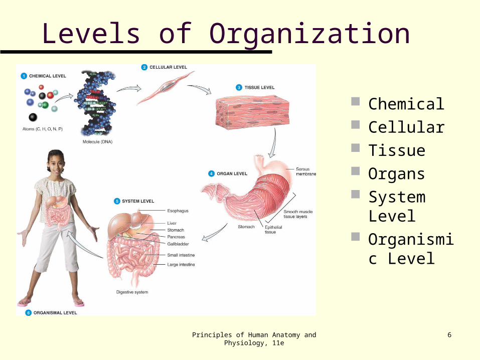
Levels of Organization
Chemical Cellular Tissue Organs System
Level Organismic
Level
Principles of Human Anatomy and Physiology, 11e
7
LEVELS OF ORGANIZATION The human body consists of several levels of
structural organization (Figure 1.1). The chemical level
atoms, the smallest units of matter that participate in chemical reactions, and molecules, two or more atoms joined together.
Cells the basic structural and functional units of an
organism. (Cell Theory) Tissues
groups of similarly specialized cells and the substances surrounding them that usually arise from common embryological tissue and perform certain special functions.
Principles of Human Anatomy and Physiology, 11e
8
LEVELS OF ORGANIZATION Tissues
groups of similarly specialized cells and the substances surrounding them that usually arise from a common ancestor and perform certain special functions.
Organs structures of definite form that are composed of
two or more different tissues and have specific functions.
Systems related organs that have a common function.
The human organism a collection of structurally and functionally
integrated systems; any living individual.
Principles of Human Anatomy and Physiology, 11e
9
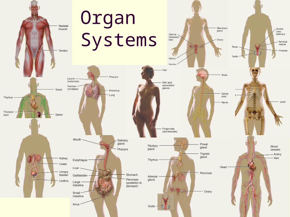
Organ Systems
Principles of Human Anatomy and Physiology, 11e
LEVELS OF ORGANIZATION
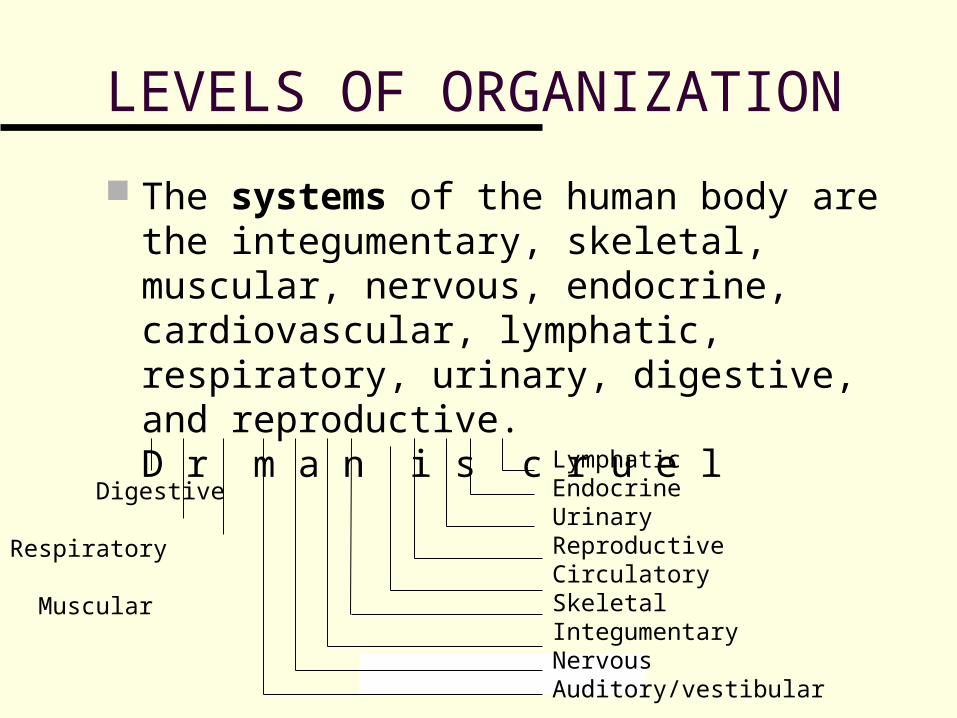
The systems of the human body are the integumentary, skeletal, muscular, nervous, endocrine, cardiovascular, lymphatic, respiratory, urinary, digestive, and reproductive.D r m a n i s c r u e l
Digestive Respiratory Muscular
LymphaticEndocrineUrinaryReproductiveCirculatorySkeletalIntegumentaryNervousAuditory/vestibular
11
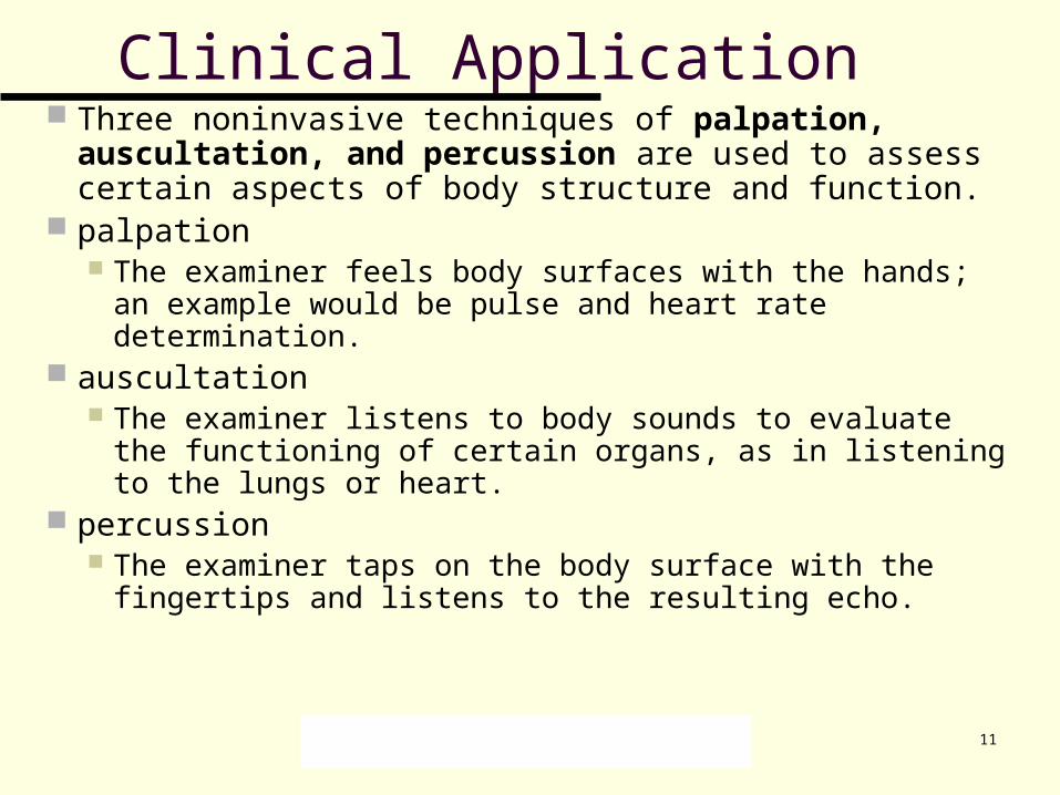
Clinical Application Three noninvasive techniques of palpation, auscultation,
and percussion are used to assess certain aspects of body structure and function.
palpation The examiner feels body surfaces with the hands; an
example would be pulse and heart rate determination. auscultation
The examiner listens to body sounds to evaluate the functioning of certain organs, as in listening to the lungs or heart.
percussion The examiner taps on the body surface with the fingertips
and listens to the resulting echo.
Principles of Human Anatomy and Physiology, 11e
12
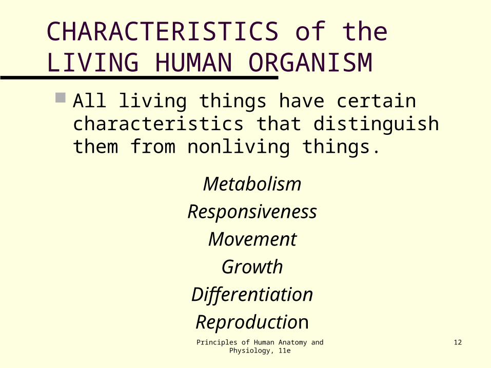
CHARACTERISTICS of the LIVING HUMAN ORGANISM All living things have certain characteristics
that distinguish them from nonliving things.
Metabolism
Responsiveness
Movement
Growth
Differentiation
Reproduction
Principles of Human Anatomy and Physiology, 11e
13
Basic Life Processes All living things have certain characteristics that
distinguish them from nonliving things. Metabolism is the sum of all chemical processes
that occur in the body, including catabolism and anabolism.
Responsiveness is the ability to detect and respond to changes in the external or internal environment.
Movement includes motion of the whole body, individual organs, single cells, or even organelles inside cells.
Principles of Human Anatomy and Physiology, 11e
14
Basic Life Processes Growth refers to an increase in size and
complexity, due to an increase in the number of cells, size of cells, or both.
Differentiation is the change in a cell from an unspecialized state to a specialized state.
Reproduction refers either to the formation of new cells for growth, repair, or replacement, or the production of a new individual.
An autopsy (see text) is a postmortem examination of the body and dissection of its internal organs to confirm or determine the cause of death.
Principles of Human Anatomy and Physiology, 11e
15
HOMEOSTASIS
Homeostasis is a condition of equilibrium in the body’s internal environment produced by the ceaseless interplay of all the body’s regulatory processes.
Principles of Human Anatomy and Physiology, 11e
16
Homeostasis
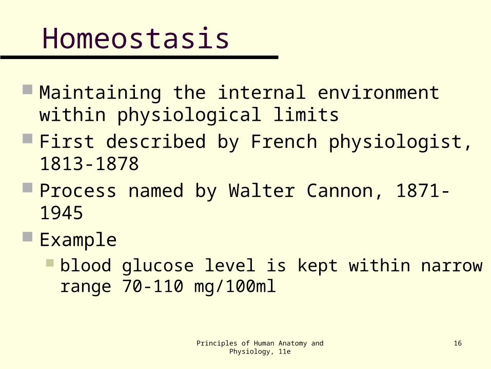
Maintaining the internal environment within physiological limits
First described by French physiologist, 1813-1878 Process named by Walter Cannon, 1871-1945 Example
blood glucose level is kept within narrow range 70-110 mg/100ml
Principles of Human Anatomy and Physiology, 11e
17
Body Fluids For the body’s cells to survive, the composition of
the surrounding fluids must be precisely maintained at all times.
Fluid inside body cells is called intracellular fluid. Fluid outside body cells is called extracellular fluid
(ECF) and is found in two principal places. ECF filling the narrow spaces between cells of
tissues is called interstitial fluid, intercellular fluid, or tissue fluid.
ECF in blood vessels is termed plasma. Since ECF is in constant motion throughout the
body and also surrounds all body cells, it is often called the body’s internal environment.
Principles of Human Anatomy and Physiology, 11e
18
Control of Homeostasis
Homeostasis is continually being disrupted by external stimuli
intense heat, cold , and lack of oxygen internal stimuli
psychological stresses exercise
Disruptions are usually mild & temporary If homeostasis is not maintained, death may result
Principles of Human Anatomy and Physiology, 11e
19
CONTROL OF HOMEOSTASIS Homeostatic imbalances occur because of
disruptions from the external or internal environments. Homeostasis is regulated by the nervous system
and endocrine system, acting together or independently.
The nervous system detects changes and sends nerve impulses to counteract the disruption.
The endocrine system regulates homeostasis by secreting hormones.
Whereas nerve impulses cause rapid changes, hormones usually work more slowly.
Examples: CO2, O2, temperature, pH, blood pressure, …
Principles of Human Anatomy and Physiology, 11e
20
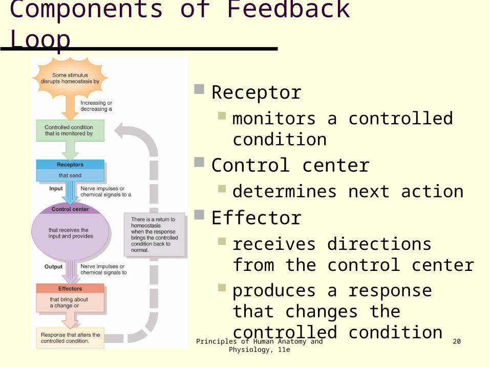
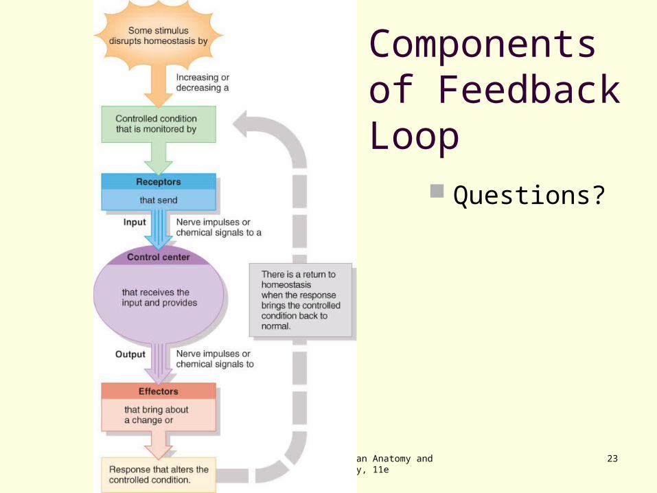
Components of Feedback Loop
Receptor monitors a controlled condition
Control center determines next action
Effector receives directions from the
control center produces a response that
changes the controlled condition
Principles of Human Anatomy and Physiology, 11e
21
Feedback Systems
General Principles A feedback system is a cycle of events in
which information about the status of a condition is continually monitored and fed back (reported) to a central control region (Figure 1.2).
Any disruption that changes a controlled condition is called a stimulus.
Principles of Human Anatomy and Physiology, 11e
22
Feedback Systems
A feedback system consists of three basic components. A receptor monitors changes in a controlled condition
and sends input in the form of nerve impulses or chemical signals to a control center.
The control center sets the range of values within which a controlled condition should be maintained, evaluates the input it receives from the receptors, and generates output commands when they are needed.
An effector is a body structure that receives output from the control center and produces a response or effect that changes the controlled condition.
Principles of Human Anatomy and Physiology, 11e
23
Components of Feedback Loop
Questions?
Principles of Human Anatomy and Physiology, 11e
24
Feedback Systems
If a response reverses the original stimulus, the system is a negative feedback system.
If a response enhances the original stimulus, the system is a positive feedback system.
Principles of Human Anatomy and Physiology, 11e
25
Negative Feedback Systems
A negative feedback system reverses a change in a controlled condition.
Homeostasis of Blood Pressure (BP): Negative Feedback (Figure 1.3) The activity of the effector produces a result, a
drop in blood pressure, that opposes the stimulus, an increase in blood pressure.
Principles of Human Anatomy and Physiology, 11e
26
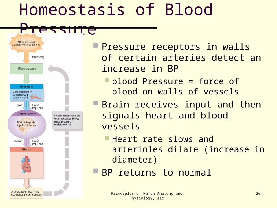
Homeostasis of Blood Pressure
Pressure receptors in walls of certain arteries detect an increase in BP blood Pressure = force of blood
on walls of vessels Brain receives input and then
signals heart and blood vessels Heart rate slows and arterioles
dilate (increase in diameter) BP returns to normal
Principles of Human Anatomy and Physiology, 11e
27
Positive Feedback System
Normal childbirth provides a good example of a positive feedback system (Figure 1.4).
The positive feedback system reinforces a change in a controlled condition.
Principles of Human Anatomy and Physiology, 11e
28
Positive Feedback during Childbirth
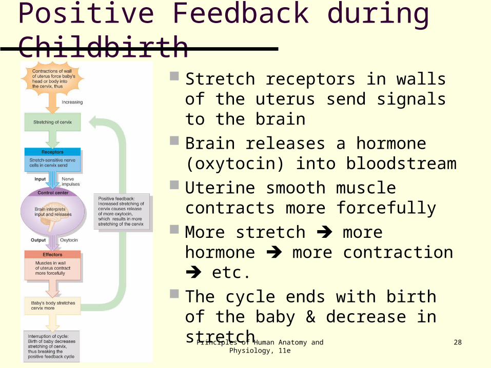
Stretch receptors in walls of the uterus send signals to the brain
Brain releases a hormone (oxytocin) into bloodstream
Uterine smooth muscle contracts more forcefully
More stretch more hormone more contraction etc.
The cycle ends with birth of the baby & decrease in stretch
Principles of Human Anatomy and Physiology, 11e
29
Homeostatic Imbalances Disruption of homeostasis can lead to disease
and death. Disorder is a general term for any derangement of
abnormality of function. Disease is a more specific term for an illness
characterized by a recognizable set of signs and symptoms. A local disease is one that affects one part or a
limited region of the body. A systemic disease affects either the entire body or
several parts.
Principles of Human Anatomy and Physiology, 11e
30
Homeostatic Imbalances Disease is a more specific term for an illness
characterized by a recognizable set of signs and symptoms. Signs are objective changes that a clinician can
observe and measure; e.g., fever or rash. Symptoms are subjective changes in body
functions that are not apparent to an observer; e.g., headache or nausea.
Diagnosis is the art of distinguishing one disease from another or determining the nature of a disease; a diagnosis is generally arrived at after the taking of a medical history and the administration of a physical examination.
Principles of Human Anatomy and Physiology, 11e
31
Aging and Homeostasis
Aging is characterized by a progressive decline in the body’s responses to restore homeostasis
These changes are apparent in all body systems. crinkled skin, gray hair, loss of bone mass, …
Principles of Human Anatomy and Physiology, 11e
32
BASIC ANATOMICAL TERMINOLOGY
Anatomical position
Regions of the body
Anatomical planes, sections
and directional terms
Principles of Human Anatomy and Physiology, 11e
33
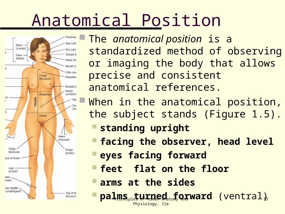
Anatomical Position The anatomical position is a
standardized method of observing or imaging the body that allows precise and consistent anatomical references.
When in the anatomical position, the subject stands (Figure 1.5). standing upright facing the observer, head level eyes facing forward feet flat on the floor arms at the sides palms turned forward (ventral)

Principles of Human Anatomy and Physiology, 11e
34
Reclining Position
If the body is lying face down, it is in the prone position.
If the body is lying face up, it is in the supine position.
Principles of Human Anatomy and Physiology, 11e
35
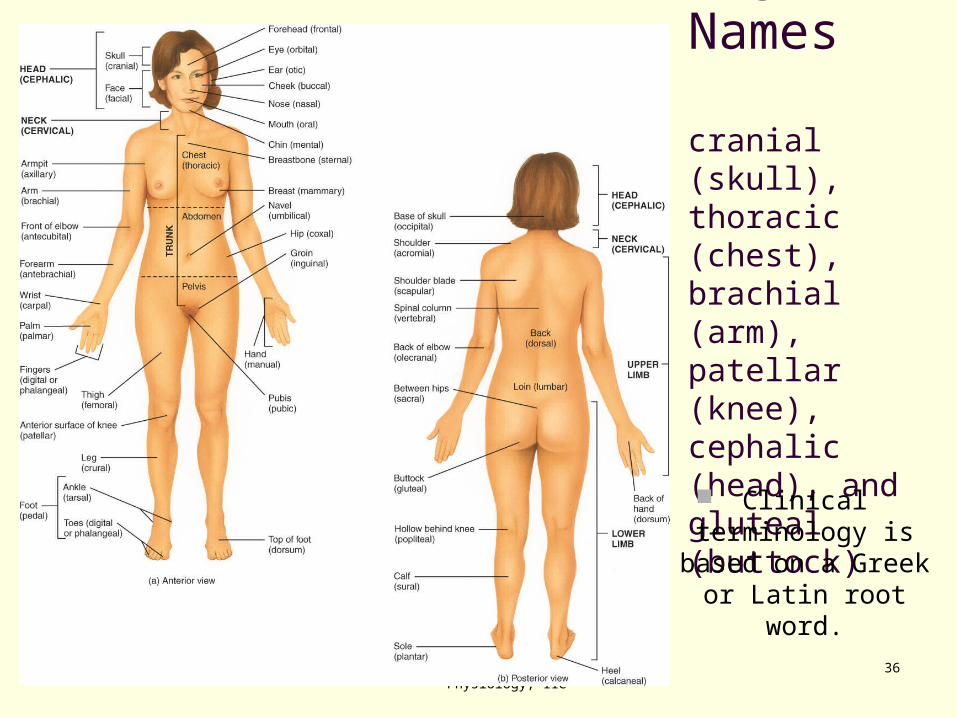
Regional Names
Regional names are names given to specific regions of the body for reference.
Examples of regional names include:
Principles of Human Anatomy and Physiology, 11e
36
Common Regional Names
cranial (skull), thoracic (chest), brachial (arm), patellar (knee), cephalic (head), and gluteal (buttock)
Clinical terminology is
based on a Greek or Latin root word.
Principles of Human Anatomy and Physiology, 11e
37
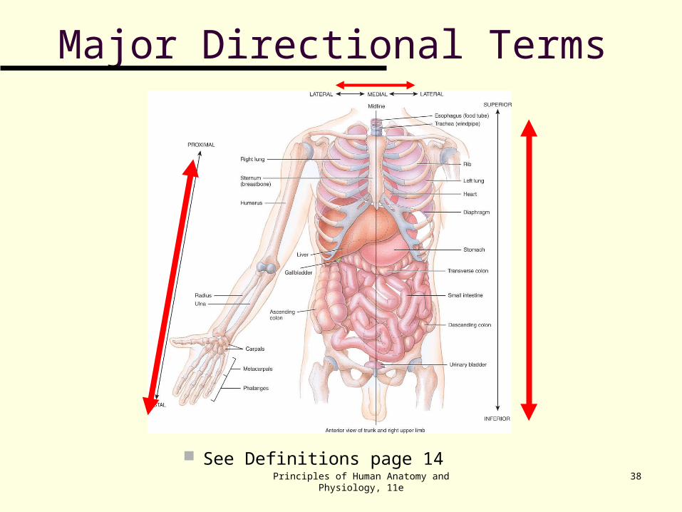
Directional Terms
Directional terms are used to precisely locate one part of the body relative to another and to reduce length of explanations.
Commonly used directional terms: dorsal, superior, medial, and distal summarized in Exhibit 1.1 and Figure 1.6.
Principles of Human Anatomy and Physiology, 11e
38
Major Directional Terms
See Definitions page 14
Principles of Human Anatomy and Physiology, 11e
39
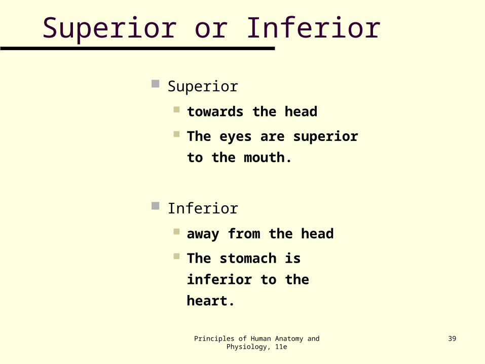
Superior or Inferior
Superior
towards the head
The eyes are superior
to the mouth.
Inferior
away from the head
The stomach is inferior
to the heart.
Principles of Human Anatomy and Physiology, 11e
40
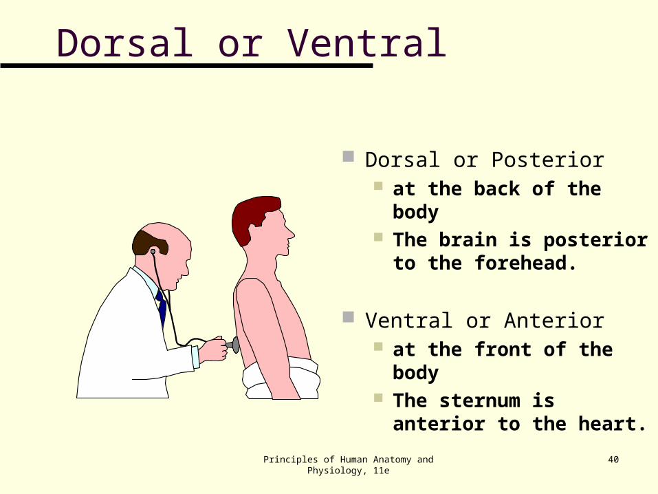
Dorsal or Posterior at the back of the body The brain is posterior to
the forehead.
Ventral or Anterior at the front of the body The sternum is anterior to
the heart.
Dorsal or Ventral
Principles of Human Anatomy and Physiology, 11e
41
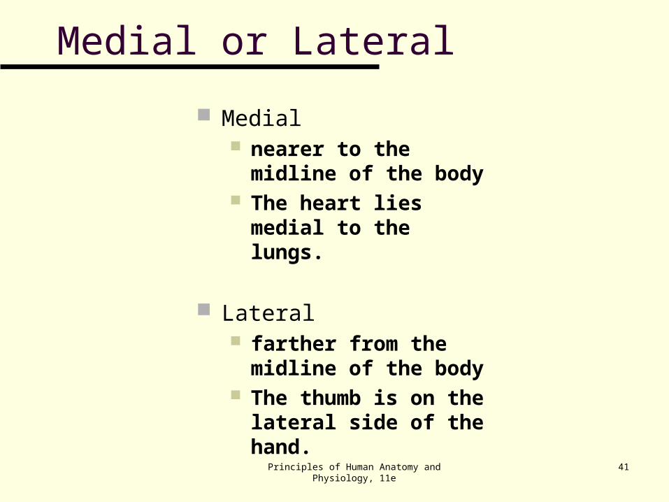
Medial or Lateral
Medial nearer to the midline of
the body The heart lies medial to
the lungs.
Lateral farther from the midline
of the body The thumb is on the
lateral side of the hand.
Principles of Human Anatomy and Physiology, 11e
42
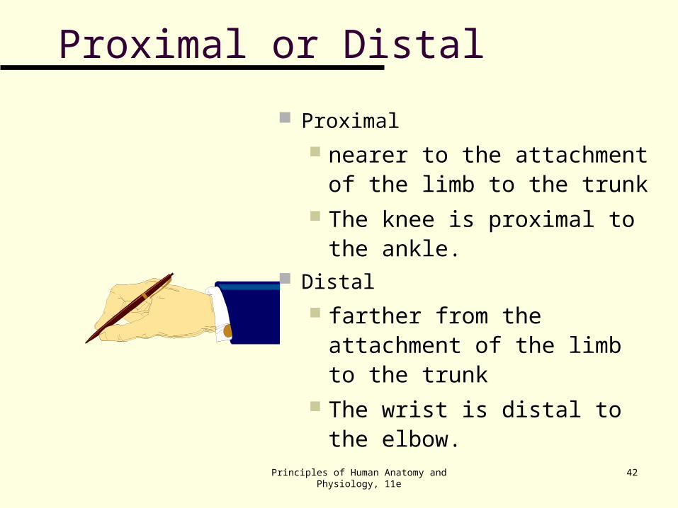
Proximal or Distal
Proximal
nearer to the attachment of the limb to the trunk
The knee is proximal to the ankle.
Distal
farther from the attachment of the limb to the trunk
The wrist is distal to the elbow.
Principles of Human Anatomy and Physiology, 11e
43
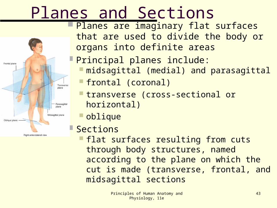
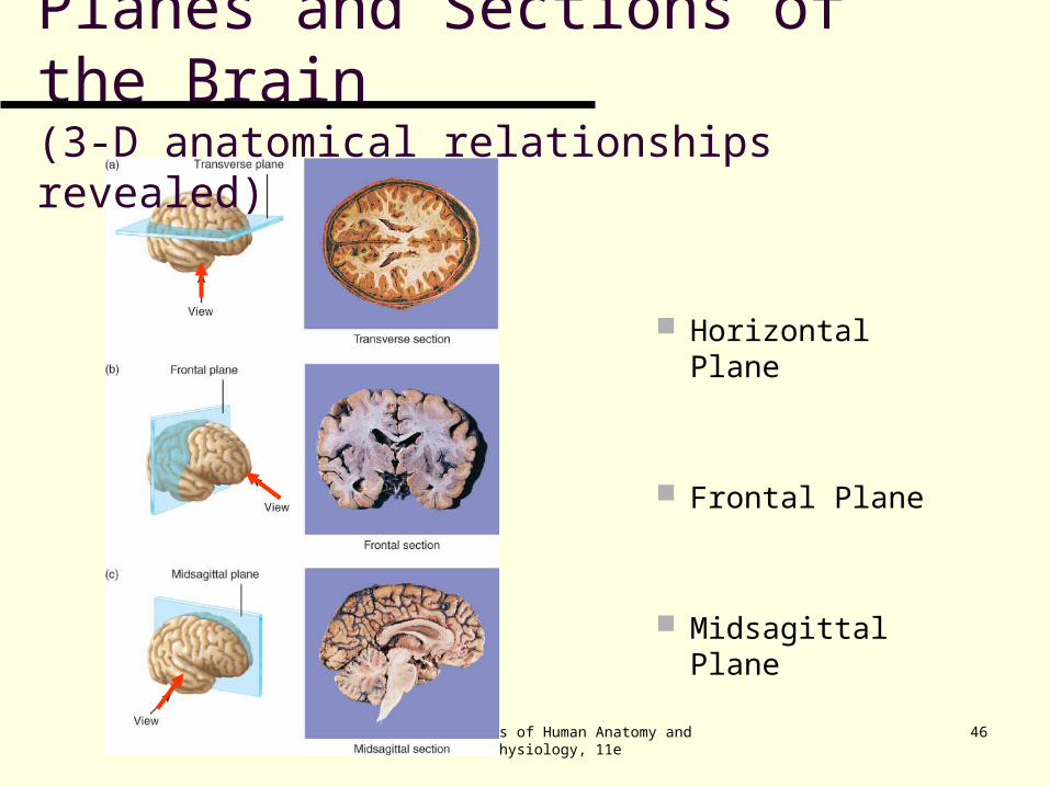
Planes and Sections Planes are imaginary flat surfaces that are
used to divide the body or organs into definite areas
Principal planes include: midsagittal (medial) and parasagittal frontal (coronal) transverse (cross-sectional or horizontal) oblique
Sections flat surfaces resulting from cuts through
body structures, named according to the plane on which the cut is made (transverse, frontal, and midsagittal sections
Principles of Human Anatomy and Physiology, 11e
44
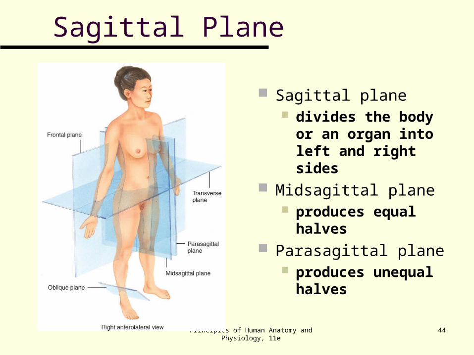
Sagittal Plane
Sagittal plane divides the body or
an organ into left and right sides
Midsagittal plane produces equal
halves Parasagittal plane
produces unequal halves
Principles of Human Anatomy and Physiology, 11e
45
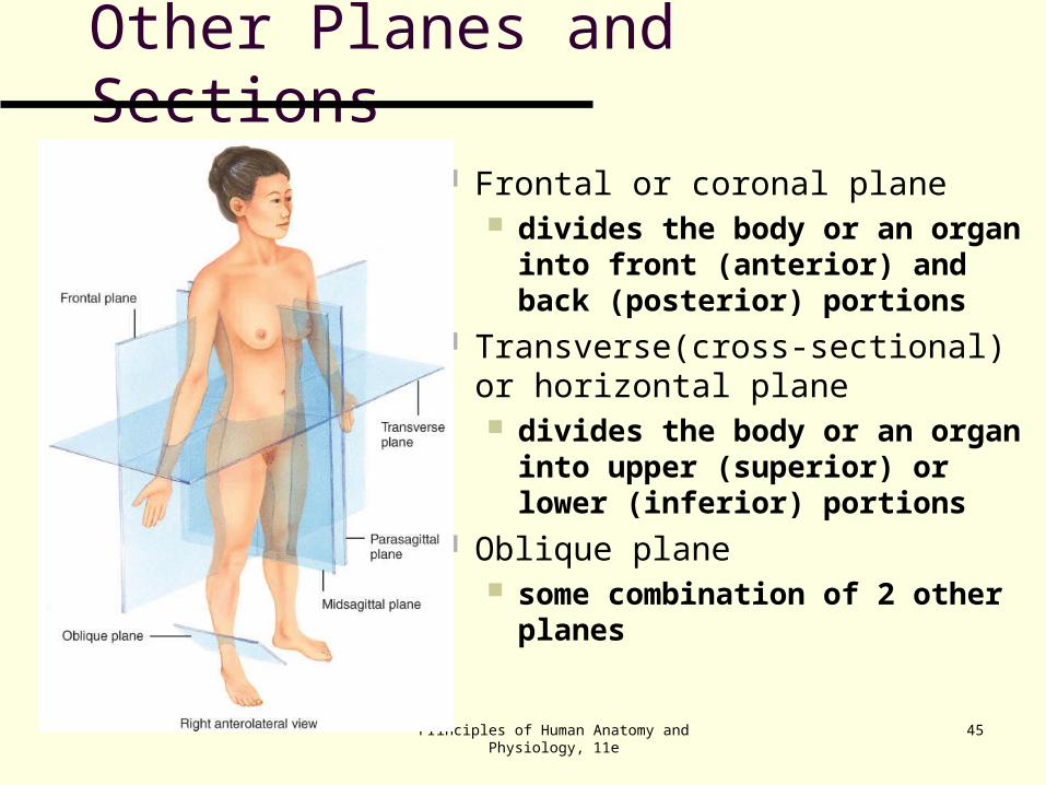
Other Planes and Sections
Frontal or coronal plane divides the body or an organ into
front (anterior) and back (posterior) portions
Transverse(cross-sectional) or horizontal plane divides the body or an organ into
upper (superior) or lower (inferior) portions
Oblique plane some combination of 2 other
planes
Principles of Human Anatomy and Physiology, 11e
46
Planes and Sections of the Brain(3-D anatomical relationships revealed)
Horizontal Plane
Frontal Plane
Midsagittal Plane
Principles of Human Anatomy and Physiology, 11e
47
Body Cavities
Body cavities are spaces within the body that help protect, separate, and support internal organs.
Principles of Human Anatomy and Physiology, 11e
48
Dorsal Body Cavity
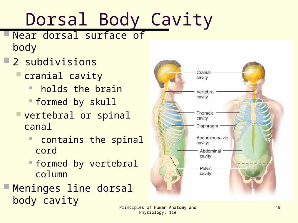
The dorsal body cavity is located near the dorsal surface of the body and has two subdivisions, the cranial cavity and the vertebral canal. (Figure 1.9)
The cranial cavity is formed by the cranial bones and contains the brain.
The vertebral (spinal) canal is formed by the bones of the vertebral column and contains the spinal cord.
Three layers of protective tissue, called meninges, line the dorsal body cavity.
Principles of Human Anatomy and Physiology, 11e
49
Dorsal Body Cavity Near dorsal surface of
body 2 subdivisions
cranial cavity holds the brain formed by skull
vertebral or spinal canal contains the spinal
cord formed by vertebral
column Meninges line dorsal body
cavity
Principles of Human Anatomy and Physiology, 11e
50
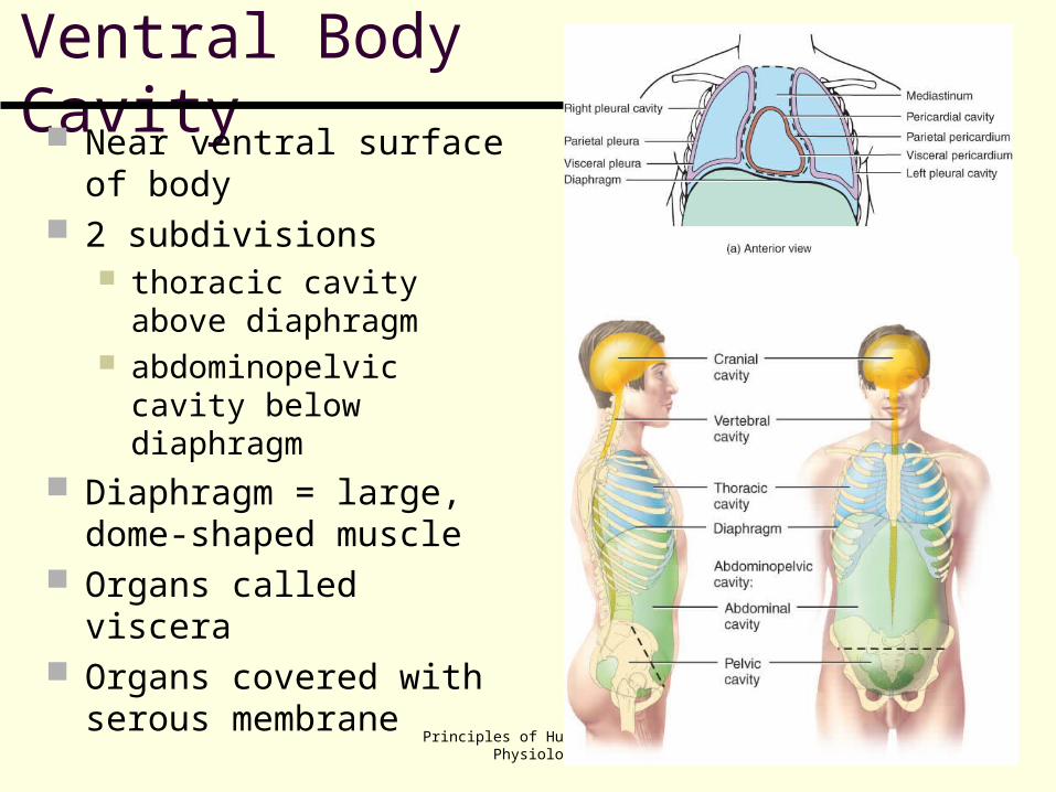
Ventral Body Cavity Near ventral surface of
body 2 subdivisions
thoracic cavity above diaphragm
abdominopelvic cavity below diaphragm
Diaphragm = large, dome-shaped muscle
Organs called viscera Organs covered with
serous membrane
Principles of Human Anatomy and Physiology, 11e
51
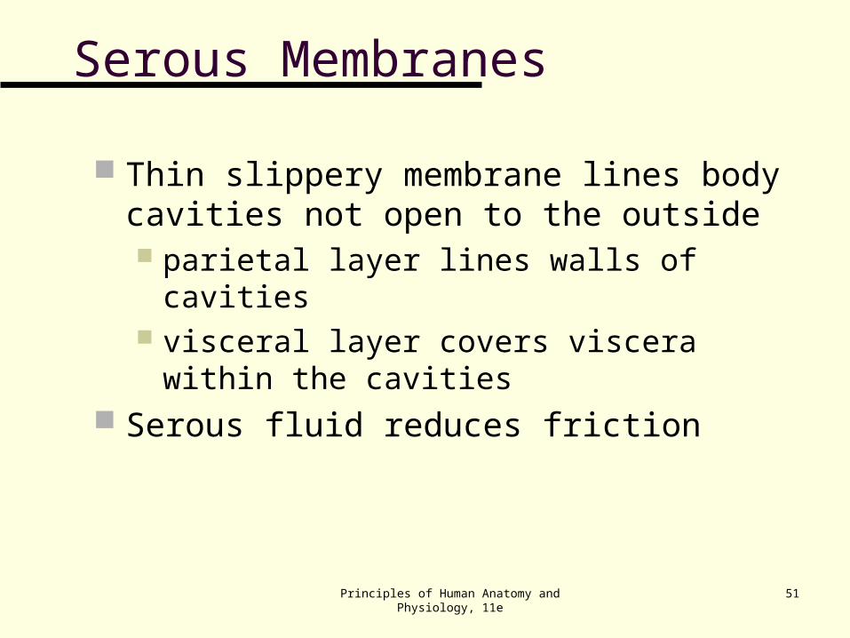
Serous Membranes
Thin slippery membrane lines body cavities not open to the outside parietal layer lines walls of cavities visceral layer covers viscera within the cavities
Serous fluid reduces friction
Principles of Human Anatomy and Physiology, 11e
52
Ventral Body Cavity The thoracic cavity contains two pleural cavities, and
the mediastinum, which includes the pericardial cavity (Figure 1.10). The pleural cavities enclose the lungs. The pericardial cavity surrounds the heart.
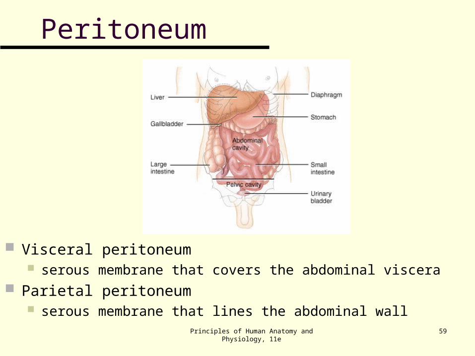
The abdominopelvic cavity is divided into a superior abdominal and an inferior pelvic cavity (Figure 1.9). Viscera of the abdominal cavity include the stomach,
spleen, pancreas, liver, gallbladder, small intestine, and most of the large intestine (Figure 1.11).
Viscera of the pelvic cavity include the urinary bladder, portions of the large intestine and internal female and male reproductive structures.
Principles of Human Anatomy and Physiology, 11e
53
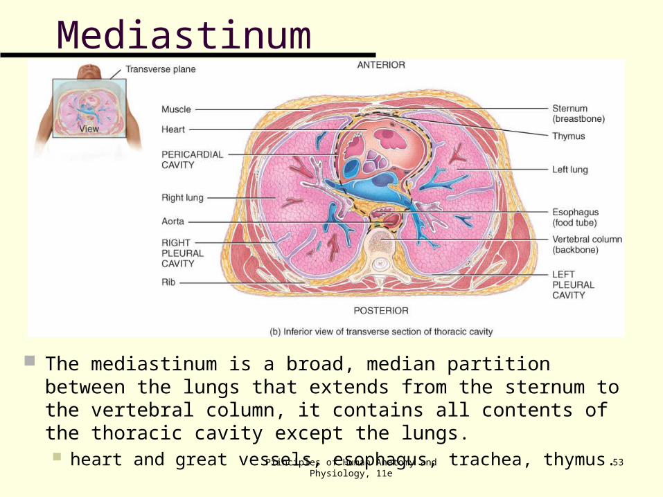
Mediastinum
The mediastinum is a broad, median partition between the lungs that extends from the sternum to the vertebral column, it contains all contents of the thoracic cavity except the lungs. heart and great vessels, esophagus, trachea, thymus.
Principles of Human Anatomy and Physiology, 11e
54
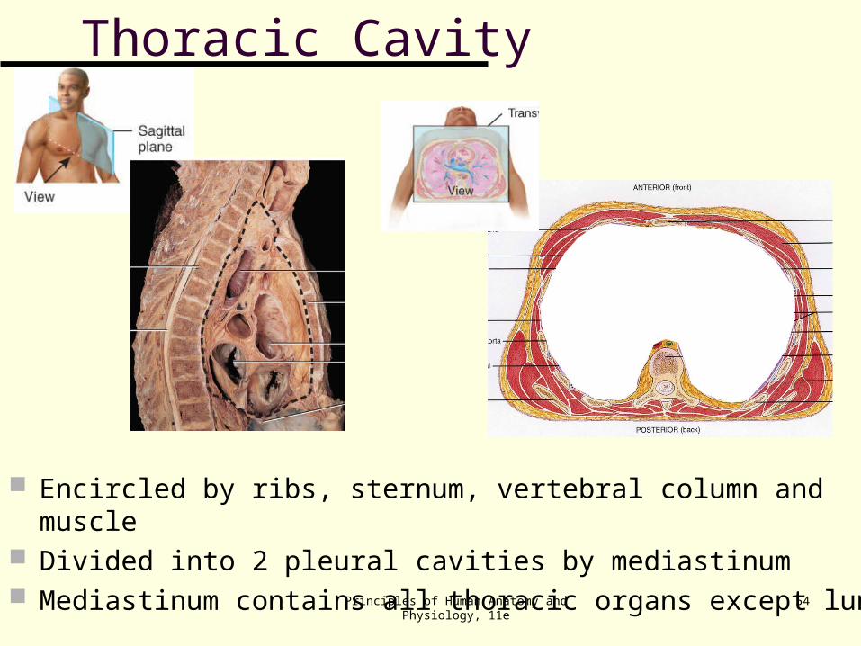
Thoracic Cavity
Encircled by ribs, sternum, vertebral column and muscle Divided into 2 pleural cavities by mediastinum Mediastinum contains all thoracic organs except lungs
Principles of Human Anatomy and Physiology, 11e
55
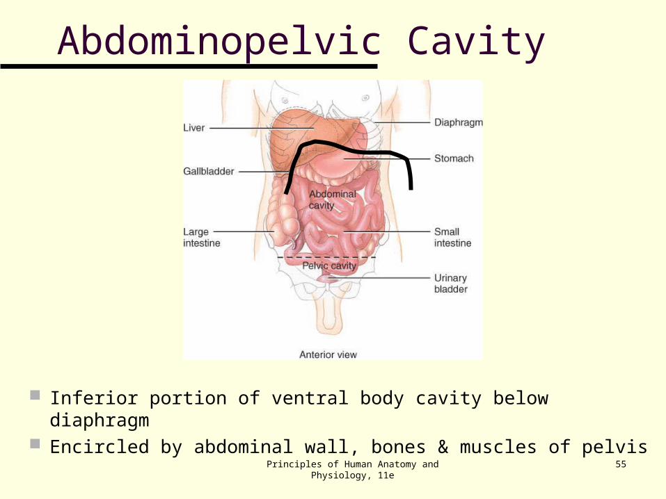
Abdominopelvic Cavity
Inferior portion of ventral body cavity below diaphragm Encircled by abdominal wall, bones & muscles of pelvis
Principles of Human Anatomy and Physiology, 11e
56
Thoracic and Abdominal Cavity Membranes
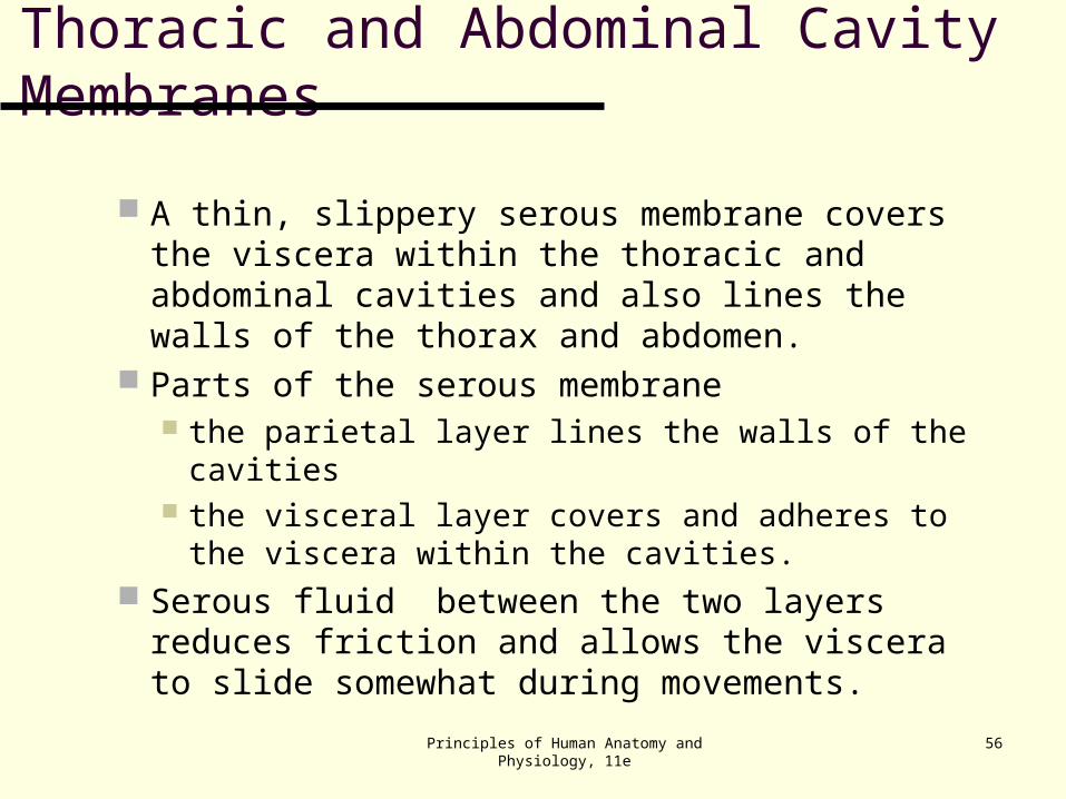
A thin, slippery serous membrane covers the viscera within the thoracic and abdominal cavities and also lines the walls of the thorax and abdomen.
Parts of the serous membrane the parietal layer lines the walls of the cavities the visceral layer covers and adheres to the
viscera within the cavities. Serous fluid between the two layers reduces
friction and allows the viscera to slide somewhat during movements.
Principles of Human Anatomy and Physiology, 11e
57
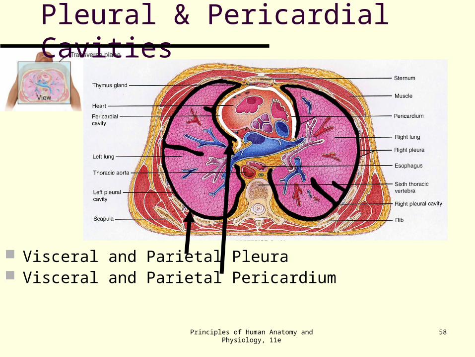
serous membranes
The serous membranes include the pleura, pericardium and peritoneum (Table 1.3).
The pleural membrane surrounds the lungs visceral pleura clings to the surface of the lungs parietal pleura lines the chest wall
The pericardium is the serous membrane of the pericardial cavity visceral pericardium covers the surface of the heart parietal pericardium lines the chest wall
The peritoneum is the serous membrane of the abdominal cavity visceral peritoneum covers the abdominal viscera parietal peritoneum lines the abdominal wall
Principles of Human Anatomy and Physiology, 11e
58
Pleural & Pericardial Cavities
Visceral and Parietal Pleura Visceral and Parietal Pericardium
Principles of Human Anatomy and Physiology, 11e
59
Peritoneum
Visceral peritoneum serous membrane that covers the abdominal viscera
Parietal peritoneum serous membrane that lines the abdominal wall
Principles of Human Anatomy and Physiology, 11e
60
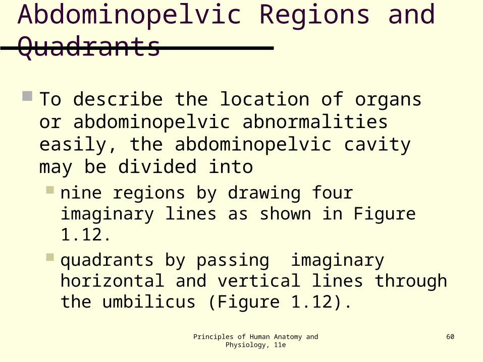
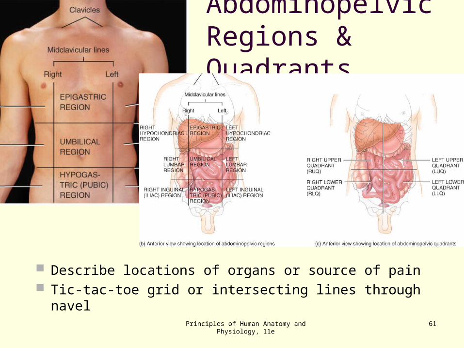
Abdominopelvic Regions and Quadrants
To describe the location of organs or abdominopelvic abnormalities easily, the abdominopelvic cavity may be divided into nine regions by drawing four imaginary lines as
shown in Figure 1.12. quadrants by passing imaginary horizontal and
vertical lines through the umbilicus (Figure 1.12).
Principles of Human Anatomy and Physiology, 11e
61
Abdominopelvic Regions & Quadrants
Describe locations of organs or source of pain Tic-tac-toe grid or intersecting lines through navel
Principles of Human Anatomy and Physiology, 11e
62
Clinical Application: Autopsy
An autopsy is a postmortem examination of the body and dissection of the internal organs to confirm or determine the cause of death.
An autopsy supplies information relating to the deceased individual.
Principles of Human Anatomy and Physiology, 11e
63
MEDICAL IMAGING
A specialized branch of anatomy and physiology that is essential for the diagnosis of many disorders is medical imaging, one division of which is radiography, which includes the use of x-rays.
Medical imaging techniques allow physicians to peer inside the body to provide clues to abnormal anatomy and deviations from normal physiology in order to help diagnose disease.
Table 1.4 describes some commonly used medical imaging techniques.
Principles of Human Anatomy and Physiology, 11e
64
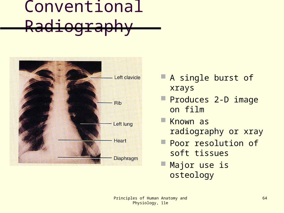
Conventional Radiography
A single burst of xrays Produces 2-D image
on film Known as
radiography or xray Poor resolution of soft
tissues Major use is
osteology
Principles of Human Anatomy and Physiology, 11e
65
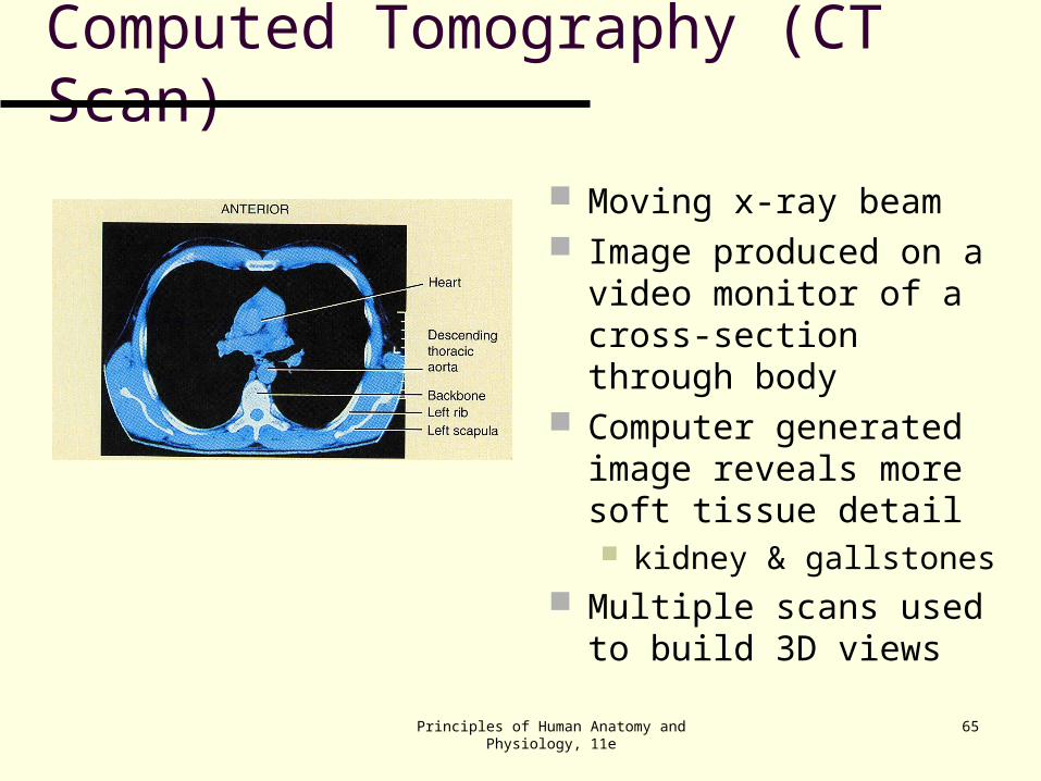
Computed Tomography (CT Scan)
Moving x-ray beam Image produced on a
video monitor of a cross-section through body
Computer generated image reveals more soft tissue detail kidney & gallstones
Multiple scans used to build 3D views
Principles of Human Anatomy and Physiology, 11e
66
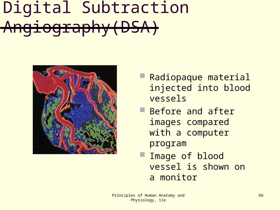
Digital Subtraction Angiography(DSA)
Radiopaque material injected into blood vessels
Before and after images compared with a computer program
Image of blood vessel is shown on a monitor
Principles of Human Anatomy and Physiology, 11e
67
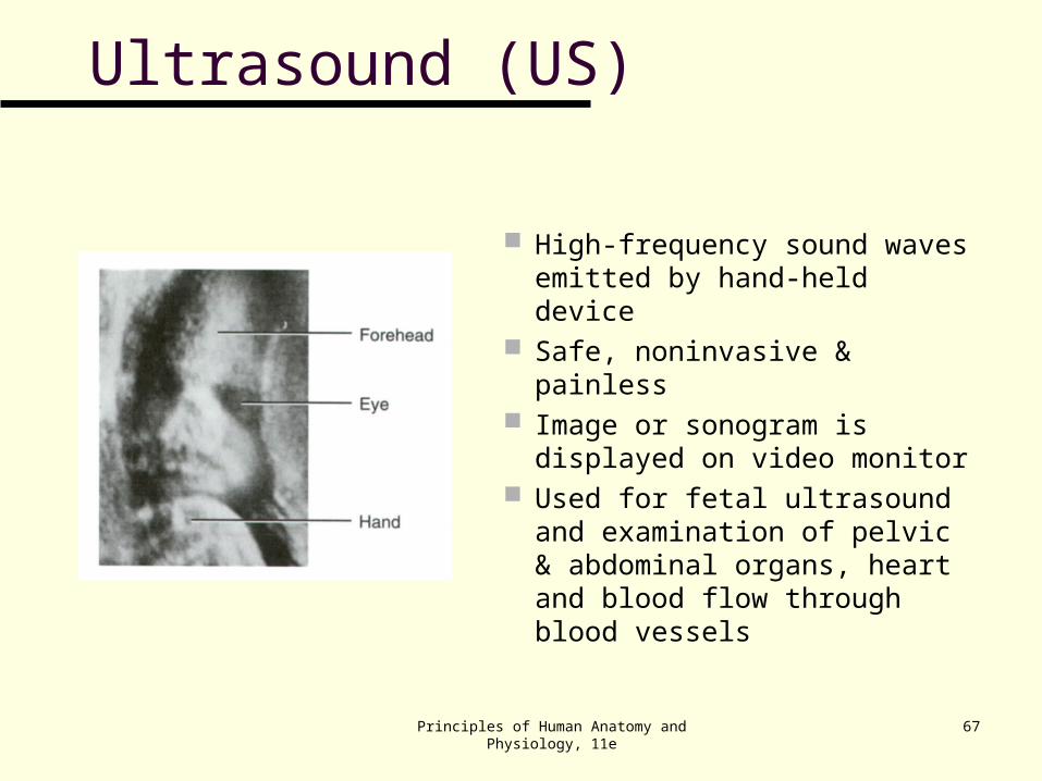
Ultrasound (US)
High-frequency sound waves emitted by hand-held device
Safe, noninvasive & painless Image or sonogram is
displayed on video monitor Used for fetal ultrasound and
examination of pelvic & abdominal organs, heart and blood flow through blood vessels
Principles of Human Anatomy and Physiology, 11e
68
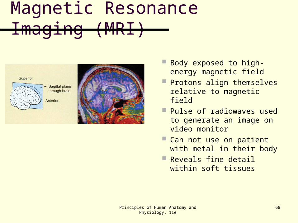
Magnetic Resonance Imaging (MRI)
Body exposed to high-energy magnetic field
Protons align themselves relative to magnetic field
Pulse of radiowaves used to generate an image on video monitor
Can not use on patient with metal in their body
Reveals fine detail within soft tissues
Principles of Human Anatomy and Physiology, 11e
69
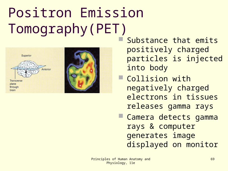
Positron Emission Tomography(PET) Substance that emits
positively charged particles is injected into body
Collision with negatively charged electrons in tissues releases gamma rays
Camera detects gamma rays & computer generates image displayed on monitor