Embed Size (px)
Citation preview

PULMONARY CIRCULATION AND RIGHT VENTRICULAR FUNCTION IN EXPERIMENTAL
ACUTE HIGH-ALTITUDE PULMONARY EDEMA
E. M. Ismailov
KEY WORDS:
UDC 616.24-005.98-092.9-02:612.275. 1]-07:[616.24-005+616.124.3-008.1
hemodynamic shift; pulmonary edema; circulation; heart.
One of the gaps in our knowledge of the pathogenesis of acute high-altitude pulmonary edema (AAPE) is uncertainty regarding the role of the different parts of the heart in the gen-
esis of the circulatory disturbances [6-8].
The object of this investigation was to study the functional load on the left and right sides of the heart in this syndrome and to examine the causes of the hemodynamic disturbances
in the pulmonary circulation.
EXPERIMENTAL METHOD
Experiments were carried out on 20 male chinchilla rabbits weighing 2.9-4.6 kg. The ani- mals were kept in a climatic pressure chamber at an "altitude" of 5.5-6 km, where the meteoro- logical factors corresponding to this altitude were reproduced (low temperature, wind velocity, air humidity, UV irradiation). To assess cardiovascular function, the ECG was recorded in standard lead II on the 6NEK-401 apparatus, the left and right sides of the heart and the pul- monary artery and aorta were catheterized, and the blood pressure recorded by means of electro- manometers (Mingograph-81, from Elema, Sweden). The velocity of the blood flow was measured in the right heart-ear sector by injecting dye and noting its appearance by means of transdu- cers of an oxyhemograph attached to the lobe of the ear. The central blood volume also was measured by the dye dilution method [2]. The parameters studied were measured every 60 min for 6 h. To determine the degree of pulmonary edema, the animals'were decapitated at the end of the experiments and the relative weight of the lungs and their dry residue were determined
[I] and histological sections prepared.
EXPERIMENTAL RESULTS
When the animals were taken up to a high altitude increased activity of the oxygen supply systems was observed. The heart rate was high throughout the period of exposure, 10-15% above
the initial level. Both the systolic blood volume and the cardiac output (CO) were increased and did not fall until the 300th minute of observation.
With an increase in CO, the arterial pressure also rose a little in the systemic circula- tion. The diastolic pressure reached its peak after 360 min, whereas the systolic pressure had a tendency to fall. These changes are evidence of compensatory changes aimed at retaining a large volume of blood in the peripheral vessels, on account of overfilling of the pulmonary circulation. As Table 1 shows, there was a parallel increase in the pressure in the pulmonary artery on account of its constriction. This measure for preventing a considerable inflow of blood into the pulmonary circulation was unable to reduce the central blood volume. Measure- ment of its values showed a gradual rise in this parameter, especially toward the end of expo-
sure, when the pulmonary blood volume was more than twice its initial value.
It is difficult to explain the hemodynanic phenomena described above simply by adaptive opening of reserve capillaries and an increase in the pulmonary vascular bed during hypoxemia. The possibility of changes in tone of the pulmonary veins may perhaps be assumed to have con- tributed under these circumstances to the increase in the pulmonary blood volume and the con- gestive manifestations in the lungs, although there is no direct evidence in support of such an assumption. It is logical to presume that they may have constricted in response to under-
Combined Laboratory of Experimental Cardiology, Kirghiz Research Institute of Cardiology, and Central Research Laboratory, Kirghiz Medical Institute, Frunze. (Presented by Academician of the Academy of M~dical Sciences of the USSR A. M. Chernukh.) Translated from Byulleten' Eksperimental'noi Biologii i Meditsiny, Vol. 94, No. 8, pp. 18-20, August, 1982. Original
article submitted April 7, 1982.
0007-4888/82/9408-1031507.50 �9 1983 Plenum Publishing Corporation 1031
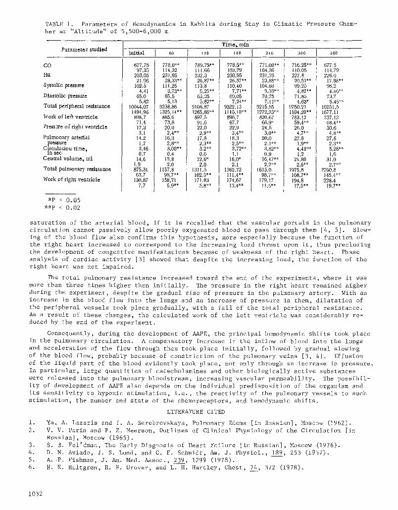
TABLE I. Parameters of Hemodynamics in Rabbits during Stay in Climatic Pressure Cham- ber at "Altitude" of 5,500-6,000 m
Time,, min Parametex studied
initial 60 120 J 8o 24 D 300 360
CO
HR
Systolic pressure
Diastolic pressure
Total peripheral resistance
Work of left ventricle
Pressure of right ventricle
Pulmonary arterial pressure
Circulation time, in see
Central volume, ml
Total pulmonary resistance
Work of right ventricle
677,75 97,35
203,05 21,96
102,5 4,41
65,0 5,82
10044,07 1494,96 898,7 71,4 17,3 3,1
14,2 1,7 3,46 0,7
14,6 1,9 875,31 63,7
130,87 7,7
778,0** 114,32 231,95 28,33**
111,25 3,73**
65,5 5,13
9238,86 1325,14'* 885,6 73,8 20,0 2,4**
16;3 2,8** 3,00"* 0,4
13,8 2,0
1157,8 98,7 ~*
158,71 6,9**
789,75 ~'* 111,66 232,3 26,87**
113,8 5,25**
63,35 5,82**
9106,87 1265,86'* 897, 5 91,6 22,0 2,9**
17,8 2,3 *~ 3,2** 0,6
12,6" 2,0
1311,5 102,5'* 171,83
5,8**
778,5** 103,79 230,95 26,57**
110,40 7,71"*
69,05 7,24**
9322,13 1115,10"* 896,7 67,7 22,9 3,4**
18,3 2,5** 3,72** 1,1
16,0' 2,1
1362,73 111,4"* 174,67 13,4"-*
771,00"* 104,36 231,25 23,88**
104,60 9,33**
70,75 7,11"*
9215,55 1272,23'* 820,67 66,9* 24,5 3,0**
20,0 2,t** 3,82** 0,9
16,47"* 2,2**
1615,0 98,7**
179,17 tt,6"*
716,25"* 110,05 227,8 20,51"* 99,25 4,82**
71,85 4,63*
9750,21 1594,20 * * 783,12
59,4** 26,0 4,7**
22,8 1,9"* 4,41"* 1,2
21,80 2,6**
1975,8 108,7"* 194,8 17,5"*
677,5 114,79 226,0
17,98"* 96,2 4,46**
73,7 5,45**
10251,5 1677,11 737,12 68,4** 30,6 4,8**
27,8 2,3** 5,26** 1,6
31,0 2,7**
2760, 8 145,4"* 228,4
19,7"*
*P < 0.05 **P < 0.02
saturation of the arterial blood, if it is recalled that the vascular portals in the pulmonary circulation cannot passively allow poorly oxygenated blood to pass through them [4, 5]. Slow- ing of the blood flow also confirms this hypothesis, more especially because the function of the right heart increased to correspond to the increasing load thrust upon it, thus precluding the development of congestive manifestations because of weakness of the right heart. Phase analysis of cardiac activity [3] showed that despite the increasing load, the function of the right heart was not impaired.
The total pulmonary resistance increased toward the end of the experiments, where it was more than three times higher than initially. The pressure in the right heart remained higher during the experiment, despite the gradual rise of pressure in the pulmonary artery. With an increase in the blood flow into the lungs and an increase of pressure in them, dilatation of the peripheral vessels took place gradually, with a fall of the total peripheral resistance. As a result of these changes, the calculated work of the left ventricle was considerably re- duced by the end of the experiment.
Consequently, during the development of AAPE, the principal hemodynamic shifts took place in the pulmonary circulation. A compensatory increase in the inflow of blood into the lungs and acceleration of the flow through them took place initially, followed by gradual slowing of the blood flow, probably because of constriction of the pulmonary veins [3, 4]. Effusion of the liquid part of the blood evidently took place, not only through an increas~ in pressure. In particular, large quantities of catecholamines and other biologically active substances were released into the pulmonary bloodstream, increasing vascular permeability. The possibil- ity of development of AAPE also depends on the individual predisposition of the organism and its sensitivity to hypoxic stimulation, i.e., the reactivity of the pulmonary vessels to such stimulation, the number and state of the chemoreceptors, and hemodynamic shifts.
LITERATURE CITED
i. Ya. A. Lazaris and I. A. Serebrovskaya, Pulmonary Edema [in Russian], Moscow (1962). 2. V.V. Parin and F. Z. Meerson, Outlines of Clinical Physiology of the Circulation [in
Russian], Moscow (1965). 3. S. B. Fel'dman, The Early Diagnosis of Heart Failure [in Russian], Moscow (1976). 4. D.M. Aviado, J. S. Lund, and C. F. Schmidt, Am. J. Physiol., 189, 253 (1957). 5. A. -P. Fishman, J. Am. Med. Assoc., 239, 1299 (]978). 6. H.N. Hultgren, R. F. Grover, and L. H. Hartley, Chest, 74, 372 (1978).
1032
7.
8.
D. Penaloza and F. Sime, Bull. Physiol-Pathol. Resp., 4, p. 17 and Discussions, p. 42 (1968). R. Sujoy et al., Br. Heart J., 31, 52 (1969).
INOTROPIC EFFECT OF RHYTHM DISPERSION
V. Ya. Izakov, Yu. L. Protsenko, F. A. Blyakhman, B~ L. Bykov, O. N. Bershitskaya, V. S. Markhasin, L. T. Lysenko, and A. V. Trubetskoi
UDC 612.172.2
KEY WORDS: isometric contraction; rhythm variance; chronotropic and inotropic effects.
Dependence of the force (and other parameters) of contractions on the frequency of stim- ulation (chronotropic and inotropic effects) is an important characteristic of the myocardium which enables it to change its contractility rapidly from cycle to cycle [2, 5]. The type of relationship between frequency and steady-state force in the myocardium of warm- and cold- blooded animals has been shown to be essentially nonlinear [3, 4]. Chronotropic and inotropic ~ffects of the myocardium are manifested not only in the steady-state frequency--force relation- ship, but also in the dependence of force on interstimulus interval, in the form of such well- known phenomena as the extrasystolic reduction in force and its postextrasystolic potentiation. Dependence of the force of contractions on the extrasystolic and postextrasystolic intervals likewise is essentially nonlinear. In such'a nonlinear system, the actual character of the interstimulus sequence may exert an additional effect, manifested as an increase or decrease in the force of contractions. If the stimulus sequence is a random process, as is the case in arrhythmias, the mean force of contractions in a fixed time interval may depend on several statistics of the interstimulus sequence: not only, moreover, on the mean value (which is trivial), but also on the second and third statistical moments (variance, asymmetry), and auto- correlation. Dependence of the force of contractions on rhythm dispersion is of definite prac- tical interest, for the heart under natural conditions (and even more, in pathological arrhyth- mias) works under random conditions [I]. Moreover, investigation of the statistical features of stochastic distributions of R-R intervals on the ECG in some extremal situations and during physiological and pathological arrhythmias has shown that the statistics of the rhythm are
highly sensitive to changes in autonomic regulation [i].
The aim of this investigation was an experimental study of the inotropic effects of rhythm
dispersion.
EXPERIMENTAL METHOD
Experiments were carried out on papillary muscles and trabeculae of rabbit and rat atria. Contractions were recorded under isometric conditions. The composition of the basic solution and other technical details were described previously [2]. The preparations were first stimu- lated at constant frequency (determined rhythm). A random gaussian uncorrelated sequence of pulses of the same mean frequency, but with controllable dispersion, was then applied from a special generator. The coefficient of variation of the rhythm CVT, equal to OT/T (where o T is the standard deviation of interpulse intervals, T the mean interpulse interval) was assigned between 8 and 40%. After a random realization had been recorded with between i00 and 200 cy- cles, a return was made to a steady rhythm. Only those realizations in which the force of con- tractions before and after application of the random rhythm remained constant were used in the
calculstions (realizations with time drift were excluded).
Laboratory of Biophysics, Sverdlovsk Research Institute of Work Hygiene and Occupational Diseases. Laboratory of Regulation of the Heart and Coronary Circulation, All-Union Cardio- logic Scientific Center, Academy of Medical Sciences of the USSR, Moscow. (Presented by Acad- emician of the Academy of Medical Sciences of the USSR I. K. Shkhvatsabaya.) Translated from Byulleten' Eksperimeatal'noi Biologii i Meditsiny, Vol. 94, No. 8, pp. 20-22, August, 1982.
Original article submitted March 16, 1982.
0007-.4888/82/9408-1033507.50 �9 1983 Plenum Publishing Corporation 1033