Embed Size (px)
Citation preview
• Radio activity is important in medicine
because it is used in diagnosing and therapy.
• Therapy is treatment such as treating cancer. Tumours are destroyed by shooting gamma rays at tumours. In this presentation will not be talking about that. We will be specializing in imaging. Radio activity is used for tracing materials around the body. This presentation is going to specialize in diagnostic imaging . Diagnosing means finding out the cause of the persons illness or injury. The images are collected and passed on to the doctor who carries out the correct treatment.
• PROCEDURE AND RADIOACTIVE ISTOPES USED IN NUCLEAR MEDICINE
• HOW NUCLEAR MEDICINE DETECTS DIFFERENT ORGANS
•ADVANTAGES AND ADVANTAGES AND DISADVANTAGES OF DISADVANTAGES OF NUCLEAR MEDICINE NUCLEAR MEDICINE
•THE USES OF THE USES OF NUCLEAR MEDICINENUCLEAR MEDICINE
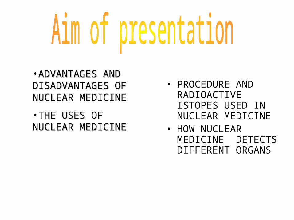
How radio nuclides are produced
• How radio nuclides are produced
• Most medical radio nuclides are prepared by neutrons bombardment in a nuclear reactor.
• Iodine 131 (131I) is an example of a radio nuclide that is produced in this way. Tellurium 136 (136Te) is placed in a nuclear reactor and under the intense bombardment eventually captures a neutron. The resulting nuclide then decays to 131I by the emission of beta radiation as the equation shows:
• 13052Te+1
0n 13153I+0
1 β
Diagram of a nuclear reactor:
Half life & elution of radio nuclides
Technetium 99m If the radio nuclide has a long half
life, the patient and others would be subjected to a continuous radiation dose. A short half life such as the six hour half life of technetium 99m, limits the radiation dose but can give problems of the time required to transport the radio nuclide from the production site to the hospital.
Elution
Saline solution is passed through the column; the technetium which is being produced by the beta decays because of the molybdenum is soluble in saline but the molybdenum itself is not. The process is known as elution and results in a solution of sodium pertechnetate,which is collected in an output vial the content of the output vial can be diluted and split in to a number of patients doses the total amount of radioactivity is goverened by the half life of molybdenum. The generator is eluted daily until the radiation activity concentration falls to the level that is too low to be useful. Which happens after 2 or 3 half lifes this means that delivery of generator to the hospital is necessary on weekly basis.
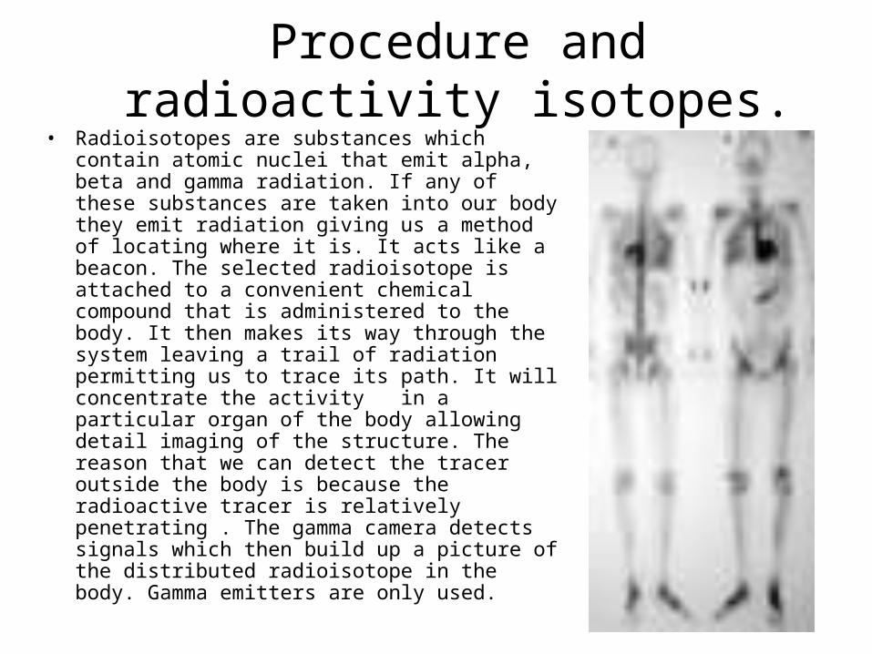
Procedure and radioactivity isotopes.
• Radioisotopes are substances which contain atomic nuclei that emit alpha, beta and gamma radiation. If any of these substances are taken into our body they emit radiation giving us a method of locating where it is. It acts like a beacon. The selected radioisotope is attached to a convenient chemical compound that is administered to the body. It then makes its way through the system leaving a trail of radiation permitting us to trace its path. It will concentrate the activity in a particular organ of the body allowing detail imaging of the structure. The reason that we can detect the tracer outside the body is because the radioactive tracer is relatively penetrating . The gamma camera detects signals which then build up a picture of the distributed radioisotope in the body. Gamma emitters are only used.
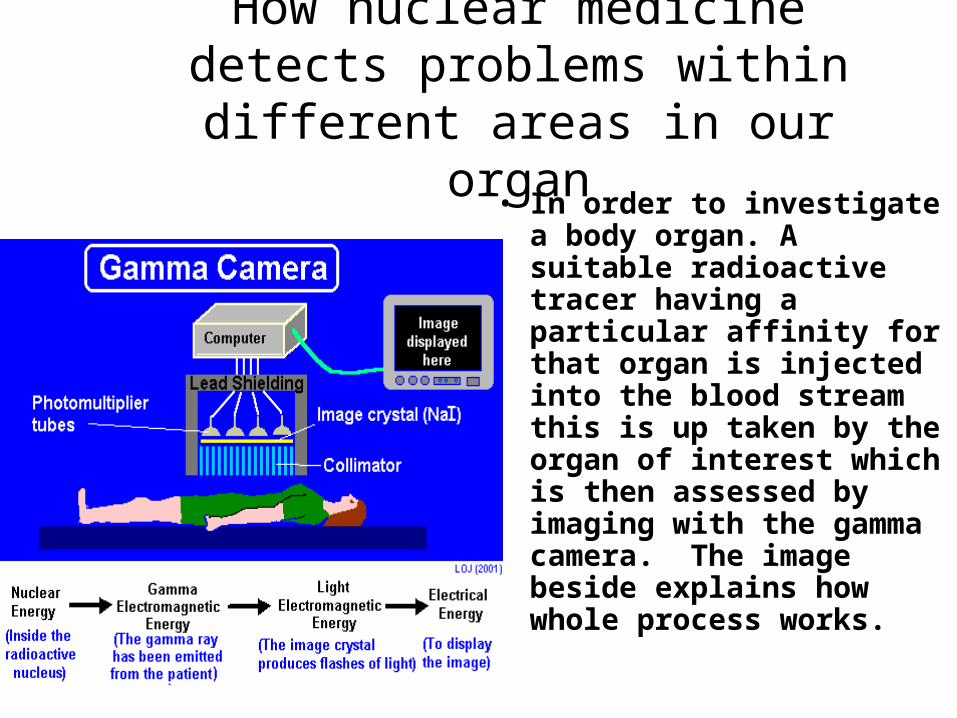
How nuclear medicine detects problems within different areas in our organ
• In order to investigate a body organ. A suitable radioactive tracer having a particular affinity for that organ is injected into the blood stream this is up taken by the organ of interest which is then assessed by imaging with the gamma camera. The image beside explains how whole process works.
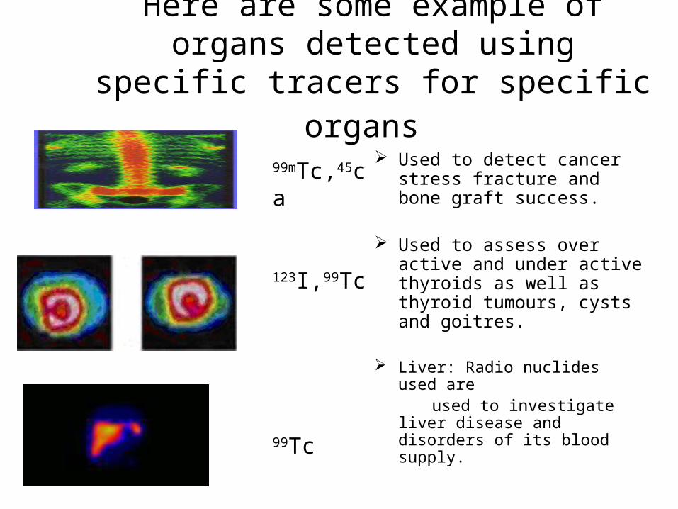
Here are some example of organs detected using specific tracers for specific organs
Used to detect cancer stress fracture and bone graft success.
Used to assess over active and under active thyroids as well as thyroid tumours, cysts and goitres.
Liver: Radio nuclides used are used to investigate liver disease
and disorders of its blood supply.
99mTc,45ca
123I,99Tc
99Tc
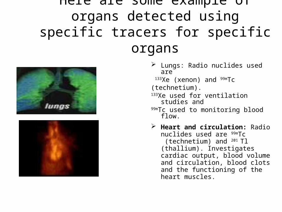
Here are some example of organs detected using specific tracers for specific organs
Lungs: Radio nuclides used are 133Xe (xenon) and 99mTc(technetium).133Xe used for ventilation studies and99mTc used to monitoring blood flow.
Heart and circulation: Radio nuclides used are 99mTc (technetium) and 201
Tl (thallium). Investigates cardiac output, blood volume and circulation, blood clots and the functioning of the heart muscles.
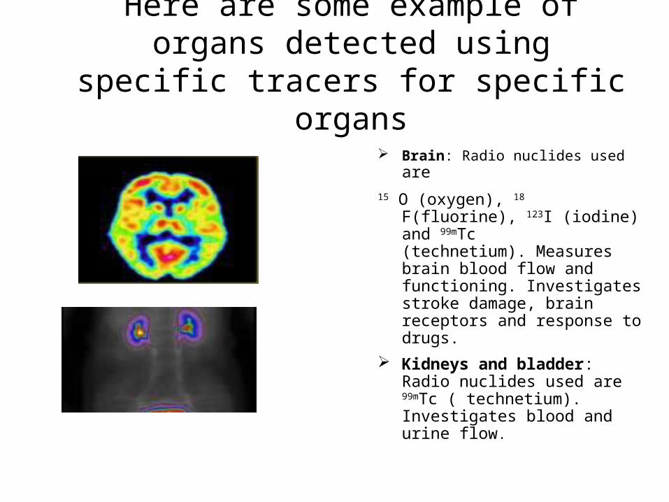
Here are some example of organs detected using specific tracers for specific organs
Brain: Radio nuclides used are 15 O (oxygen), 18 F(fluorine), 123I (iodine)
and 99mTc (technetium). Measures brain blood flow and functioning. Investigates stroke damage, brain receptors and response to drugs.
Kidneys and bladder: Radio nuclides used are 99mTc ( technetium). Investigates blood and urine flow.
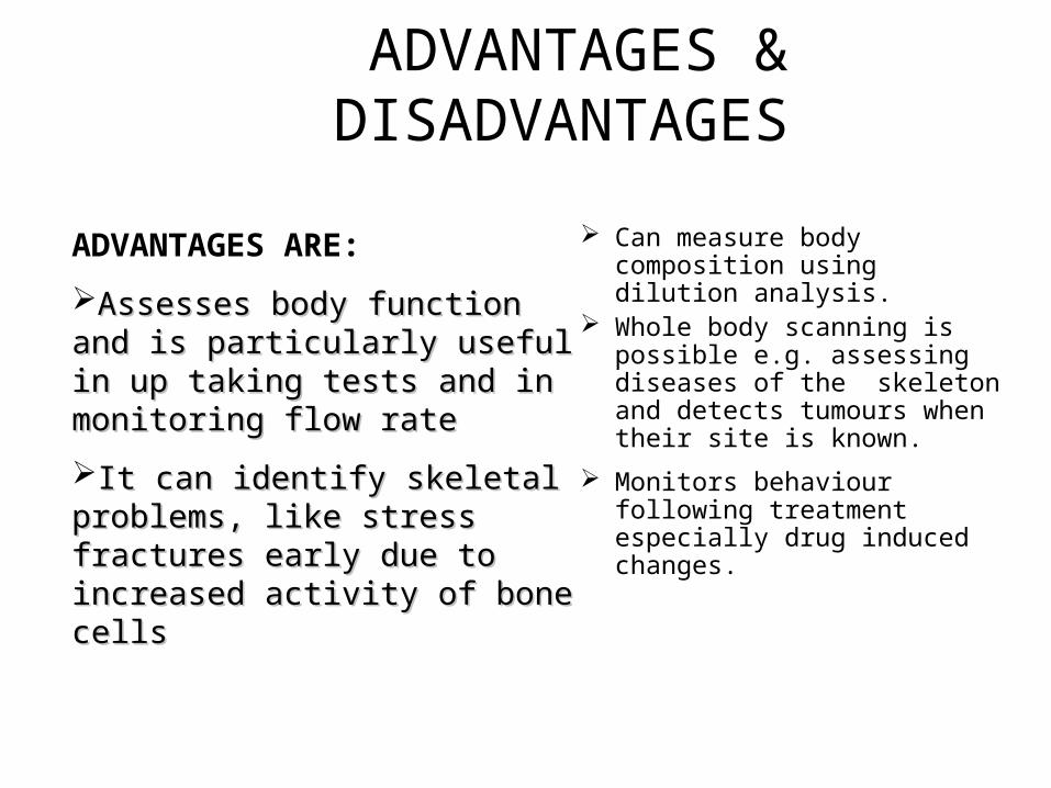
ADVANTAGES & DISADVANTAGES
Can measure body composition using dilution analysis.
Whole body scanning is possible e.g. assessing diseases of the skeleton and detects tumours when their site is known.
Monitors behaviour following treatment especially drug induced changes.
ADVANTAGES ARE:
Assesses body function and Assesses body function and is particularly useful in up is particularly useful in up taking tests and in monitoring taking tests and in monitoring flow rateflow rate
It can identify skeletal It can identify skeletal problems, like stress fractures problems, like stress fractures early due to increased activity early due to increased activity of bone cellsof bone cells
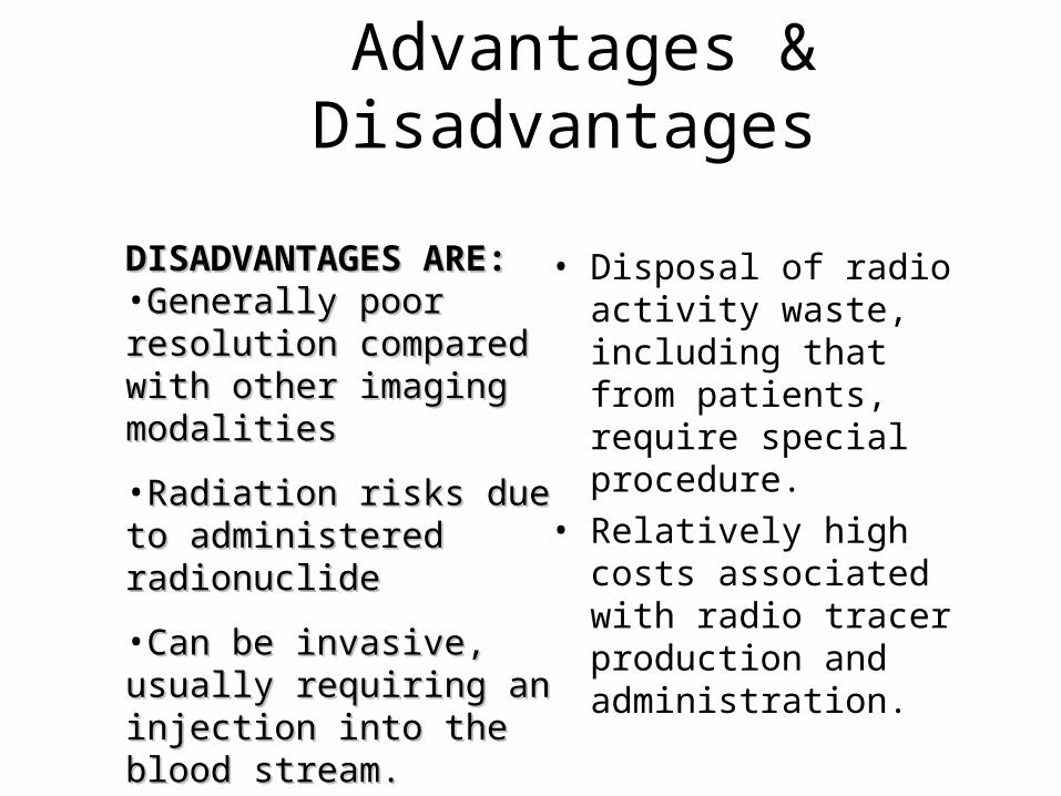
Advantages & Disadvantages
• Disposal of radio activity waste, including that from patients, require special procedure.
• Relatively high costs associated with radio tracer production and administration.
DISADVANTAGES ARE:DISADVANTAGES ARE: •Generally poor Generally poor resolution compared resolution compared with other imaging with other imaging modalitiesmodalities
•Radiation risks due to Radiation risks due to administered administered radionuclide radionuclide
•Can be invasive, Can be invasive, usually requiring an usually requiring an injection into the blood injection into the blood stream. stream.
Safety and risk of Nuclear Medicine
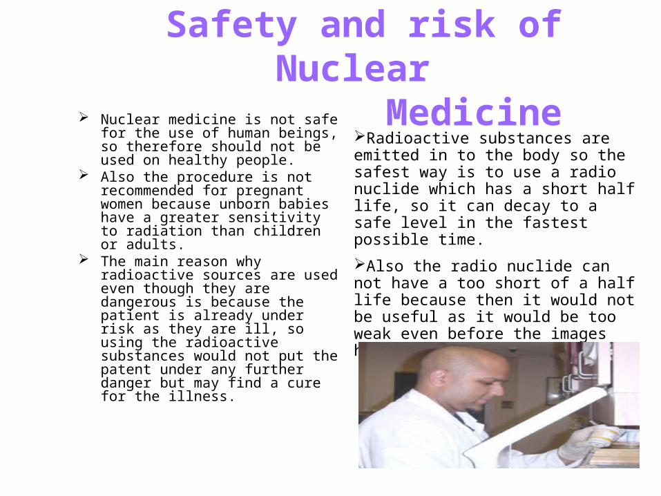
Nuclear medicine is not safe for the use of human beings, so therefore should not be used on healthy people.
Also the procedure is not recommended for pregnant women because unborn babies have a greater sensitivity to radiation than children or adults.
The main reason why radioactive sources are used even though they are dangerous is because the patient is already under risk as they are ill, so using the radioactive substances would not put the patent under any further danger but may find a cure for the illness.
Radioactive substances are emitted in to the body so the safest way is to use a radio nuclide which has a short half life, so it can decay to a safe level in the fastest possible time.
Also the radio nuclide can not have a too short of a half life because then it would not be useful as it would be too weak even before the images have been taken.
Safety and risk of Nuclear Medicine
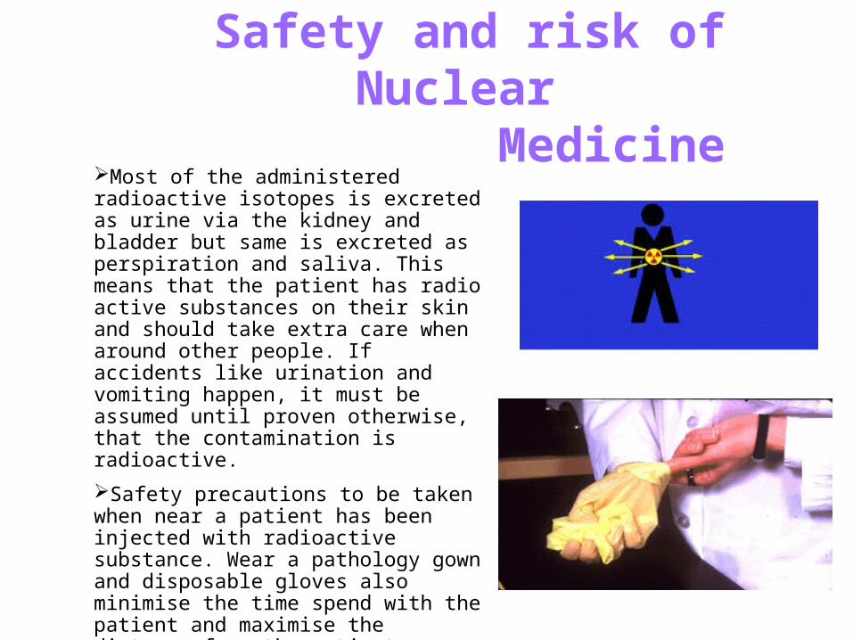
Most of the administered radioactive isotopes is excreted as urine via the kidney and bladder but same is excreted as perspiration and saliva. This means that the patient has radio active substances on their skin and should take extra care when around other people. If accidents like urination and vomiting happen, it must be assumed until proven otherwise, that the contamination is radioactive.
Safety precautions to be taken when near a patient has been injected with radioactive substance. Wear a pathology gown and disposable gloves also minimise the time spend with the patient and maximise the distance from the patient.