Embed Size (px)
Citation preview
Respiratory System
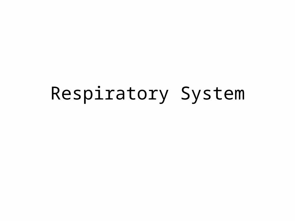
Respiratory System: Overview
Figure 17-2 b: Anatomy Summary
3
Respiration Includes• Pulmonary ventilation
– Air moves in and out of lungs– Continuous replacement of gases in alveoli (air sacs)
• External respiration– Gas exchange between blood and air at alveoli– O2 (oxygen) in air diffuses into blood– CO2 (carbon dioxide) in blood diffuses into air
• Transport of respiratory gases– Between the lungs and the cells of the body– Performed by the cardiovascular system– Blood is the transporting fluid
• Internal respiration– Gas exchange in capillaries between blood and tissue cells– O2 in blood diffuses into tissues– CO2 waste in tissues diffuses into blood
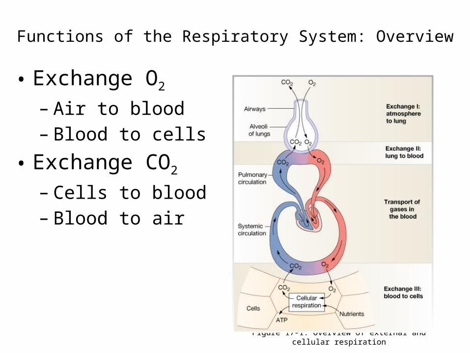
Functions of the Respiratory System: Overview
Figure 17-1: Overview of external and cellular respiration
• Exchange O2
– Air to blood– Blood to cells
• Exchange CO2 – Cells to blood– Blood to air
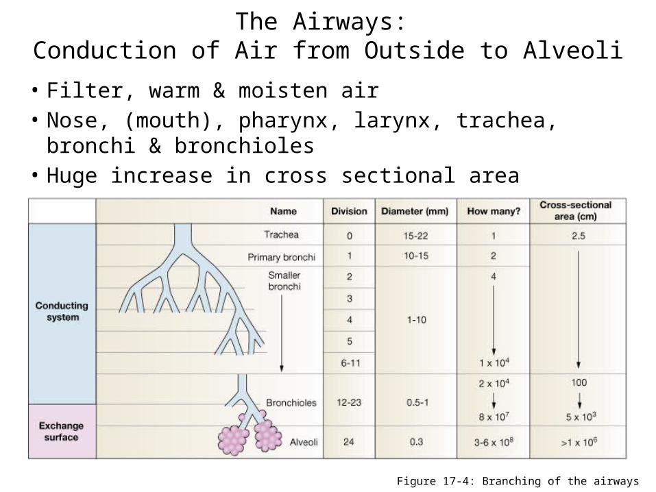
• Filter, warm & moisten air• Nose, (mouth), pharynx, larynx, trachea, bronchi & bronchioles • Huge increase in cross sectional area
The Airways: Conduction of Air from Outside to Alveoli
Figure 17-4: Branching of the airways
6
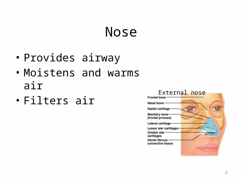
Nose
• Provides airway• Moistens and warms air• Filters air External nose
7
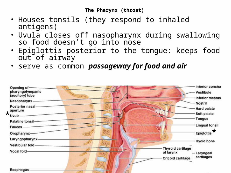
The Pharynx (throat)
• Houses tonsils (they respond to inhaled antigens)• Uvula closes off nasopharynx during swallowing so food doesn’t
go into nose• Epiglottis posterior to the tongue: keeps food out of airway• serve as common passageway for food and air
*
*
8
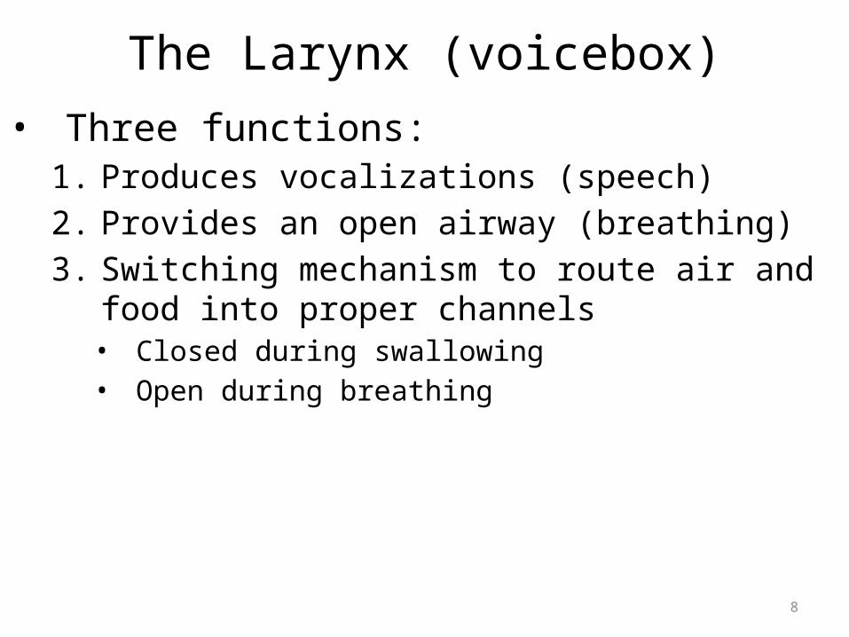
The Larynx (voicebox)• Three functions:
1. Produces vocalizations (speech)2. Provides an open airway (breathing)3. Switching mechanism to route air and food into proper
channels• Closed during swallowing• Open during breathing
9
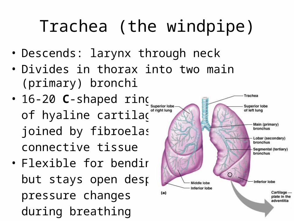
Trachea (the windpipe)• Descends: larynx through neck • Divides in thorax into two main (primary) bronchi• 16-20 C-shaped rings
of hyaline cartilage joined by fibroelastic connective tissue
• Flexible for bendingbut stays open despitepressure changesduring breathing
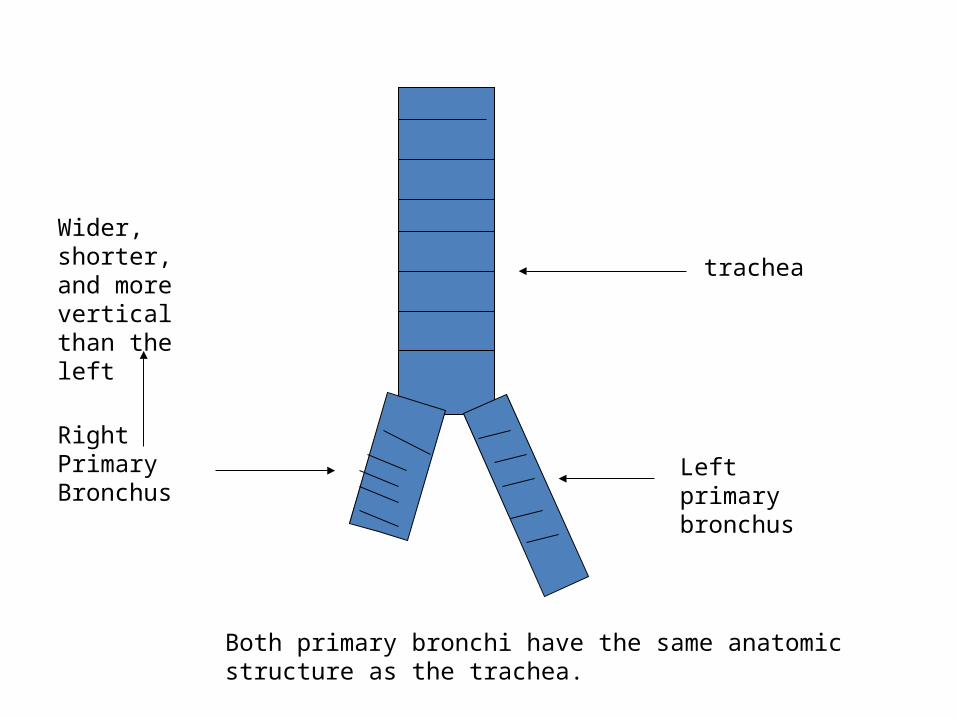
trachea
Left primary bronchus
Right Primary Bronchus
Wider, shorter, and more vertical than the left
Both primary bronchi have the same anatomic structure as the trachea.
Bronchi
• The primary bronchi divide to form SECONDARY BRONCHI
• There is one secondary bronchus for each lobe of the lungs.
• There are 2 lobes on the left lung.• There are 3 lobes on the right lung.• These also have the same anatomy as the trachea.
Bronchi, continued
• The secondary bronchi branch to form TERTIARY BRONCHI.
• They continue to branch.• As they get smaller, they lose their cartilage.• When they lose their cartilage, they are called
BRONCHIOLES which are microscopic.
13
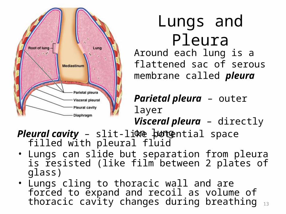
Lungs and Pleura
Pleural cavity – slit-like potential space filled with pleural fluid• Lungs can slide but separation from pleura is resisted (like film
between 2 plates of glass)• Lungs cling to thoracic wall and are forced to expand and
recoil as volume of thoracic cavity changes during breathing
Around each lung is a flattened sac of serous membrane called pleura
Parietal pleura – outer layerVisceral pleura – directly on lung
14
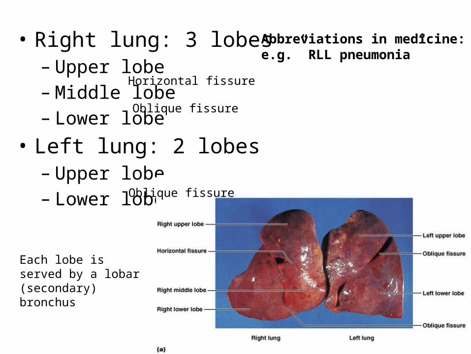
• Right lung: 3 lobes– Upper lobe– Middle lobe– Lower lobe
• Left lung: 2 lobes– Upper lobe– Lower lobe Oblique fissure
Oblique fissure
Horizontal fissure
Abbreviations in medicine:e.g.” RLL pneumonia”
Each lobe is served by a lobar (secondary) bronchus


![Anatomy and Physiology Respiratory System [Tab 2] Respiratory System](https://img.pdfslide.net/doc/110x75/56649ebd5503460f94bc631f/anatomy-and-physiology-respiratory-system-tab-2-respiratory-system.jpg)