Embed Size (px)
Citation preview
Vol. 59, No. 7INFECTION AND IMMUNITY, JUlY 1991, p. 2418-24260019-9567/91/072418-09$02.00/0Copyright X) 1991, American Society for Microbiology
Cell-Mediated Immune Responses to Babesia bovis MerozoiteAntigens in Cattle following Infection with Tick-Derived or
Cultured ParasitesWENDY C. BROWN,* KATHLEEN S. LOGAN, GERALD G. WAGNER, AND CHRISTINE L. TETZLAFF
Department of Veterinary Pathobiology, Texas A&M University, College Station, Texas 77843
Received 20 February 1991/Accepted 26 April 1991
Peripheral blood mononuclear cells from cattle experimentally infected with Babesia bovis were examined forparasite-specific cell-mediated immune responses. Unfractionated merozoites and soluble and membranefractions derived from merozoites were all antigenic for imimune cattle, although the membrane fraction was
the most stimulatory. Cattle responded to different antigenic fractions in a differential manner, and only thatanimal immunized with autologous cultured parasites responded to parasitized erythrocyte culture superna-
tants. Plastic-adherent cells (presumably monocytes/macrophages) were required for a proliferative response
to babesial antigens but not to the T-cell mitogen concanavalin A, suggesting that babesial proteins are notsimply mitogenic for T cells. Lymphocyte responses directed against a different hemoparasite from Mexico,Babesia bigemina, indicate that this parasite shares cross-reactive T-cell epitopes with B. bovis. These studiesdefine a system whereby T lymphocytes from babesia-immune cattle can be used in proliferation assays toidentify babesial merozoite antigens which are immunogenic for T cells. Because identification of helper T-celiepitopes is important for the design of a babesial subunit vaccine which will evoke anamnestic responses, thestudies described here provide a basis for such experiments.
Babesiosis is a major hemoparasitic disease of cattle insemitropical regions of the world which results in severeeconomic losses to the livestock industry. Transmitted bythe ixodid tick Boophilus microplus, Babesia bovis infec-tions in susceptible cattle are characterized by fever, ano-rexia and cachexia, low parasitemia, and a generalizedcirculatory disturbance which includes sequestration ofparasitized erythrocytes in the capillary beds of the brainand lung. Death is often caused by a respiratory distresssyndrome associated with massive infiltration of neutrophilsand parasitized erythrocytes into the lung capillaries, result-ing in vascular permeability and edema (47, 48). Cytokinesreleased by parasite-activated T cells and monocytes aremost likely involved in the pathophysiology of this disease(47, 48), as has been demonstrated with plasmodium-in-fected mice (7, 14, 27).Animals that survive natural infection, or recover from
experimental infection with attenuated parasites, are pro-tected against virulent challenge (3), and partial immunityagainst homologous and heterologous challenge can be in-duced by vaccination with crude parasite extracts (24, 25)and soluble culture-derived exoantigens (18, 28, 39, 42).However, vaccination with living parasites poses a numberof problems, not the least of which is establishment of acarrier state which permits continual babesial transmission.These observations provide the basis for developing a non-live vaccine for babesiosis.Although antibody is important in controlling babesial
infection (23), antibody alone cannot account for the pat-terns of immunity observed in response to infection andchallenge with heterologous B. bovis parasites (22) and doesnot always correlate with protection (41). In addition toproviding B-cell help, T cells are the primary effector cells in
* Corresponding author.
protective immunity against many protozoal infections (9,17, 29, 36, 37, 38), including Babesia microti infections (33).Few published studies have examined the role of T lym-
phocytes in immunity to Babesia bovis. Timms et al. (43)reported that peripheral blood mononuclear cells (PBMC)from cattle protectively immunized with attenuated liveparasites exhibited weak and short-lived proliferative re-sponses to a crude soluble parasite extract. Animals immu-nized with exoantigen had stronger cell-mediated responsesbut were less protected upon challenge. However, in bothgroups of cattle, lymphocyte proliferation and antibodyresponses correlated with protection when cattle were chal-lenged at different times after vaccination. As a strategy fordeveloping vaccines which would include T-cell epitopesimportant in the induction of a protective immune response(6), we have focused our research on characterizing T-cell-immunodominant antigens and T-cell responses in cattleprotectively immunized against B. bovis. In the studiesreported here, we have examined proliferative responses ofPBMC against crude membrane and soluble parasite extractsand soluble exoantigen from cattle immunized with virulenttick-derived parasites or avirulent cultured parasites. Vigor-ous proliferation was observed in response to crude parasiteantigens for at least 2 years following challenge (as of thiswriting). These experiments provide the basis for an in-depthfunctional analysis of defined populations of T cells andmonocytes from these and other babesia-immune cattle inresponse to defined babesial antigens.
MATERIALS AND METHODS
Babesial parasite strains and in vitro cultivation. The B.bovis parasites used for in vivo infection and challengestudies originated from a Mexican isolate, designated isolate222, obtained from naturally infected cattle in a babesia-endemic area of Mexico in 1978. Parasites were maintainedin a tick colony at the U.S. Department of Agriculture
2418
on October 4, 2018 by guest
http://iai.asm.org/
Dow
nloaded from

LYMPHOCYTE RESPONSES TO B. BOVIS 2419
laboratory in Mission, Tex. In vitro erythrocyte culturesderived from this isolate of B. bovis were maintained asdescribed by Holman et al. (16), using a modification of themicroaerophilus stationary-phase (MASP) culture system(20). In vitro cultures of Babesia bigemina parasites weremaintained under the same culture conditions. The B.bigemina parasites were obtained in 1982 from a naturallyinfected animal in northeastern Mexico. Parasites werepassaged four times in splenectomized calves by subinocu-lation of blood containing parasitized erythrocytes, andwashed, packed erythrocytes obtained during peak para-sitemia were mixed with an equal volume of phosphate-buffered saline (PBS) containing 4 M dimethyl sulfoxide(Sigma Chemical Co., St. Louis, Mo.), aliquoted, and cryo-preserved in liquid nitrogen. This parasite stock, or stabilate(21), was then used to initiate the parasite cultures.
Experimental cattle. Animal C15, a spleen-intact, 2-year-old European crossbred cow, was inoculated intravenouslytwo times, 1 month apart, with 2 x 10⁶ and 4 x 10⁶erythrocytes derived from MASP cultures of autologous, B.bovis-infected erythrocytes. Three years later, the cow wasinoculated again with 7 x 10⁹ cultured parasitized erythro-cytes, and PBMC were monitored thereafter for responses toparasite antigens. Animals C97 and C88, 9-month-old spleen-intact Brahman-European crossbred cows, were infectedwith the Mexican isolate of B. bovis by infestation with B.bovis-infected Boophilus microplus tick larvae (1 g; approx-imately 20,000). Both animals experienced clinical babesio-sis and were treated by intramuscular inoculation of dimina-zene aceturate (Ganaseg; 3 mg/kg; Squibb and Sons, MexicoCity, Mexico) diluted in H20. Three months later, theanimals were challenged by intramuscular inoculation of 2.5ml of ground-up tick stabilate (GUTS) prepared from B.bovis-infected ticks collected from animal C97 on day 3postinfection. GUTS was prepared in the following way.Engorged ticks (2 g; approximately 10,000) were homoge-nized with a loose-fitting Ten-Broeck tissue homogenizer in15 ml of RPMI 1640 medium (GIBCO, Grand Island, N.Y.)containing 3.5% bovine serum albumin. The homogenatewas centrifuged for 5 min at 250 x g, and the supernatantwas collected, layered over 3 ml of Ficoll-Hypaque (Histo-paque-1077; Sigma), and centrifuged for 5 min at 150 x g.The supernatant above the Ficoll-Hypaque interface wascollected, diluted in an equal volume of RPMI 1640 mediumcontaining 4 M dimethyl sulfoxide, aliquoted, and rapidlyfrozen in liquid nitrogen. Animal C110, a spleen-intact,9-month-old steer, served as a nonimmune control to assessthe virulence of the GUTS challenge. Age-matched animalC99, a Brahman-European crossbred steer, served as anoninfected control for all experiments. All cattle weremonitored daily for at least 2 weeks after infection for signsof infection by determining rectal temperature, packederythrocyte volume (PCV), and peripheral parasitemia inGiemsa-stained thin blood films.
Parasite antigens. Parasites were cultured at 37°C in 5%CO2 in air in 24-well plates (1.25 ml; Costar, Cambridge,Mass.) or in 25-cm2 flasks (12.5 ml; Costar) containing a 5%(vol/vol) concentration of erythrocytes from animal C15 inmedium 199 (GIBCO) supplemented with 40% autologousbovine serum. Merozoites were harvested following CO2deprivation (46) of cultures after the relative percentage ofparasitized erythrocytes was increased by sequential reduc-tion of the concentration of erythrocytes (13), with thefollowing modifications. Infected erythrocyte cultures con-taining at least 15% parasitized erythrocytes were incubatedfor 18 h in serum-free HL-1 medium (Ventrex Laboratories,
Inc., Portland, Maine) in 100% medical-grade oxygen at37°C. Merozoites were recovered from the culture superna-tant following a series of 10-min (140, 140, 180, and 220 x g)and 15-min (5,000 x g) centrifugations of the supernatant,followed by two 15-min (6,500 x g) centrifugations of theparasite pellet resuspended in PBS. Merozoites prepared inthis manner contained some erythrocyte membranes butwere relatively pure. Culture supernatants collected from B.bovis MASP cultures contain soluble parasite antigens des-ignated exoantigens (32). The culture supernatant was col-lected after the 5,000 x g centrifugation and is designatedISUP. Supernatants prepared in the same way from autolo-gous, uninfected cultured erythrocytes are designatedUSUP. Merozoites (designated MER) were resuspended inPBS containing the protease inhibitors E-64 and Antipain(Boehringer Mannheim Biochemicals, Indianapolis, Ind.) at25 ,ug/ml. To prepare subcellular fractions, merozoites col-lected from 12 25-cm2 flasks were disrupted by two passagesthrough a French pressure cell (SLM Instruments, Inc.,Urbana, Ill.) under a chamber pressure of 1,500 lb/in2, andthe homogenate was centrifuged for 1 h at 145,000 x g. Thesoluble high-speed supernatant fraction (designated HSSfraction) and membrane pellet resuspended in an equalvolume of PBS (designated CM fraction) were collected.Fractions prepared in this manner generally yielded approx-imately 6 mg of protein in the CM fraction and 3 mg ofprotein in the HSS fraction. Ghosts prepared from unin-fected erythrocytes (referred to as URBC) and a CM andHSS fraction derived therefrom were also prepared. Proteinconcentrations in the parasite or erythrocyte fractions weredetermined as described previously (2), using bovine immu-noglobulin G (Bio-Rad Laboratories, Richmond, Calif.) as aprotein standard. All fractions were stored at -80°C inaliquots.SDS-PAGE and Western blotting. Sodium dodecyl sulfate-
polyacrylamide gel electrophoresis (SDS-PAGE) was per-formed as described previously (19), using a Bio-Rad minigelapparatus, 10% acrylamide gels, and 10 ,ug of protein (con-sisting of babesial or uninfected erythrocyte antigens) perlane. Immunoblotting onto nitrocellulose was performed asdescribed previously (44) for 1 h at 100 V, using the Bio-RadTransblot apparatus. Blots were reacted with sera fromnormal cattle (C99 or preinfection sera) or B. bovis-immunecattle diluted 1:100 in Tris-buffered saline (10 mM Tris, 150mM NaCl, pH 7.5) containing 1% gelatin, and serologicallyreactive proteins were detected with a 1:5,000 dilution ofalkaline phosphatase-conjugated goat anti-bovine immuno-globulin G (Kirkegaard & Perry Laboratories, Gaithersburg,Md.) and substrates nitroblue tetrazolium and 5-bromo-4-chloro-3-indolyl phosphate (Sigma) as instructed by themanufacturers.Lymphocyte proliferation assays. PBMC were prepared by
Ficoll-Hypaque density gradient centrifugation of blood di-luted 1:1 in Alsever's solution (10). PBMC were tested forreactivity to parasite antigens in a proliferation assay per-formed for 5, 6, or 7 days. However, in one experiment,lymphoproliferation was compared in duplicate assays har-vested after 3 or 6 days. In another experiment, PBMC (2 x10⁶ cells per ml in a 25-cm2 flask at 37°C) were allowed toadhere for 2 h to polystyrene, and the nonadherent fractionof the cells was compared with unseparated PBMC forlymphoproliferative responses. For all experiments, cellswere cultured in complete medium, which consisted ofRPMI 1640 medium containing 25 mM N-2-hydroxyethylpi-perazine-N-2-ethanesulfonic acid (HEPES; GIBCO) andsupplemented with 10% heat-inactivated fetal bovine serum
VOL. 59, 1991
on October 4, 2018 by guest
http://iai.asm.org/
Dow
nloaded from

2420 BROWN ET AL.
b c d e
TABLE 1. Comparison of proliferative responses of normal andimmune PBMC cultured for 3 or 6 days with
B. bovis antigen or mitogen
Radioactivity (cpm) incorporated by PBMC withCattle indicated antigen or mitogen'
URBC CM B. bovis CM ConA
C99 (control)Day 3 382 + 24 389 ± 30 28,750 ± 4,435Day 6 431 ± 60 1,290 ± 715 40,306 ± 9,050
C15 (immune)Day 3 1,011 ± 164 3,080 ± 294 119,530 ± 5,485Day 6 3,683 ± 2,956 77,502 ± 8,798 59,202 ± 4,372
C97 (immune)Day 3 480 ± 27 961 ± 165 126,858 ± 3,258Day 6 673 ± 25 16,457 ± 3,990 55,182 ± 2,865a A total of 2 x 10⁵ PBMC were cultured in wells of half-area 96-well plates
with antigen or ConA. Background responses (medium) were always less thanthe responses to URBC CM. After 3 or 6 days, the cells were radiolabeled for4 h with '25IUDR and then harvested and counted as described in the text.Results are means ± standard deviations of triplicate cultures. ConA waspresent at a final concentration of 1 iLg/ml; URBC and B. bovis CM antigenswere present at a final concentration of 25 ,ug/ml.
_a 4
I
---31-
FIG. 1. Serologic reactivity of B. bovis-immune sera with babe-sial antigens. Western blots were performed by using preimmune(lanes e to h) or postchallenge (lanes a to d) sera from animal C15 (A)or C97 (B). (A) Lanes: a and h, URBC; b and g, parasite HSS; c andf, parasite CM; d and e, MER. (B) Lanes: a and h, URBC; b and g,MER; c and f, parasite CM; d and e, parasite HSS. Molecular sizestandards are indicated on the left.
(Hyclone, Logan, Utah), 2 mM L-glutamine (GIBCO), 5 x10⁻⁵ M 2-mercaptoethanol (Sigma), and 50 ,ug of gentamicinsulfate (GIBCO) per ml. Proliferation assays were performedin 96-well flat-bottom plates (200 [lI per well) or half-areaplates (100 RIl per well; Costar) with a final concentration of2 x 106 PMBC per ml of complete medium. Concanavalin A(ConA; Sigma) or antigen (B. bovis MER, CM, and HSS; B.bigemina MER, URBC, and URBC CM) were added to theassays at final concentrations of 0.2 to 25 Fg of protein perml of complete medium. Exoantigens (ISUP or USUP) wereadded at a final concentration of 50 or 250 ,ug/ml. Whenpresent, indomethacin (Sigma) was added to the assays at afinal concentration of 0.5 ,ug/ml. The cells were radiolabeledfor the last 4 or 18 h of culture with 0.25 or 0.5 ,uCi of125iododeoxyuridine (125IUDR; ICN Biomedicals, Inc.,Costa Mesa, Calif.) and harvested onto glass filters with anautomated cell harvester (Skatron Instruments, Inc., Ster-ling, Va.). Proliferation was determined by measuring theincorporation of 125IUDR by counting the radioactivity onthe filters in a gamma counter (Packard, Laguna Hills,Calif.). Results are expressed as the mean radioactivity incounts per minute of triplicate samples ± standard devia-tion.
RESULTS
Clinical responses of cattle infected and challenged with B.bovis. Animal C15, inoculated with autologous, cultured B.bovis-infected erythrocytes, experienced mild signs of infec-
tion after the primary inoculation, with a 24% reduction inPCV on day 7 postinoculation. There were no clinical signsof babesiosis following the secondary and tertiary inocula-tions. This cow was not treated with Ganaseg. Animals C88and C97 developed severe babesiosis following infestationwith B. bovis-infected ticks. Parasites were observed inblood films from C88 on days 8 to 13 postinfestation, and onday 10 the PCV was reduced by 48%. Parasites weredetected in blood films from C97 on days 13 and 14 postin-festation, and on day 13 the PCV fell by 50%. Both animalswere treated with Ganaseg. Three months later, C88 and C97were challenged with an infective tick stabilate, and bothanimals were solidly immune to challenge. They experiencedno reduction in PCV, whereas the nonimmune control ani-mal C110 experienced severe babesiosis with a 57% reduc-tion in PCV on day 14 postinoculation. This animal recov-ered without treatment.
Serologic responses of B. bovis-infected cattle. All fouranimals were serologically positive for B. bovis, as deter-mined by indirect immunofluorescence staining of smears ofcultured parasites (not shown). Postchallenge sera from allanimals reacted on immunoblots with numerous bandspresent in whole merozoites and soluble and membranesubfractions of merozoites, whereas there was no reactivityagainst proteins present in uninfected, cultured erythro-cytes. Figure 1 is an immunoblot of preimmune (lanes e to h)and postchallenge (lanes a to d) sera of animals C15 (Fig. 1A)and C97 (Fig. 1B) incubated with URBC and B. bovis MER,CM, and HSS. Major protein bands in the MER recognizedby C97 antiserum had approximate molecular masses of 42,60, 85, 120, and 225 kDa (Fig. 1B, lane b), whereas C15immune serum reacted most strongly with bands of 42 and116 kDa (Fig. 1A, lane d). Several protein bands recognizedby immune sera from C15 and C97 were present in bothsoluble and membrane fractions, including the immunodom-inant 42-kDa (12, 15), 60-kDa (12, 40), and 85-kDa (12)proteins. Serum from normal animal C99 did not react withany babesial protein, and none of the sera reacted with B.bigemina merozoites on immunoblots (data not shown).Examination of babesia-specific proliferative responses over
time. To determine the optimal assay time for babesia-specific responses, proliferation assays of control (C99) or
A
kD a
116-
66-
43-
31-
BKD
200
97-
66-
43--
INFECT. IMMUN.
on October 4, 2018 by guest
http://iai.asm.org/
Dow
nloaded from

LYMPHOCYTE RESPONSES TO B. BOVIS 2421
C8B PBUC
40-
30-
20-
60AZoll- 10
zD ~~~~C15SPBMC
o) 100
80-
a-20
C97 PBUC
C110 PBMC
14 15 23 29 32 35 38 42
WEEKS FOLLOWING CHALLENGE
FIG. 2. Time course of the proliferative response of PBMC from B. boiis-immune cattle. PBMC (4 x 10⁵) from immune cattle wereassessed for proliferative responses against B. bovis MER (gray bars), URBC (white bars), or medium control (black bars) at various weeksfollowing the last challenge inoculation as indicated on the abscissa. Cells were cultured for 6 or 7 days with indomethacin, radiolabeled for4 h, harvested, and counted as described in the text. The data shown are for optimal concentrations of URBC or MER antigen in the culturewells, which were as follows: C88, 6.2 ,ug/ml; C97, 25 ,ug/ml; C15, 1.0 to 6.2 ,ug/ml; and C110, 25 ,ug/ml.
immune (C97 and C15) PBMC were performed for 3 or 6days (Table 1). There was no response to URBC at eitherday. Little or no reactivity against babesial antigen hadoccurred by day 3, but strong responses were present by day6. In contrast, strong responses to the T-cell mitogen ConAwere present by day 3. Subsequent assays were conductedfor 5 to 7 days.Immune cattle were monitored for proliferative responses
to babesial and control antigens for weeks or months follow-ing the last challenge inoculation. Figure 2 presents theresponses of PBMC from the four immunized cattle tooptimal antigen concentrations of purified merozoites oruninfected erythrocytes on various weeks after challenge.Vigorous proliferative responses to unfractionated merozo-ites were consistently observed with C15 and C110 PBMCafter 10 to 15 weeks postchallenge. Animals C88 and C97 hadinconsistent and often undetectable responses to merozoitesover time. However, responses to the CM fraction wereconsistently positive in all immune animals and after 2 yearscould still be induced in PBMC from animals C97 and C15,which were chosen for prolonged study.
Differential responses of PBMC from babesia-sensitizedcattle to different antigenic fractions. PBMC from B. bovis-immune and control cattle were compared for stimulation bydifferent crude preparations of uninfected erythrocytes andB. bovis merozoites. Figure 3 compares proliferation ofPBMC from three immune cattle and normal animal C99
against four concentrations of URBC, MER, CM, and HSSfractions. PBMC from all animals were unresponsive toURBC (Fig. 3) and soluble and membrane fractions preparedfrom URBC (not shown). PBMC from all immune cattleresponded vigorously to the parasite CM fraction in adose-dependent manner, with maximal proliferation occur-ring at a concentration of 25 ,ug of protein per ml. SolubleHSS was weakly stimulatory for C97 PBMC and moderatelystimulatory for C110 PBMC, but it induced strong responsesin C15 PBMC at the higher concentrations of antigen (5 and25 ,ug of protein per ml). MER induced intermediate levels ofproliferation in C97 and C110 PBMC, in a dose-dependentfashion, whereas the proliferative response of C15 PBMC toMER was routinely highest with the lowest concentration ofprotein assayed (in this case, 0.2 ,ug/ml). Occasional re-sponses of control C99 lymphocytes to CM or HSS antigenwere observed, but these responses were neither consistentnor dose dependent. Nevertheless, the possibility that mero-zoites contain mitogenic components must be considered.
Effect of indomethacin on parasite-specific responses. Weobserved large, activated mononuclear cells in cultures ofPBMC stimulated with merozoites. This, together with thefinding that high concentrations of merozoite antigen had aninhibitory effect on proliferation of C15 PBMC (Fig. 3),prompted us to test the effect of the prostaglandin E2 (PGE2)inhibitor indomethacin on parasite-induced proliferation. Ata final concentration of 0.5 ,ug/ml, indomethacin has been
IVOL. 59, 1991
on October 4, 2018 by guest
http://iai.asm.org/
Dow
nloaded from
2422 BROWN ET AL.
C15 PBLIC C97 PBMC
I
1 10 100
Ci1o PBSC
100-
80-
60-
40
20
ol. _........
PROTEIN CONCENTRATION (MICROGRAMS PER ML)FIG. 3. Dose-dependent proliferative responses against merozoite fractions. PBMC (4 x 10⁵) from one normal (C99) and three immune
cattle were stimulated for 6 days with a final concentration of 0.2, 1, 5, or 25 ,ug of protein per ml of URBC (solid circles), B. bovis MER (opencircles), B. bovis CM (triangles), or B. bovis HSS (squares). Indomethacin was included in the assay.
shown to augment interleukin-2 (IL-2) production by bovineT cells (35). When added to proliferation assays, this con-centration of indomethacin enhanced the responses ofPBMC from all immune cattle to MER and the CM fraction;an eightfold increase was observed in the proliferation ofC97 PBMC to CM with indomethacin (Table 2). There wasless effect on the response to HSS and no effect on theresponse to URBC.
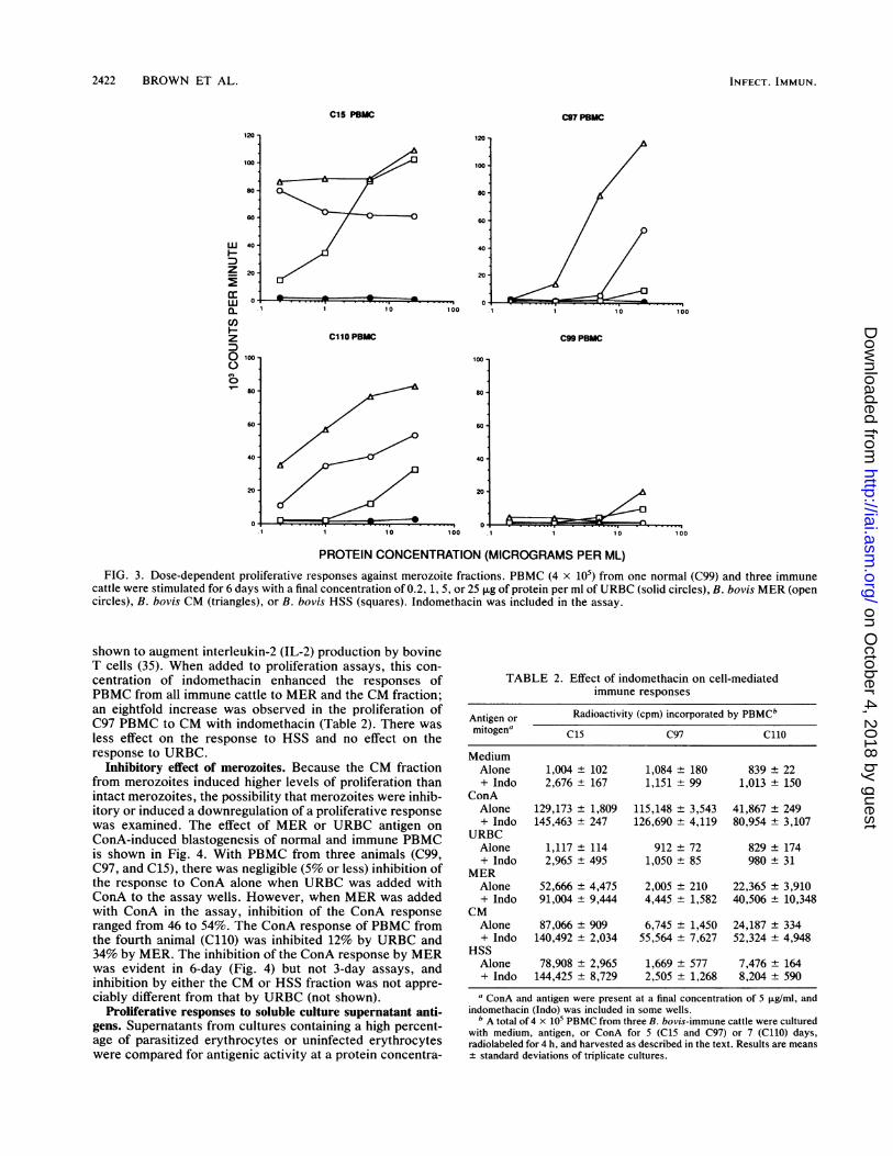
Inhibitory effect of merozoites. Because the CM fractionfrom merozoites induced higher levels of proliferation thanintact merozoites, the possibility that merozoites were inhib-itory or induced a downregulation of a proliferative responsewas examined. The effect of MER or URBC antigen onConA-induced blastogenesis of normal and immune PBMCis shown in Fig. 4. With PBMC from three animals (C99,C97, and C15), there was negligible (5% or less) inhibition ofthe response to ConA alone when URBC was added withConA to the assay wells. However, when MER was addedwith ConA in the assay, inhibition of the ConA responseranged from 46 to 54%. The ConA response of PBMC fromthe fourth animal (C110) was inhibited 12% by URBC and34% by MER. The inhibition of the ConA response by MERwas evident in 6-day (Fig. 4) but not 3-day assays, andinhibition by either the CM or HSS fraction was not appre-ciably different from that by URBC (not shown).
Proliferative responses to soluble culture supernatant anti-gens. Supernatants from cultures containing a high percent-age of parasitized erythrocytes or uninfected erythrocyteswere compared for antigenic activity at a protein concentra-
TABLE 2. Effect of indomethacin on cell-mediatedimmune responses
Antigen or Radioactivity (cpm) incorporated by PBMCbmitogen' C15 C97 Ci1oMediumAlone 1,004 ± 102 1,084 ± 180 839 ± 22+ Indo 2,676 ± 167 1,151 ± 99 1,013 ± 150
ConAAlone 129,173 ± 1,809 115,148 ± 3,543 41,867 ± 249+ Indo 145,463 ± 247 126,690 ± 4,119 80,954 ± 3,107
URBCAlone 1,117 ± 114 912 ± 72 829 ± 174+ Indo 2,965 ± 495 1,050 ± 85 980 ± 31
MERAlone 52,666 ± 4,475 2,005 ± 210 22,365 ± 3,910+ Indo 91,004 ± 9,444 4,445 + 1,582 40,506 ± 10,348
CMAlone 87,066 ± 909 6,745 ± 1,450 24,187 ± 334+ Indo 140,492 ± 2,034 55,564 ± 7,627 52,324 ± 4,948
HSSAlone 78,908 ± 2,965 1,669 ± 577 7,476 ± 164+ Indo 144,425 ± 8,729 2,505 ± 1,268 8,204 ± 590
a ConA and antigen were present at a final concentration of 5 jig/ml, andindomethacin (Indo) was included in some wells.
b A total of 4 x 10⁵ PBMC from three B. bovis-immune cattle were culturedwith medium, antigen, or ConA for 5 (C15 and C97) or 7 (C110) days,radiolabeled for 4 h, and harvested as described in the text. Results are means+ standard deviations of triplicate cultures.
120
100'
60
60'
W 40H
Z 20
0
a-C)z
0 1X
80v- so0
60
40
20
100
C99 PBMC
10 10010 100
I--if .- ___.
-0 ...I
INFECT. IMMUN.
Ax--
on October 4, 2018 by guest
http://iai.asm.org/
Dow
nloaded from

LYMPHOCYTE RESPONSES TO B. BOVIS 2423
C99
clio
4
z4C
C97
C15
'II
TABLE 3. Proliferative responses of PBMC toB. bigemina merozoites
Radioactivity (cpm) incorporatedAntigen and concn by PBMC'
(pLg/ml)C15 C97
Medium 966 ± 308 647 ± 66URBC CM
5 626 83 777 31625 834 ± 391 535 ± 33
B. bovis CM5 20,396 ± 1,422 3,421 ± 1,14125 22,831 ± 756 8,147 ± 3,596
B. bigemina MER5 15,703 ± 1,786 5,018 ± 1,62325 14,386 ± 1,948 793 ± 262
0 1 0 20 30 40 50 60 a A total of 2 x 10⁵ PBMC were cultured in half-area 96-well plates withantigen for 6 days, radiolabeled for 4 h with 125IUDR, harvested, and counted
PERCENTAGE INHIBITION as described in the text. All wells contained indomethacin. Results are meansstandard deviations of trinlicate cultures
FIG. 4. Inhibition of ConA-induced proliferation by merozoites.PBMC (4 x 10⁵) from one normal (C99) and three immune cattlewere stimulated for 6 days with ConA (at a final concentration of 1p.g/ml), MER (at a final concentration of 25 ,ug/ml), URBC (at a finalconcentration of 25 ,ug/ml), or a combination of ConA plus MER orConA plus URBC. Percent inhibition of the proliferative response toConA alone is shown for PBMC cultured with ConA plus MER(hatched bars) and ConA plus URBC (white bars). Indomethacinwas included in the assay.
tion of 250 ,ug/ml (Fig. 5). PBMC from animal C15, whichwas immunized with cultured parasites, were consistentlyresponsive to ISUP and, at a protein concentration of 50,ug/ml, incorporated 36,715 ± 3,403 cpm. In contrast, PBMCfrom cattle infected with tick-derived parasites (C97 andC110) did not respond to the lower antigen concentration andresponded only weakly to the higher concentration of ISUP.
Proliferative responses to unrelated B. bigemina merozoites.B. bigemina merozoites also induced proliferation of PBMC
80
,, 60
z
44U)
1~~~_I-_
z
0
_ 20
0
015 097 0110 099
ANIMAL
FIG. 5. Proliferative responses to culture supernatant antigen.PBMC from one normal (C99) and three immune cattle werestimulated for 6 days with medium alone (black bars) or withsupernatants USUP (white bars) or ISUP (gray bars). The responsesshown were elicited with a final protein concentration of 250 p.g/ml.Indomethacin was included in the assay.
from animals C15 and C97, although these animals wereserologically unreactive with this parasite on immunoblots.The response of C15 PBMC was always vigorous to thisparasite, whereas the response of C97 PBMC was routinelyweaker (Table 3).
Role of monocytes/macrophages in the response to babesialantigens. Other protozoan parasites, including Plasmodiumfalciparum (45, 49) and Theileria parva (1, 11, 30, 31), havebeen shown to induce nonspecific lymphocyte proliferation.To examine the possibility that babesial parasites were alsomitogenic, PBMC depleted of monocytes by 2 h of adher-ence to polystyrene (10) were compared with unseparatedPBMC for the ability to respond to Babesia antigen or ConA(Table 4). Adherent cells were required for a babesia-specificresponse in both immune animals but not for a mitogenicresponse to ConA. In undepleted PBMC, both specific andnonspecific responses were markedly enhanced by the addi-tion of indomethacin. However, in monocyte-depleted cells,indomethacin had little effect on the ConA response and noeffect on the babesia-specific response. These data suggestthat monocytes/macrophages are required as antigen-pre-senting cells for the induction of lymphocyte blastogenesis tobabesial antigens.
DISCUSSIONThe results of this study demonstrate that infection of
cattle with B. bovis-infected ticks, tick stabilate, or culturedmerozoites elicits strong, cell-mediated immune responsesto merozoite antigens. All four immunized animals, but notthe control animal, responded to parasite antigens in adose-dependent manner. Animals C15 and C97 have beenmonitored for 2 years since the last parasite inoculation, andthey continue to respond vigorously to parasite antigen.Since both animals received viable parasites upon challenge,these long-lived responses may be due to chronic infectionand continual release of parasite antigen or to a memoryT-cell response.When the proliferative responses against crude, subcellu-
lar fractions of merozoites were compared, we found thatcattle infected with tick-derived parasites preferentially re-sponded to an antigenic fraction enriched in parasite mem-branes. In contrast, the animal that was inoculated withcultured merozoites responded vigorously to the membrane-enriched fraction, the soluble merozoite fraction, and solu-
I
Irol NO,
a
VOL. 59, 1991
-..- - -- I - .. -
on October 4, 2018 by guest
http://iai.asm.org/
Dow
nloaded from

2424 BROWN ET AL.
TABLE 4. Requirement of plastic-adherent PBMC forB. bovis-specific proliferative responses
Radioactivity (cpm) incorporated by PBMC withCattle and indicated antigen or mitogen'PBMC
Medium B. bovis MER ConA
C88UnseparatedAlone 2,022 ± 297 4,517 + 656 51,905 ± 676+ Indob 7,424 ± 2,954 35,580 ± 7,752 119,952 ± 3,966
Nonadherent'Alone 793 ± 86 1,026 ± 123 111,483 ± 1,368+ Indo 716 ± 66 881 ± 117 117,363 + 3,240
C97UnseparatedAlone 8,875 ± 110 48,749 ± 8,945 62,056 ± 2,364+ Indo 7,231 ± 958 71,027 + 7,504 98,306 ± 5,355
NonadherentAlone 438 + 38 705 ± 123 84,387 ± 5,595+ Indo 279 ± 197 660 ± 155 91,160 ± 5,270
a A total of 4 x 10⁵ PBMC were cultured in 96-well plates with medium,antigen, or ConA for 7 days. The cells were radiolabeled for 18 h with1251UDR, harvested, and counted as described in the text. Results are means± standard deviations of triplicate cultures. ConA was present at a finalconcentration of 5 ,ug/ml; B. bovis MER antigen was present at a finalconcentration of 6.2 jig/ml.
b Indomethacin (Indo) was included in the assay.c A total of 5 x 10⁷ PBMC were incubated for 2 h at 37°C in a 25-cm2 flask
(10 ml), and the nonadherent cells were used in the assay.
ble culture supernatant antigens. Responses of animals C97and C110 to the soluble merozoite fraction were weaker,even in the presence of indomethacin, and these cattle didnot react significantly with culture supernatant exoantigens.These studies suggest that antigens most immunogenic for Tcells, and those which induce an anamnestic response fromcattle exposed to babesia-infected ticks, are found in para-site membrane-enriched fractions. Soluble antigens and an-tigens secreted or shed into the culture supernatant mostlikely contain some of these membrane-associated proteins,as shown by the reactivity on immunoblots of immune serawith protein bands common to CM and HSS fractions.However, the use of soluble parasite extracts to induceT-cell blastogenesis may explain the weak and short-livedresponses in B. bovis-immune cattle observed by Timms andcoworkers (43). Furthermore, the unique response ofPBMCfrom animal C15 to culture supernatant antigens may bedirected primarily against cultured merozoite antigens usedfor immunization. The response of B. bovis-immune PBMCto B. bigemina parasites is likely directed against epitopesshared by the two parasites, and species-common polypep-tides have been demonstrated by immunoprecipitation withimmune sera (26). The lack of serological reactivity onimmunoblots with C97 and C15 immune sera may reflect adifference in T- and B-cell epitopes on these putative cross-reactive proteins.The optimal time of 5 to 7 days for blastogenesis to occur
in response to babesial antigen is consistent with an antigen-induced and not a mitogen-induced response. Furthermore,comparison of proliferative responses from immune andnormal cattle indicates that these responses were evoked inan antigen-specific manner, since babesial antigens stimu-lated little or no proliferation in PBMC from the nonimmunecontrol. Additional evidence for the antigen-specific natureof the proliferative response was demonstrated by the inabil-ity of PBMC depleted of plastic-adherent cells to respond toantigen, with no reduction in the response to ConA. Because
the majority of monocytes are removed by this procedure(10), these results indicate that monocytes are required asantigen-presenting cells in the babesia-specific response.The regulation of lymphocyte proliferation in response to
babesial antigens appears quite complex. As well as theapparent requirement for monocytes as accessory or anti-gen-presenting cells, monocytes appear to inhibit immuneresponses through the production of PGE2. This autocoidhas been shown to play a key role in immune downregulationby inhibiting both IL-1 and IL-2 production (reviewed inreferences 4 and 8), possibly through the induction ofsuppressor T cells (5). In our studies, the PGE2 inhibitorindomethacin enhanced both babesia-specific and ConAresponses but did not reverse the inability of uninfectederythrocyte membranes to stimulate PBMC. An interpreta-tion of these results is that babesial merozoite antigensinduce the production of PGE2 by monocytes, which maydownregulate IL-1- and IL-2-induced lymphocyte prolifera-tive responses. Our findings are consistent with those ofRuebush and coworkers (34), who showed that B. microtiinfection of mice elicited PGE2 synthesis by macrophages,which resulted in suppression of delayed-type hypersensitiv-ity responses to parasite antigens.
Unfractionated merozoites both stimulated proliferationof antigen-primed lymphocytes and inhibited the prolifera-tive response to the T-cell mitogen ConA. This inhibitoryeffect was observed in the presence or absence of indometh-acin, implying that factors in addition to PGE2 are involvedin suppressing proliferation. We are exploring the possibilitythat tumor necrosis factor alpha, released by parasite-acti-vated macrophages, may play a role in downregulating T-cellproliferation. Inhibition of ConA-induced proliferation wasnot caused by antigen-specific T cells, since PBMC fromnonimmune animal C99 were inhibited to the same extent asthose from babesia-sensitized animals. Soluble factors pro-duced by T. parva-infected T cells were similarly shown tosuppress proliferation of T cells from normal cattle (11).These studies provide a foundation for further analyses of
the cellular immune response which is undoubtedly involvedin protective immunity against Babesia spp. and all hemo-parasites. To unravel the complex interactions between hostlymphocytes, monocytes, and parasite antigens, definedsubpopulations of T cells and biochemically purified orrecombinant parasite antigens must be used. Experimentsdesigned to identify T-cell-immunodominant babesial anti-gens by using functionally characterized babesia-specificT-cell lines and clones are in progress and will be the subjectof future publications.
ACKNOWLEDGMENTS
We thank Ken Waldrup for veterinary assistance, David Cruz forinfesting cattle with babesia-infected ticks, and Patricia Holman forinstruction on the MASP culture system.
This work was supported by the Texas Agricultural ExperimentStation projects 6964 and 6980 and by Public Health Service grantAI30136-01 (W.C.B.) from the National Institute of Allergy andInfectious Diseases.
REFERENCES1. Brown, W. C., and K. S. Logan. 1986. Bovine T-cell clones
infected with Theileria parva produce a factor with IL 2-likeactivity. Parasite Immunol. 8:189-192.
2. Brown, W. C., C. Sugimoto, P. A. Conrad, and D. J. Grab. 1989.Differential response of bovine T-cell lines to membrane andsoluble antigens of Theileria parva schizont-infected cells. Par-asite Immunol. 11:567-583.
3. Callow, L. L. 1977. Vaccination against bovine babesiosis, p.
INFECT. IMMUN.
on October 4, 2018 by guest
http://iai.asm.org/
Dow
nloaded from
LYMPHOCYTE RESPONSES TO B. BOVIS 2425
121-149. In L. H. Miller, J. A. Pino, and J. J. McKelvey (ed.),Immunity to blood parasites of animals and man. PlenumPublishing Corp., New York.
4. Chang, J., and A. J. Lewis. 1984. Prostaglandins and cyclooxy-genase inhibitors, p. 649467. In R. L. Fenichel and M. A.Chirigos (ed.), Immune modulation agents and their mecha-nisms. Marcel Dekker, New York.
5. Chouaib, S., L. Chatenoud, D. Klatzmann, and D. Fradelizi.1984. The mechanisms of inhibition of human IL 2 production.II. PGE2 induction of suppressor T lymphocytes. J. Immunol.132:1851-1857.
6. Chulay, J. D. 1989. Development of sporozoite vaccines formalaria. Trans. R. Soc. Trop. Med. Hyg. 83(Suppl.):61-66.
7. Clark, I. A., W. B. Cowden, G. A. Butcher, and N. H. Hunt.1987. Possible roles of tumor necrosis factor in the pathology ofmalaria. Am. J. Pathol. 129:192-199.
8. Cueppens, J. L., and J. S. Goodwin. 1984. Prostaglandins asmodulators of T and B lymphocyte function, p. 627-648. InR. L. Fenichel and M. A. Chirigos (ed.), Immune modulationagents and their mechanisms. Marcel Dekker, New York.
9. Emery, D. L. 1981. Adoptive transfer of immunity to infectionwith Theileria parva (East Coast fever) between cattle twins.Res. Vet. Sci. 30:364-367.
10. Goddeeris, B. M., C. L. Baldwin, 0. ole-MoiYoi, and W. I.Morrison. 1986. Improved methods for purification and deple-tion of monocytes from bovine peripheral blood mononuclearcells. Functional evaluation of monocytes in response to lectins.J. Immunol. Methods 89:165-173.
11. Goddeeris, B. M., and W. I. Morrison. 1987. The bovineautologous Theileria mixed leukocyte reaction: influence ofmonocytes and phenotype of the parasitized stimulator cell onproliferation and parasite specificity. Immunology 60:90-96.
12. Goff, W. L., W. C. Davis, G. H. Palmer, T. F. McElwain, W. C.Johnson, J. F. Bailey, and T. C. McGuire. 1988. Identification ofBabesia bovis merozoite surface antigens by using immunebovine sera and monoclonal antibodies. Infect. Immun. 56:2363-2368.
13. Goff, W. L., and C. E. Yunker. 1986. Babesia bovis: increasedpercentage of parasitized erythrocytes in cultures and assess-ment of growth by incorporation of (3H) hypoxanthine. Exp.Parasitol. 62:202-210.
14. Grau, G. E., L. F. Fajardo, P. F. Piguet, B. Allet, P. H. Lambert,and P. Vassali. 1987. Tumor necrosis factor (cachectin) as anessential mediator in murine cerebral malaria. Science 237:1210-1212.
15. Hines, S. A., T. F. McElwain, G. M. Buening, and G. H. Palmer.1989. Molecular characterization of Babesia bovis merozoitesurface proteins bearing epitopes immunodominant in protectedcattle. Mol. Biochem. Parasitol. 37:1-10.
16. Holman, P. J., K. A. Waldrup, and G. G. Wagner. 1988. In vitrocultivation of a Babesia isolated from a white-tailed deer(Odocoileus virginianus). J. Parasitol. 74:111-115.
17. Kumar, S., M. F. Good, F. Dontfraid, J. M. Vinetz, and L. H.Miller. 1989. Interdependence of CD4+ T cells and malarialspleen in immunity to Plasmodium vinckei vinckei. J. Immunol.143:2017-2023.
18. Kuttler, K. L., M. G. Levy, M. A. James, and M. Ristic. 1982.Efficacy of a nonviable culture-derived Babesia bovis vaccine.Am. J. Vet. Res. 43:281-284.
19. Laemmli, U. K. 1970. Cleavage of structural proteins during theassembly of the head of bacteriophage T4. Nature (London)227:680-685.
20. Levy, M. G., and M. Ristic. 1980. Babesia bovis: continuouscultivation in a microaerophilus stationary phase culture. Sci-ence 207:1218-1220.
21. Lunsden, W. H. R., and G. J. C. Hardy. 1965. Nomenclature ofliving parasite material. Nature (London) 205:1032.
22. Mahoney, D. F. 1986. Studies on the protection of cattle againstBabesia bovis infection, p. 539-554. In W. I. Morrison (ed.),The ruminant immune system in health and disease. CambridgeUniversity Press, Cambridge.
23. Mahoney, D. F., J. D. Kerr, B. V. Goodger, and I. G. Wright.1979. The immune response of cattle to Babesia bovis (syn. B.
argentina): studies on the nature and specificity of protection.Int. J. Parasitol. 9:297-306.
24. Mahoney, D. F., and I. G. Wright. 1976. Babesia argentina:immunization of cattle with a killed antigen against infectionwith a heterologous strain. Vet. Parasitol. 2:273-306.
25. Mahoney, D. F., I. G. Wright, and B. V. Goodger. 1981. Bovinebabesiosis: the immunization of cattle with fractions of eryth-rocytes infected with Babesia bovis (syn. B. argentina). Vet.Immunol. Immunopathol. 2:145-156.
26. McElwain, T. F., G. H. Palmer, W. L. Goff, and T. C. McGuire.1988. Identification of Babesia bigemina and Babesia bovismerozoite proteins with isolate- and species-common epitopesrecognized by antibodies in bovine immune sera. Infect. Im-mun. 56:1658-1660.
27. Miller, K. L., P. H. Silverman, B. Kullgren, and L. J. Mahl-mann. 1989. Tumor necrosis factor alpha and the anemiaassociated with murine malaria. Infect. Immun. 57:1542-1546.
28. Montenegro-James, S., and M. Ristic. 1985. Heterologous strainimmunity in bovine babesiosis using a culture-derived solubleBabesia bovis immunogen. Vet. Parasitol. 18:321-337.
29. Morrison, W. I., B. M. Goddeeris, W. C. Brown, C. L. Baldwin,and A. J. Teale. 1989. Theileria parva in cattle: characterizationof infected lymphocytes and the immune responses they pro-voke. Vet. Immunol. Immunopathol. 20:213-237.
30. Pearson, T. W., L. B. Lundin, T. T. Dolan, and D. A. Stagg.1979. Cell-mediated immunity to Theileria-transformed celllines. Nature (London) 281:678-680.
31. Pinder, M., S. Kar, K. S. Withey, L. B. Lundin, and G. E.Roelants. 1981. Proliferation and lymphocyte stimulatory capac-ity of Theileria-infected lymphoblastoid cells before and afterthe elimination of intracellular parasites. Immunology 44:51460.
32. Ristic, M., and I. Kakoma. 1988. Exoantigens of Babesia, p.131-141. In M. Ristic (ed.), Babesiosis of domestic animals andman. CRC Press, Inc., Boca Raton, Fla.
33. Ruebush, M. J., and W. L. Hanson. 1980. Thymus dependenceof resistance to infection with Babesia microti of human originin mice. Am. J. Trop. Med. Hyg. 29:507-515.
34. Ruebush, M. J., L. K. Steel, and D. A. Kennedy. 1986. Il.Prostaglandin-mediated suppression of delayed-type hypersen-sitivity to infected erythrocytes during Babesia microti infectionof mice. Cell. Immunol. 98:300-310.
35. Sambhara, S. R., and E. L. Belden. 1988. Bovine interleukin-2:production and characterization. Vet. Immunol. Immunopathol.18:165-172.
36. Schofield, L., J. Villaquiran, A. Ferreira, H. Schellekens, R.Nussenzweig, and V. Nussenzweig. 1987. Gamma interferon,CD8+ T cells and antibodies required for immunity to malariasporozoites. Science 330:664467.
37. Scott, P., P. Caspar, and A. Sher. 1990. Protection againstLeishmania major in Balb/c mice by adoptive transfer of a T cellclone recognizing a low molecular weight antigen released bypromastigotes. J. Immunol. 144:1075-1079.
38. Scott, P., P. Natovitz, R. L. Coffman, E. Pearce, and A. Sher.1988. Immunoregulation of cutaneous leishmaniasis. T cell linesthat transfer protective immunity or exacerbation belong todifferent T helper subsets and respond to distinct parasiteantigens. J. Exp. Med. 168:1675-1684.
39. Smith, R. D., J. Carpenter, A. Cabrera, S. D. Gravely, E. E.Erp, M. Osorno, and M. Ristic. 1979. Bovine babesiosis: vacci-nation against tick-borne challenge exposure with culture-de-rived Babesia bovis immunogens. Am. J. Vet. Res. 40:1678-1682.
40. Suarez, C. E., G. H. Palmer, D. P. Jasmer, S. A. Hines, L. E.Perryman, and T. F. McElwain. 1991. Characterization of thegene encoding a 60 kilodalton Babesia bovis merozoite proteinwith conserved and surface exposed epitopes. Mol. Biochem.Parasitol. 46:45-52.
41. Timms, P. 1989. Development of babesial vaccines. Trans. R.Soc. Trop. Med. Hyg. 83(Suppl.):73-79.
42. Timms, P., J. J. Dagliesh, D. N. Barry, C. K. Dimmock, andB. J. Rodweli. 1973. Babesia bovis: comparison of culture-derived parasites, non-living antigen and conventional vaccinein the protection of cattle against heterologous challenge. Aust.
VOL. 59, 1991
on October 4, 2018 by guest
http://iai.asm.org/
Dow
nloaded from

INFECT. IMMUN.
Vet. J. 60:75-77.43. Timms, P., N. P. Stewart, B. J. Rodwell, and D. N. Barry. 1984.
Immune responses of cattle following vaccination with livingand non-living Babesia bovis antigens. Vet. Parasitol. 16:243-251.
44. Towbin, H., T. Staehelin, and J. Gordon. 1979. Electrophoretictransfer of proteins from polyacrylamide gels to nitrocellulosesheets: procedure and some applications. Proc. Natl. Acad. Sci.USA 76:4350-4354.
45. Troye-Blomberg, M., H. Perlmann, E. Patarroyo, and P. Perl-mann. 1983. Regulation of the immune response in Plasmodinmfalciparum malaria. II. Antigen specific proliferative responsesin vitro. Clin. Exp. Immunol. 53:345-353.
46. Winger, C. M., E. U. Canning, and J. D. Culverhouse. 1987.
Induction of protective immunity to Babesia divergens in mon-golian gerbils. Merionces unguiculatus, using culture-derivedimmunogens. Vet. Parasitol. 26:43-53.
47. Wright, I. G., B. V. Goodger, G. D. Buffington, I. A. Clark, F.Parrodi, and D. J. Waltisbuhl. 1989. Immunopathophysiology ofbabesial infections. Trans. R. Soc. Trop. Med. Hyg. 83(Suppl.):11-13.
48. Wright, I. G., B. V. Goodger, and I. A. Clark. 1988. Immuno-pathophysiology of Babesia bovis and PlasmnodiUOI falcipariuninfections. Parasitol. Today 4:214-218.
49. Wyler, D. J., and J. Brown. 1977. Malaria antigen-specific T-cellresponsiveness during infection with Plasmodiuin falciparum.Clin. Exp. Immunol. 29:401-407.
2426 BROWN ET AL.
on October 4, 2018 by guest
http://iai.asm.org/
Dow
nloaded from