Embed Size (px)
Citation preview
Retrospective and Prospective Case Review of Chronic Inflammatory
Demyelinating Polyradiculoneuropathy at the Johannesburg Hospital
Dr David G Anderson
University of the Witwatersrand Department of Neurosciences
Division of Neurology
A research report submitted to the Faculty of Health Sciences, University of the
Witwatersrand, Johannesburg, in partial fulfilment of the requirements for the degree of Master of Science in Medicine in Neurology.
Johannesburg 2008
1

Declaration
I, David Graham Anderson, declare that this research report is my own work. It is being
submitted for the degree of Master of Science in Medicine in the division of Neurology, at
the University of the Witwatersrand, Johannesburg.
This work has never been submitted before for any degree or examination at this or any
other university.
_____________________
David Graham Anderson
___ Day of ____________ 2008
2
Details Title A Retrospective and Prospective Case Review of Chronic Inflammatory Demyelinating
Polyradiculoneuropathy at the Johannesburg Hospital
Contact Details
Student Name: David Graham Anderson
Student Number: 9501360R
Office Number: 011-726-8741
Cell Number: 083-556-7769
Email: [email protected]
Ethics Number
M050711
Supervisors
Primary supervisor
Dr Andre Mochan
Contact: 011-488-4432
Secondary Supervisor
Professor Girish Modi
Contact: 011-488-4432
3

Dedication This dissertation has been 3 years in the making and has been hard fought. I have much to be thankful for and many people to thank. Firstly my supervisors: To Andre Mochan, who nurtured my love of peripheral neurology and guided me through this thesis. You have been both a mentor and a friend. To Prof Modi, thank you for the spark that triggered my interest in CIDP, the hours of putting up with my bad English and helping me to be the Neurologist I am today. I need to thank the entire Neurology Department at the Johannesburg Hospital, Chris Hani Baragwanith and Helen Joseph for all the referrals and tolerance that you showed with my obsession. A special thanks to Mrs. Cook and Myles Connor who kept me sane at full moon. The neurophysiology department knows more about CIDP than most. Thank you for putting up with my banter and special thanks to Khadija for always keeping the machines warmed up for me. To the staff of 586, I would go home and you would be the ones to stay and take care of all these patients. You have been my teachers and my family for the last four years and for this I will be eternally grateful. My friends have been very patient for many years now. When I would want to work they would wait, but only round the corner. Adam, Brett, Dion, Pia and Jed, thank you. To Tony for all the crossed t’s... I am forever in your debt. To my family: I am everything and have everything today because of you. To my beautiful sister thank you for your love and support. I dedicate this MMED to my Mom and Dad. Thank you for being my anchor and my sail.
4
Abbreviations AAN- American Academy of Neurologists
ACTH- Adrenocorticotropic Hormone
AIDP- Acute Inflammatory Demyelinating Polyneuropathy
AIDS- Acquired immune Deficiency Syndrome
BBB- Blood Brain Barrier
CIDP- Chronic Inflammatory Demyelinating Polyneuropathy
CMAP- Compound Muscle Action Potential
CSF- Cerebrospinal Fluid
DADS- Distal Acquired Demyelinating Symmetrical
EFNS- European Federation of Neurological Societies
ESR- Erythrocyte Sedimentation Rates
FBC- Full Blood Count
GALOP- Gait disorder Auto antibody Late age Onset Polyneuropathy
HIV- Human immunodeficiency virus
INCAT- Inflammatory Neuropathy Cause And Treatment
MADSAM-Multi-focal Acquired Demyelinating Sensory And Motor
MGUS- Monoclonal gammopathy of unknown significance
MMN- Multifocal Motor Neuropathy
POEMS- Polyneuropathy Organomegaly Endocrineopathy M-protein Skin changes
TNF- Tumour Necrosis Factor
5
Index Declaration ............................................................................................................................ 2
Details.................................................................................................................................... 3
Dedication.............................................................................................................................. 4
Abbreviations ........................................................................................................................ 5
1. Introduction ....................................................................................................................... 9
1.1. Definition........................................................................................................................ 9
1.2. Literature Review ........................................................................................................... 9
1.2.1. Historical Background................................................................................................. 9
1.2.2. Epidemiology .............................................................................................................. 9
Table 1: Comparison of demographic information of CIDP............................................... 10
1.2.3. Development of diagnostic criteria to aid the diagnosis of CIDP ............................. 11
Table 2: Comparison of the diagnostic criteria of CIDP (Adapted from Koller et al.) ....... 17
1.2.4. Pathophysiology ........................................................................................................ 18
1.2.5. Pathology................................................................................................................... 18
1.2.6. Disease course ........................................................................................................... 19
1.2.7. CIDP Subtypes .......................................................................................................... 19
Table 4: CIDP subtypes (Adapted from http://www.neuro.wustl.edu/neuromuscular) ...... 20
1.2.8. CIDP and concurrent illness ...................................................................................... 21
1.2.9. Treatment................................................................................................................... 24
1.2.10. Prognosis ................................................................................................................. 24
2. Aim of the Study ............................................................................................................ 25
3. Design.............................................................................................................................. 25
3.1. Population and case ascertainment ............................................................................... 25
6
3.2. Duration of the study .................................................................................................... 26
3.3. Assessment of patients.................................................................................................. 26
3.4. Inclusion Criteria .......................................................................................................... 26
3.5. Ethics ............................................................................................................................ 27
3.6. Statistical analysis ........................................................................................................ 27
4. Results ............................................................................................................................. 27
4.1. Demographics............................................................................................................... 27
Graph 1. ............................................................................................................................... 28
4.2. Latency ......................................................................................................................... 28
4.3. Course........................................................................................................................... 28
4.4. Motor findings .............................................................................................................. 29
4.5. Sensory Findings .......................................................................................................... 30
4.6. Cerebrospinal Fluid Findings ....................................................................................... 30
Graph 4. ............................................................................................................................... 30
Table 6. Blood Brain Barrier results.................................................................................... 31 4.7. Concurrent illness ......................................................................................................... 31
4.8. Electrophysiology......................................................................................................... 32
Table 5: Nerve Motor Conduction Velocity results ............................................................ 32 4.9. Antibodies..................................................................................................................... 33
4.10. HIV and CIDP ............................................................................................................ 33
4.10.1. Demographics.......................................................................................................... 34
4.10.2. History and course of weakness .............................................................................. 34
4.10.3. CIDP Subtype.......................................................................................................... 35
4.10.4. Motor ....................................................................................................................... 35
4.10.5. Sensory .................................................................................................................... 36
4.10.6. Cerebrospinal fluid .................................................................................................. 36
7
5.1. Demographics............................................................................................................... 38
5.1.1 Gender Ratio............................................................................................................... 38
5.1.2. Ethnicity .................................................................................................................... 38
5.1.3. Age ............................................................................................................................ 39
5.1.4. Latency ...................................................................................................................... 39
5.1.5. Course........................................................................................................................ 40
5.2. Motor ............................................................................................................................ 41
5.3. Sensory ......................................................................................................................... 41
5.4. Cerebrospinal fluid ....................................................................................................... 42
5.5. Concurrent illness ......................................................................................................... 43
5.6. Antibodies..................................................................................................................... 43
5.7. HIV and CIDP .............................................................................................................. 44
6. Conclusion ....................................................................................................................... 46
Search 1. .............................................................................................................................. 54
References for Search 1....................................................................................................... 55
Search 2. .............................................................................................................................. 56
References for Search 2:...................................................................................................... 56
8
1. Introduction
1.1. Definition
Chronic Inflammatory Demyelinating Polyradiculoneuropathy (CIDP) is an immune
mediated neuropathy with variable presentations, ranging from symmetrical paralysis to a
variety of focal manifestations and which may progress slowly or in a fluctuating pattern.
(Dyck, Lais, Ohta et al., 1975).
1.2. Literature Review
1.2.1. Historical Background
A description of CIDP first appeared in the literature more than half a century after
Guillain, Barré and Strohl described areflexic ascending paralysis. James Austin first
reported two cases of ACTH-responsive polyneuropathy and reviewed the findings in nine
other cases (Austin, 1958). Twenty-seven years later Peter J Dyck et al. described in great
detail 53 patients with an ascending progressive paralysis from the Mayo Clinic (Dyck et
al, 1975).
1.2.2. Epidemiology
Epidemiological studies for CIDP are few. A large community based study from southeast
England Four Thames area found the adult prevalence to be 1 per 100 000 (Lunn, Manji,
Choudhary et al, 1999) (table 1). A second study in New South Wales, Australia, found the
9
adult CIDP community-based prevalence to be 1.9 per 100 000 (McLeod, Pollard, Macaskill et
al, 1999). The data from the Australian study was then used in a separate study to work out the
childhood crude prevalence as 0,46 per 100 000, where childhood was defined as aged below
20 years (Connolly, 2001). A study based in Norway used a county neuropathy database to
estimate prevalence of CIDP at 7.7 per 100 000 (Mygland & Monstad, 2001). In this
retrospective study there was the potential for selection bias as it was a hospital based study
using a specialised neuropathy unit’s database and this was then correlated with the area’s
population and therefore this figure may be an over estimation. There are no studies for CIDP
in Africa (Search 1-Apendix page 49).
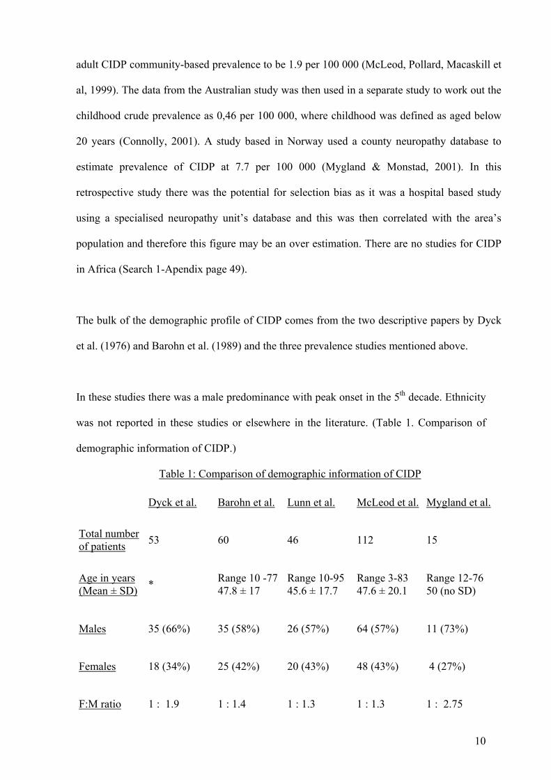
The bulk of the demographic profile of CIDP comes from the two descriptive papers by Dyck
et al. (1976) and Barohn et al. (1989) and the three prevalence studies mentioned above.
In these studies there was a male predominance with peak onset in the 5th decade. Ethnicity
was not reported in these studies or elsewhere in the literature. (Table 1. Comparison of
demographic information of CIDP.)
Table 1: Comparison of demographic information of CIDP
Dyck et al. Barohn et al. Lunn et al. McLeod et al. Mygland et al.
Total number of patients
53 60 46 112 15
Age in years (Mean ± SD)
* Range 10 -7747.8 ± 17
Range 10-95 45.6 ± 17.7
Range 3-83 47.6 ± 20.1
Range 12-76 50 (no SD)
Males 35 (66%) 35 (58%) 26 (57%) 64 (57%) 11 (73%)
Females 18 (34%) 25 (42%) 20 (43%) 48 (43%) 4 (27%)
F:M ratio 1 : 1.9 1 : 1.4 1 : 1.3 1 : 1.3 1 : 2.75
10
* No mean was available from this paper but the highest frequency of the disorder was reported in the 5th and 6th decade. 1.2.3. Development of diagnostic criteria to aid the diagnosis of CIDP
In Dyck’s paper the distinction was made between idiopathic inflammatory
polyradiculopathies and those associated with systemic illness because this was helpful in
making the diagnosis and in predicting out come. It also divided them into motor, sensory
and mixed types. A time frame of 6 months before the neurological deficit had “crested”
was used to divide the neuropathies into acute and chronic. The chronic form was then
found to follow a steadily progressive, recurrent, stepwise progressive or monophasic
course (Dyck et al, 1975).
The diagnostic criteria evolved during the decade that the Mayo group collected their data.
(1) No toxic or other disease could explain the neuropathy.
(2) There was a history of preceding illness or immunization.
(3) The patient had neurological deterioration that continued beyond 6 months.
(4) Involvement was usually symmetrical, with proximal and distal weakness seen.
(5) Papilloedema and essential tremor are occasionally seen.
(6) Electrodiagnostic study conduction velocities are generally slowed and may
even be blocked proximally and there is often a disproportion between clinical
signs and nerve conductions.
(7) There is cytoalbuminologic dissociation at some point during the course of the
illness and the ү-globulin may be elevated in the CSF.
(8) Full blood count (FBC) and erythrocyte sedimentation rates (ESR) are normal.
Many of these criteria have stood the test of time.
11
Since Dyck’s seminal paper there have been many attempts to redefine the definitions and
the diagnostic criteria for CIDP. A sensitive and specific set of diagnostic criteria is
important because CIDP represents up to 21% of undiagnosed neuropathies and is a
treatable condition (Dyck, Oviatt, & Lambert, 1981). By increasing the number of patients
diagnosed, more patients could get treated and therefore avoid the chronic morbidity
associated with this condition.
In 1989 an American group published a retrospective report of 60 patients with CIDP seen
in the Neuromuscular Division at the Ohio State University over a ten-year period (Barohn,
Kissel, Warmolts et al, 1989). Recommendations were given to expand the only diagnostic
criteria for CIDP that had been published at that time. These new criteria were broader and
therefore would allow for the heterogeneity seen in CIDP. One of the changes was the to
the to the progression of weakness beyond 6 months. This was reduced to 2 months because
they found that no patients with Acute Inflammatory Demyelinating Polyneuropathy
(AIDP) showed progression of weakness after 6 weeks (Mendell, Barohn, Freimer et al,
2001). This had the advantage of allowing earlier treatment of CIDP.
Later, an American Academy of Neurologists (AAN) task force developed diagnostic
criteria that are based on the history, neurological examination and nerve conduction
studies (Ad Hoc Subcommittee of the American Academy of Neurology AIDS Task Force,
1991). Cerebrospinal fluid analysis and sural nerve biopsy are mandatory for the diagnosis
of CIDP when using the AAN criteria.
12
The clinical features first described by Dyck are common to all currently available
diagnostic criteria (Dyck et al., 1975; Ad Hoc Subcommittee of the American Academy of
Neurology AIDS Task Force. 1991) All criteria require a patient to have at least two
months of progressive weakness, and symmetrical proximal and distal weakness is
considered a major diagnostic feature. The hallmark of CIDP is hyporeflexia or areflexia.
Although CIDP is a predominantly motor condition, the majority of CIDP patients also
have at least some sensory involvement. Numbness to pain and temperature testing may be
present in a stocking distribution and there may be associated paraesthesias in the same
regions. Proprioception may be lost in the lower limbs in classical CIDP (Dyck et al, 1975;
Barohn et al, 1989).
The AAN electrophysiological criteria require at least 3 of the following four criteria: (1)
Partial conduction block must be present in at least 1 motor nerve. This may be subdivided
into definite, probably or possible partial conduction block and defined as a greater than
20% drop in negative peak area or peak-to-peak amplitude, plus a less than 15% change in
duration between proximal and distal sites (partial conduction block) or a greater than 15%
change in duration between proximal and distal sites (possible conduction block/temporal
dispersion). Conduction block and temporal dispersion are only considered in the following
nerve segments: peroneal nerve between ankle and fibular head, median nerve between
wrist and elbow, and ulnar nerve between wrist and below elbow. (2) Conduction velocity
must be abnormal in at least 2 motor nerves. This is defined as a reduction in velocity less
than 80% of the lower limit of normal if the compound muscle action potential amplitude
(CMAP) amplitude is greater than 80% of the lower limit of normal or as a reduction in
velocity less than 70% of the lower limit of normal if the CMAP amplitude is less than 80%
of the lower limit of normal. (3) The distal latency must be abnormally increased in at least
13
two nerves. This is defined as prolonged more than 125% of the upper limit of normal if the
CMAP is greater than 80% of the lower limit of normal and l50% if the CMAP is less than
80% lower than normal. (4) F-wave latency must be abnormal in at least two motor nerves.
This is defined as an absent or prolonged F-wave more than 125% of the upper limit of
normal if the CMAP amplitude is more than 80% of the lower limit of normal or more than
150% of the upper limit of normal if the CMAP amplitude is less than 80% of the lower
limit (Ad Hoc Subcommittee of the American Academy of Neurology AIDS Task Force.
1991) . (Table 2: Comparison of the diagnostic criteria)
Only one to two thirds of patients with the diagnosis of CIDP made by a neuromuscular
specialist fulfil the AAN electrodiagnostic criteria (Magda, Latov, Brannagan III et al,
2003; Sander & Latov, 2003). There are several reasons for this. (1) There are insufficient
fibres affected. (2) The demyelination is proximal and therefore out of the field of study. (3)
There is severe secondary axonal damage that precludes accurate evaluation of nerve
conduction velocities. (4) In sensory nerves the action potentials may be absent due to
temporal dispersion demyelination and therefore not be documented (Sander et al, 2003).
The restrictive nature of the AAN criteria led to other groups developing more sensitive
criteria. These include the Nicolas, INCAT (Inflammatory Neuropathy Cause And
Treatment) and Saperstein Criteria (Hughes, Bensa, Willison et al, 2001; Saperstein, Katz,
Amato et al, 2001; Nicolas, Maisonobe, Le et al, 2002). The Nicolas criteria are purely
electrodiagnostic and therefore not appropriate to this clinically based study. Therefore they
will not be considered further.
14
The INCAT criteria are less stringent than the AAN or Saperstein criteria and do not
require CSF testing or nerve biopsy. The Saperstein criteria are similar to the AAN criteria
but only require two of the electrodiagnostic features and a biopsy is not mandatory for the
diagnosis.
Since Dyck et al. described the first cases of CIDP in 1975 the CSF parameters have been a
major component of the diagnostic criteria. In the 53 patients collected over 10 years, 44
had CSF results. The CSF protein was raised in 40 of the 44 patients (90%) at some stage in
their disease, where a protein of more than 0.45g/l was considered increased. The average
was 1.4g/l. The electrophoresis of the CSF protein showed raised IgG. The average number
of white cells, including lymphocytes and neutrophils was 4.26 /mm3. In central nervous
system infections typically the CSF protein and white cell count increase together, but in
CIDP the protein rises out of proportion to the cell count. This is referred to as
cytoalbuminological dissociation.
In the second of the large studies that provided a detailed demographic profile of patients
with CIDP, Barohn et al. examined CSF protein levels in 59 of the 60 patients in the study.
A raised protein was considered to be greater than 0.45g/l. The CSF protein was raised in
56 out of the 59 patients (95%) and the average white cell count was 1.7/mm3. They did not
perform protein electrophoresis on the CSF (Barohn et al, 1989).
The AAN Criteria require the white-cell count to be less than 10/mm3, a negative syphilis
serology test, and protein above 0.45g/l to diagnose CIDP. These criteria were used in two
large prevalence studies from England and Australia but the CSF protein means were not
reported (Lunn et al, 1999; McLeod et al, 1999).
15
The INCAT criteria recommend a raised CSF protein for the diagnosis but it is not
mandatory. In the Saperstein criteria the CSF protein should be more than 0.45 g/l and a
white-cell count of less than 10/mm3 is supportive.
A nerve biopsy is considered an essential part of the AAN criteria (1991). Molenaar et al.
combined clinical features, nerve conduction tests, CSF proteins and treating neurologists
clinical opinion and assessed whether the a sural nerve biopsy added to, or changed the
diagnosis (Molenaar, Vermeulen, & de Haan, 1998). They concluded that nerve biopsy did
not add any additional value when making the diagnosis of CIDP.
Although research criteria for enrolment in clinical studies need to have a high specificity,
clinical criteria should be more sensitive to allow the identification of patients who may
need treatment (Magda et al, 2003). Therefore the choices of criteria used in this study are
based on our departmental guidelines. We routinely do electrodiagnostic testing and CSF
analysis in our department and use the Saperstein criteria for the diagnosis of CIDP.
However, if the clinical history, examination and the nerve conduction studies fulfil the
criteria and the CSF is normal, we are inclined to ignore it as we feel uneasy denying
patients’ treatment in the face of other convincing evidence of CIDP. We do not perform
nerve biopsies routinely in keeping with the findings of Molenaar et al. (Molenaar et al,
1998)
More recently the European Federation of Neurological Societies has published a
consensus set of diagnostic criteria. These were not available at the time that this study was
initiated and therefore were not used. They closely resemble the INCAT criteria. These
16
guidelines are very useful in the treatment of chronic inflammatory demyelinating
polyradiculoneuropathy and are based on the available evidence and, where adequate
evidence was not available, consensus (EFNS/PNS CIDP Guidelines; 2005)
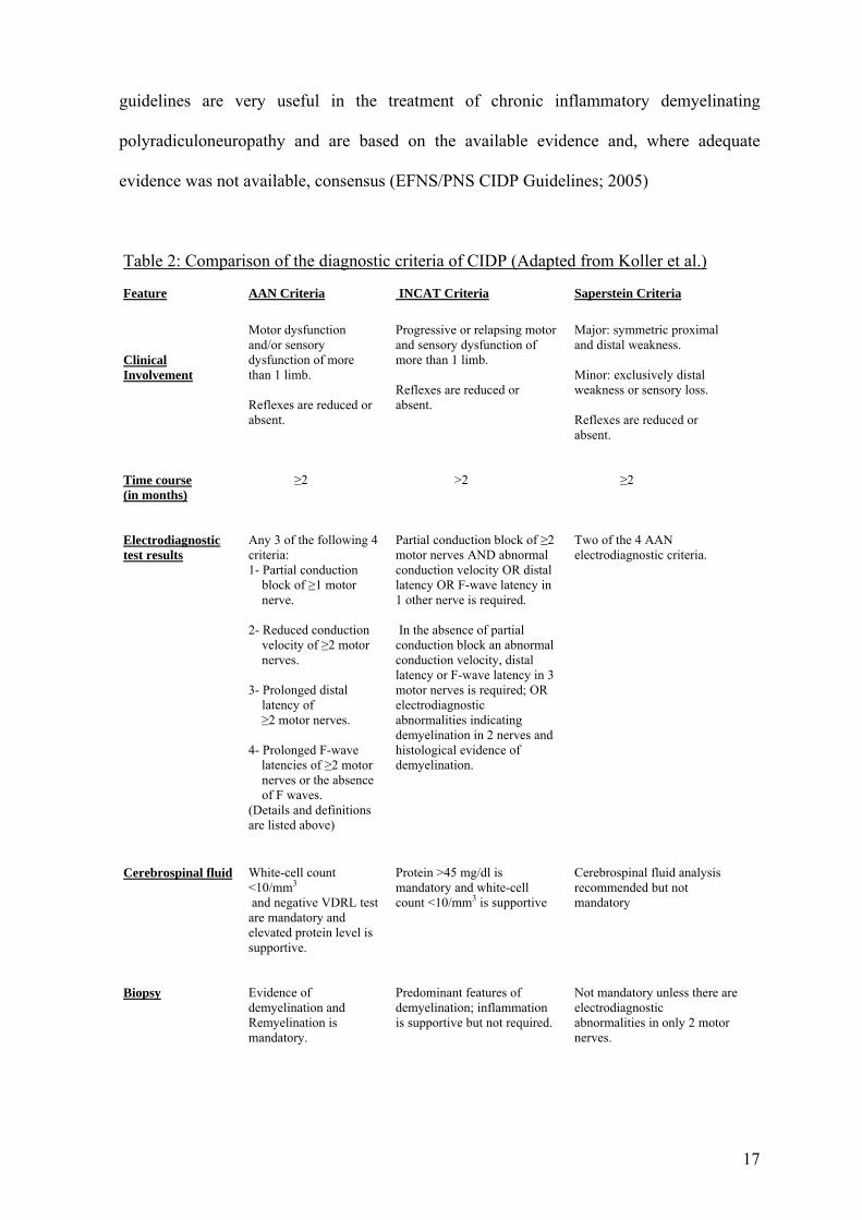
Table 2: Comparison of the diagnostic criteria of CIDP (Adapted from Koller et al.) Feature
AAN Criteria
INCAT Criteria
Saperstein Criteria
Clinical Involvement
Motor dysfunction and/or sensory dysfunction of more than 1 limb. Reflexes are reduced or absent.
Progressive or relapsing motor and sensory dysfunction of more than 1 limb. Reflexes are reduced or absent.
Major: symmetric proximal and distal weakness. Minor: exclusively distal weakness or sensory loss. Reflexes are reduced or absent.
Time course (in months)
≥2
>2
≥2
Electrodiagnostic test results
Any 3 of the following 4 criteria: 1- Partial conduction
block of ≥1 motor nerve.
2- Reduced conduction
velocity of ≥2 motor nerves.
3- Prolonged distal
latency of ≥2 motor nerves. 4- Prolonged F-wave latencies of ≥2 motor
nerves or the absence of F waves.
(Details and definitions are listed above)
Partial conduction block of ≥2 motor nerves AND abnormal conduction velocity OR distal latency OR F-wave latency in 1 other nerve is required. In the absence of partial conduction block an abnormal conduction velocity, distal latency or F-wave latency in 3 motor nerves is required; OR electrodiagnostic abnormalities indicating demyelination in 2 nerves and histological evidence of demyelination.
Two of the 4 AAN electrodiagnostic criteria.
Cerebrospinal fluid
White-cell count <10/mm3 and negative VDRL test are mandatory and elevated protein level is supportive.
Protein >45 mg/dl is mandatory and white-cell count <10/mm3 is supportive
Cerebrospinal fluid analysis recommended but not mandatory
Biopsy
Evidence of demyelination and Remyelination is mandatory.
Predominant features of demyelination; inflammation is supportive but not required.
Not mandatory unless there are electrodiagnostic abnormalities in only 2 motor nerves.
17
1.2.4. Pathophysiology
The aetiology of acquired demyelinating polyneuropathies is presumed to be autoimmune
or dysimmune (Koller, Kieseier, Jander et al, 2005). Unlike AIDP there is no clear
association between the CIDP and antecedent infections. It is likely that myelin proteins act
as a target for the immune system in CIDP. When mice are inoculated with the P0 protein, a
major myelin protein, they develop demyelination and conduction blocks (Yan, Archelos,
Hartung et al, 2001). It is thought that an auto-antigen activates T lymphocytes in the in the
blood. These activated T lymphocytes then cross the blood–nerve barrier in a complex
process involving cellular adhesion molecules, matrix metalloproteinases, and chemokines
(Quattrini, Previtali, Kieseier et al, 2003). Within the peripheral nervous system, T cells
activate macrophages that enhance phagocytic activity, cytokine production, and the release
of toxic mediators, including nitric oxide, tumour necrosis factor alpha and interferon
gamma (Oka, Akiguchi, Kawasaki et al, 1998). These activated T cells also induce
autoantibody production. These are produced by plasma cells and contribute to
demyelination and axonal damage via the complement pathway or direct adhesion to
membrane channels (Quattrini et al., 2003).
1.2.5. Pathology
The hallmark of CIDP is demyelination that is multifocal. In one large series of biopsies in
patients with CIDP, demyelinating features were seen in only 48%, 21% had predominantly
axonal changes, 13% had mixed demyelinating and axonal changes, and 18% were normal
(Barohn et al, 1989). There may be other evidence of inflammation like endoneural and
subepineural oedema, T lymphocyte and macrophage infiltrates and histochemical staining
18
may be positive for cytokines like TNF alpha. Remyelination is seen in advanced cases of
CIDP and is evidenced by onion bulb formation (Dyck et al, 1975).
1.2.6. Disease course
It has been noted that CIDP can follow a steadily progressive, recurrent, stepwise
progressive or monophasic course. In two studies patients were divided into recurrent and
not recurrent (Table 3. Clinical Course). There is a large variation between each of the
papers and this is in part due to patients being treated prior to the studies and this changes
the course of the illness (Lunn et al, 1999; McLeod et al, 1999). The variation or difference
found might be explained, at least in part, by prior treatment. In an attempt to avoid the
impact of treatment on our patients’ course, we documented their temporal course prior to
initiating treatment.
1.2.7. CIDP Subtypes
Since the 1950’s other forms of acquired demyelinating polyneuropathies have been
described that differ from classic chronic inflammatory demyelinating polyneuropathy,
both with respect to clinical presentation and in their response to treatment. It is not clear
whether these conditions are variants of chronic inflammatory demyelinating
polyneuropathy or distinct disease entities as there pathophysiology is not fully understood.
These conditions are occasionally classified together because they have similar
electrophysiological findings and can be treated with immunotherapy.
(http://neuromuscular.wustl.edu/antibody/pnimdem.html) (Table 4. CIDP Subtypes)
19
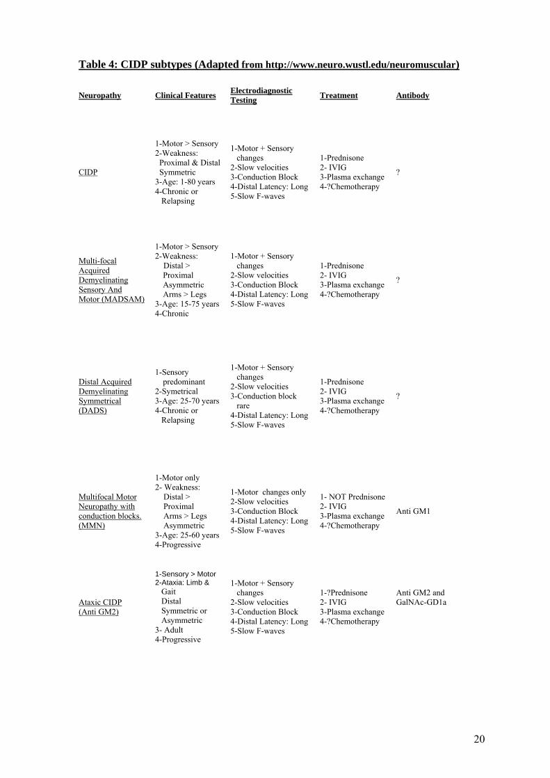
Table 4: CIDP subtypes (Adapted from http://www.neuro.wustl.edu/neuromuscular)
Neuropathy Clinical Features Electrodiagnostic Testing Treatment Antibody
CIDP
1-Motor > Sensory 2-Weakness: Proximal & Distal Symmetric 3-Age: 1-80 years 4-Chronic or Relapsing
1-Motor + Sensory changes 2-Slow velocities 3-Conduction Block 4-Distal Latency: Long5-Slow F-waves
1-Prednisone 2- IVIG 3-Plasma exchange 4-?Chemotherapy
?
Multi-focal Acquired Demyelinating Sensory And Motor (MADSAM)
1-Motor > Sensory 2-Weakness: Distal >
Proximal Asymmetric Arms > Legs
3-Age: 15-75 years 4-Chronic
1-Motor + Sensory changes 2-Slow velocities 3-Conduction Block 4-Distal Latency: Long5-Slow F-waves
1-Prednisone 2- IVIG 3-Plasma exchange 4-?Chemotherapy
?
Distal Acquired Demyelinating Symmetrical (DADS)
1-Sensory predominant
2-Symetrical 3-Age: 25-70 years 4-Chronic or Relapsing
1-Motor + Sensory changes 2-Slow velocities 3-Conduction block rare 4-Distal Latency: Long5-Slow F-waves
1-Prednisone 2- IVIG 3-Plasma exchange 4-?Chemotherapy
?
Multifocal Motor Neuropathy with conduction blocks. (MMN)
1-Motor only 2- Weakness: Distal > Proximal Arms > Legs Asymmetric 3-Age: 25-60 years 4-Progressive
1-Motor changes only 2-Slow velocities 3-Conduction Block 4-Distal Latency: Long5-Slow F-waves
1- NOT Prednisone 2- IVIG 3-Plasma exchange 4-?Chemotherapy
Anti GM1
Ataxic CIDP (Anti GM2)
1-Sensory > Motor 2-Ataxia: Limb & Gait Distal Symmetric or Asymmetric 3- Adult 4-Progressive
1-Motor + Sensory changes 2-Slow velocities 3-Conduction Block 4-Distal Latency: Long5-Slow F-waves
1-?Prednisone 2- IVIG 3-Plasma exchange 4-?Chemotherapy
Anti GM2 and GalNAc-GD1a
20
Neuropathy Clinical Features Electrodiagnostic Testing Treatment Antibody
Gait disorder Auto antibody Late age Onset Polyneuropathy (GALOP)
1-Sensory > Motor 2-Ataxic gait Distal Symmetric 3-Age: > 50 years 4-?
1-Motor + Sensory changes 2-Slow velocities 3-Conduction Block 4-Distal Latency: Long5-Slow F-waves
1-?Prednisone 2- ?IVIG 3-Plasma exchange 4-?Chemotherapy
Membrane Sulphatide anti- body
Polyneuropathy Organomegaly Endocrineopathy M-protein Skin changes (POEMS)
1- Sensory and Motor 2-Symmetric 3-Age: 25-60 years
1-Motor + Sensory changes 2-Slow velocities 3-No Conduction Block 4-Distal Latency: Long5-Slow F-waves
1-Prednisone 2- IVIG 3-Plasma exchange 4-?Chemotherapy 5- Removal of tumour
?
1.2.8. CIDP and concurrent illness
CIDP may be also associated with concurrent illness for example, viral diseases like HIV
and hepatitis C, inflammatory diseases like Sjögren’s syndrome and inflammatory bowel
disease and neoplasms like melanoma and lymphoma. The relevance of such concurrent
diseases is unclear. Monoclonal gammopathy of unknown significance (MGUS) is also
associated with CIDP and here the pathology may be due to myelin and auto-antibody
interaction (Gorson, Allam, & Ropper, 1997).
The association with diabetes mellitus is very important because CIDP occurs more
commonly among patients with diabetes (Stewart, McKelvey, Durcan et al, 1996). This
21
creates diagnostic and management difficulty as diabetic patients often have pre-existing
neuropathy and use of prednisone in the treatment of CIDP makes glycaemic control
difficult.
CIDP may develop in conjunction with another polyneuropathy, even one with a hereditary
basis, such as Charcot–Marie–Tooth disease (Ginsberg, Malik, Kenton et al, 2004).
While the association between acute inflammatory demyelinating polyradiculoneuropathy
(AIDP) and HIV is well established, the association of CIDP and HIV is less so. The first
case of AIDP associated with AIDS was described in 1985. Cornblath described three
patients with AIDP preceding the diagnosis of AIDS in 1987 (Ferrari, Vento, Monaco et al,
2006). Later studies confirmed the observation that AIDP occurs early in HIV-1 infection
before severe immunosuppression (Vendrell, Heredia, Pujol et al, 1987). In a large series of
32 patients with AIDP in Zimbabwe, all 16 HIV-1-positive patients developed AIDP before
the diagnosis of AIDS (Thornton, Latif, & Emmanuel, 1991). The original description of
HIV and CIDP occurring together was in 1987 (Cornblath, McArthur, Kennedy et al,
1987). (Search 2)
In a study from 2003, 10 patients with AIDP also had HIV and only 40% developed AIDP
after onset of AIDS (CD4 T-cell count <200/ml). Three of these patients went on to
develop CIDP. This study also showed that cerebrospinal fluid pleocytosis was not always
present in HIV-associated AIDP (Brannagan III & Zhou, 2003). The CSF pleocytosis in
HIV associated CIDP was considered one of the distinguishing features from CIDP patients
without HIV.
22
Little has been published about the South African experience of CIDP. The Kwazulu-Natal
experience has been alluded to in a recent review paper. Twenty-four HIV positive patients
were seen over a 10-year period with CIDP. Clinical features were identical in the HIV and
non-HIV patients except for the presence of a mild lymphocytic pleocytosis in the CSF in
the HIV positive patients. All patients responded well to treatment. The description does
not provide details of the patients’ CD4 counts, race, age or gender (Bhigjee, 2005).
Despite there being very little in the literature about the HIV-associated CIDP there are
some important differences that have been noted. The CSF protein level is often raised
throughout the course of HIV (Marshall, Brey, Cahill et al, 1988). The protein level does
not however correlate with the degree of the distal sensory polyneuropathy seen in HIV
(Barohn, Gronseth, Amato et al, 1996). The classic cytoalbuminological dissociation is not
always seen in HIV-associated CIDP (Cornblath et al, 1987). HIV has been isolated from
nerves of patients with CIDP (Dalakas & Pezeshkpour, 1988). Cytomegalovirus was
isolated from nerves of patients with HIV-associated CIDP but not in the non-HIV type
(Grafe & Wiley, 1989). Pathologically these nerve biopsies are similar, showing segmental
demyelination with an inflammatory infiltration of monocytes in the endo- and epineurium
(Cornblath et al, 1987).
In the original article describing the association between CIDP and HIV, Cornblath et al.
found a male predominance. The majority of these patients had risk factors for HIV
namely being male homosexuals or intravenous drug users. The prognosis for patients with
both HIV and CIDP seems to be similar to the non-HIV patients with CIDP. Patients who
show a progressive course are amenable to the standard forms of treatment but given the
23
immunosuppressive nature of these, Cornblath recommended that patients should be
monitored closely (Cornblath et al, 1987).
1.2.9. Treatment
Corticosteroids, plasmapheresis, and IVIg are all effective treatments in CIDP. Individual
patients, however, may differ in response to any one of these treatments (Gorson et al,
1997). A single randomised controlled trial provided weak evidence to support the use of
corticosteroids in CIDP and subsequent studies have shown that there is no significant
difference between the treatment modalities (Gorson et al, 1997; Sghirlanzoni, Solari,
Ciano et al, 2000; Mehndiratta & Hughes, 2002). The principal of treatment is to block the
inflammatory process and thereby prevent further demyelination and secondary axonal loss
leading to permanent disability (Gorson et al, 1997; Sghirlanzoni et al, 2000; Koller et al,
2005).
1.2.10. Prognosis
In a series of 83 patients evaluated on average 6 years after onset, 56% had good outcome,
24% deteriorated and failed to respond to all treatments, and 11% died of complications of
the disease. Axonal loss on the nerve biopsy correlated with poorer outcome (Bouchard,
Lacroix, Plante et al, 1999; Sghirlanzoni et al, 2000). In a more recent study the prognosis
seemed to be more favourable with 39% of patients still requiring immune treatments and
13% having severe disabilities (Kuwabara, Misawa, Mori et al, 2006). The literature
suggests that early treatment may be helpful in improving prognosis by avoiding axonal
damage, though there are no randomised data yet to support this.
24
2. Aim of the Study
In the literature are several studies that describe the clinical presentation and diagnostic
criteria for CIDP. However, these studies have been done in high-income countries and in
predominantly caucasian populations. South Africa, a middle-income country with a high
prevalence of HIV and multiethnic population provides an ideal setting to add information
to the literature.
Our aim was to describe the clinical features, cerebrospinal fluid findings and
electrophysiological examination in an urban, hospital-based, South African population.
3. Design
This is a descriptive study combining retrospective case review and prospective assessment of
patients with CIDP referred to the Johannesburg Hospital Division of Neurology.
3.1. Population and case ascertainment
We based our study at the Johannesburg Hospital, a 1088 bed academic referral hospital
that provides health care to predominantly indigent patients. The hospital also provides care
at a primary and secondary level to much of the population of Johannesburg. Patients with
CIDP were ascertained from the Johannesburg, Chris Hani Baragwanath and Helen Joseph
Hospital neurology services. Patients with CIDP reached these services either by direct
referral or indirectly via other medical departments.
25
3.2. Duration of the study
Patients were ascertained and assessed from 1st January 2005 to 31st December 2006 (24
months).
3.3. Assessment of patients
All patients were examined by at least one neurologist. Nerve conduction studies were done
at the Johannesburg Hospital by a neurologist or a neurology registrar who was supervised
by a neurologist. Lumbar punctures and blood tests were done at the Johannesburg Hospital
and analysed at the National Health Sciences Laboratory. The first four patients were
studied retrospectively the remaining 22 were studied prospectively.
3.4. Inclusion Criteria
Patients were included if they gave informed consent to take part in the study, provided
they fulfilled the Saperstein criteria for CIDP. These criteria include mandatory clinical,
electrodiagnostic, and supportive cerebrospinal fluid criteria, as well as a time course of
greater than two months progression. Patients were entitled to withdraw consent or refuse
any of the investigations. If they did this we excluded them from the study. One patient
refused to take part but received the same standard treatment.
26
3.5. Ethics
Approval from the Johannesburg Hospital was obtained in writing to proceed with the
study. The study protocol, data collection sheet, consent form and information sheet were
approved by the University of the Witwatersrand Ethics Committee and Postgraduate
Committee. All patients, both retrospectively and prospectively studied signed consent.
3.6. Statistical analysis
Statistical analysis was performed using Stata (Stata Corp. 2007. Stata Statistical Software:
Release 10. College Station, TX: Stata Corp LP.).
4. Results
4.1. Demographics
Number of patients: Twenty-six patients were diagnosed with CIDP over a two-year period
(1 January 2005 to 31 December 2006).
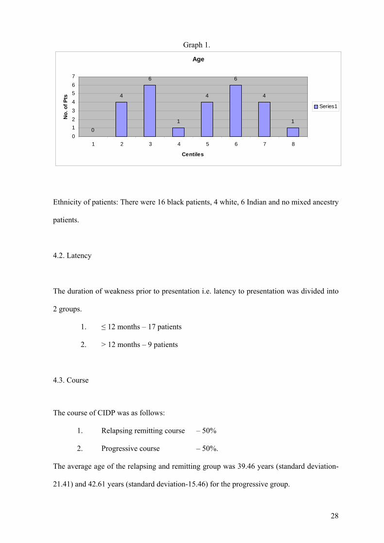
Gender of patients: Eight were male and 18 were female. (Ratio male to female - 1:2.25)
Age of patients: The range of the ages was 12 to 74 years of age. The average age was 41
years with a standard deviation of 18, 37 years (Graph 1).
27
Graph 1.
Age
0
4
6
1
4
6
4
1
01234567
1 2 3 4 5 6 7 8
Centiles
No.
of P
ts
Series1
Ethnicity of patients: There were 16 black patients, 4 white, 6 Indian and no mixed ancestry
patients.
4.2. Latency
The duration of weakness prior to presentation i.e. latency to presentation was divided into
2 groups.
1. ≤ 12 months – 17 patients
2. > 12 months – 9 patients
4.3. Course The course of CIDP was as follows:
1. Relapsing remitting course – 50%
2. Progressive course – 50%.
The average age of the relapsing and remitting group was 39.46 years (standard deviation-
21.41) and 42.61 years (standard deviation-15.46) for the progressive group.
28
Analysis of the association between latency to presentation and time course of illness by
Pearson chi-square test reveals a statistically significant correlation with 65% of patients
with a progressive course presenting within one year of symptom onset (p = 0.039).
No statistically significant difference could be found by two-sample t-test when comparing
the age of patients with the course of their illness. This was confirmed using the two-
sample Wilcoxon rank-sum (Mann-Whiney) test.
4.4. Motor findings
The majority of the patients had the characteristic motor signs of proximal and distal
weakness in the lower limbs and only distal weakness in the upper limbs except for three.
All three of these patients had distal weakness as the dominant feature and two of them also
had an asymmetrical presentation and were male. Areflexia was common to all 26 patients.
Neck flexion weakness was found in 19 of the patients. There were 8 patients who
presented unable to stand, 3 could stand unaided but not walk, 11 had a high stepping gait,
3 had a completely normal gait and 1 was ataxic and could stand. Patients were grouped
into ambulatory (14 patients) and non-ambulatory (12 patients) for statistical analysis.
Using the Pearson chi-square test no association was found between latency to presentation
and severity in terms of ambulation.
29
4.5. Sensory Findings
Most of the patients had mild sensory signs of loss to pain and temperature except 3
patients who had no sensory signs. These included the two male patients mentioned above
who had an asymmetrical presentation. Another patient had a predominantly ataxic
presentation with loss of joint position sense up to the knee and pseudoathetosis.
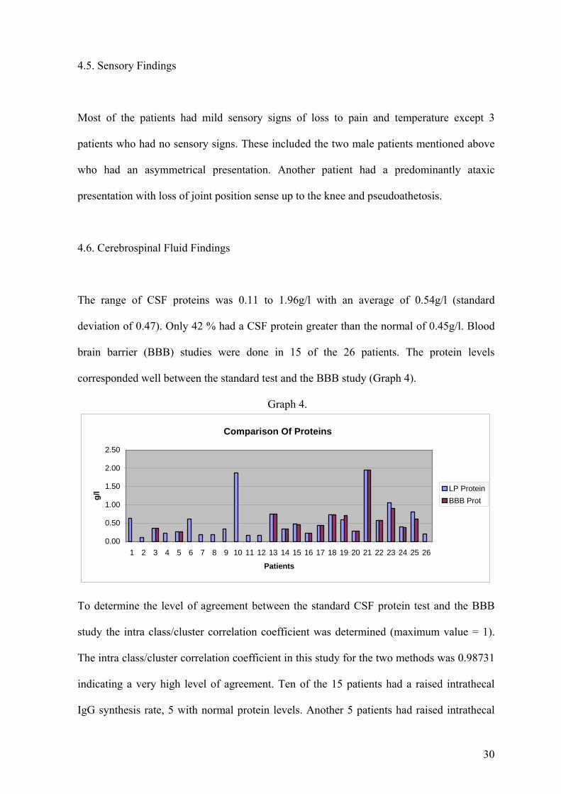
4.6. Cerebrospinal Fluid Findings
The range of CSF proteins was 0.11 to 1.96g/l with an average of 0.54g/l (standard
deviation of 0.47). Only 42 % had a CSF protein greater than the normal of 0.45g/l. Blood
brain barrier (BBB) studies were done in 15 of the 26 patients. The protein levels
corresponded well between the standard test and the BBB study (Graph 4).
Graph 4.
Comparison Of Proteins
0.00
0.50
1.00
1.50
2.00
2.50
1 2 3 4 5 6 7 8 9 10 11 12 13 14 15 16 17 18 19 20 21 22 23 24 25 26
Patients
g/l LP Protein
BBB Prot
To determine the level of agreement between the standard CSF protein test and the BBB
study the intra class/cluster correlation coefficient was determined (maximum value = 1).
The intra class/cluster correlation coefficient in this study for the two methods was 0.98731
indicating a very high level of agreement. Ten of the 15 patients had a raised intrathecal
IgG synthesis rate, 5 with normal protein levels. Another 5 patients had raised intrathecal
30
IgG synthesis rates and raised CSF proteins. The remaining 5 either had BBB damage or
were indeterminate. (Table 6)
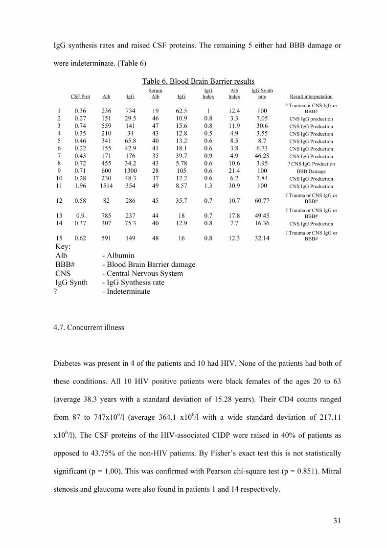
Table 6. Blood Brain Barrier results
Key:
CSF Prot Alb IgG
Serum Alb IgG
IgG Index
Alb Index
IgG Synth rate Result interpretation
1 0.36 236 734 19 62.5 1 12.4 100
? Trauma or CNS IgG or BBB#
2 0.27 151 29.5 46 10.9 0.8 3.3 7.05 CNS IgG production 3 0.74 559 141 47 15.6 0.8 11.9 30.6 CNS IgG Production 4 0.35 210 34 43 12.8 0.5 4.9 3.55 CNS IgG Production 5 0.46 341 65.8 40 13.2 0.6 8.5 8.7 CNS IgG Production 6 0.22 155 42.9 41 18.1 0.6 3.8 6.73 CNS IgG Production 7 0.43 171 176 35 39.7 0.9 4.9 46.28 CNS IgG Production 8 0.72 455 34.2 43 5.78 0.6 10.6 3.95 ? CNS IgG Production 9 0.71 600 1300 28 105 0.6 21.4 100 BBB Damage
10 0.28 230 48.3 37 12.2 0.6 6.2 7.84 CNS IgG Production 11 1.96 1514 354 49 8.57 1.3 30.9 100 CNS IgG Production
12 0.58 82 286 45 35.7 0.7 10.7 60.77
? Trauma or CNS IgG or BBB#
13 0.9 785 237 44 18 0.7 17.8 49.45
? Trauma or CNS IgG or BBB#
14 0.37 307 75.3 40 12.9 0.8 7.7 16.36 CNS IgG Production
15 0.62 591 149 48 16 0.8 12.3 32.14 ? Trauma or CNS IgG or
BBB#
Alb - Albumin BBB# - Blood Brain Barrier damage CNS - Central Nervous System IgG Synth - IgG Synthesis rate ? - Indeterminate
4.7. Concurrent illness
Diabetes was present in 4 of the patients and 10 had HIV. None of the patients had both of
these conditions. All 10 HIV positive patients were black females of the ages 20 to 63
(average 38.3 years with a standard deviation of 15.28 years). Their CD4 counts ranged
from 87 to 747x106/l (average 364.1 x10⁶/l with a wide standard deviation of 217.11
x106/l). The CSF proteins of the HIV-associated CIDP were raised in 40% of patients as
opposed to 43.75% of the non-HIV patients. By Fisher’s exact test this is not statistically
significant (p = 1.00). This was confirmed with Pearson chi-square test (p = 0.851). Mitral
stenosis and glaucoma were also found in patients 1 and 14 respectively.
31
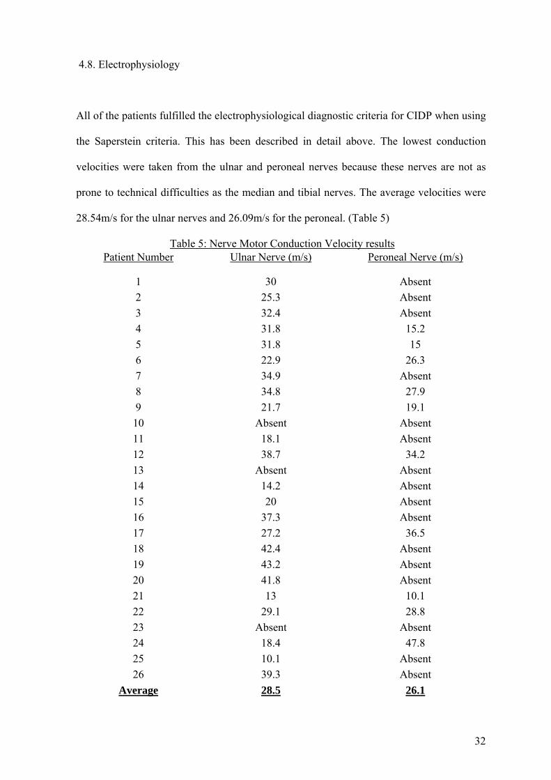
4.8. Electrophysiology
All of the patients fulfilled the electrophysiological diagnostic criteria for CIDP when using
the Saperstein criteria. This has been described in detail above. The lowest conduction
velocities were taken from the ulnar and peroneal nerves because these nerves are not as
prone to technical difficulties as the median and tibial nerves. The average velocities were
28.54m/s for the ulnar nerves and 26.09m/s for the peroneal. (Table 5)
Table 5: Nerve Motor Conduction Velocity results Patient Number Ulnar Nerve (m/s) Peroneal Nerve (m/s)
1 30 Absent 2 25.3 Absent 3 32.4 Absent 4 31.8 15.2 5 31.8 15 6 22.9 26.3 7 34.9 Absent 8 34.8 27.9 9 21.7 19.1 10 Absent Absent 11 18.1 Absent 12 38.7 34.2 13 Absent Absent 14 14.2 Absent 15 20 Absent 16 37.3 Absent 17 27.2 36.5 18 42.4 Absent 19 43.2 Absent 20 41.8 Absent 21 13 10.1 22 29.1 28.8 23 Absent Absent 24 18.4 47.8 25 10.1 Absent 26 39.3 Absent
Average 28.5 26.1
32
Normal Velocity Values: 1- ulnar nerve - >45.0 m/s
2- peroneal nerve - >40.0 m/s
Reference: Preston D & Shapiro B: Electromyography and Neuromuscular Disorders
Clinical-Electrophysiological Correlations, Second Edition 2005
4.9. Antibodies
There were only 3 patients that had an asymmetrical presentation. All three were males who
did not have the classic mixed motor and sensory CIDP but clinically had multifocal motor
neuropathy with conduction blocks. None of these patients received steroids. There was one
patient with the rare ataxic variant of CIDP. None of our patients had DADS or MADSAM
neuropathy.
Two patients had positive antibodies on serum testing. Patient 9 was a 53-year-old Indian
male and was positive for GM1 antibodies. He was diagnosed with multifocal motor
neuropathy with conduction blocks. There were two other patients who had pure motor
syndromes clinically and electrophysiologically but both were negative for GM1
antibodies. The second patient with a specific antibody was patient number 6, a white 68-
year-old female who presented with a severe sensory ataxia. She tested positive for GM2
anti bodies.
4.10. HIV and CIDP
Ten of our 26 patients with CIDP were HIV infected. There are several differences between
the HIV negative and positive groups.
33
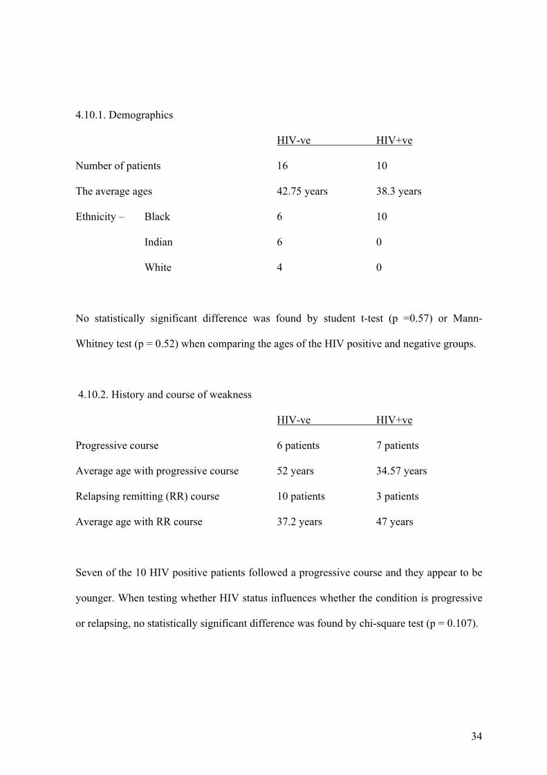
4.10.1. Demographics
HIV-ve HIV+ve
Number of patients 16 10
The average ages 42.75 years 38.3 years
Ethnicity – Black 6 10
Indian 6 0
White 4 0
No statistically significant difference was found by student t-test (p =0.57) or Mann-
Whitney test (p = 0.52) when comparing the ages of the HIV positive and negative groups.
4.10.2. History and course of weakness
HIV-ve HIV+ve
Progressive course 6 patients 7 patients
Average age with progressive course 52 years 34.57 years
Relapsing remitting (RR) course 10 patients 3 patients
Average age with RR course 37.2 years 47 years
Seven of the 10 HIV positive patients followed a progressive course and they appear to be
younger. When testing whether HIV status influences whether the condition is progressive
or relapsing, no statistically significant difference was found by chi-square test (p = 0.107).
34
To analyse whether there is an association between HIV status and the age of patients with
a progressive or a relapsing course a two way anova was performed. Although not
statistically significant (p = 0.191) there is a strong indication of interaction between
progression and HIV status being present for age. In particular a progressive course is more
likely in older HIV positive patients.
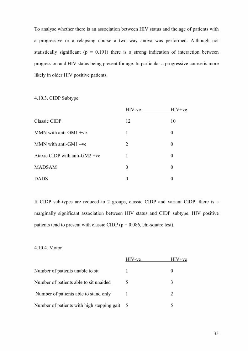
4.10.3. CIDP Subtype
HIV-ve HIV+ve
Classic CIDP 12 10
MMN with anti-GM1 +ve 1 0
MMN with anti-GM1 –ve 2 0
Ataxic CIDP with anti-GM2 +ve 1 0
MADSAM 0 0
DADS 0 0
If CIDP sub-types are reduced to 2 groups, classic CIDP and variant CIDP, there is a
marginally significant association between HIV status and CIDP subtype. HIV positive
patients tend to present with classic CIDP (p = 0.086, chi-square test).
4.10.4. Motor
HIV-ve HIV+ve
Number of patients unable to sit 1 0
Number of patients able to sit unaided 5 3
Number of patients able to stand only 1 2
Number of patients with high stepping gait 5 5
35
Number of patients with an ataxic gait 1 0
Number of patients with a normal gait 3 3
4.10.5. Sensory
All of the HIV positive patients had a glove and stocking sensory loss to pain and
temperature.
4.10.6. Cerebrospinal fluid
HIV-ve HIV+ve
Average protein 0.51g/l 0.57g/l
Number of patients with protein > 0.45g/l 7 4
Average Glucose 3.89 mmol/l 3.16 mmol/l
Average Chloride 121.4 mmol/l 127.2 mmol/l
Average Polymorphs count 0.19 x 10⁶/l 0.5 x 10⁶/l
Average Lymphocytes count 0.81 x 10⁶/l 16.6 x 10⁶/l
By two sample t-test with equal variances there were no statistically significant differences
between HIV positive and HIV negative patients with respect to CSF protein (p = 0.381) or
glucose (p = 0.967). CSF chloride was significantly higher in the HIV positive group (p =
0.023), but both were in within the normal range for our laboratory (116-130mmol/l).
Cytoalbuminological dissociation was present in all 16 HIV negative patients and in 7 of
the 10 HIV positive patients. Using Pearson’s chi-square test, the absence of
36
cytoalbuminological dissociation is strongly associated with HIV positive status (p =
0.020).
5. Discussion
CIDP remains a relatively unknown condition. Apart from neurologists, most clinicians are
unaware of the existence of the condition. The old term of “Chronic Guillain-Barré
Syndrome” is still adhered to, other than in neurological circles. There is a lack of
awareness of CIDP variants, which now include specific antibody testing. The evolution of
this disease has created problems with the diagnosis and diagnostic criteria as described
above. This problem is primarily responsible for what may be an underestimation of the
condition’s prevalence. (Dyck, Lais, Ohta et al, 1975; Dyck, Oviatt, & Lambert, 1981;
Gorson, Allam & Ropper, 1997; Koller, Kieseier, Jander et al, 2005). There are two
prevalence studies, one from Australia the other from the United Kingdom (Lunn, Manji,
Choudhary et al, 1999; McLeod, Pollard, Macaskill et al, 1999). Both of these studies used
very strict diagnostic criteria for CIDP. The prevalence is thought to be about 1: 100 000.
These studies are also based on a limited number of cases.
There is very little information about the condition in Africa and no studies have been done
at all in Sub Saharan Africa (Search 1.)
Obtaining large number of cases of CIDP is difficult. In the original Dyck et al. paper from
1975, 53 patients were collected over a 10-year period from several centres and Barohn et
al. (1989) collected 60 patients from a specialized neuromuscular unit over 10 years. We
were able to collect 26 patients over a 2-year period from the greater Johannesburg area.
37
This number may be an insufficient sample size to derive robust statistical information but
this study will nevertheless contribute numbers to the CIDP body of knowledge and
particularly on CIDP in Africans.
5.1. Demographics
5.1.1 Gender Ratio
According to the literature there is a male predominance in patients with CIDP of
approximately 2:1. In our case series we found that 8 out of the 26 patients were male
which gives a reverse ratio of 2.25 females to one male.
This may be co-incidental and be a reflection of small sample size. A hypothetical alternate
explanation may be the relationship to the HIV epidemic. As in the rest of sub-Saharan
Africa, the epidemic in South Africa disproportionately affects women. Young women (15–
24 years) are four times more likely to be HIV-infected than are young men: in 2004,
prevalence among young women was 17% compared with 4.4% among young men
(Connolly, Shisana, Colvin et al, 2004). We found that 38.46% of our patients were HIV
positive and all were female. There were 16 HIV negative patients, with 8 males and 8
females, giving a 1:1 ratio that is closer to the gender ratio seen in the literature.
5.1.2. Ethnicity
38
Ethnicity has not previously been documented in the literature and all the pivotal studies do
not mention ethnic backgrounds of patients.
In our study we divided patients on the basis of ethnic backgrounds and found that there
were 16 Black patients, 4 White, 6 Indian and no patients of mixed ancestry. This
demographic distribution is a fair reflection of the population in South Africa.
Patients with HIV and CIDP were all black. This again may be co-incidental or more likely
is a reflection of the HIV epidemic in South Africa (Connolly, 2001).
5.1.3. Age
Our patients had an earlier age of onset compared to the literature. The average age of our
group was 41 years. In the international literature the age of onset is older than this, usually
in the 50th to 60th decades (Table 2). When the HIV positive patients are separated from the
rest of the group they are slightly younger at 38.3 years compared to 41.2 years in the HIV
negative group (Graph 2). Overall, our patients are younger even when the HIV patients are
discounted. This may be co-incidental or represent the profile of CIDP in African patients.
5.1.4. Latency
The duration of weakness prior to presentation was divided into 4 groups. Twelve patients
presented from 2 to 6 months from the onset of their symptoms, 5 patients from 6 to 12
months, 2 patients from 12 to 24 months and 7 patients presented more than two years from
the onset of their symptoms.
39
To see if there is a correlation between the latency and severity of the CIDP, the time to
presentation was allied to the patients’ ability to walk at presentation.
Twelve patients (46%) presented within 6 months of onset of their illness. Five of these
patients could only sit, one could only stand and not walk, 5 had a high stepping gait, and
one had a severe ataxic syndrome and this prevented ambulation.
Five patients (19%) presented between 6 and 12 months from the onset of the illness. Two
could only sit, one could only stand but not walk, one had a high stepping gait and one had
a normal gait.
Two patients (8%) presented in the 12 to 24-month group. One was paralysed completely
and could not sit unaided and the other had a high stepping gait.
The last 7 patients presented after 24 months. One could sit only, one could stand unaided,
3 had a high stepping gait and 2 had a normal gait and.
The correlation between latency to presentation and the severity of the illness is not clear
due to the small numbers in the study but it is noted that the majority of the patients
presented in the first 6 months from onset of disease (46%). Patients who presented later
were generally ambulatory.
5.1.5. Course
40
There was an even split between patients with a progressive course and a relapsing
remitting course. In the literature there is a large variation between the numbers of patients
with a relapsing remitting or progressive course. This may be due to different criteria used
in each of the studies to define the course and due to the small number of patients in each of
the studies. In the large prevalence study in the United Kingdom there was a 50% split
between the two groups. The distribution between the two groups demonstrated in our data
most closely resembles that of the largest study found in the literature to date (McLeod et
al, 1999).
According to the literature the patients with a relapsing remitting course are usually
younger. In our study the average age of the relapsing remitting group of patients was 39.46
years and 42.61 years for the progressive group. This included HIV positive and negative
patients. There does not appear to be a significant difference between the ages of the
relapsing or progressive groups. This may be due to the history of the course of the illness
being incorrectly documented as several of our patients could not speak English and a
translator was required. It may be that our numbers are too small and they do not reflect
CIDP in our population.
5.2. Motor
The majority of our patients presented with the classic motor signs and areflexia.
5.3. Sensory
41
Most of the patients had mild sensory signs of loss to pain and temperature except 3
patients who had no sensory signs.
5.4. Cerebrospinal fluid
The CSF protein is quoted as being raised in 80-90% of CIDP cases (Barohn, Kissel,
Warmolts et al, 1989). However, only 42% of our patients had a raised protein in the CSF.
To confirm the CSF protein levels, blood brain barrier studies were performed in 15
patients. It was found that 52% of these patients had raised CSF proteins. This verified the
lower than expected CSF protein levels in our patient group.
In the original article describing HIV associated CIDP the CSF protein level was raised in 5
of the 6 patients with CIDP (Cornblath, McArthur, Kennedy et al, 1987). In our group of 10
HIV positive patients with CIDP, 4 had a raised CSF protein.
The blood brain barrier tests also showed a raised intrathecal IgG synthesis rate in 10 of the
15 patients. Five of these had normal CSF protein levels. This implies that despite a normal
CSF protein there was an intrathecal inflammatory process. Another 5 patients had both
raised intrathecal IgG synthesis rates and raised CSF protein levels as expected. The
remaining 5 either had BBB damage or were indeterminate (Table 6.).
The pathological hallmark of CIDP is segmental demyelination that occurs from the roots
along the entire path of the peripheral nerve. The raised CSF protein seen in CIDP derives
42
from inflammation of the myelin sheaths of the anterior and posterior roots (Dyck et al,
1975). It is possible that in our patients the inflammation occurred more distally and
therefore CSF protein may not be raised, or that the intrathecal protein synthesis
documented in the BBB studies is insufficient to raise the CSF protein above normal levels.
Further research is required to confirm this and investigate why our patients have lower
than expected CSF protein levels. It should be noted however that when investigating for
CIDP, a normal CSF protein should not refute the diagnosis at our centre.
5.5. Concurrent illness
Diabetes mellitus and HIV were the main concurrent illnesses. There were 4 diabetic
patients. All were males between the ages of 51 and 63. Two were Indian and two were
black. All 4 had classic CIDP with regards to their motor and sensory findings. Two had
raised CSF protein levels (Table 7.). These 4 patients conformed to the typical CIDP patient
profile.
5.6. Antibodies
CIDP is an autoimmune condition as described above. Specific antibody testing has
revolutionised other autoimmune conditions like rheumatoid arthritis and myasthenia gravis
in both diagnosing the conditions and in potential therapeutic strategies. In the last few
years there have been advances in antibody testing for autoimmune peripheral neuropathies.
There are several CIDP syndromes that use specific antibodies to support their diagnosis.
The majority of these antibodies are directed against gangliosides along the nerve axon.
Multifocal Motor Neuropathy (MMN) and its association with Anti GM1 has been one of
43
the most useful advances in peripheral nerve antibody testing. Early on in the condition
MMN can mimic Classic CIDP but treating MMN like classic CIDP, with steroids is
contraindicated. By using the specific antibody test MMN can be differentiated and
properly treated.
Antiganglioside antibody testing is available in Johannesburg. If indicated, patients serum
is tested for specific antibodies to aid in diagnosis.
Two patients had positive antibodies on serum testing. Patient 9 was a 53-year-old Indian
male and was positive for GM1 antibodies. He was diagnosed with multifocal motor
neuropathy with conduction blocks. There were two other patients who had pure motor
syndromes clinically and electrophysiologically but both were negative for GM1
antibodies. The second patient with a specific antibody was patient number 6, a white 68-
year-old female who presented with a severe sensory ataxia. She tested positive for GM2
anti bodies. This is an extremely rare condition and has only been described a few times in
the literature.
With the advances in the antiganglioside antibody testing more subgroups of CIDP may
come to light and target specific therapies may be discovered.
5.7. HIV and CIDP
The clinical and demographic information of HIV associated CIDP has been described in 2
papers (Cornblath et al, 1987; Brannagan III & Zhou, 2003). There were 6 patients in the
44
1987 paper and 3 in one from 2003 making a total of 9 patients. Ten of our CIDP patients
were HIV positive, doubling the number of cases reported in the literature.
Cornblath described 6 patients of which 5 were male patients and one was female. Ethnicity
was not described. The patients that were described all had risk factors for HIV namely
being male homosexuals or intravenous drug users. All 10 of our HIV positive patients
were black female with an average age of 38 years. The patients from Johannesburg fall in
to the highest demographic risk group for HIV in Sub-Saharan Africa i.e. young black
females. They have no other risk factors for HIV.
Most of the HIV positive patients followed a progressive course. This was unlike the HIV
negative group where 10 of the 16 patients had a relapsing remitting course. There is no
data on the clinical course in either of the two studies on HIV associated CIDP. In the
literature the relapsing remitting group is usually younger but in our HIV positive patients
who had CIDP the relapsing and remitting group (47 years) was older than the group with a
progressive course (34 years). The average age of the HIV positive patients with a
progressive course was 34 years compared to the HIV negative group that had an average
age of 52 years. The significance of this is uncertain but it would appear that our patients
with HIV and CIDP they are younger and have a progressive course.
All of our CIDP patients who were HIV positive had a classic CIDP with symmetrical
proximal and distal weakness, hyporeflexia and numbness to pain and temperature in a
stocking distribution.
All of our HIV positive patients fulfilled the Saperstein criteria for CIDP.
45
In our group of 10 HIV positive patients with CIDP 4 had a raised CSF protein. In the
original article describing HIV associated CIDP the CSF protein level was raised in 5 of the
6 patients with CIDP (Cornblath et al., 1987). The glucose and chloride were normal in
both the HIV positive and HIV negative groups. Although Cornblath described a
pleocytosis in the CSF of patients with HIV and CIDP we did not find this in the majority
of our patients. Brannagan III also found this lack of CSF pleocytosis in two of his three
patients (Brannagan, III & Zhou, 2003). The reason for these differences is not clear.
Over all our patients with CIDP who were HIV positive had some clear differences to that
previously documented. Namely there was a black female predominance and they had
lower than expected CSF protein levels.
6. Conclusion
Much had been learned about CIDP since the first large series in 1975. The clinical and
laboratory features have been well documented and effective therapy is available and
constantly being advanced. Over the last two decades research has focused on the diagnosis
of less obvious cases in order to start treatment earlier and prevent morbidity. There has
been no research in Africa about CIDP. South Africa with its multiethnic population and a
high prevalence of HIV provides an opportunity to study CIDP outside of high-income
predominantly Caucasian populations and to add to the literature of CIDP in HIV positive
patients.
46
We looked at our patients retrospectively and prospectively over a two-year period and
documented their clinical, biochemical and electrophysiological features and compared
them to the available literature. Some interesting differences were noted namely that more
of our patients were female, 38% of our patients also had HIV all of whom were black
females and that our patients had lower than expected CSF protein levels. The demographic
differences between our patients and the rest of the world may be coincidental due to the
small sample size or reflect the HIV epidemic in South Africa. The lower than expected
CSF protein levels need to be confirmed in further studies and if verified diagnostic criteria
should be modified accordingly.
We collected a surprisingly large number of patients with CIDP over a short period of time.
This suggests that a prevalence study needs to be done in CIDP in the Johannesburg area to
determine the costs that this condition may have on health and social services. Further
research in HIV and CIDP may be undertaken and specific antibody testing may further our
understanding of these diseases and how they interact.
47
7. References
Research criteria for diagnosis of chronic inflammatory demyelinating polyneuropathy
(CIDP). Report from an Ad Hoc Subcommittee of the American Academy of
Neurology AIDS Task Force. 1991. Neurology, vol. 41, pp. 617-618.
Austin, J.H. 1958. Recurrent polyneuropathies and their corticosteroid treatment; with five-
year observations of a placebo-controlled case treated with corticotrophin,
cortisone, and prednisone. Brain, vol. 81, pp. 157-192.
Barohn, R.J., Gronseth, G.S., Amato, A.A. et al. 1996. Cerebrospinal fluid and nerve
conduction abnormalities in HIV positive individuals. J Neurol Sci, vol. 136, pp.
81-85.
Barohn, R.J., Kissel, J.T., Warmolts, J.R. et al. 1989. Chronic inflammatory demyelinating
polyradiculoneuropathy. Clinical characteristics, course, and recommendations for
diagnostic criteria. Arch Neurol, vol. 46, pp. 878-884.
Bhigjee, A.I. 2005. Neurological manifestations of HIV infection in Kwazulu-Natal South
Africa. J Neurovirol, vol. 11 Suppl 1, pp. 17-21.
Bouchard, C., Lacroix, C., Plante, V. et al. 1999. Clinicopathologic findings and prognosis
of chronic inflammatory demyelinating polyneuropathy
Neurology, vol. 52, pp. 498-503.
48
Brannagan, T.H., III & Zhou, Y. 2003. HIV-associated Guillain-Barre syndrome. J Neurol
Sci, vol. 208, pp. 39-42.
Connolly, A.M. 2001. Chronic inflammatory demyelinating polyneuropathy in childhood.
Pediatr Neurol, vol. 24, pp. 177-182.
Connolly, C., Shisana, O., Colvin, M. et al. 2004. Epidemiology of HIV in South Africa--
results of a national, community-based survey
S Afr Med J, vol. 94, pp. 776-781.
Cornblath, D.R., McArthur, J.C., Kennedy, P.G. et al. 1987. Inflammatory demyelinating
peripheral neuropathies associated with human T-cell lymphotropic virus type III
infection. Ann Neurol, vol. 21, pp. 32-40.
Dalakas, M.C. & Pezeshkpour, G.H. 1988. Neuromuscular diseases associated with human
immunodeficiency virus infection. Ann Neurol, vol. 23 Suppl, pp. S38-S48.
Dyck, P.J., Lais, A.C., Ohta, M. et al. 1975. Chronic Inflammatory
Polyradiculoneuropathy. Mayo Clinic Proceedings, vol. 50, pp. 621-637.
Dyck, P.J., Oviatt, K.F. & Lambert, E.H. 1981. Intensive evaluation of referred
unclassified neuropathies yields improved diagnosis. Ann Neurol, vol. 10, pp. 222-
226.
49
EFNS/PNS CIDP Guidelines. European Federation of Neurological Societies/Peripheral
Nerve Society Guideline on management of chronic inflammatory demyelinating
polyradiculoneuropathy. Report of a joint task force of the European Federation
of Neurological Societies and the Peripheral Nerve Society; Journal of the
Peripheral Nervous System 10:220–228 (2005)
Ferrari, S., Vento, S., Monaco, S. et al. 2006. Human immunodeficiency virus-associated
peripheral neuropathies. Mayo Clin Proc, vol. 81, pp. 213-219.
Ginsberg, L., Malik, O., Kenton, A.R. et al. 2004. Coexistent hereditary and inflammatory
neuropathy. Brain, vol. 127, pp. 193-202.
Gorson, K.C., Allam, G. & Ropper, A.H. 1997. Chronic inflammatory demyelinating
polyneuropathy: clinical features and response to treatment in 67 consecutive
patients with and without a monoclonal gammopathy. Neurology, vol. 48, pp. 321-
328.
Grafe, M.R. & Wiley, C.A. 1989. Spinal cord and peripheral nerve pathology in AIDS: the
roles of cytomegalovirus and human immunodeficiency virus. Ann Neurol, vol. 25,
pp. 561-566.
Hughes, R., Bensa, S., Willison, H. et al. 2001. Randomized controlled trial of intravenous
immunoglobulin versus oral prednisolone in chronic inflammatory demyelinating
polyradiculoneuropathy
Ann Neurol, vol. 50, pp. 195-201.
50
Koller, H., Kieseier, B.C., Jander, S. et al. 2005. Chronic inflammatory demyelinating
polyneuropathy
N Engl J Med, vol. 352, pp. 1343-1356.
Kuwabara, S., Misawa, S., Mori, M. et al. 2006. Long term prognosis of chronic
inflammatory demyelinating polyneuropathy: a five year follow up of 38 cases
J Neurol Neurosurg Psychiatry, vol. 77, pp. 66-70.
Lunn, M.P.T., Manji, H., Choudhary, P.P. et al. 1999. Chronic inflammatory
demyelinating polyradiculoneuropathy: a prevalence study in south east England. J
Neurol Neurosurg Psychiatry, vol. 66, pp. 677-680.
Magda, P., Latov, N., Brannagan, T.H., III et al. 2003. Comparison of electrodiagnostic
abnormalities and criteria in a cohort of patients with chronic inflammatory
demyelinating polyneuropathy. Arch Neurol, vol. 60, pp. 1755-1759.
Marshall, D.W., Brey, R.L., Cahill, W.T. et al. 1988. Spectrum of cerebrospinal fluid
findings in various stages of human immunodeficiency virus infection. Arch
Neurol, vol. 45, pp. 954-958.
McLeod, J.G., Pollard, J.D., Macaskill, P. et al. 1999. Prevalence of chronic inflammatory
demyelinating polyneuropathy in New South Wales, Australia
Ann Neurol, vol. 46, pp. 910-913.
51
Mehndiratta, M.M. & Hughes, R.A. 2002. Corticosteroids for chronic inflammatory
demyelinating polyradiculoneuropathy
Cochrane Database Syst Rev, pp. CD002062-
Mendell, J.R., Barohn, R.J., Freimer, M.L. et al. 2001. Randomized controlled trial of IVIg
in untreated chronic inflammatory demyelinating polyradiculoneuropathy.
Neurology, vol. 56, pp. 445-449.
Molenaar, D.S., Vermeulen, M. & de Haan, H.R. 1998. Diagnostic value of sural nerve
biopsy in chronic inflammatory demyelinating polyneuropathy
J Neurol Neurosurg Psychiatry, vol. 64, pp. 84-89.
Mygland, A. & Monstad, P. 2001. Chronic polyneuropathies in Vest-Agder, Norway. Eur J
Neurol, vol. 8, pp. 157-165.
Neuromuscular Disease Centre, Washington University, St. Louis, MO USA, Website;
http://neuromuscular.wustl.edu/antibody/pnimdem.html Nicolas, G., Maisonobe, T., Le, F.N. et al. 2002. Proposed revised electrophysiological
criteria for chronic inflammatory demyelinating polyradiculoneuropathy
Muscle Nerve, vol. 25, pp. 26-30.
Oka, N., Akiguchi, I., Kawasaki, T. et al. 1998. Tumour necrosis factor-alpha in peripheral
nerve lesions. Acta Neuropathol (Berl), vol. 95, pp. 57-62.
Quattrini, A., Previtali, S.C., Kieseier, B.C. et al. 2003. Autoimmunity in the peripheral
nervous system
Crit Rev Neurobiol, vol. 15, pp. 1-39.
52
Sander, H.W. & Latov, N. 2003. Research criteria for defining patients with CIDP
Neurology, vol. 60, pp. S8-15.
Saperstein, D.S., Katz, J.S., Amato, A.A. et al. 2001. Clinical spectrum of chronic acquired
demyelinating polyneuropathies
Muscle Nerve, vol. 24, pp. 311-324.
Sghirlanzoni, A., Solari, A., Ciano, C. et al. 2000. Chronic inflammatory demyelinating
polyradiculoneuropathy: long-term course and treatment of 60 patients. Neurol Sci,
vol. 21, pp. 31-37.
Stewart, J.D., McKelvey, R., Durcan, L. et al. 1996. Chronic inflammatory demyelinating
polyneuropathy (CIDP) in diabetics
J Neurol Sci, vol. 142, pp. 59-64.
Thornton, C.A., Latif, A.S. & Emmanuel, J.C. 1991. Guillain-Barre syndrome associated
with human immunodeficiency virus infection in Zimbabwe
Neurology, vol. 41, pp. 812-815.
Vendrell, J., Heredia, C., Pujol, M. et al. 1987. Guillain-Barre syndrome associated with
seroconversion for anti-HTLV-III. Neurology, vol. 37, pp. 544-
Yan, W.X., Archelos, J.J., Hartung, H.P. et al. 2001. P0 protein is a target antigen in
chronic inflammatory demyelinating polyradiculoneuropathy
Ann Neurol, vol. 50, pp. 286-292.
53

Search 1.
This search was performed on the 30th of November 2006. The strategy was to find any
literature that described CIDP in Southern Africa. Entrez Pubmed was used as the search
tool.
Search:
((Peripheral nervous system disease OR neuropathy OR demyelinating neuropathy OR
demyelinating polyneuropathy OR CIDP OR chronic inflammatory demyelinating
polyneuropathy OR chronic inflammatory demyelinating polyradiculoneuropathy) AND
(negro/ OR ethnic group/ OR race/ OR ethnic difference/ OR ethnology/ OR (blacks OR
negro*[tw] OR african[tw]) OR africa/ OR africa south of the sahara/ OR developing
country/ OR underdeveloped country/ OR low income country/ OR sub saharan[tw] OR
(ethiopia OR ghana OR kenya OR mozambique OR nigeria OR senegal OR south africa
OR sudan OR united republic of cameroon OR zaire OR congo OR zambia OR zimbabwe)
There were 1217 publications found of which none were directly relevant to CIDP in
southern Africa but 30 articles showed some relationship. Of these 30, only 2 mentioned
CIDP at all (Thomas, Valentine, & Youl 1996;Thornton, Latif, & Emmanuel 1991).
54
References for Search 1
Thomas, P. K., Valentine, A., & Youl, B. D. 1996, "Chronic inflammatory demyelinating
polyneuropathy with multifocal CNS demyelination in an Afrid
Journal of Neurology, Neurosurgery, and Psychiatry, vol. 61, no. 5, pp. 529-530.
Thornton, C. A., Latif, A. S., & Emmanuel, J. C. 1991, "Guillain-Barre syndrome
associated with human immunodeficiency virus infection in Zimbabwe
Neurology, vol. 41, no. 6, pp. 812-815.
55
Search 2.
This search was performed on the 20 December 2006. The strategy was to find any
literature that described HIV and CIDP together. Entrez Pubmed was used as the search
tool.
Search:
(cidp OR chronic inflammatory demyelinating polyradiculoneuropathy OR neuropathy OR
peripheral nervous system disease OR CIDP) AND (hiv OR aids OR HIV OR AIDS OR
immune compromise OR CD4)
There were 954 publications found of which 89 had some relevance to CIDP with
concurrent HIV. Only 3 were directly relevant (Chimowitz et al. 1989;Cornblath et al.
1987;Thornton, Latif, & Emmanuel 1991). Note the Cornblath article was not seen in the
search because it refers to HIV as human T-cell lymphotropic virus type III.
References for Search 2:
Chimowitz, M. I., Audet, A. M., Hallet, A., & Kelly, J. J., Jr. 1989, "HIV-associated
CIDP", Muscle Nerve, vol. 12, no. 8, pp. 695-696.
Cornblath, D. R., McArthur, J. C., Kennedy, P. G., Witte, A. S., & Griffin, J. W. 1987,
"Inflammatory demyelinating peripheral neuropathies associated with human T-cell
lymphotropic virus type III infection", Ann.Neurol, vol. 21, no. 1, pp. 32-40.
56

57
Thornton, C. A., Latif, A. S., & Emmanuel, J. C. 1991, "Guillain-Barre syndrome
associated with human immunodeficiency virus infection in Zimbabwe, Neurology, vol.
41, no. 6, pp. 812-815.