Embed Size (px)
Citation preview

1Stroke Research Centre,
Department of Brain repair and
Rehabilitation, University College
London Institute of Neurology,
London, UK2Stroke Research Centre, Institute
of Neuroscience and Newcastle
University Institute for Ageing,
Newcastle Upon Tyne, UK3Neuroradiological Academic
Unit, University College London
Institute of Neurology, London,
UK
Correspondence to
Professor David J Werring, Stroke
Research Centre, University
College London Institute of
Neurology, London, UK;
Accepted 8 May 2017
" http://dx.doi.org/10.1136/
practneurol-2017-001692
To cite: Evans MRB, White P,
Cowley P, et al. Pract Neurol
2017;17:252–265.
Revolution in acute ischaemic stroke
care: a practical guide to mechanical
thrombectomy
Matthew R B Evans,1 Phil White,2 Peter Cowley,1,3 David J Werring1
ABSTRACT
Rapid, safe and effective arterial recanalisation to
restore blood flow and improve functional
outcome remains the primary goal of hyperacute
ischaemic stroke management. The benefit of
intravenous thrombolysis with recombinant
tissue-type plasminogen activator for patients
with severe stroke due to large artery occlusion is
limited; early recanalisation is generally less than
30% for carotid, proximal middle cerebral artery
or basilar artery occlusion. Since November 2014,
nine positive randomised controlled trials of
mechanical thrombectomy for large vessel
occlusion in the anterior circulation have led to a
revolution in the care of patients with acute
ischaemic stroke. Its efficacy is unmatched by any
previous therapy in stroke medicine, with a
number needed to treat of less than 3 for
improved functional outcome. With effectiveness
shown beyond any reasonable doubt, the key
challenge now is how to implement accessible,
safe and effective mechanical thrombectomy
services. This review aims to provide neurologists
and other stroke physicians with a summary of
the evidence base, a discussion of practical
aspects of delivering the treatment and future
challenges. We aim to give guidance on some of
the areas not clearly described in the clinical trials
(based on evidence where available, but if not,
on our own experience and practice) and
highlight areas of uncertainty requiring further
research.
INTRODUCTION
In the UK, stroke is the most commonserious neurological disease (incidence
115–150 per 100 000 population)1 2 and
a leading cause of death;3 there are morethan 1.2million stroke survivors,4–7 of
whom more than 50% have a disability.8
Improving outcome from stroke is thus akey healthcare priority. About 80% of
acute strokes are ischaemic,9 mainly fromlarge vessel occlusion due to either
artery-to-artery embolism or cardiacembolism. Early treatment is critical to
rescue potentially salvageable tissue (‘time
is brain’)10 11: safe, rapid and effectivearterial recanalisation to restore blood
flow and improve functional outcome
remains the primary goal of hyperacuteischaemic stroke management.12
Until recently, the only licensed treat-
ment for acute ischaemic stroke wasintravenous thrombolysis with recombi-
nant tissue-type plasminogen activator
(IV r-tPA). However, since November2014, nine positive randomised
controlled trials of mechanical thrombec-
tomy have been published (table 1),leading to a revolution in the care of
patients with acute ischaemic stroke due
to large vessel occlusion in the anteriorcirculation. The efficacy of this treatment
is unmatched by any previous therapy in
stroke medicine, with a number neededto treat of less than 3 for improved func-
tional outcome.13 With effectiveness
shown beyond any reasonable doubt, thekey challenge is how to implement safe
and effective services accessible to the
patients who need it.
BACKGROUND: THE EVIDENCE
Intravenous recombinant tissue-type plasmin-
ogen activator (alteplase) and its limitations
Intravenous recombinant tissue-type
plasminogen activator (IV r-tPA) 0.9mg/kg is licensed for use in the UK up to
4.5 hours post symptom onset.14 15 In a
meta-analysis of 6756 patients in ninerandomised trials comparing alteplase
with placebo or open control, treatment
within 3 hours resulted in goodoutcome for 259 (32.9%) of 787
patients who received alteplase
compared with 176 (23.1%) of 762
252 Evans MRB, et al. Pract Neurol 2017;17:252–265. doi:10.1136/practneurol-2017-001685
REVIEW
on 13 June 2018 by guest. Protected by copyright.
http://pn.bmj.com
/P
ract Neurol: first published as 10.1136/practneurol-2017-001685 on 24 June 2017. D
ownloaded from

who received control (OR 1.75, 95%CI 1.35 to
2.27).13 Rapid delivery of intravenous thrombolysis
after stroke onset is crucial: the number needed to
treat for an excellent outcome roughly doubles
from 5 (for treatment within 90min) to 9 (when
treatment is given at 3.0–4.5 hours).13 However, the
relative benefit of IV r-tPA appears to be consistent
regardless of age or stroke severity.13 Stroke services
burgeoned around intravenous thrombolysis, with
development of hyperacute stroke units, pathways
and protocols, emergency stroke teams and public
awareness campaigns, in order to allow populations
to access this effective treatment as quickly as
possible.
However, the benefit of IV r-tPA for patients with
severe stroke due to large artery occlusion is vari-
able, due largely to failure of early recanalisation
(generally less than 30% for carotid, proximal
middle cerebral artery or basilar artery occlusion).16
More importantly, there is a good clinical outcome
in only ~25%of patients (at best) with proximal
anterior circulation or basilar artery occlusion.17
Important independent risk factors predicting poor
outcome post intravenous thrombolysis are the
length18–22 and location23 24 of the arterial
thrombus. This lack of efficacy of the only licensed
treatment led to efforts to remove larger arterial
clots using intra-arterial techniques, initially using
lytic but then mechanical means.
Intra-arterial versus intravenous thrombolysis
The PROACT II trial randomised 180 patients with
acute ischaemic stroke due to proven occlusion of the
middle cerebral artery and without haemorrhage or
major early infarction signs on CT scan to heparin
and intra-arterial pro-urokinase or heparin alone;
40% in the intervention arm achieved a good
outcome compared with 25% in the control arm.25
This promising endovascular approach led to the
development of mechanical thrombectomy.
Mechanical thrombectomy
The introduction of mechanical intra-arterial clot
retrieval into clinical practice heralds a new age in the
acute management of ischaemic stroke for patients
with acute large artery occlusive stroke. The Food and
Drug Administration gave clearance to the first endo-
vascular device: Merci Retrieval System (MERCI), in
August 2004.26 The MERCI trial27 demonstrated a
recanalisation rate (including the basilar artery) of
46% by MERCI device alone and 60.8% when
combined with adjuvant intra-arterial recombinant
tissue-type plasminogen activator. Intracranial
haemorrhage occurred in 7.8%. The MultiMERCI
trial28 used a later-generation MERCI device and
demonstrated 69.5% recanalisation after device and
adjunctive lytic (intra-arterial or intravenous) with
favourable clinical outcomes in 34%, but there was
no control medical therapy group.
Table 1 Details of the nine positive thrombectomy trials
Trial Trial dates Centres Participants Primary outcome measure Age (years)
Onset ofsymptoms
NIHSSIV r-tPA MT
MR CLEAN35 2010–14 16 502 mRS at 90 days �18 �4.5 �6 >1
REVASCAT36* 2012–14 4 206 mRS at 90 days 18–80z �4.5 �8 >5
EXTEND 1A37† 2012–14 10 70 Reperfusion at 24 hours,NIHSS at day 3
�18 �4.5 �6 No restriction
SWIFT-prime38† 2012–14 39 196 mRS at 90 days 18–80 �3.5 �6 8–29
ESCAPE39† 2013–14 22 316 mRS at 90 days �18 �4.5 �12 >5
THRACE40† 2010–15 26 402 mRS�2 at 90 days 18–80 �4 �5 10–25
THERAPY41* 2012–14 36 108 mRS�2 at 90 days 18–85 �4.5x �8¶ >7
PISTE42 2013–15 10 65 mRS�2 at 90 days �18 �4.5 6 No restriction
EASI43* 2013–14 1 77 mRS�2 at 3 months �18 <3 �6 >7**
*Enrolment was halted early after positive results for thrombectomy were reported from other similar trials.†Trial stopped early due to efficacy.zAfter enrolling 160 patients, inclusion criteria were modified to include patients up to the age of 85 years with an ASPECTS >8.xThree-hour limit if patient>80 with diabetes, previous stroke, previous anticoagulation and NIHSS>25.¶Revised protocol reduced cut-off to 5 hours.
**Or the presence of clinical imaging mismatch, and suspected or proven occlusion of the M1 or M2 segments of the middle cerebral artery, supraclinoidinternal carotid artery or basilar artery.IV r-tPA, intravenous thrombolysis with recombinant tissue-type plasminogen activator; mRS, modified Rankin Scale; MT, mechanical thrombectomy;NIHSS, National Institutes of Health Stroke Scale.
Evans MRB, et al. Pract Neurol 2017;17:252–265. doi:10.1136/practneurol-2017-001685 253
REVIEW
on 13 June 2018 by guest. Protected by copyright.
http://pn.bmj.com
/P
ract Neurol: first published as 10.1136/practneurol-2017-001685 on 24 June 2017. D
ownloaded from

Optimism about thrombectomy was diminished
when three early randomised controlled trials
published in 201329–31 failed to show improved
efficacy of endovascular clot retrieval compared
with intravenous thrombolysis. However, the study
designs were criticised because of the following:
limitations in patient selection (in one of the
studies,29 documented large vessel occlusion was not
required), use of older technology (mainly first-
generation clot retrieval devices) and a long delay
from stroke onset to intervention. Nevertheless, in a
post hoc subgroup analysis of those with CT angio-
gram-proven large vessel occlusion, there was a
statistical benefit from endovascular treatment
within 90min of IV r-tPA.32
New-generation stent retriever devices (the Solitaire
FR Revascularisation Device and Trevo ProVue
Retriever) were studied in two small randomised
controlled trials,33 34 which showed significantly
better recanalisation compared with the older MERCI
device; indeed, the SWIFT study33 was stopped early
due to significantly better recanalisation with Solitaire
(83% vs 48.1% with MERCI), as well as reduced
mortality at 3months (17.2% vs 38.2%) and better
neurological outcome at 90 days.
Everything changed with the publication, in rapid
succession, of nine landmark randomised controlled
trials,35–43 testing new-generation stent retriever
devices (between December 2010 and February
2015), which showed the consistently clear superi-
ority of endovascular clot retrieval over standard
medical care alone in reducing disability at 90 days in
patients with ischaemic stroke due to anterior circula-
tion large vessel occlusion, as measured by the
modified Rankin Scale (mRS; the primary outcome
measure). The first study to report was the Multi-
center Randomised Clinical Trial of Endovascular
Treatment for Acute Ischaemic Stroke in the Nether-
lands (MR CLEAN),35 with subsequent studies all
stopped early due to efficacy, loss of equipoise or
both (and it should be noted that stopping early might
have caused the later trials to overestimate the effect
size of the treatment). Tables 1–3 summarise some key
features of these studies. Note that, unlike the
previous neutral trials, these all selected patients with
proven large artery occlusion using CT angiography
Table 2 Treatment details for participants in each cohort
Trial
Mechanical thrombectomy cohort IV r-tPA cohort
Treatment n Age(±SD)
MedianNIHSS(IQR)
Treatment n Age(±SD)
MedianNIHSS(IQR)
MR CLEAN35 �IV r-tPA + MT � (IA r-tPA or intra-arterial urokinase)
233 65.8(54.5–76)z
17 (14–21)
�IV r-tPA 267 65.7 (55.5–76.4)z
18 (14–22)
REVASCAT36* �IV r-tPA + M.T. 103 65.7(�11.3)¶
17 (14–20)
�IV r-tPA 103 67.2(�9.5)¶
17 (12–19)
EXTEND1A37†
IV r-tPA � M.T. 35 68.6(�12.3)¶
17 (13–20)
IV r-tPA 35 70.2(�11.8)¶
13 (9–19)
SWIFT-prime38†
IV r-tPA � M.T. 98 65.0(�12.5)¶
17 (13–20)
IV r-tPA 98 66.3(�11.3)¶
17 (13–19)
ESCAPE39† M.T. � IV r-tPA 165 71 (60–81)z
16 (13–20)
�IV r-tPA 150 70 (60–81)z 17 (12–20)
THRACE40† IV r-tPA � M.T. 200 66 (54–74)z
18 (15–21)
IV r-tPA 202 68 (54–75)z 17 (13–20)
THERAPY41* IV r-tPA � M.T. 55 67 (�11)¶ 17 (13–22)
IV r-tPA 53 70 (�10)¶ 18 (14–22)
PISTE42 IV r-tPA � M.T. 33 67 (�17)¶ 18 (6–24)x
IV r-tPA 32 64 (�16)¶ 14 (6–29)x
EASI43* IV r-tPA � M.T. 40 74 (62.7–80)z
18 (13–21.75)
IV r-tPA 37 71 (59–79)z 20 (12–23)
*Enrolment was halted early after positive results for thrombectomy were reported from other similar trials.†Trial stopped early due to efficacy.zMedian (IQR).xMedian (�range).¶Mean (�SD).IV r-tPA, intravenous recombinant tissue-type plasminogen activator; IA r-tPA, intra-arterial recombinant tissue-type plasminogen activator;MT, mechanical thrombectomy; NIHSS, National Institutes of Health Stroke Scale; IQR, interquartile range; SD, standard deviation.
254 Evans MRB, et al. Pract Neurol 2017;17:252–265. doi:10.1136/practneurol-2017-001685
REVIEW
on 13 June 2018 by guest. Protected by copyright.
http://pn.bmj.com
/P
ract Neurol: first published as 10.1136/practneurol-2017-001685 on 24 June 2017. D
ownloaded from

and mostly randomised patients within 6 hours of
stroke onset (table 1).
Powerful evidence for the safety and efficacy ofmechanical thrombectomy comes from the ‘Highly
Effective Reperfusion Evaluated in Multiple Endovas-
cular Stroke Trials’ (HERMES) collaboration meta-
analysis of the first five positive studies.44 By pooling
individual data from 1287 patients, the meta-analysis
could also investigate the efficacy of thrombectomy in
subgroups that were too small to investigate in the indi-
vidual trials. HERMES showed that the proportions of
patients achieving a good (independent) functional
outcome (mRS 0–2 at 90 days) were 46.0% (mechan-
ical thrombectomy) vs 26.5% (best medical treatment).
IV r-tPA was given to 83% of patients in the thrombec-
tomy population and 87% of those in the control
population. The number needed to treat for patients to
achieve a reduction of 1 or more points on mRS was
2.6. Reassuringly, mortality at 90 days and risk of
symptomatic intracerebral haemorrhage did not differ
between patients receiving IV r-tPA and thrombectomy
versus IV r-tPA alone. The benefit remained in
subgroups of patients >80 years of age and those who
did not receive IV r-tPA. Thrombectomy led to consis-
tent benefit across National Institutes of Health Stroke
Scale (NIHSS) scores, from milder to more severe
strokes. Although there was no statistical heterogeneity
of effect by the degree of early brain ischaemia
measured by the Alberta Stroke Programme Early CT
score (ASPECTS), there was clear benefit only for
ASPECTS >5 (indicating a limited extent of early
ischaemic tissue injury). However, there were only a
few patients with ASPECTS <5 included. Other recent
meta-analyses have confirmed the key findings fromHERMES.45–47
Based on evidence from these trials, updated practice
guidelines were rapidly published in the USA,48
Canada,49 Europe50 and in the UK,51 52 recommending
that mechanical thrombectomy should be provided to
patients with occlusion of the internal carotid artery orproximal middle cerebral artery who have received
treatment with IV r-tPA within 4.5 hours of onset53 andwho can undergo the procedure (arterial puncture)
within 6 hours of symptom onset.
A further meta-analysis of the five studies54 showedimproved outcomes when thrombectomy was
performed up to 7.3 hours after symptom onset, in
patients satisfying imaging criteria for the randomisedtrials, but there was still clearly greater benefit with
faster intervention (<2 hours). Patients with moderate
infarct core volumes (ASPECTS 7–8) had a shallowerdecline in benefit with longer symptom onset to
reperfusion than patients with minor infarct core
volumes (ASPECTS 9–10). The important messagehere is that, just as for IV r-tPA, speed of delivery of
mechanical thrombectomy is key to achieving the best
possible outcomes. However, the time window fortreatment may be longer for those with smaller irre-
versibly damaged ischaemic core.
HOW TO SELECT PATIENTS
The decision to proceed with mechanical thrombec-
tomy should be made by a physician trained in the
diagnosis and treatment of stroke, in conjunction witha neurointerventionist who has the relevant brain and
arterial imaging available for review. It is essential to
Table 3 Effect of mechanical thrombectomy compared with best medical therapy on good functional outcome (modified Rankin Score�2* at 90 days)
Trial Mechanical thrombectomy Best medical therapyAdjusted OR (95%CI)p value
MR CLEAN35 76 (32.6) 51 (19.1) 2.16 (1.39–3.38)
REVASCAT36 45 (43.7) 29 (28.2) 2.1 (1.1–4.0)
EXTEND 1A37 25 (71) 14 (40) 4.2 (1.4–12) p=0.01
SWIFT-prime38 59 (60) 33 (35) 1.70 (1.23–2.33) p<0.001
ESCAPE39 87 (53.0) 43 (29.3) 1.7 (1.3–2.2)
THRACE40 106 (53) 85 (42) 1.55 (1.05–2.30) p=0.028†
THERAPY41 19 (38) 14 (30) 1.4 (0.60–3.3) p=0.55
PISTE42 17 (57) 10 (35) 4.92 (1.23–19.69) p=0.021z
EASI43 20 (50)x 14 (38)¶ p=0.36
Figures are numbers of patients achieving a good functional outcome at 90 days after stroke (%).
*This corresponds to slight or no residual disability as a result of the stroke.†Value at 30 days.zPer protocol population analysis.x19/35 anterior circulation, 1/5 posterior circulation.¶14/32 anterior circulation, 0/5 posterior circulation.OR = odds ratio.
Evans MRB, et al. Pract Neurol 2017;17:252–265. doi:10.1136/practneurol-2017-001685 255
REVIEW
on 13 June 2018 by guest. Protected by copyright.
http://pn.bmj.com
/P
ract Neurol: first published as 10.1136/practneurol-2017-001685 on 24 June 2017. D
ownloaded from

have rapid, expert clinical assessment—for stroke diag-
nosis, localisation, severity stratification (NIHSS) and
assessment of pre-stroke functional status (modified
Rankin score) and comorbidities—and adequate brain
and vascular imaging acquisition (typically CT and CT
angiography) and interpretation. It is crucially impor-
tant to have interaction, discussion and teamwork
between stroke physician and neurointerventionist to
make what are often complex and time-sensitive deci-
sions. Extracranial vessel imaging (easily obtained with
the same CT angiogram) is essential to determine the
feasibility of access to the target artery occlusion. The
selection criteria applied in practice should parallel
those of the successful trials, including the following:
" documented large vessel anterior circulation occlu-sion (middle cerebral artery, M1 or internal carotidartery)
" significant clinical deficit at the time of treatment(this might be NIHSS>5or a lower score that isfunctionally significant for the patient; note thateven mild deficit from proven large vessel occlusionhas a high risk of clinical deterioration)
" lack of extensive early ischaemic change (thosewith ASPECTS more than 5 on plain CT clearlybenefit)
" pre-stroke functional status and lack of seriouscomorbidities indicating potential to benefit fromtreatment (note that age>80 years alone is NOT acontraindication to treatment)
" treatment with intravenous thrombolysis within4.5 hours (although patients ineligible for intrave-nous thrombolysis due to bleeding risk were also
included in some of the trials and might alsoreasonably be offered treatment)
" thrombectomy can be performed within 6 hours" good collateral circulation (though benefit in
patients with poor collaterals remains uncertain).
Areas of remaining uncertainty include patients with
more distal occlusions (eg, M2); there was no statis-
tical evidence of treatment effect heterogeneity in
patients with M2 occlusion, but only 94 such patients
were included in the clinical trials. Patients with
substantial symptoms and technically accessible occlu-
sions in proximal M2 might thus be reasonable to
treat, but we need more evidence. There is also still
only limited evidence for thrombectomy in basilar
occlusion. The role of more advanced imaging also
remains to be defined (beyond the mandatory CT and
CT angiography, eg, CT perfusion, MRI DWI and
PWI, which can more accurately define ischaemic
core volume as well as potentially salvageable brain).
Nevertheless, good outcomes have been achieved
in the Netherlands and the UK using pragmatic CT
angiogram-based patient selection (MR CLEAN and
PISTE).
HOW IT IS DONE
Devices, technique and clot types
After the positive randomised trials, the Solitaire FR
stent retriever device became the benchmark for
mechanical thrombectomy. However, rapid and safe
recanalisation and reperfusion of brain is the key
Figure 1 A range of different clot types, which have different physical properties, potentially requiring a range of thrombectomy
techniques. These are experimental clot analogues, primarily from ovine blood. Image provided courtesy Neuravi.84
256 Evans MRB, et al. Pract Neurol 2017;17:252–265. doi:10.1136/practneurol-2017-001685
REVIEW
on 13 June 2018 by guest. Protected by copyright.
http://pn.bmj.com
/P
ract Neurol: first published as 10.1136/practneurol-2017-001685 on 24 June 2017. D
ownloaded from
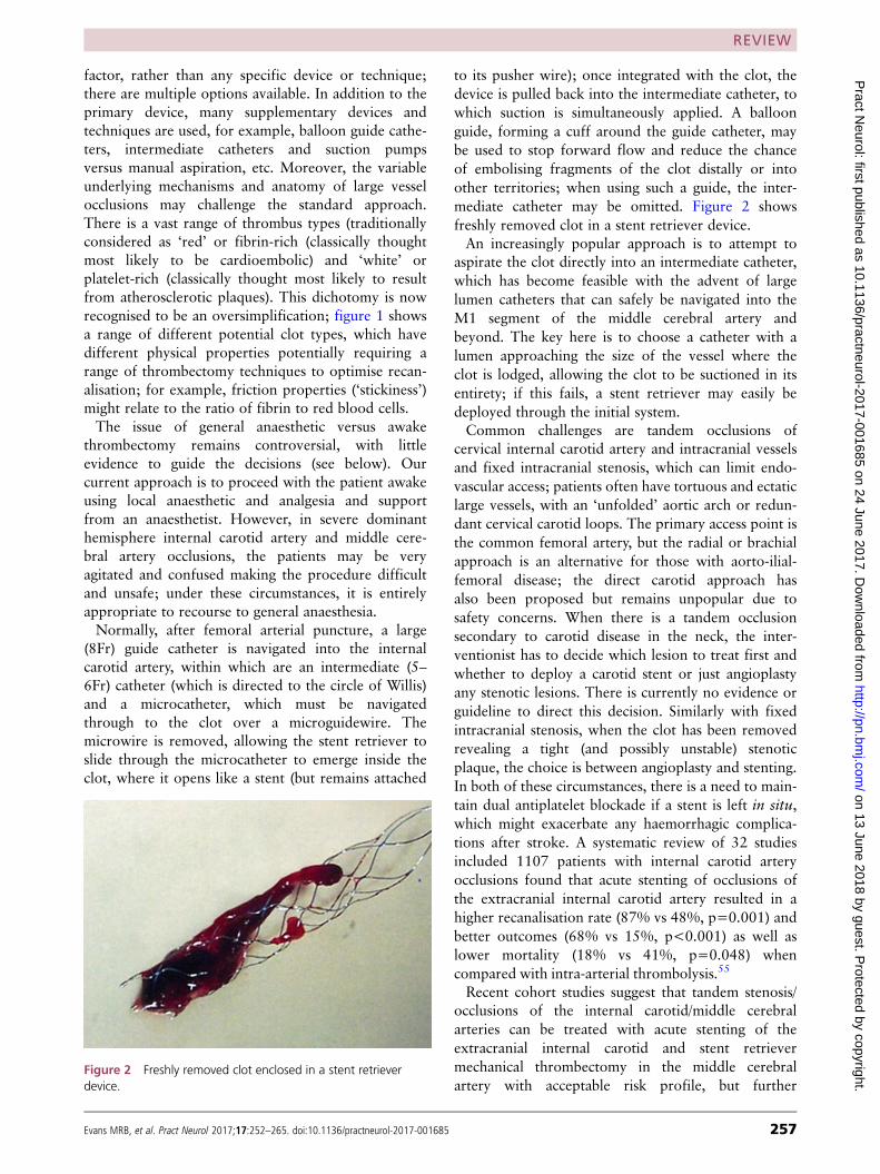
factor, rather than any specific device or technique;
there are multiple options available. In addition to the
primary device, many supplementary devices and
techniques are used, for example, balloon guide cathe-
ters, intermediate catheters and suction pumps
versus manual aspiration, etc. Moreover, the variable
underlying mechanisms and anatomy of large vessel
occlusions may challenge the standard approach.
There is a vast range of thrombus types (traditionally
considered as ‘red’ or fibrin-rich (classically thought
most likely to be cardioembolic) and ‘white’ or
platelet-rich (classically thought most likely to result
from atherosclerotic plaques). This dichotomy is now
recognised to be an oversimplification; figure 1 shows
a range of different potential clot types, which have
different physical properties potentially requiring a
range of thrombectomy techniques to optimise recan-
alisation; for example, friction properties (‘stickiness’)
might relate to the ratio of fibrin to red blood cells.
The issue of general anaesthetic versus awake
thrombectomy remains controversial, with little
evidence to guide the decisions (see below). Our
current approach is to proceed with the patient awake
using local anaesthetic and analgesia and support
from an anaesthetist. However, in severe dominant
hemisphere internal carotid artery and middle cere-
bral artery occlusions, the patients may be very
agitated and confused making the procedure difficult
and unsafe; under these circumstances, it is entirely
appropriate to recourse to general anaesthesia.
Normally, after femoral arterial puncture, a large
(8Fr) guide catheter is navigated into the internal
carotid artery, within which are an intermediate (5–
6Fr) catheter (which is directed to the circle of Willis)
and a microcatheter, which must be navigated
through to the clot over a microguidewire. The
microwire is removed, allowing the stent retriever to
slide through the microcatheter to emerge inside the
clot, where it opens like a stent (but remains attached
to its pusher wire); once integrated with the clot, the
device is pulled back into the intermediate catheter, to
which suction is simultaneously applied. A balloon
guide, forming a cuff around the guide catheter, may
be used to stop forward flow and reduce the chance
of embolising fragments of the clot distally or into
other territories; when using such a guide, the inter-
mediate catheter may be omitted. Figure 2 shows
freshly removed clot in a stent retriever device.
An increasingly popular approach is to attempt toaspirate the clot directly into an intermediate catheter,
which has become feasible with the advent of large
lumen catheters that can safely be navigated into the
M1 segment of the middle cerebral artery and
beyond. The key here is to choose a catheter with a
lumen approaching the size of the vessel where the
clot is lodged, allowing the clot to be suctioned in its
entirety; if this fails, a stent retriever may easily be
deployed through the initial system.Common challenges are tandem occlusions of
cervical internal carotid artery and intracranial vessels
and fixed intracranial stenosis, which can limit endo-
vascular access; patients often have tortuous and ectatic
large vessels, with an ‘unfolded’ aortic arch or redun-
dant cervical carotid loops. The primary access point is
the common femoral artery, but the radial or brachial
approach is an alternative for those with aorto-ilial-
femoral disease; the direct carotid approach has
also been proposed but remains unpopular due to
safety concerns. When there is a tandem occlusion
secondary to carotid disease in the neck, the inter-
ventionist has to decide which lesion to treat first and
whether to deploy a carotid stent or just angioplasty
any stenotic lesions. There is currently no evidence or
guideline to direct this decision. Similarly with fixed
intracranial stenosis, when the clot has been removed
revealing a tight (and possibly unstable) stenotic
plaque, the choice is between angioplasty and stenting.
In both of these circumstances, there is a need to main-
tain dual antiplatelet blockade if a stent is left in situ,
which might exacerbate any haemorrhagic complica-
tions after stroke. A systematic review of 32 studies
included 1107 patients with internal carotid artery
occlusions found that acute stenting of occlusions of
the extracranial internal carotid artery resulted in a
higher recanalisation rate (87% vs 48%, p=0.001) and
better outcomes (68% vs 15%, p<0.001) as well as
lower mortality (18% vs 41%, p=0.048) when
compared with intra-arterial thrombolysis.55
Recent cohort studies suggest that tandem stenosis/
occlusions of the internal carotid/middle cerebral
arteries can be treated with acute stenting of the
extracranial internal carotid and stent retriever
mechanical thrombectomy in the middle cerebral
artery with acceptable risk profile, but furtherFigure 2 Freshly removed clot enclosed in a stent retriever
device.
Evans MRB, et al. Pract Neurol 2017;17:252–265. doi:10.1136/practneurol-2017-001685 257
REVIEW
on 13 June 2018 by guest. Protected by copyright.
http://pn.bmj.com
/P
ract Neurol: first published as 10.1136/practneurol-2017-001685 on 24 June 2017. D
ownloaded from

research of the safety profile and benefit of thisapproach is needed.56–60
Complications and how they are managed
Complications of endovascular procedures can result
from direct device-related vascular injury, vascular
access and the use of radiological contrast media.Stent retriever devices are generally safe61 with lower
complication rates than first-generation devices. The
most common complications include the following:vessel perforation,62–64 which occurred in 1.6%
patients in the five recent positive endovascular trials
(range 0.9%–4.9%); symptomatic intracranialhaemorrhage (3.6%–9.3%); subarachnoid haemor-
rhage (0.6%–4.9%); arterial dissection (0.6%–3.9%);
emboli to new territories (1.0%–8.6% in randomisedcontrolled trials); vasospasm; and vascular access site
complications (including dissection, pseudoaneurysm,
retroperitoneal haematoma and infection). Theoverall procedural complication rate from recent
randomised controlled trials is in the range of 15%,
but it must be emphasised that many do not adverselyaffect clinical outcome. Stent retriever detachment65
66 is an uncommon complication (about 2%–3%with
first-generation Solitaire FR device, but anecdotallymuch lower with the latest versions).
The key strategy to minimise complications is
obvious and simple: for thrombectomy to be onlyperformed in high-volume centres by trained physi-
cians competent in intracranial endovascularprocedures and undertaking them regularly to maintain
skills, as recommended by various multidisciplinary
guidelines.51 52 Mechanical thrombectomy should onlybe performed by a multidisciplinary team operating
within comprehensive stroke centres with adequate
neurointerventional procedural volumes (eg, >200 perannum), of which a reasonable proportion are
mechanical thrombectomy and undertaking regularassessment/audit of technical and clinical results,
process times and complications. When complications
do occur, the immediate availability of neurocriticalcare and (less frequently required) neurosurgical
support are mandatory and may be lifesaving. Figures
3–5 present three examples of thrombectomy proce-dures to demonstrate some of the potential
complexities of the procedure.
How are patients cared for before, during and after the
procedure
Based on the published trial evidence, treatment
should ideally be undertaken in major neurointerven-tional centres with well-functioning hyperacute stroke
units and with rapid access to neurosurgical and
neurointensive care facilities. Currently intravenousthrombolysis is typically administered as soon as the
diagnosis of ischaemic stroke is made, if the patient is
within 4.5 hours and there are no contraindications.Evidence for ‘primary’ thrombectomy without intra-
venous thrombolysis remains limited but there are
trials both ongoing and proposed.
Anaesthesia
The use of general versus local anaesthesia (conscious
sedation) currently varies; each strategy has potentialadvantages. General anaesthesia reduces subject
distress and movement, and it can make the technical
aspects easier; on the other hand, conscious sedationallows continuous neurological monitoring for
complications, and it avoids any potential hazard of
general anaesthetic agents. Retrospective datacomparing general with local anaesthesia during the
procedure found that general anaesthesia, often asso-
ciated with systolic blood pressure<140mm Hg, wasassociated with a poor functional outcome (mRS >2)
Figure 3 Plain CT scan of head (a) and prethrombectomy (b) and post-thrombectomy (c) digital subtraction angiograms in a 49-
year-old woman with sudden onset left hemiparesis and confusion. Plain CT scan of head shows hyperdense clot in the right middle
cerebral artery (red arrow) and early perisylvian loss of grey–white matter differentiation. Prethrombectomy digital subtraction
angiogram shows occluded right proximal middle cerebral artery (blue arrow). The catheter is visible passing through the occlusion.
Post-procedure imaging shows good filling of all middle cerebral artery branches (yellow arrow). There was complete resolution of
neurological signs and symptoms following aspiration thrombectomy.
258 Evans MRB, et al. Pract Neurol 2017;17:252–265. doi:10.1136/practneurol-2017-001685
REVIEW
on 13 June 2018 by guest. Protected by copyright.
http://pn.bmj.com
/P
ract Neurol: first published as 10.1136/practneurol-2017-001685 on 24 June 2017. D
ownloaded from

at 90 days.67 However, a recent single-centre rando-
mised trial in 150 patients with acute ischaemic strokefound similar early (24 hours) outcomes (measured as
NIHSS change) from general anaesthesia with intuba-
tion or conscious sedation without intubation duringthrombectomy.68 Moreover, two studies presented at
the 3rd European Stroke Organisation Conference
(ESOC) in 2017 (GOLIATH and ANSTROKE) bothsuggested that general anesthaesia and conscious
sedation are equally safe. Thus, either approachcurrently seems reasonable, and the choice should be
guided by careful consideration of each individual
patient (eg, agitation, neurological or haemodynamicstability, ease of vascular access to the target lesion,
etc). Ongoing trials of general anaesthesia versus
conscious sedation should provide a clearer answer.
Blood pressure
Based on several neutral randomised trials of blood
pressure lowering, guidelines suggest that lowering
blood pressure in acute ischaemic stroke should bepostponed, at least for a day or two, unless it is
severely elevated (>220/120mm Hg, or >200/
100mm Hg with acute kidney injury, aortic dissec-tion, cardiac ischaemia, hypertensive encephalopathy
or pulmonary oedema).69 Following thrombectomy,
medical and nursing teams are often uncertain how to
manage blood pressure. However, there is limited
evidence to guide how blood pressure should bemanaged before, during and after thrombectomy. It
has been suggested that the poorer functional
outcome (mRS>2) at 90 days associated with generalanaesthesia might relate to the generally lower blood
pressure (usually <140mm Hg systolic), but this
study could not account for confounding factors.68
Given the lack of evidence, we currently recommend
maintaining blood pressure within a physiologicalrange (typically 110–160mm Hg systolic) in a high
dependency (eg, neurocritical care) setting following
thrombectomy. Specific situations (eg, critical extra-cranial or intracranial stenosis with haemodynamic
failure or post-procedure intracranial bleeding) may
require different blood pressure targets.
Antithrombotic treatments
There is little evidence on optimum antithrombotic
treatment during and after thrombectomy. Urgent
anticoagulation is not generally recommended inacute ischaemic stroke due to the risk of intracranial
haemorrhage. Aspirin is not recommended within
24 hours of IV r-tPA but should be started orally (orvia nasogastric tube) within 24–48 hours after stroke
onset. Randomised trials and registries do not give
consistent data or recommendations regarding
Figure 4 Plain CT scan of head (a) and prethrombectomy (b) and post-thrombectomy (c, e, f) digital subtraction angiograms in a 58-
year-old man with a short history of visual symptoms and vertigo followed by a rapid drop in conscious level. Plain CT scan of head (a)
shows thrombus in the basilar artery (red arrow) with complex plaque at the vertebral artery origin, confirmed on digital subtraction
angiography (b). Following successful thrombectomy (c), with removal of a large cast of thrombus (d) by aspiration, a stent was
deployed across the unstable stenotic plaque at the vertebral artery origin (blue arrows, e and f). Basilar thrombi can often be
removed in bulk like this, possibly because of their physical composition.
Evans MRB, et al. Pract Neurol 2017;17:252–265. doi:10.1136/practneurol-2017-001685 259
REVIEW
on 13 June 2018 by guest. Protected by copyright.
http://pn.bmj.com
/P
ract Neurol: first published as 10.1136/practneurol-2017-001685 on 24 June 2017. D
ownloaded from

antithrombotic use in mechanical thrombectomy.
Some units give a single procedural dose of heparin,
but they avoid antiplatelet medication or further anti-
coagulation for 24 hours from stroke symptom onset,
and they suggest follow-up brain imaging with CT or
MRI to exclude haemorrhagic complications, but
practice varies. If we do not deploy a stent, after
24 hours and satisfactory clinical progress and follow-
up imaging to exclude significant haemorrhage, we
then give aspirin 300mg for up to 2weeks, followed
by long-term secondary prevention. This will depend
on stroke mechanism: usually clopidogrel or aspirin
for non-cardioembolic and oral anticoagulation for
atrial fibrillation or other cardioembolic sources. If we
do deploy a stent, we recommend acutely starting
treatment with aspirin and clopidogrel (or equivalent)
dual antiplatelet therapy for at least 3–6months. For
stents in patients requiring anticoagulation we gener-
ally switch to a single antiplatelet agent for long-term
secondary prevention.In the hyperacute clinical setting of mechanical
thrombectomy, it is easy to forget the important task,
as in all patients with acute stroke, of working out the
likely causative processes and mechanism(s) to opti-
mise preventive treatment. For example, this mightinvolve specific investigations for arterial dissection,
detailed cardiac structure and rhythm evaluation and
investigations for thrombophilias or systemic disease.
CHALLENGES AND THE FUTURE
Thrombectomy with reperfusion �6hours aftersymptom onset, alongside IV r-tPA, is clearly the new
standard of care for the treatment of acute ischaemic
stroke due to large vessel occlusion in the anteriorcirculation. The UK National Institute for Health and
Care Excellence (NICE) now approves the use of
mechanical thrombectomy in stroke.70 However,many challenges remain, including, crucially, its prac-
tical implementation.
Can mechanical thrombectomy be delivered in the UK?
The PISTE trial (run in 10 English neuroscience
centres) data confirm the generalisability of the
compelling randomised trial results and show that
Figure 5 Plain CT scan of head (a) and prethrombectomy (b, c), during thrombectomy (d, e, f) and post-thrombectomy (g, h) digital
subtraction angiogram images in a 61-year-old man who presented with a 10min seizure, followed by left-sided weakness and
neglect. Plain CT scan of head shows hyperdense thrombus in the right middle cerebral artery (red arrow, a) with angiogram
identifying a critical stenosis of the internal carotid artery origin (blue arrow, b). We performed middle cerebral artery thrombectomy
using stent retriever technique (e and f). An internal carotid artery stent was inserted (green arrow, d) complicated by an iatrogenic
dissection (yellow arrow, e) necessitating stenting (purple arrow, h).
260 Evans MRB, et al. Pract Neurol 2017;17:252–265. doi:10.1136/practneurol-2017-001685
REVIEW
on 13 June 2018 by guest. Protected by copyright.
http://pn.bmj.com
/P
ract Neurol: first published as 10.1136/practneurol-2017-001685 on 24 June 2017. D
ownloaded from

thrombectomy can be safely and effectively deliveredwithin the National Health Service (NHS).42 At the
time of writing, only two UK centres offer 24 hour
endovascular clot retrieval, with most othersproviding the service only during working hours.
Modelling work based on Sentinel Stroke National
Audit Programme (SSNAP) data, randomisedcontrolled trials and other high-quality evidence indi-
cates that, based on the current criteria and guidelineslisted above, around 10% of all stroke admissions in
the UK (around 9500 patients) would be eligible for
thrombectomy annually. The great bulk of thosewould come from patients presenting to hospital
within 4.5 hours of stroke onset. However, ongoing
randomised trials may well expand those eligibilitycriteria further over the next 3–5 years, for example,
for strokes in the 6–12hour window, in stroke of
unknown time onset and in mild strokes (NIHSS<6).
What are the challenges in delivering thrombectomy as
standard clinical practice in the UK?
Rapid access to appropriate imaging (non-contrast CTscan of head and CT angiogram at a minimum) is
mandatory in selecting patients appropriate for endo-
vascular treatment. Although this has been recentlyrecommended in UK national guidelines,71 it is not
yet standard care in all acute stroke centres, and so
routine availability of CT angiography for acutestroke needs to be rapidly increased. Making CT angi-
ography a routine acute stroke investigation can bring
major gains in speeding up diagnostic pathways, asstroke teams and radiographers develop expertise and
familiarity with the processes. Although there have
been attempts to perform CT angiography only inmore severe strokes (who are more likely to have a
large vessel occlusion), patients with milder strokes
commonly have large vessel occlusion (about 10% ofthose with NIHSS <6) with a high risk of clinical
worsening.72 Once a potentially treatable large vessel
occlusion is identified (which requires 24/7 access totrained neuroradiologists or stroke physicians), treat-
ment must then be delivered quickly. In the positive
trials detailed above, median time between symptomonset to femoral artery puncture was less than
4 hours; median time from symptom onset to recanali-
sation was 4 1/3 hours. This timeframe is currently achallenge in certain parts of the UK, particularly
outside standard working hours, and will require
innovation in local imaging acquisition and interpreta-tion, as well as emergency transport services. Once a
patient eligible for endovascular clot retrieval is iden-
tified, there should be no delay in transferring themto an appropriate centre; however, administration of
IV r-tPA should not be delayed, since this is still the
cornerstone of initial treatment (and given in around
90% of patients in the nine recently published clinicaltrials of thrombectomy).73 We need meticulous orga-
nisation and robust, well-audited care pathways to
enable safe and rapid transfer. The two potentialmodels for providing thrombectomy can be described
as ‘drip and ship’ (initial transfer to a local stroke
centre for diagnosis and intravenous thrombolysis,followed by rapid transfer to a specialist thrombec-
tomy centre) and ‘mothership’ (transfer immediatelyto a specialist comprehensive stroke centre able to
undertake thrombectomy and other required neuro-
science support services). The optimal model will varyaccording to local geography including population
density, transport infrastructure and distance from
specialist centres able to deliver the treatment safelyand effectively. ‘Drip and ship’ might be the more
appropriate solution for more remote areas, while a
‘mothership’ model might be a good solution forurban city populations.
How (and by whom) should thrombectomy be delivered?
Endovascular clot retrieval is safe, but only inexperienced, appropriately trained, competent
hands. It is a complex procedure requiring an
experienced team to deliver, and it needs to beperformed with great rapidity. Therefore, throm-
bectomy delivery will need to be centralised so
that centres and teams can develop expertisequickly and maintain 24/7 services robustly and so
that neurointerventionists can undertake a sufficient
number of cases to maintain expertise. In the UK,published guidance on training and competencies
for thrombectomy is helpful here, and it
indicates that a caseload of at least 40 intracranialendovascular interventions per year is required to
maintain competency in neurointervention.74 We
need new ways of thinking about care deliveryboth before and after completion of training to
expand the pool of skilled neurointerventionists.
Although there has been considerable interest inwhether thrombectomy could be safely delivered
by other (non-interventional neuroradiology)
specialties, the skills required to open cerebralarteries quickly, safely and effectively might not be
generic across these other specialties; for example,
although coronary and cerebral arteries are ofsimilar calibre, brain arteries are more delicate
with a thinner tunica media and adventitia, often
with proximal ectasia and tortuosity, making navi-gation with a catheter both hazardous and
challenging. Moreover, dealing with sudden neuro-
logical complications requires great skill andexpertise in navigating the complex cerebral vascu-
lature. Indeed, the best results in clinical trials and
clinical practice were achieved by experienced
Evans MRB, et al. Pract Neurol 2017;17:252–265. doi:10.1136/practneurol-2017-001685 261
REVIEW
on 13 June 2018 by guest. Protected by copyright.
http://pn.bmj.com
/P
ract Neurol: first published as 10.1136/practneurol-2017-001685 on 24 June 2017. D
ownloaded from

neurointerventionists in high-volume centres.75 Weneed international mechanical thrombectomy regis-
tries to identify whether the real-world experience
is commensurate with that seen in the positiveclinical trials; SITS thrombectomy is one such
registry.
Is mechanical thrombectomy cost effective?
One study76 modelled the hyperacute managementof stroke using intravenous thrombolysis and
mechanical thrombectomy in the UK (compared
with intravenous thrombolysis alone) using Markovsimulations of estimated lifetime costs and quality-
adjusted life years (QALYs), based on pooled
outcome data of five randomised controlled trials.This study found an incremental cost per QALY
gained of mechanical thrombectomy over a 20 year
period of $11 651 (£7061). A more recent studythat modelled the intervention in a US setting
found an incremental cost-effectiveness ratio for
endovascular treatment (compared with standardcare) of $3110/QALY (about £2500 per QALY) in
all simulations, although cost effectiveness was
lower in more distal (M2) occlusion and with estab-lished ischaemic injury (ASPECTS score �5). Both
of these studies show that the cost of mechanical
thrombectomy is well below the frequently applied£30 000 per QALY threshold used by National Insti-
tute for Health and Care Excellence (NICE) to
evaluate new treatments.
Future research questions
There are many remaining questions regarding throm-bectomy. We have few data on thrombectomy for
basilar artery thrombosis; some registry data suggest
that a high proportion of patients (68%) have a pooroutcome (mRS>3), with no difference according to the
use of intravenous thrombolysis or mechanical throm-
bectomy.77 As in the anterior circulation, recanalisationis a key prognostic factor: a recent meta-analysis of 45
observational studies (n=2056) of reperfusion
versus no reperfusion of acute basilar occlusion showeda number needed to treat to decrease death or depen-
dency of 3.78 Small single-centre studies reported good
functional outcomes following basilar thrombectomy,ranging from 30% to 48%.79–82 The time window may
be longer for basilar thrombosis (possibly up to 12–
24 hours), perhaps relating to the tissue properties, clotcomposition and haemodynamics of the collateral
vascular supply in the posterior circulation. We need
randomised trials of thrombectomy, but it might bechallenging because of lack of clinical equipoise (given
the clear benefits of thrombectomy in the anterior
circulation and the devastating outcome from
untreated basilar thrombosis); one trial of treatment
within 6 hours is underway.83
The optimum form of imaging acquisition and proc-
essing (to determine the extent of ischaemic core,
potentially salvageable tissue, collateral supply, etc.)
requires further study. Is MRI better than CT? Is
perfusion imaging required, or will collateral assess-
ment and APSECTS suffice? This is a very important
question for thrombectomy implementation as
imaging triage is likely to be critical in the ‘drip and
ship’ service model in particular. For many logistic
reasons, it would be preferable to secondarily transfer
for thrombectomy only those patients who are very
likely to benefit. We also need to know whether all
patients need advanced brain imaging or just some,
and if some, who? The PISTEai (advanced imaging)
trial is proposed in the UK to answer some of these
questions.Another critical question is whether we should use
advanced imaging in more delayed (including ‘wake-
up’ stroke) presentations. Trials are ongoing, including
POSITIVE (6–12hour time window with appropriate
image selection). The DAWN trial (6–24 hour time
window, including wake-up stroke) selected patients
with substantial clinical deficit but a small ischaemic
core on CT-perfusion imaging and randomised them
to mechanical thrombectomy with the Trevo device,
or to medical therapy alone. The DAWN trial was
stopped early on 9/3/2017 after ~200/500 patients
had been recruited. Data from the DAWN trial
Key points
" Thrombectomy for anterior circulation stroke due to provenproximal major vessel (carotid or M1) occlusion within 6hours of stroke onset is safe and highly effective, and setsthe new standard of care
" In a meta-analysis of randomised trials, the proportions ofpatients achieving a good (independent) functional outcome(mRS 0–2 at 90 days) were 46.0% (mechanical thrombec-tomy) vs 26.5% (best medical treatment); most patients alsoreceived intravenous thrombolysis
" Favourable outcome from mechanical thrombectomy isstrongly time dependent (’time is brain’), with the bestresults achieved when there is no evidence of extensive earlyischaemic brain injury (e.g. ASPECTS score >5); if goodrecanalisation is achieved within 4.5 hours, the absolute rateof good functional outcome is 61%
" Complications of endovascular procedures can follow device-related vessel injury (perforation, dissection, subarachnoidhaemorrhage), vascular access or radiological contrast media
" Thrombectomy must be delivered by appropriately trainedinterventionists
" The next challenge is in delivering the treatment acrosshealthcare systems; the optimal solution (eg, ’drip and ship’versus ’mothership’) may differ according to geography andpopulation density
262 Evans MRB, et al. Pract Neurol 2017;17:252–265. doi:10.1136/practneurol-2017-001685
REVIEW
on 13 June 2018 by guest. Protected by copyright.
http://pn.bmj.com
/P
ract Neurol: first published as 10.1136/practneurol-2017-001685 on 24 June 2017. D
ownloaded from

presented at the European Stroke Organisation
Conference (ESOC) in May 2017 indicated that at 90
days, 48.6% of patients in the intervention arm
achieved functional independence, compared to
13.1% in the control medical therapy arm. Evidence
of an extended time window for mechanical throm-
bectomy potentially means that more stroke patients
might be eligible for endovascular treatment.
However, these data should not detract from the key
message that the most rapid treatment possible
remains the key aim to optimise outcomes for all
reperfusion therapies in acute stroke.
There are also trials proposed of thrombectomy inpatients with milder stroke. The role of direct throm-
bectomy (without intravenous thrombolysis) also
remains to be defined in randomised controlled trials.Many of these pressing remaining questions about
mechanical thrombectomy will probably be answered
within the next 3–5 years. All patients undergoing
mechanical thrombectomy should also be prospectively
included in registries to obtain further evidence on
effectiveness and safety in ‘real world’ practice.
CONCLUSION
Mechanical thrombectomy is a highly successful,
safe and cost-effective treatment for patients with
large artery occlusive stroke. It is therefore a ‘no
brainer’ that the UK NHS and other healthcare
systems need to deliver it as soon as practicable.
However, that will inevitably require reorganisation
of UK stroke services and that will require substan-
tial investment, great attention to care pathways
and extensive cooperation between services
including ambulance services and hospitals.
Contributors ME, PC, PWand DW wrote the manuscript and areresponsible for the content.
Funding DWreceives research funding support from the British
Heart Foundation and the Stroke Association (TSA BHF 2009/01;
PPA 2015/04). This work was undertaken at University College
London Hospitals NHS Foundation Trust/University CollegeLondon who received a proportion of funding from the
Department of Health’s National Institute for Health Research
Biomedical Research Centers funding scheme.
Competing interests None declared.
Provenance and peer review Commissioned; externally peer
reviewed. This paper was reviewed by William Whiteley, Edinburgh,UK.
Open Access This is an Open Access article distributed in
accordance with the terms of the Creative Commons Attribution
(CC BY 4.0) license, which permits others to distribute, remix,
adapt and build upon this work, for commercial use, provided theoriginal work is properly cited. See: http://creativecommons.org/
licenses/by/4.0/
© Article author(s) (or their employer(s) unless otherwise stated in
the text of the article) 2017. All rights reserved. No commercial useis permitted unless otherwise expressly granted.
REFERENCES
1 Feigin VL, Forouzanfar MH, Krishnamurthi R, et al. Global and
regional burden of stroke during 1990–2010: findings from the
Global Burden of Disease Study 2010. Lancet
2014;383:245–55.
2 Wang Y, Rudd AG, Wolfe CD. Age and ethnic disparities in
incidence of stroke over time: the South London Stroke
Register. Stroke 2013;44:3298–304.
3 Mechanical clot retrieval for treating acute ischaemic stroke.
National Institute for Health and Care Excellence (NICE).
Available: https://www.nice.org.uk/guidance/ipg548.
4 Health and Social Care Information Centre. Quality and
Outcomes Framework (QOF)—2014-15. 2015, Available:
http://bit.ly/1PhPYmL.
5 NHS Wales Informatics Service Information and Statistics.
Hospital Admissions for Stroke. 2015.
6 Department of Health, Social Services and Public Safety. QOF
Achievement Data. 2014. Available: http://www.dhsspsni.gov.
uk/index/statistics/qof/qof- achievement.htm.
7 World Health Organisation. The top 10 causes of death. 2014.
Available: http://www.who.int/mediacentre/factsheets/fs310/en/.
8 Adamson J, Beswick A, Ebrahim S. Is stroke the most common
cause of disability? J Stroke Cerebrovasc Dis 2004;13:171–7.
9 Luengo-Fernandez R, Gray AM, Bull L, et al. Quality of life
after TIA and stroke: ten-year results of the Oxford Vascular
Study. Neurology 2013;81:1588–95.
10 Saver JL. Time is brain—quantified. Stroke 2006;37:263–6.
11 Khatri P, Yeatts SD, Mazighi M, et al. Time to angiographic
reperfusion and clinical outcome after acute ischaemic stroke:
an analysis of data from the Interventional Management of
Stroke (IMS III) phase 3 trial. Lancet Neurol 2014;13:
567–74.
12 del Zoppo GJ, Poeck K, Pessin MS, et al. Recombinant tissue
plasminogen activator in acute thrombotic and embolic stroke.
Ann Neurol 1992;32:78–86.
13 Emberson J, Lees KR, Lyden P, et al. Effect of treatment delay,
age, and stroke severity on the effects of intravenous
thrombolysis with alteplase for acute ischaemic stroke: a meta-
analysis of individual patient data from randomised trials.
Lancet 2014;384:1929–35.
14 Alteplase for treating acute ischaemic stroke. London (UK):
National Institute for Health and Care Excellence (NICE).
2012. Available: https://www.guideline.gov/summaries/
summary/38443/alteplase-for-treating-acute-ischaemic-stroke-
review-of-technology-appraisal-guidance-122.
15 Hacke W, Kaste M, Bluhmki E, et al. Thrombolysis with
alteplase 3 to 4.5 hours after acute ischemic stroke. N Engl J
Med 2008;359:1317–29.
16 Bhatia R, Hill MD, Shobha N, et al. Low rates of acute
recanalization with intravenous recombinant tissue plasminogen
activator in ischemic stroke: real-world experience and a call for
action. Stroke 2010;41:2254–8.
17 Fischer U, Arnold M, Nedeltchev K, et al. NIHSS score and
arteriographic findings in acute ischemic stroke. Stroke
2005;36:2121–5.
18 Mishra SM, Dykeman J, Sajobi TT, et al. Early reperfusion rates
with IV tPA are determined by CTA clot characteristics. AJNR
Am J Neuroradiol 2014;35:2265–72.
19 Elofuke P, Reid JM, Rana A, et al. Disappearance of the
hyperdense MCA sign after stroke thrombolysis: implications
Evans MRB, et al. Pract Neurol 2017;17:252–265. doi:10.1136/practneurol-2017-001685 263
REVIEW
on 13 June 2018 by guest. Protected by copyright.
http://pn.bmj.com
/P
ract Neurol: first published as 10.1136/practneurol-2017-001685 on 24 June 2017. D
ownloaded from

for prognosis and early patient selection for clot retrieval. J R
Coll Physicians Edinb 2016;46:81–6.
20 Kamalian S, Morais LT, Pomerantz SR, et al. Clot length
distribution and predictors in anterior circulation stroke:
implications for intra-arterial therapy. Stroke 2013;44:
3553–6.
21 Riedel CH, Jensen U, Rohr A, et al. Assessment of thrombus in
acute middle cerebral artery occlusion using thin-slice
nonenhanced Computed Tomography reconstructions. Stroke
2010;41:1659–64.
22 Riedel CH, Zimmermann P, Jensen-Kondering U, et al. The
importance of size: successful recanalization by intravenous
thrombolysis in acute anterior stroke depends on thrombus
length. Stroke 2011;42:1775–7.
23 Hirano T, Sasaki M, Mori E, et al. Residual vessel length on
magnetic resonance angiography identifies poor responders to
alteplase in acute middle cerebral artery occlusion patients:
exploratory analysis of the Japan Alteplase Clinical Trial II.
Stroke 2010;41:2828–33.
24 Saarinen JT, Sillanp€a€a N, Rusanen H, et al. The mid-M1
segment of the middle cerebral artery is a cutoff clot location
for good outcome in intravenous thrombolysis. Eur J Neurol
2012;19:1121–7.
25 Furlan A, Higashida R, Wechsler L, et al. Intra-arterial
prourokinase for acute ischemic stroke. The PROACT II study:
a randomized controlled trial. Prolyse in Acute Cerebral
Thromboembolism. JAMA 1999;282:2003–11.
26 Felten RP, Ogden NR, Peña C, et al. The Food and Drug
Administration medical device review process: clearance of a
clot retriever for use in ischemic stroke. Stroke 2005;36:
404–6.
27 Smith WS, Sung G, Starkman S, et al. Safety and efficacy of
mechanical embolectomy in acute ischemic stroke: results of the
MERCI trial. Stroke 2005;36:1432–8.
28 Smith WS, Sung G, Saver J, et al. Mechanical thrombectomy
for acute ischemic stroke: final results of the Multi MERCI
trial. Stroke 2008;39:1205–12.
29 Broderick JP, Palesch YY, Demchuk AM, et al. Endovascular
therapy after intravenous t-PA versus t-PA alone for stroke. N
Engl J Med 2013;368:893–903.
30 Ciccone A, Valvassori L, Nichelatti M, et al. Endovascular
treatment for acute ischemic stroke. N Engl J Med
2013;368:904–13.
31 Kidwell CS, Jahan R. Endovascular treatment of acute ischemic
stroke. Neurol Clin 2015;33:401–20.
32 Demchuk AM, Goyal M, Yeatts SD, et al. Recanalization and
clinical outcome of occlusion sites at baseline CT angiography in
the Interventional Management of Stroke III trial. Radiology
2014;273:202–10.
33 Saver JL, Jahan R, Levy EI, et al. Solitaire flow restoration
device versus the Merci Retriever in patients with acute
ischaemic stroke (SWIFT): a randomised, parallel-group, non-
inferiority trial. Lancet 2012;380:1241–9.
34 Nogueira RG, Lutsep HL, Gupta R, et al. Trevo versus Merci
retrievers for thrombectomy revascularisation of large vessel
occlusions in acute ischaemic stroke (TREVO 2): a randomised
trial. Lancet 2012;380:1231–40.
35 Berkhemer OA, Fransen PS, Beumer D, et al. A randomized
trial of intraarterial treatment for acute ischemic stroke. N Engl
J Med 2015;372:11–20.
36 Jovin TG, Chamorro A, Cobo E, et al. Thrombectomy within 8
hours after symptom onset in ischemic stroke. N Engl J Med
2015;372:2296–306.
37 Campbell BC, Mitchell PJ, Kleinig TJ, et al. Endovascular
therapy for ischemic stroke with perfusion-imaging selection. N
Engl J Med 2015;372:1009–18.
38 Saver JL, Goyal M, Bonafe A, et al. Stent-retriever
thrombectomy after intravenous t-PA vs. t-PA alone in stroke. N
Engl J Med 2015;372:2285–95.
39 Goyal M, Demchuk AM, Menon BK, et al. Randomized
assessment of rapid endovascular treatment of ischemic stroke.
N Engl J Med 2015;372:1019–30.
40 Bracard S, Ducrocq X, Mas JL, et al. Mechanical thrombectomy
after intravenous alteplase versus alteplase alone after stroke
(THRACE): a randomised controlled trial. Lancet Neurol
2016;15:1138–47.
41 Mocco J, Zaidat OO, von Kummer R, et al. Aspiration
thrombectomy after intravenous alteplase versus intravenous
alteplase alone. Stroke 2016;47:2331–8.
42 Muir KW, Ford GA, Messow CM, et al. Endovascular therapy
for acute ischaemic stroke: the Pragmatic Ischaemic Stroke
Thrombectomy Evaluation (PISTE) randomised, controlled
trial. J Neurol Neurosurg Psychiatry 2017;88:38–44.
43 Khoury NN, Darsaut TE, Ghostine J, et al. Endovascular
thrombectomy and medical therapy versus medical therapy
alone in acute stroke: a randomized care trial. J Neuroradiol
2017: S01509861pii):30120–1.
44 Goyal M, Menon BK, van Zwam WH, et al. Endovascular
thrombectomy after large-vessel ischaemic stroke: a meta-
analysis of individual patient data from five randomised trials.
Lancet 2016;387:1723–31.
45 Badhiwala JH, Nassiri F, Alhazzani W, et al. Endovascular
thrombectomy for acute ischemic stroke: a meta-analysis. JAMA
2015;314:1832–43.
46 Bush CK, Kurimella D, Cross LJ, et al. Endovascular treatment
with stent-retriever devices for acute ischemic stroke: a meta-
analysis of randomized controlled trials. PLoS One
2016;11:e0147287.
47 Rodrigues FB, Neves JB, Caldeira D, et al. Endovascular
treatment versus medical care alone for ischaemic stroke:
systematic review and meta-analysis. BMJ 2016;353:i1754.
48 Powers WJ, Derdeyn CP, Biller J, et al. 2015 American Heart
Association/American Stroke Association Focused Update of the
2013 Guidelines for the Early Management of Patients With
Acute Ischemic Stroke Regarding Endovascular Treatment: A
Guideline for Healthcare Professionals From the American
Heart Association/American Stroke Association. Stroke
2015;46:3020–35.
49 Casaubon LK, Boulanger JM, Glasser E, Blacquiere D, et al.
Canadian Stroke Best Practice Recommendations: Acute
Inpatient Stroke Care Guidelines, Update 2015. Int J Stroke
2016;11:239–52.
50 Wahlgren N, Moreira T, Michel P, et al. Mechanical
thrombectomy in acute ischemic stroke: consensus statement by
ESO-Karolinska Stroke Update 2014/2015, supported by ESO,
ESMINT, ESNR and EAN. Int J Stroke 2016;11:134–47.
51 White PM, Bhalla A, Dinsmore J, et al. Standards for providing
safe acute ischaemic stroke thrombectomy services (September
2015). Clin Radiol. The Royal College of Radiologists 2015;72,
e1-175–e9.
264 Evans MRB, et al. Pract Neurol 2017;17:252–265. doi:10.1136/practneurol-2017-001685
REVIEW
on 13 June 2018 by guest. Protected by copyright.
http://pn.bmj.com
/P
ract Neurol: first published as 10.1136/practneurol-2017-001685 on 24 June 2017. D
ownloaded from
52 Intercollegiate Stroke Working Party. National Clinical
Guideline for Stroke. Royal College of Physicians 2012; 232.
Available: http://www.rcplondon.ac.uk/sites/default/files/
national-clinical-guidelines-for-stroke-fourth-edition.pdf.
53 Demchuk AM, Coutts SB. Alberta Stroke Program Early CT
Score in acute stroke triage. Neuroimaging Clin N Am
2005;15:409–19.
54 Saver JL, Goyal M, van der Lugt A, et al. Time to treatment
with endovascular thrombectomy and outcomes from ischemic
stroke: a meta-analysis. JAMA 2016;316:1279–88.
55 Kappelhof M, Marquering HA, Berkhemer OA, et al. Intra-
arterial treatment of patients with acute ischemic stroke and
internal carotid artery occlusion: a literature review. J
Neurointerv Surg 2015;7:8–15.
56 Cohen JE, Gomori JM, Rajz G, et al. Extracranial carotid artery
stenting followed by intracranial stent-based thrombectomy for
acute tandem occlusive disease. J Neurointerv Surg
2015;7:412–7.
57 Stampfl S, Ringleb PA, M€ohlenbruch M, et al. Emergency
cervical internal carotid artery stenting in combination with
intracranial thrombectomy in acute stroke. AJNR Am J
Neuroradiol 2014;35:741–6.
58 Maurer CJ, Joachimski F, Berlis A. Two in one: endovascular
treatment of acute tandem occlusions in the anterior circulation.
Clin Neuroradiol 2015;25:397–402.
59 Lescher S, Czeppan K, Porto L, et al. Acute stroke and
obstruction of the extracranial carotid artery combined with
intracranial tandem occlusion: results of interventional
revascularization. Cardiovasc Intervent Radiol 2015;38:304–13.
60 Dababneh H, Bashir A, Hussain M, et al. Endovascular
treatment of tandem internal carotid and middle cerebral artery
occlusions. J Vasc Interv Neurol 2014;7:26–31.
61 Campbell BC, Hill MD, Rubiera M, et al. Safety and Efficacy of
Solitaire stent thrombectomy: individual patient data meta-
analysis of randomized trials. Stroke 2016;47:798–806.
62 Gascou G, Lobotesis K, Machi P, et al. Stent retrievers in acute
ischemic stroke: complications and failures during the
perioperative period. AJNR Am J Neuroradiol 2014;35:734–40.
63 Leishangthem L, Satti SR. Vessel perforation during withdrawal
of Trevo ProVue stent retriever during mechanical
thrombectomy for acute ischemic stroke. J Neurosurg
2014;121:995–8.
64 Mokin M, Fargen KM, Primiani CT, et al. Vessel perforation
during stent retriever thrombectomy for acute ischemic stroke:
technical details and clinical outcomes. J Neurointerv Surg
2016:neurintsurg-2016-012707.
65 Kim ST, Jin SC, Jeong HW, et al. Unexpected detachment of
Solitaire stents during mechanical thrombectomy. J Korean
Neurosurg Soc 2014;56:463–8.
66 Akpinar S, Yilmaz G. Spontaneous SolitaireÉ AB thrombectomy
stent detachment during stroke treatment. Cardiovasc Intervent
Radiol 2015;38:475–8.
67 Davis MJ, Menon BK, Baghirzada LB, et al. Anesthetic
management and outcome in patients during endovascular
therapy for acute stroke. Anesthesiology 2012;116:396–405.
68 Sch€onenberger S, Uhlmann L, Hacke W, et al. Effect of
conscious sedation vs general anesthesia on early neurological
improvement among patients with ischemic stroke undergoing
endovascular thrombectomy: a randomized clinical trial. JAMA
2016;316:1986–96.
69 Jauch EC, Saver JL, Adams HP, et al. Guidelines for the early
management of patients with acute ischemic stroke: a guideline
for healthcare professionals from the American Heart
Association/American Stroke Association. Stroke
2013;44:870–947.
70 National Institute of Health and Care Excellence (NICE).
Mechanicalclot retrieval for treating acute ischaemic stroke.
Interventional procedures guidance. 2016.
71 Royal College of Physicians. National clinical guideline for
stroke, Fifth Edition, 2016. Available: https://www.strokeaudit.
org/SupportFiles/Documents/Guidelines/2016-National-Clinical-
Guideline-for-Stroke-5t-(1).aspx.
72 Coutts SB, Modi J, Patel SK, et al. CT/CT angiography and
MRI findings predict recurrent stroke after transient ischemic
attack and minor stroke: results of the prospective CATCH
study. Stroke 2012;43:1013–7.
73 Meurer WJ, Barth BE, Gaddis G, et al. Rapid systematic review:
intra-arterial thrombectomy ("clot retrieval") for selected
patients with acute ischemic stroke. J Emerg Med
2017;52:255–61.
74 Lenthall R, McConachie N, White P, et al. BSNR training
guidance for mechanical thrombectomy. Clin Radiol 2017;72,
e11–175, e18.
75 Gupta R, Horev A, Nguyen T, et al. Higher volume
endovascular stroke centers have faster times to treatment,
higher reperfusion rates and higher rates of good clinical
outcomes. J Neurointerv Surg 2013;5:294–7.
76 Ganesalingam J, Pizzo E, Morris S, et al. Cost-utility analysis of
mechanical thrombectomy using stent retrievers in acute
ischemic stroke. Stroke 2015;46:2591–8.
77 Schonewille WJ, Wijman CA, Michel P, et al. Treatment and
outcomes of acute basilar artery occlusion in the Basilar Artery
International Cooperation Study (BASICS): a prospective
registry study. Lancet Neurol 2009;8:724–30.
78 Kumar G, Shahripour RB, Alexandrov AV. Recanalization of
acute basilar artery occlusion improves outcomes: a meta-
analysis. J Neurointerv Surg 2015;7:868–74.
79 Nagel S, Kellert L, M€ohlenbruch M, et al. Improved clinical
outcome after acute basilar artery occlusion since the
introduction of endovascular thrombectomy devices.
Cerebrovasc Dis 2013;36(5-6):394–400.
80 M€ohlenbruch M, Stampfl S, Behrens L, et al. Mechanical
thrombectomy with stent retrievers in acute basilar artery
occlusion. AJNR Am J Neuroradiol 2014;35:959–64.
81 Baek JM, Yoon W, Kim SK, et al. Acute basilar artery occlusion:
outcome of mechanical thrombectomy with Solitaire stent
within 8 hours of stroke onset. AJNR Am J Neuroradiol
2014;35:989–93.
82 Mourand I, Machi P, Nogu�e E, et al. Diffusion-weighted
imaging score of the brain stem: A predictor of outcome in
acute basilar artery occlusion treated with the Solitaire FR
device. AJNR Am J Neuroradiol 2014;35:1117–23.
83 van der Hoeven EJ, Schonewille WJ, Vos JA, et al. The Basilar
Artery International Cooperation Study (BASICS): study
protocol for a randomised controlled trial. Trials 2013;
14:200.
84 Duffy S, Farrell M, McArdle K, et al. Novel methodology to
replicate clot analogs with diverse composition in acute
ischemic stroke. J Neurointerv Surg 2017;9:486–91.
Evans MRB, et al. Pract Neurol 2017;17:252–265. doi:10.1136/practneurol-2017-001685 265
REVIEW
on 13 June 2018 by guest. Protected by copyright.
http://pn.bmj.com
/P
ract Neurol: first published as 10.1136/practneurol-2017-001685 on 24 June 2017. D
ownloaded from