Embed Size (px)
Citation preview
Journal of
Clinical Medicine
Review
Risk and Protective Environmental Factors Associatedwith Autism Spectrum Disorder: Evidence-BasedPrinciples and Recommendations
Leonardo Emberti Gialloreti 1,*, Luigi Mazzone 2, Arianna Benvenuto 2, Alessio Fasano 3 ,Alicia Garcia Alcon 4 , Aletta Kraneveld 5 , Romina Moavero 2,6, Raanan Raz 7 ,Maria Pia Riccio 8, Martina Siracusano 1,9, Ditza A. Zachor 10, Marina Marini 11 andPaolo Curatolo 2
1 Department of Biomedicine and Prevention, Tor Vergata University of Rome, 00133 Rome, Italy;[email protected]
2 Child Neurology and Psychiatry Unit, Systems Medicine Department, Tor Vergata University of Rome,00133 Rome, Italy; [email protected] (L.M.); [email protected] (A.B.);[email protected] (R.M.); [email protected] (P.C.)
3 Division of Pediatric Gastroenterology and Nutrition, Massachusetts General Hospital for Children,Harvard Medical School, Boston, MA 02114, USA; [email protected]
4 Hospital General Universitario Gregorio Marañón. 28009 Madrid, Spain; [email protected] Division of Pharmacology, Utrecht Institute for Pharmaceutical Sciences, Faculty of Science,
Utrecht University, 3584 Utrecht, The Netherlands; [email protected] Child Neurology Unit, Neuroscience and Neurorehabilitation Department,
Bambino Gesù Children’s Hospital, IRCCS, 00165 Rome, Italy7 Braun School of Public Health and Community Medicine, Hebrew University, Hadassah 99875, Israel;
[email protected] Child and Adolescent Neuropsychiatry, Federico II University, 80133 Naples, Italy; [email protected] Department of Biotechnological and Applied Clinical Sciences, University of L’Aquila, 67100 L’Aquila, Italy10 The Autism Center/ALUT, Assaf Harofeh Medical Center, Sackler Faculty of Medicine, Tel Aviv University,
69978 Tel Aviv, Israel; [email protected] DIMES, Bologna University, 40126 Bologna, Italy and IRCCS Fondazione Don Carlo Gnocchi,
20148 Milan, Italy; [email protected]* Correspondence: [email protected]; Tel./Fax: +39-0672-596-614
Received: 8 January 2019; Accepted: 5 February 2019; Published: 8 February 2019�����������������
Abstract: Autism Spectrum Disorder (ASD) is a complex condition with early childhood onset,characterized by a set of common behavioral features. The etiology of ASD is not yet fully understood;however, it reflects the interaction between genetics and environment. While genetics is now awell-established risk factor, several data support a contribution of the environment as well. This papersummarizes the conclusions of a consensus conference focused on the potential pathogenetic roleof environmental factors and on their interactions with genetics. Several environmental factorshave been discussed in terms of ASD risk, namely advanced parental age, assisted reproductivetechnologies, nutritional factors, maternal infections and diseases, environmental chemicals andtoxicants, and medications, as well as some other conditions. The analysis focused on their specificimpact on three biologically relevant time windows for brain development: the periconception,prenatal, and early postnatal periods. Possible protective factors that might prevent or modify anASD trajectory have been explored as well. Recommendations for clinicians to reduce ASD risk or itsseverity have been proposed. Developments in molecular biology and big data approaches, whichare able to assess a large number of coexisting factors, are offering new opportunities to disentanglethe gene–environment interplay that can lead to the development of ASD.
J. Clin. Med. 2019, 8, 217; doi:10.3390/jcm8020217 www.mdpi.com/journal/jcm
J. Clin. Med. 2019, 8, 217 2 of 23
Keywords: Autism Spectrum Disorder; risk factors; protective factors; environment; genetics;medications; toxicants; recommendations
1. Introduction
Autism Spectrum Disorder (ASD) is a complex biological condition characterized by a commonset of behavioral features with early childhood onset, reflecting the interaction between differentgenetic and environmental risk factors [1].
At present, there is no ultimate treatment for the core features of ASD. Nevertheless, autisticsymptoms can be reduced by early behavioral interventions [2,3], and some pharmacological therapiesare available for the treatment of psychiatric comorbidities [4].
ASD prevalence seems to be increasing: most recent estimates suggest a prevalence of 1 in 59among 8-year-old children from the USA (https://www.cdc.gov/ncbddd/autism/data.html) [5].Another study estimated a 3.5 prevalence increase between 2001 and 2011 in 2- to 17-year-oldchildren [6]. What caused this increased prevalence, beyond a broadening of ASD diagnostic criteriaand a better ascertainment of cases, is still unclear. Still, as ASD is the final consequence of cascadeevents impacting brain development from gestation to early postnatal life [7], it is possible that a truerise is related to these complex events.
While the etiology of ASD is not fully understood, genetics is a well-established risk factor [8].Twin studies suggested a 76% concordance in monozygotic twins, confirming a strong genetichereditability for ASD, but also supporting an important contribution of environmental factors [9].
Genetic defects in more than 100 genes and loci, and hundreds of copy number variants (CNVs)and single nucleotide (SNVs) polymorphisms (SNPs) have been implicated in about 20% of ASDcases [10–13]. DNA microarrays enable the discovery of rare and recurrent CNVs as importantcontributors to ASD and lead to gains in the understanding of autism genetics and to the identificationof individuals who might be genetically susceptible to autism. Hotspots of recurrent CNVs, including16p11.2, 22q11.2, 1q21.1, 7q11.23, and 15q11–q13, have been shown to be strongly associated withASD [14]. Next-generation sequencing (NGS) methods revolutionized ASD gene discovery and havealso substantially contributed to functional genetic data, linking mutations frequently associated withASD with genes involved in the regulation of brain transcriptional networks during brain developmentand early synaptogenesis, thus throwing some light on the understanding of the neurobiologicalconsequences of the disruption of these ASD-associated genes [12,15]. Nevertheless, also single-genessyndromes have been associated with ASD, including Fragile-X (FMR1), Tuberous Sclerosis Complex(TSC1-2) and PTEN syndrome [16,17].
Nonetheless, the heterogeneous clinical and biological phenotypes observed in ASD stronglysuggest that, in genetically susceptible individuals, environmental risk factors also combine orsynergize to generate a “threshold point” that might determine a dysfunction. While progress has beenmade towards gaining an understanding of genetic and epigenetic factors, environmental risk factorsare less understood [18]. Actually, recent studies have demonstrated that during critical periods ofcentral nervous system development, early exposure to a variety of environmental factors, rangingfrom microbes (bacteria and viruses) to medications, from chemicals to physical agents, can affectneurobiological development, including effects relevant to ASD [19,20].
In October 2018, international ASD experts convened in Rome to discuss the potential pathogeneticrole of environmental factors, as well as their interactions with genetic susceptibility, focusing on threebiologically relevant windows for brain development: the periconception, prenatal and early postnatalperiods. From the epidemiological point of view, the identification of the exact timing of action ofeach environmental factor, as well as its consequences in the neurodevelopmental pathways, remainselusive. Nevertheless, it is now possible to establish some differentiations among risk factors that canassist in developing detection and personalized follow-up of populations at higher risk for ASD.
J. Clin. Med. 2019, 8, 217 3 of 23
In this paper, we summarize the results of this consensus conference and put forward clinicalrecommendations for clinicians to reduce ASD risk and/or its severity.
2. Conception Period
Advanced parental age: The association between older parental age at conception andneuropsychiatric disorders in offspring is now well documented [21,22]. In the case of ASD, bothadvanced maternal and paternal age at time of birth (≥35 years) were associated with an increasedrisk of ASD [23–26]. Emerging evidence also confirms a combined parental age effect, which is highestwhen both parents are in the older age range and increases with increasing differences in parentalages [27]. Both human and animal model studies support the hypothesis of an association betweenelevated rates of de novo mutations in older fathers and increased risk of ASD [28,29]. It has been alsosuggested that maternal mechanisms mediating the effects of advanced maternal age on ASD risk areassociated not only with chromosomal or genetic modifications, but also with a higher prevalenceof chronic diseases and a less favorable uterine environment, often resulting in more obstetricalcomplications, which might eventually lead to an increased risk of adverse birth outcomes [26].
Use of hormonal induction and/or assisted reproductive technologies: Assisted ReproductiveTechnologies (ART) now account for 1–3% of all live births in the Western world (https://www.cdc.gov/reproductivehealth/index.html) [30]. Several procedures that are used in the ART process, suchas hormonal stimulation, egg retrieval, in vitro fertilization (IVF), intra-cytoplasmic sperm injection(ICSI), micro-manipulation of gametes and exposure to culture medium, could subject the gametesand early embryos to environmental stress and may be associated with an increased risk of birthdefects and low birth weight (LBW) [31]. Children conceived using ART are also at a higher risk forcongenital anomalies including a two-fold increase in the central nervous system and epigenetic andimprinted disorders [32–34]; there is some evidence that ART might have an impact on imprintingthrough DNA methylation [35]. Actually, assisted conception and ASD share several risk factors.In both cases, hormonal disturbances, especially in testosterone/androgen regulation, along with highrates of advanced parental ages, preterm deliveries, and LBW, have been reported [6,24,36,37].
Additionally, a recent meta-analysis indicated that the use of ART may be associated with ahigher risk of ASD in the offspring [31]. In a previous case-control study conducted on a large Israelipopulation [38], a higher ART prevalence (IVF and ICSI) (10.7%) even in young mothers (<29 years)was reported among ASD children compared to the overall ART rate. In addition, the study ruled outthe hypothesis that ART was associated with unique autism symptomatology (i.e. autism severityand adaptive functioning, a history of developmental regression) that may represent a distinct clinicalphenotype in this group. The study results indicated that although assisted conception may be a riskfactor for ASD, this group did not appear to represent a separate clinical phenotype within the autismspectrum. These findings suggest that the increased recent prevalence of both ART and ASD mightbe related.
Environmental chemical and toxicant factors: There is some evidence that exposure tochemical pollutants at critical developmental stages may affect neural and behavioral development.The pathogenetic mechanisms of environmental chemical factors can involve neurotoxicity but canalso extend to pathways of immune dysregulation, altered lipid metabolism, and mitochondrialdysfunction. To date, the strongest evidence of association is shown by traffic-related air pollutantsand pesticides at different times of exposures [39,40].
Maternal nutritional status: Maternal nutritional status and body mass index before pregnancyhave been considered as environmental factors that can influence normal brain development throughexcess or deficit of micronutrients and growth factors, which can affect neurodevelopmental outcomesof offspring [41,42]. In this view, both maternal obesity and underweight have been associated withan increased risk of ASD [42,43]. Maternal obesity results in activation of the maternal immunesystem and in a chronic inflammation of the uterine environment potentiating abnormal neuronalgrowth and differentiation in the fetus, with consequent neurodevelopmental impairments in the
J. Clin. Med. 2019, 8, 217 4 of 23
offspring [44]. At the same level, maternal undernutrition may elicit a physiological stress responseleading to neuronal damage through a disproportionate release of proinflammatory factors [45].
A large number of recent studies have suggested association between pre-conception intake offolate and risk of ASD onset in newborns [46]. A significantly higher rate of ASD has been found inchildren not exposed to folic acid (FA) compared to in children of mothers who took it. Conversely,some apparently conflicting results were reported by other studies that related an increased riskfor ASD and neurocognitive impairments in children of mothers who used dietary supplements ofsynthetic FA [47–49]. A possible explanation of these diverging results might be offered by the differentcompositions between the FA used in supplements (pteroylmonoglutamic) and the one from naturalfood sources (ormyl-tetrahydropteroylglutamates). High levels of pteroylmonoglutamic acid, whichdepend on liver-based metabolism, could result in high levels of unmetabolized and non-useful FA inthe blood, which can cause changes in brain synaptic transmission and dysregulation of expression ofmany genes associated with ASD [50–52].
Another important micronutrient potentially linked to the neurodevelopmental alterations inASD is iron. The importance of a correct intake of iron is evident already from the peri-conceptionperiod [53]. In the brain, iron contributes to neurotransmitter production, myelination and immunefunction. In this view, iron deficiency in this period could result not only in impairment in the generaldevelopment of cognitive, motor and language skills, but also in deficit in social orientation andengagement that could lead to ASD [53].
Medications: A growing number of researches highlighted the potential association ofprenatal exposure to Selective Serotonin Reuptake Inhibitors (SSRIs) with the onset of ASD,hypothesizing a pathogenetic link between alterations in serotonin pathways and ASD neurobiologicalabnormalities [54–56]; exposure during the preconception period or the first trimester seems tobe associated with a higher risk compared to the other two trimesters [57]. Others have foundthat antidepressants, regardless of their composition, might be associated with increased ASDrisk [58]. Thus, some diverging results have been found in relation to both antidepressant types anddosages [58,59]. Furthermore, a Danish longitudinal study, with a follow-up of 5,057,282 person-years,did not detect a significant association between maternal use of SSRIs during pregnancy and ASDin the offspring [60]. Moreover, another large research did not find, after controlling for severalconfounding factors, a significant association between prenatal exposure to antidepressant medicationand ASD [61]. Lastly, a “confounding by indication” cannot be excluded, raising the possibility that itis depression and anxiety that might be risk factors for offspring ASD, rather than antidepressantsper se [58]. Nevertheless, even if evidence is still conflicting, the recommendation is to proceed toapply the precautionary principle, balancing the use of antidepressants against the substantial adverseconsequences of untreated maternal depression.
3. Prenatal Period
Environmental chemicals and toxicants: In the last few years, epidemiologic investigationsindicated that prenatal exposure to chemical and toxic factors such as air pollution, pesticides,materials used in the plastic industry and heavy metals may increase the risk of ASD [39,40,62–64].Possible mechanisms behind the association between these environmental risk factors and ASD arenot only their interactions with genetic factors, and/or epigenetic marks leading to a diminishedability to detoxify xenobiotics [65,66] but also their potential role in triggering neuro-inflammationand oxidative stress that lead to neurobiological and neurotransmitter alterations and abnormalities insignaling pathways [63].
Air pollution: Air pollution is probably the chemical risk factor with the strongest evidence ofassociation with ASD, especially for exposures in the third trimester [40,67,68]. Multiple variables,such as metrics of exposure, type of pollution, time of exposure, could influence the risk of ASDand its clinical outcome [69–71]. It should be noted, however, that despite positive associations thatwere observed in many countries like the USA, Canada, Taiwan and Israel, European studies did
J. Clin. Med. 2019, 8, 217 5 of 23
not find any association [71–73]. Recently, in a Canadian population-based birth cohort, a significantassociation between exposure to nitric oxide and ASD was found. No association was found betweenASD and particulate matter with a diameter of <2.5 µm or nitrogen dioxide [74]. These contradictoryresults might be due to the fact that studies of air pollution have been often limited by indirect andcross-sectional methods of exposure measurement, by different metrics of exposure, by differentevaluations of outcomes, and by focusing on different pollutants. Notwithstanding the need for furtherinvestigation, and even if some unanswered questions remain, prenatal air pollution exposure hasemerged as a potentially modifiable risk factor for ASD.
Pesticides: Exposure to organochlorine pesticides (measured using geographical mapping)increases the risk of offspring ASD [75,76]. Additionally, studies that examined risk of ASD in relationto prenatal levels of poly-chlorinated biphenyls (PCBs) reported a suggestive association with specificPCBs [20,77,78]. Organophosphate exposure during pregnancy increases the risk of autistic symptomsin the offspring, at 2–3 years of age [79]. In particular, residential proximity to organophosphatesat some point during pregnancy is associated with a 60% increased risk for offspring to developASD [80]. Conversely, a pilot case-control study investigating risk associated with exposure to organicpollutants (including a variety of PCB congeners, DDT - dichloro-diphenyl-trichloroethane, and DDE -Dichloro-2,2-bis(p-chlorophenyl)-ethylene) measured in archived maternal serum and diagnosis ofASD in children did not find significant differences in odds ratios for ASD [77]. Most pesticides ofcurrent use are neurotoxic, may target the developing brain [81] and are prone to cause oxidativestress [82]. The widely used pyrethroids have been associated to ASD and neurodevelopmentaldelay [83]. Nevertheless, for the same reasons described before, when talking about air pollution, anassociation between pesticide exposure and ASD is not yet confirmed [78].
Phthalates: Phthalates are a class of chemicals used as plasticizers, solvents, and lubricants, andas enteric coatings on pharmaceuticals and nutritional supplements. Few studies have addressedthe relationship between ASD and prenatal exposure to phthalates (3rd trimester) with contrastingresults [78].
Heavy metal exposure: Little evidence for an association between hair metal concentration ofmercury, copper, cadmium, selenium, chromium and autistic symptoms has emerged until now [84].Moreover, as most of these studies only measured biomarkers and do not ascertain actual exposuresources, temporality of association is unknown. Some studies examined exposure in relation tomaternal dental amalgam fillings and maternal or child consumption of seafood with inconsistentfindings [19]. A meta-analysis found not only consistent evidence for lack of association betweenchildhood thimerosal exposure and ASD, but also an increased risk of ASD following a higher level ofinorganic mercury exposure [85].
Medications: The association between ASD and prenatal exposure to drugs is increasinglyinvestigated; a specific area of interest was the study of antiepileptic and antidepressant agents [54].
Among antiepileptic drugs (AED), valproate showed the strongest association withneurodevelopmental outcome, in terms of cognitive disabilities, developmental delay, and ASD [86].It is therefore contraindicated as a first-line antiepileptic or mood stabilizer in pregnant women orin those who plan pregnancy. Moreover, other AED, as oxcarbazepine and lamotrigine (alone orcombined with valproate), have been found to be associated with the onset of ASD in the offspring [87].Findings across several meta-analyses examining the association between antidepressant exposureduring pregnancy and ASD are reasonably consistent showing an increased risk [88]. Additionally,maternal psychiatric disorders could play a critical role in the development of ASD; thus, these havebeen considered also as a potential confounding or addictive risk factor for exposure to antidepressantsalone [88–90].
Some studies suggested also a possible link between prenatal or early-life antibiotic use andASD [91], but too limited information is currently available to draw conclusions. Recently, however, ithas been demonstrated that low-dose antibiotic exposure in late pregnancy and early postnatal life inmice induces impaired social behaviors and aggression in mice associated with changes in the intestinal
J. Clin. Med. 2019, 8, 217 6 of 23
microbiome [92]. On the other hand, supplementation with the probiotic Lactobacillus RhamnosusJB-1 might prevent the early-life antibiotic-induced aberrant behaviors. Taken together, these resultsmerit further research on the potential role of early-life antibiotic exposure in the development of ASD.
Substance abuse: A large number of studies examined prenatal exposure to substance abuse asheavy tobacco smoke, alcohol, or cocaine and ASD. Association between high amounts of alcoholconsumption in pregnancy and ASD in offspring (especially those with Fetal Alcohol Syndrome) isdocumented [93–95]. On the other hand, association between moderate alcohol intake in pregnancyand ASD is unlikely [94].
An association between smoking during pregnancy and risk of childhood autism has beensuggested [96], but in this case, results are conflicting, with two meta-analyses in a total of 15 studiesreporting no association with overlapping odds ratios [97,98]. Therefore, at present, insufficient datahave been found to support an association.
Nutritional factors: Epidemiological studies and data obtained in humans have provided evidencethat mother’s diet during pregnancy plays a critical role in the development of the neural circuitrythat regulates behavior, thus determining persistent behavioral effects in the offspring [48]. Generally,it is known that some elements of maternal diet during pregnancy, such as FA, vitamin D, iron andfatty acids, are associated with higher or lower incidence of ASD or autistic traits in the offspring [99].Specifically, low concentrations of vitamin D and FA are associated with an increased risk of ASDdiagnosis, in particular if these deficiencies are present in the mid-gestational period [100,101].In addition, a maternal diet with high levels of methanol and aspartame during gestation couldbe linked to an increased risk of ASD [102].
A poor omega-3 intake during gestation and maternal high-fat diet during pregnancy hasbeen associated with the risk of ASD and other neurodevelopmental disorders [19,103]. In fact,high-fat consumption during pregnancy is strongly associated with activation of several of thesame inflammatory cytokines (e.g., interleukins IL-4, and IL-5) that are elevated during gestationin mothers of children with ASD. Furthermore, high-fat diet consumption in pregnant women isassociated with modifications of the neural pathways involved in behavioral regulation, specificallythe serotoninergic system. The suppression of serotoninergic synthesis in the brain may underliethe risk of developing later behavioral disorders, as long as the offspring is exposed to maternalhigh-calorie diet during pregnancy.
Prenatal infections and maternal immune activation: Current data suggest that at least for a subsetof women, exposure to infections during pregnancy might increase ASD risk or other disorders of thecentral nervous system (CNS) in the offspring. Activation of the maternal immune response can confera risk for the onset of psychiatric disorders. In particular, exposure to prenatal infections, such as flu,rubella, measles, herpes simplex virus, and bacterial infections, may increase the risk for the offspringof developing bipolar disorder and schizophrenia [104]. More recently, some population-based cohortstudies described a potential link between autism risk and maternal infection or inflammation duringpregnancy, depending on the time of gestational exposure, the type of infective agent, and the intensityof the maternal immune response; specifically, viral infections seem to be associated to ASD risk inthe first trimester, bacterial infections in the second trimester, influenza and febrile episodes duringthe whole pregnancy but especially in the third trimester [105,106]. Fewer studies have examined thepotential impact on ASD risk of fever as such, rather than in connection with infection broadly [106].A retrospective case-control study based on maternal self-report showed an association between feverduring pregnancy and increased ASD risk [105]; it showed also that this risk was attenuated only inmothers who took anti-pyretic medications to control their fever, but not in those mothers who didnot [105]. A prospective study in Norway also found an increased risk for ASD after prenatal feverexposure, as well as evidence of a dose–response relationship, with risks rising parallel to multipleepisodes of maternal fever [107].
A prevailing concept is that maternal immune activation (MIA) may alter the expression ofinflammatory molecules in the developing fetus and that maternal-fetal immune dysregulation may
J. Clin. Med. 2019, 8, 217 7 of 23
disrupt brain development and neural connectivity, which in turn may have long-term effects on theoffspring’s mental functions [108]. Among the studies supporting a link between maternal infectionand increased risk of ASD, there are several ones carried out with the quantification of cytokine,chemokines and of other inflammatory mediators measured in the maternal serum and amnioticfluid [109]. These studies, however, have generated conflicting results [56,105]. Recently, increasedlevels of maternal cytokines and chemokines during gestation have been associated with subsequentASD with intellectual disability [110].
Maternal immune systems can be involved in increasing ASD risk, even independently fromprenatal infections. In particular, maternal autoantibodies might recognize proteins in the developingfetal brain [111]; these autoantibodies can be detected in ~20% of mothers of children at risk fordeveloping autism versus 1% of mothers of typically developing children, and defined an additionalsub-phenotype of ASD [112,113].
Individual maternal factors and diseases: Gestational diabetes has been considered a risk factorbecause it negatively affects fetal growth and it increases the rate of pregnancy complications [114–116].Moreover, it impacts long-term fine and gross motor development and leads to learning difficultiesand attention-deficit hyperactivity disorder [117]. These adverse effects of maternal diabetes onbrain development may arise from the increased fetal oxidative stress, as well as from epigeneticchanges in the expression of several genes [114,115,118]. However, the increased risk for ASD linked togestational diabetes may be related to pregnancy complications rather than to complications secondaryto hyperglycemia. Whether control of diabetes reduces ASD risk is still unknown [114,115].
Additionally, maternal melatonin levels have been investigated as potential culprits in the ASDpathogenesis [119]. Melatonin is a crucial hormone for neurodevelopment and protects from oxidativestress and neurotoxicant agents. Melatonin deficiency is frequently detected in ASD children alreadyin a very early period of life, and thus the potential implications of low maternal melatonin levels havebeen considered as a factor that might increase the susceptibility to autism [120].
4. Perinatal/Early Postnatal Period
Current research seems to suggest that obstetric risk factors occur more often in ASD childrencompared to neurotypical controls, even though these results have been challenged by otherauthors [121]. In this view, the higher prevalence of obstetric negative events in ASD could beexplained, not only by the maternal genetic/epigenetic mechanisms mentioned above, but also byhormonal factors altering the in utero environment, leading to a fertility decline and increasingpregnancy and obstetric complications, which lead to emergencies, such as caesarean sections (CSs) orpreterm births [114].
Several studies have examined the possible relationship between CS and/or induced laborand ASD, with conflicting findings [122,123]. One of the pathogenetic hypotheses is the possibleeffect of oxytocin (OT) variations during CS in the etiology of ASD. Epigenetic dysregulations of theoxytocinergic system could play a role in the behavioral dysfunctions of ASD. Perinatal alterationsof OT can also have life-long lasting effects on the development of social behaviors [124]. Within theperinatal period, various processes, like planned caesarean section, labor induced by syntheticOT or interrupted with oxytocinergic antagonists, can also alter the OT balance in the newborns,even though the implications and medium/long-term effects of these manipulations are still largelyunknown [123,125].
Other studied perinatal factors include gestational age of <36 weeks, spontaneous, induced, orno labor, breech presentation, as well as preeclampsia and fetal distress [24,26,126]. In preterm births,chorioamnionitis, acute intrapartum haemorrhage, and LBW have been associated with higher risks ofabnormal results during early autistic screenings [127]. According to a study, parity of ≥4 might be aprotective factor that decreases ASD risk [126].
Microbiome: Scientific evidence is beginning to accumulate suggesting that, within ASDpopulations, the gut microbiome shows a different composition compared to typically developing
J. Clin. Med. 2019, 8, 217 8 of 23
individuals (e.g., higher representation of Clostridia, Bacteroidetes, Desulfovibrio, and Sutterella spp),which might be responsible for frequent gastrointestinal disorders experienced by patients with ASD.Recent evidence in ASD subjects suggests that microbiota transplantation could represent a promisingapproach to improving gastrointestinal and ASD symptoms [128]; data, corroborated also by ASDanimal models, showed the potential beneficial effects of probiotics treatment and fecal microbiotatransplantation [129]. However, further research is necessary in order to evaluate the effectivelong-term improvements on the ASD clinical phenotype. Another way to target the microbiomeis dietary intervention with prebiotics, including fibers, such as galacto-oligosaccharides (GOS) thatinduces the growth and activity of beneficial bacteria [130]. Recently, it has been suggested that thecombination of exclusion diets and GOS supplementation might result in significant improvements inanti-social behavior in ASD [131].
While the fetal environment was initially thought to be entirely sterile, recent evidence suggeststhat some bacteria are present in the amniotic fluid and placenta. One implication of these newdiscoveries is that the microbial composition of the developing offspring may be sensitive toenvironmental changes even during prenatal stages of life [132]. Animal models suggest that maternalgut bacteria can promote neurodevelopmental abnormalities in offspring, possibly mediated byT-helper-17 cells with subsequent immune system activation [133].
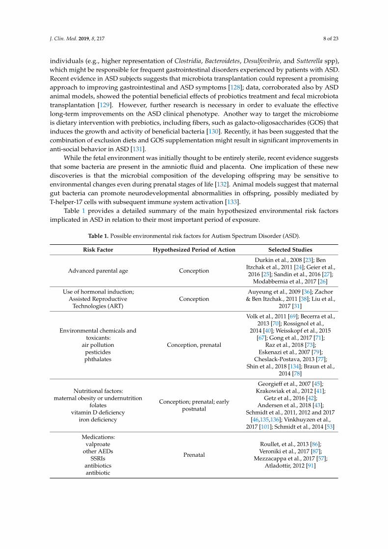
Table 1 provides a detailed summary of the main hypothesized environmental risk factorsimplicated in ASD in relation to their most important period of exposure.
Table 1. Possible environmental risk factors for Autism Spectrum Disorder (ASD).
Risk Factor Hypothesized Period of Action Selected Studies
Advanced parental age Conception
Durkin et al., 2008 [23]; BenItzchak et al., 2011 [24]; Geier et al.,2016 [25]; Sandin et al., 2016 [27];
Modabbernia et al., 2017 [26]
Use of hormonal induction;Assisted Reproductive
Technologies (ART)Conception
Auyeung et al., 2009 [36]; Zachor& Ben Itzchak., 2011 [38]; Liu et al.,
2017 [31]
Environmental chemicals andtoxicants:
air pollutionpesticidesphthalates
Conception, prenatal
Volk et al., 2011 [69]; Becerra et al.,2013 [70]; Rossignol et al.,
2014 [40]; Weisskopf et al., 2015[67]; Gong et al., 2017 [71];
Raz et al., 2018 [73];Eskenazi et al., 2007 [79];
Cheslack-Postava, 2013 [77];Shin et al., 2018 [134]; Braun et al.,
2014 [78]
Nutritional factors:maternal obesity or undernutrition
folatesvitamin D deficiency
iron deficiency
Conception; prenatal; earlypostnatal
Georgieff et al., 2007 [45];Krakowiak et al., 2012 [41];
Getz et al., 2016 [42];Andersen et al., 2018 [43];
Schmidt et al., 2011, 2012 and 2017[46,135,136]; Vinkhuyzen et al.,
2017 [101]; Schmidt et al., 2014 [53]
Medications:valproate
other AEDsSSRIs
antibioticsantibiotic
Prenatal
Roullet, et al., 2013 [86];Veroniki et al., 2017 [87];
Mezzacappa et al., 2017 [57];Atladottir, 2012 [91]
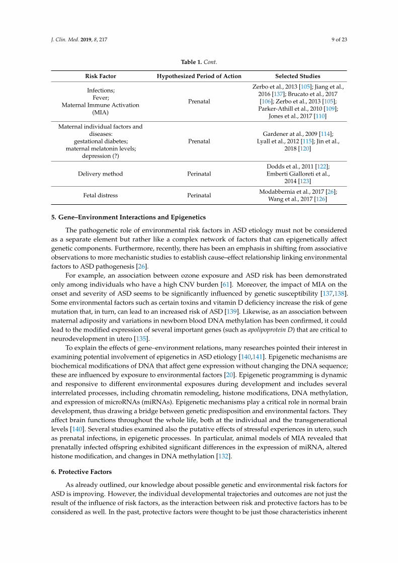
J. Clin. Med. 2019, 8, 217 9 of 23
Table 1. Cont.
Risk Factor Hypothesized Period of Action Selected Studies
Infections;Fever;
Maternal Immune Activation(MIA)
Prenatal
Zerbo et al., 2013 [105]; Jiang et al.,2016 [137]; Brucato et al., 2017[106]; Zerbo et al., 2013 [105];Parker-Athill et al., 2010 [109];
Jones et al., 2017 [110]
Maternal individual factors anddiseases:
gestational diabetes;maternal melatonin levels;
depression (?)
PrenatalGardener at al., 2009 [114];
Lyall et al., 2012 [115]; Jin et al.,2018 [120]
Delivery method PerinatalDodds et al., 2011 [122];Emberti Gialloreti et al.,
2014 [123]
Fetal distress Perinatal Modabbernia et al., 2017 [26];Wang et al., 2017 [126]
5. Gene–Environment Interactions and Epigenetics
The pathogenetic role of environmental risk factors in ASD etiology must not be consideredas a separate element but rather like a complex network of factors that can epigenetically affectgenetic components. Furthermore, recently, there has been an emphasis in shifting from associativeobservations to more mechanistic studies to establish cause–effect relationship linking environmentalfactors to ASD pathogenesis [26].
For example, an association between ozone exposure and ASD risk has been demonstratedonly among individuals who have a high CNV burden [61]. Moreover, the impact of MIA on theonset and severity of ASD seems to be significantly influenced by genetic susceptibility [137,138].Some environmental factors such as certain toxins and vitamin D deficiency increase the risk of genemutation that, in turn, can lead to an increased risk of ASD [139]. Likewise, as an association betweenmaternal adiposity and variations in newborn blood DNA methylation has been confirmed, it couldlead to the modified expression of several important genes (such as apolipoprotein D) that are critical toneurodevelopment in utero [135].
To explain the effects of gene–environment relations, many researches pointed their interest inexamining potential involvement of epigenetics in ASD etiology [140,141]. Epigenetic mechanisms arebiochemical modifications of DNA that affect gene expression without changing the DNA sequence;these are influenced by exposure to environmental factors [20]. Epigenetic programming is dynamicand responsive to different environmental exposures during development and includes severalinterrelated processes, including chromatin remodeling, histone modifications, DNA methylation,and expression of microRNAs (miRNAs). Epigenetic mechanisms play a critical role in normal braindevelopment, thus drawing a bridge between genetic predisposition and environmental factors. Theyaffect brain functions throughout the whole life, both at the individual and the transgenerationallevels [140]. Several studies examined also the putative effects of stressful experiences in utero, suchas prenatal infections, in epigenetic processes. In particular, animal models of MIA revealed thatprenatally infected offspring exhibited significant differences in the expression of miRNA, alteredhistone modification, and changes in DNA methylation [132].
6. Protective Factors
As already outlined, our knowledge about possible genetic and environmental risk factors forASD is improving. However, the individual developmental trajectories and outcomes are not just theresult of the influence of risk factors, as the interaction between risk and protective factors has to beconsidered as well. In the past, protective factors were thought to be just those characteristics inherent
J. Clin. Med. 2019, 8, 217 10 of 23
to the individual, such as a high intellectual quotient or better social skills. Now, there is an increasingunderstanding that there is the need to look also at possible pre- and postnatal environmental factors.However, despite the growing interest in the identification of environmental risk factors and theirpotential prevention in order to decrease ASD risk, to date, little is known about protective factors forASD. Nevertheless, in the last few years, increasing efforts have been made to try to identify factorsthat may improve long-term outcomes [142].
Some elements of the mother’s diet might play a protective role by countering some core autisticsymptoms. The main elements of the maternal diet that seem to play a protective role against ASD arefatty acids, vitamin D (vit. D), and iron [99].
A mean daily FA intake of ≥600 µg in the periconception period and/or during the first monthof pregnancy, but only in cases of significant mother’s fatty acids deficiency, is associated with a 40%decrease of ASD risk [100,143]. The association between fatty acids and reduced ASD risk is strongestfor mothers and children with MTHFR 677 C > T (cytosine > thymine) variant genotypes, which leadsto less efficient folate metabolism [143].
According to some studies, vitamin D supplements during pregnancy could reduce the risk ofdeveloping ASD in the offspring [144].
Higher iron intake through the end of pregnancy and particularly during breast feeding wasassociated with reduced ASD risk compared to lower intakes [53].
In addition, some meta-analyses provide evidence that breastfeeding (exclusively or accompaniedby additional supplements) may protect against ASD [145]. Breastfeeding may reflect the protectiveeffect of breast milk [145–147]; for example, breast milk contains bifidobacteria, lysozyme, lipoxins,glutathione, and anti-inflammatory cytokines. Literature suggests that, relative to controls, childrenwith ASD have lower levels or bifidobacteria and lysozyme in the digestive tract and increased levelsof inflammatory cytokines in plasma. Therefore, a number of possible components of breast milk couldplausibly be connected to a decreased ASD risk.
Additionally, the interest in polyunsaturated fatty acids (PUFA) in maternal diet is increasing,as lipid composition (lipidomics) seems crucial in psychiatric disorders [148]. The increase in PUFA,especially omega-3 fatty acids, in a prenatal maternal diet was associated with a decreased ASDrisk [19].
Another factor that could play a role as a protective agent is melatonin. Melatonin synthesisis frequently impaired in patients with ASD and in their mothers. Therefore, consumption ofthis hormone during pregnancy could act as a neuroprotective factor, decreasing the risk ofneurodevelopmental disorders, including ASD [120].
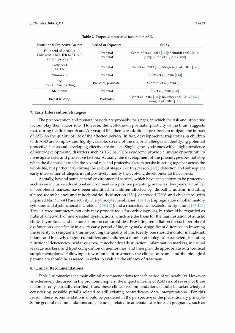
Table 2 summarizes the main known protective factors for ASD, in relation to the suggested periodof exposure. These are likely protective factors that might prevent and/or modify a poor outcomein a more positive one, thus enhancing the potential for a healthier life. For all these reasons, focuson reinforcing protective factors should be increasingly considered a key element in the preventivemethod as well as in the clinical approach to ASD.
J. Clin. Med. 2019, 8, 217 11 of 23
Table 2. Proposed protective factors for ASD.
Nutritional Protective Factors Period of Exposure Study
Folic acid of ≥600 µgFolic acid + MTHFR 677 C > T
variant genotype
PrenatalPrenatal
Schmidt et al., 2012 [143]; Schmidt et al., 2011[149]; Suren et al., 2013 [100]
Fatty acidPUFA Prenatal Lyall et al., 2013 [19]; Morgese et al., 2016 [148]
Vitamin D Prenatal Stubbs et al., 2016 [144]
IronIron + Breastfeeding Prenatal; postnatal Schmidt et al., 2014 [53]
Melatonin Prenatal Jin et al., 2018 [120]
Breast feeding Postnatal Bar et al., 2016 [146]; Boucher et al., 2017 [147];Tseng et al., 2017 [145]
7. Early Intervention Strategies
The preconception and prenatal periods are probably the stages, in which the risk and protectivefactors play their major role. However, the well-known postnatal plasticity of the brain suggeststhat, during the first month and/or year of life, there are additional prospects to mitigate the impactof ASD on the quality of life of the affected person. In fact, developmental trajectories in childrenwith ASD are complex and highly variable, so one of the major challenges is identifying potentialprotective factors and developing effective treatments. Single-gene syndromes with a high prevalenceof neurodevelopmental disorders such as TSC or PTEN syndrome provide a unique opportunity toinvestigate risks and protective factors. Actually, the development of the phenotype does not stopwhen the diagnosis is made; the several risk and protective factors persist in acting together across thewhole life, but particularly during the earliest stages. For this reason, early detection and subsequentearly intervention strategies might positively modify the evolving developmental trajectories.
Actually, beyond some general environmental aspects, which have been shown to be protective,such as an inclusive educational environment or a positive parenting, in the last few years, a numberof peripheral markers have been identified in children affected by idiopathic autism, includingaltered redox balance and mitochondrial dysfunction [150], decreased DHA and cholesterol withimpaired Na+/K+-ATPase activity in erythrocyte membranes [151,152], upregulation of inflammatorycytokines and dysfunctional microbiota [153,154], and a characteristic metabolomic signature [136,155].These altered parameters not only may provide tools for early diagnosis, but should be regarded ashubs of a network of inter-related dysfunctions, which are the basis for the manifestation of autisticclinical symptoms and its more common comorbidities. Providing remediation for such peripheraldysfunctions, specifically in a very early period of life, may make a significant difference in lesseningthe severity of symptoms, thus improving the quality of life. Ideally, one should monitor in high-riskinfants and in newly diagnosed toddlers and children, a number of biological parameters, includingnutritional deficiencies, oxidative stress, mitochondrial dysfunction, inflammation markers, intestinalleakage markers, and lipid composition of membranes, and then provide appropriate nutraceuticalsupplementations. Following a few months of treatment, the clinical outcome and the biologicalparameters should be assessed, in order to evaluate the efficacy of treatment.
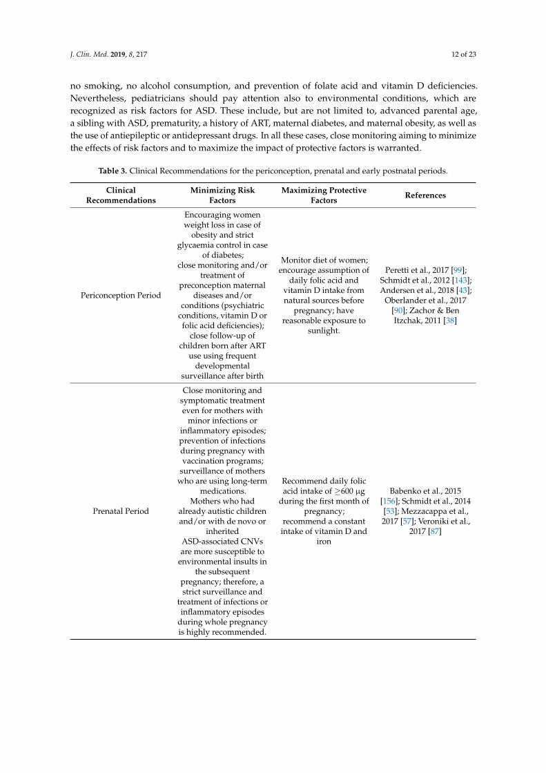
8. Clinical Recommendations
Table 3 summarizes the main clinical recommendations for each period of vulnerability. However,as extensively discussed in the previous chapters, the impact in terms of ASD risk of several of thesefactors is only partially clarified; thus, these clinical recommendations should be acknowledgedconsidering possible pitfalls related to still existing contradictory data interpretations. For thisreason, these recommendations should be pondered in the perspective of the precautionary principle.Some general recommendations are, of course, related to antenatal care for each pregnancy, such as
J. Clin. Med. 2019, 8, 217 12 of 23
no smoking, no alcohol consumption, and prevention of folate acid and vitamin D deficiencies.Nevertheless, pediatricians should pay attention also to environmental conditions, which arerecognized as risk factors for ASD. These include, but are not limited to, advanced parental age,a sibling with ASD, prematurity, a history of ART, maternal diabetes, and maternal obesity, as well asthe use of antiepileptic or antidepressant drugs. In all these cases, close monitoring aiming to minimizethe effects of risk factors and to maximize the impact of protective factors is warranted.
Table 3. Clinical Recommendations for the periconception, prenatal and early postnatal periods.
ClinicalRecommendations
Minimizing RiskFactors
Maximizing ProtectiveFactors References
Periconception Period
Encouraging womenweight loss in case of
obesity and strictglycaemia control in case
of diabetes;close monitoring and/or
treatment ofpreconception maternal
diseases and/orconditions (psychiatric
conditions, vitamin D orfolic acid deficiencies);
close follow-up ofchildren born after ART
use using frequentdevelopmental
surveillance after birth
Monitor diet of women;encourage assumption of
daily folic acid andvitamin D intake fromnatural sources before
pregnancy; havereasonable exposure to
sunlight.
Peretti et al., 2017 [99];Schmidt et al., 2012 [143];Andersen et al., 2018 [43];
Oberlander et al., 2017[90]; Zachor & BenItzchak, 2011 [38]
Prenatal Period
Close monitoring andsymptomatic treatmenteven for mothers with
minor infections orinflammatory episodes;prevention of infectionsduring pregnancy withvaccination programs;
surveillance of motherswho are using long-term
medications.Mothers who had
already autistic childrenand/or with de novo or
inheritedASD-associated CNVsare more susceptible to
environmental insults inthe subsequent
pregnancy; therefore, astrict surveillance and
treatment of infections orinflammatory episodes
during whole pregnancyis highly recommended.
Recommend daily folicacid intake of ≥600 µg
during the first month ofpregnancy;
recommend a constantintake of vitamin D and
iron
Babenko et al., 2015[156]; Schmidt et al., 2014[53]; Mezzacappa et al.,2017 [57]; Veroniki et al.,
2017 [87]
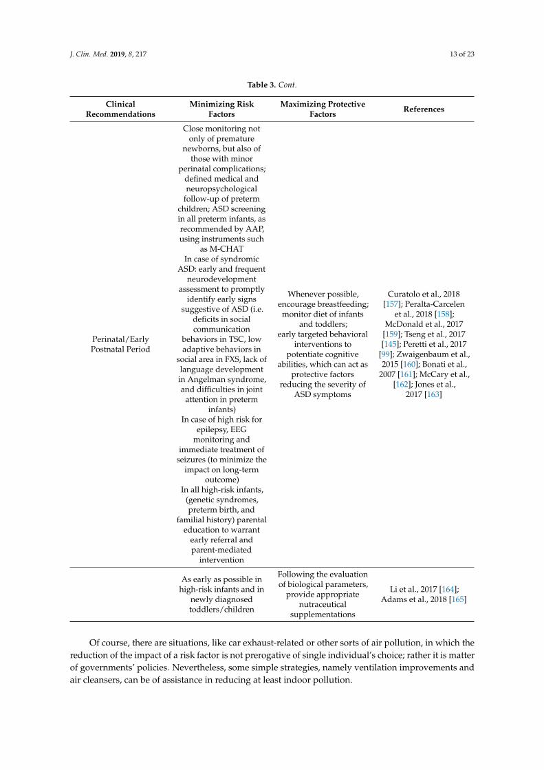
J. Clin. Med. 2019, 8, 217 13 of 23
Table 3. Cont.
ClinicalRecommendations
Minimizing RiskFactors
Maximizing ProtectiveFactors References
Perinatal/EarlyPostnatal Period
Close monitoring notonly of premature
newborns, but also ofthose with minor
perinatal complications;defined medical andneuropsychological
follow-up of pretermchildren; ASD screeningin all preterm infants, asrecommended by AAP,using instruments such
as M-CHATIn case of syndromic
ASD: early and frequentneurodevelopment
assessment to promptlyidentify early signs
suggestive of ASD (i.e.deficits in socialcommunication
behaviors in TSC, lowadaptive behaviors in
social area in FXS, lack oflanguage developmentin Angelman syndrome,and difficulties in joint
attention in preterminfants)
In case of high risk forepilepsy, EEG
monitoring andimmediate treatment of
seizures (to minimize theimpact on long-term
outcome)In all high-risk infants,
(genetic syndromes,preterm birth, and
familial history) parentaleducation to warrant
early referral andparent-mediated
intervention
Whenever possible,encourage breastfeeding;
monitor diet of infantsand toddlers;
early targeted behavioralinterventions to
potentiate cognitiveabilities, which can act as
protective factorsreducing the severity of
ASD symptoms
Curatolo et al., 2018[157]; Peralta-Carcelen
et al., 2018 [158];McDonald et al., 2017
[159]; Tseng et al., 2017[145]; Peretti et al., 2017
[99]; Zwaigenbaum et al.,2015 [160]; Bonati et al.,
2007 [161]; McCary et al.,[162]; Jones et al.,
2017 [163]
As early as possible inhigh-risk infants and in
newly diagnosedtoddlers/children
Following the evaluationof biological parameters,
provide appropriatenutraceutical
supplementations
Li et al., 2017 [164];Adams et al., 2018 [165]
Of course, there are situations, like car exhaust-related or other sorts of air pollution, in which thereduction of the impact of a risk factor is not prerogative of single individual’s choice; rather it is matterof governments’ policies. Nevertheless, some simple strategies, namely ventilation improvements andair cleansers, can be of assistance in reducing at least indoor pollution.
J. Clin. Med. 2019, 8, 217 14 of 23
Anyway, it should also be mentioned that several of these factors have been associated with otherneurodevelopmental or psychiatric disorders as well, for example with Attention Deficit HyperactivityDisorder (ADHD), conduct problems, or lower behavioral scores [166]. As a matter of fact, it isnow recognized that co-occurrence of neurodevelopmental disorders is more often the rule thanthe exception. In this perspective, one should ask not just if an environmental factor increases therisk for ASD, but also if it might impact individually just one of the different features of ASD, likesocial-communication or repetitive behavior.
In short, the synergic effects of genetic, epigenetic and environmental factors can lead to a highersusceptibility during the whole pregnancy, especially in a subset of mothers at high risk of havinga child with ASD. Although it is not possible, at present, to change the pathogenetic effects of themajority of these factors, some modifiable environmental agents could be modulated in order torestrain the severity of the disorder and, potentially, to prevent its onset.
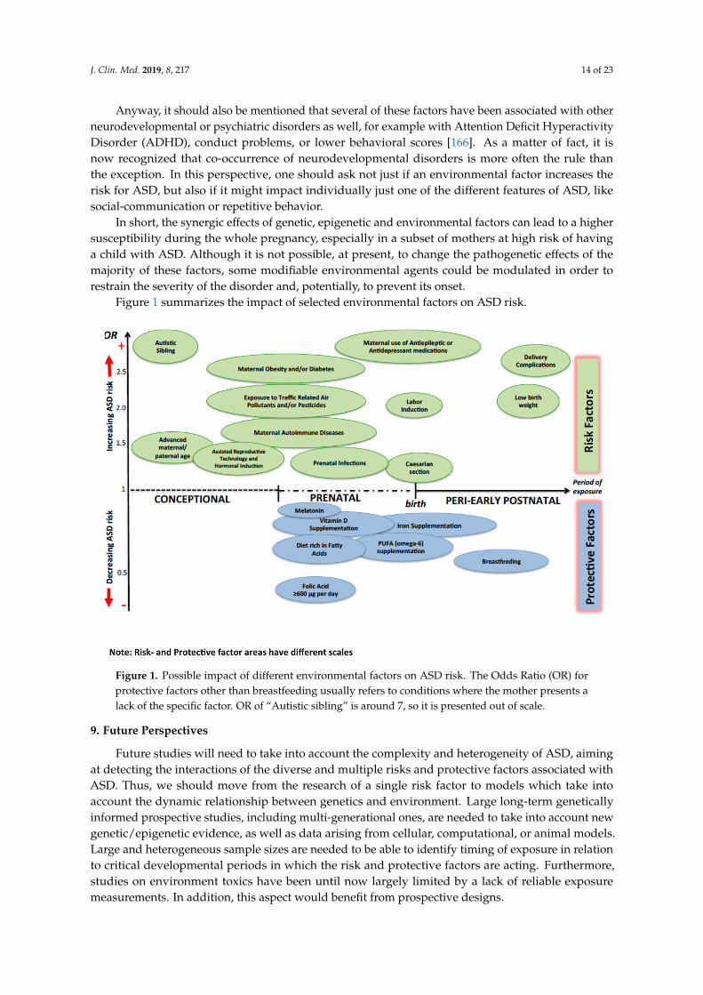
Figure 1 summarizes the impact of selected environmental factors on ASD risk.
J. Clin. Med. 2019, 8, 217 14 of 24
Anyway, it should also be mentioned that several of these factors have been associated with other neurodevelopmental or psychiatric disorders as well, for example with Attention Deficit Hyperactivity Disorder (ADHD), conduct problems, or lower behavioral scores [166]. As a matter of fact, it is now recognized that co-occurrence of neurodevelopmental disorders is more often the rule than the exception. In this perspective, one should ask not just if an environmental factor increases the risk for ASD, but also if it might impact individually just one of the different features of ASD, like social-communication or repetitive behavior.
In short, the synergic effects of genetic, epigenetic and environmental factors can lead to a higher susceptibility during the whole pregnancy, especially in a subset of mothers at high risk of having a child with ASD. Although it is not possible, at present, to change the pathogenetic effects of the majority of these factors, some modifiable environmental agents could be modulated in order to restrain the severity of the disorder and, potentially, to prevent its onset.
Figure 1 summarizes the impact of selected environmental factors on ASD risk.
Figure 1. Possible impact of different environmental factors on ASD risk. The Odds Ratio (OR) for protective factors other than breastfeeding usually refers to conditions where the mother presents a lack of the specific factor. OR of “Autistic sibling” is around 7, so it is presented out of scale.
9. Future Perspectives
Future studies will need to take into account the complexity and heterogeneity of ASD, aiming at detecting the interactions of the diverse and multiple risks and protective factors associated with ASD. Thus, we should move from the research of a single risk factor to models which take into account the dynamic relationship between genetics and environment. Large long-term genetically informed prospective studies, including multi-generational ones, are needed to take into account new genetic/epigenetic evidence, as well as data arising from cellular, computational, or animal models. Large and heterogeneous sample sizes are needed to be able to identify timing of exposure in relation to critical developmental periods in which the risk and protective factors are acting. Furthermore, studies on environment toxics have been until now largely limited by a lack of reliable exposure measurements. In addition, this aspect would benefit from prospective designs.
Figure 1. Possible impact of different environmental factors on ASD risk. The Odds Ratio (OR) forprotective factors other than breastfeeding usually refers to conditions where the mother presents alack of the specific factor. OR of “Autistic sibling” is around 7, so it is presented out of scale.
9. Future Perspectives
Future studies will need to take into account the complexity and heterogeneity of ASD, aimingat detecting the interactions of the diverse and multiple risks and protective factors associated withASD. Thus, we should move from the research of a single risk factor to models which take intoaccount the dynamic relationship between genetics and environment. Large long-term geneticallyinformed prospective studies, including multi-generational ones, are needed to take into account newgenetic/epigenetic evidence, as well as data arising from cellular, computational, or animal models.Large and heterogeneous sample sizes are needed to be able to identify timing of exposure in relationto critical developmental periods in which the risk and protective factors are acting. Furthermore,studies on environment toxics have been until now largely limited by a lack of reliable exposuremeasurements. In addition, this aspect would benefit from prospective designs.
J. Clin. Med. 2019, 8, 217 15 of 23
In conclusion, recent developments in molecular biology and big data approaches, which areable to assess a large number of coexisting factors, are offering new opportunities to disentangle thegene–environment interplay that can lead to the development of ASD.
Author Contributions: P.C., L.E.G., and L.M. conceptualized the consensus conference; P.C., and L.E.G.coordinated the implementation of the consensus conference, its infrastructure and logistics; all authors activelyparticipated in the consensus conference; P.C., L.E.G., M.M., A.B., R.M., and M.S. conceptualized, and drafted theinitial manuscript, and reviewed and revised the final manuscript; L.M., A.F., A.G.A., R.R., and D.Z. criticallyreviewed and revised the manuscript; all authors approved the final manuscript as submitted.
Conflicts of Interest: The authors declare no conflicts of interest.
References
1. Parellada, M.; Penzol, M.J.; Pina, L.; Moreno, C.; Gonzalez-Vioque, E.; Zalsman, G.; Arango, C.The neurobiology of autism spectrum disorders. Eur. Psychiatry 2014, 29, 11–19. [CrossRef] [PubMed]
2. Zachor, D.A.; Curatolo, P.; Participants of Italian-Israeli Consensus Conference. Recommendations for earlydiagnosis and intervention in autism spectrum disorders: An Italian-Israeli consensus conference. Eur. J.Paediatr. Neurol. 2014, 18, 107–118. [CrossRef]
3. Reichow, B.; Hume, K.; Barton, E.E.; Boyd, B.A. Early intensive behavioral intervention (EIBI) for youngchildren with autism spectrum disorders (ASD). Cochrane Database Syst. Rev. 2018, 5. [CrossRef] [PubMed]
4. Benvenuto, A.; Battan, B.; Porfirio, M.C.; Curatolo, P. Pharmacotherapy of autism spectrum disorders.Brain Dev. 2013, 35, 119–127. [CrossRef] [PubMed]
5. Baio, J.; Wiggins, L.; Christensen, D.L.; Maenner, M.J.; Daniels, J.; Warren, Z.; Kurzius-Spencer, M.;Zahorodny, W.; Robinson Rosenberg, C.; White, T.; et al. Prevalence of Autism Spectrum Disorder AmongChildren Aged 8 Years - Autism and Developmental Disabilities Monitoring Network, 11 Sites, United States,2014. MMWR Surveill. Summ. 2018, 67, 1–23. [CrossRef] [PubMed]
6. Idring, S.; Magnusson, C.; Lundberg, M.; Ek, M.; Rai, D.; Svensson, A.C.; Dalman, C.; Karlsson, H.; Lee, B.K.Parental age and the risk of autism spectrum disorders: Findings from a Swedish population-based cohort.Int. J. Epidemiol. 2014, 43, 107–115. [CrossRef] [PubMed]
7. Loke, Y.J.; Hannan, A.J.; Craig, J.M. The Role of Epigenetic Change in Autism Spectrum Disorders.Front. Neurol. 2015, 6, 107. [CrossRef] [PubMed]
8. Sandin, S.; Lichtenstein, P.; Kuja-Halkola, R.; Hultman, C.; Larsson, H.; Reichenberg, A. The Heritability ofAutism Spectrum Disorder. JAMA 2017, 318, 1182–1184. [CrossRef]
9. Ronald, A.; Hoekstra, R.A. Autism spectrum disorders and autistic traits: A decade of new twin studies.Am. J. Med. Genet. B Neuropsychiatr. Genet. 2011, 156B, 255–274. [CrossRef]
10. Girirajan, S.; Dennis, M.Y.; Baker, C.; Malig, M.; Coe, B.P.; Campbell, C.D.; Mark, K.; Vu, T.H.; Alkan, C.;Cheng, Z.; et al. Refinement and discovery of new hotspots of copy-number variation associated with autismspectrum disorder. Am. J. Hum. Genet. 2013, 92, 221–237. [CrossRef]
11. Sanders, S.J.; He, X.; Willsey, A.J.; Ercan-Sencicek, A.G.; Samocha, K.E.; Cicek, A.E.; Murtha, M.T.; Bal, V.H.;Bishop, S.L.; Dong, S.; et al. Insights into Autism Spectrum Disorder Genomic Architecture and Biologyfrom 71 Risk Loci. Neuron 2015, 87, 1215–1233. [CrossRef] [PubMed]
12. Ziats, M.N.; Rennert, O.M. The Evolving Diagnostic and Genetic Landscapes of Autism Spectrum Disorder.Front. Genet. 2016, 7, 65. [CrossRef] [PubMed]
13. Kushima, I.; Aleksic, B.; Nakatochi, M.; Shimamura, T.; Okada, T.; Uno, Y.; Morikawa, M.; Ishizuka, K.;Shiino, T.; Kimura, H.; et al. Comparative Analyses of Copy-Number Variation in Autism Spectrum Disorderand Schizophrenia Reveal Etiological Overlap and Biological Insights. Cell. Rep. 2018, 24, 2838–2856.[CrossRef] [PubMed]
14. Bergbaum, A.; Ogilvie, C.M. Autism and chromosome abnormalities-A review. Clin. Anat. 2016, 29, 620–627.[CrossRef] [PubMed]
15. Sanders, S.J. Next-Generation Sequencing in Autism Spectrum Disorder. Cold Spring Harb. Perspect. Med. 2018.[CrossRef] [PubMed]
16. Benvenuto, A.; Moavero, R.; Alessandrelli, R.; Manzi, B.; Curatolo, P. Syndromic autism: Causes andpathogenetic pathways. World J. Pediatr. 2009, 5, 169–176. [CrossRef] [PubMed]
J. Clin. Med. 2019, 8, 217 16 of 23
17. Saskin, A.; Fulginiti, V.; Birch, A.H.; Trakadis, Y. Prevalence of four Mendelian disorders associated withautism in 2392 affected families. J. Hum. Genet. 2017, 62, 657–659. [CrossRef]
18. Emberti Gialloreti, L.; Curatolo, P. Autism Spectrum Disorder: Why Do We Know So Little? Front. Neurol.2018, 9, 670. [CrossRef]
19. Lyall, K.; Munger, K.L.; O’Reilly, E.J.; Santangelo, S.L.; Ascherio, A. Maternal dietary fat intake in associationwith autism spectrum disorders. Am. J. Epidemiol. 2013, 178, 209–220. [CrossRef]
20. Lyall, K.; Croen, L.A.; Sjodin, A.; Yoshida, C.K.; Zerbo, O.; Kharrazi, M.; Windham, G.C. PolychlorinatedBiphenyl and Organochlorine Pesticide Concentrations in Maternal Mid-Pregnancy Serum Samples:Association with Autism Spectrum Disorder and Intellectual Disability. Environ. Health Perspect. 2017,125, 474–480. [CrossRef]
21. Janecka, M.; Mill, J.; Basson, M.A.; Goriely, A.; Spiers, H.; Reichenberg, A.; Schalkwyk, L.; Fernandes, C.Advanced paternal age effects in neurodevelopmental disorders-review of potential underlying mechanisms.Transl. Psychiatry 2017, 7, e1019. [CrossRef] [PubMed]
22. Merikangas, A.K.; Calkins, M.E.; Bilker, W.B.; Moore, T.M.; Gur, R.C.; Gur, R.E. Parental Age and OffspringPsychopathology in the Philadelphia Neurodevelopmental Cohort. J. Am. Acad. Child. Adolesc. Psychiatry2017, 56, 391–400. [CrossRef] [PubMed]
23. Durkin, M.S.; Maenner, M.J.; Newschaffer, C.J.; Lee, L.C.; Cunniff, C.M.; Daniels, J.L.; Kirby, R.S.; Leavitt, L.;Miller, L.; Zahorodny, W.; et al. Advanced parental age and the risk of autism spectrum disorder.Am. J. Epidemiol. 2008, 168, 1268–1276. [CrossRef] [PubMed]
24. Ben Itzchak, E.; Lahat, E.; Zachor, D.A. Advanced parental ages and low birth weight in autism spectrumdisorders-rates and effect on functioning. Res. Dev. Disabil. 2011, 32, 1776–1781. [CrossRef] [PubMed]
25. Geier, D.A.; Kern, J.K.; Sykes, L.K.; Geier, M.R. Examining genotypic variation in autism spectrum disorderand its relationship to parental age and phenotype. Appl. Clin. Genet. 2016, 9, 121–129. [CrossRef] [PubMed]
26. Modabbernia, A.; Velthorst, E.; Reichenberg, A. Environmental risk factors for autism: An evidence-basedreview of systematic reviews and meta-analyses. Mol. Autism 2017, 8, 13. [CrossRef] [PubMed]
27. Sandin, S.; Schendel, D.; Magnusson, P.; Hultman, C.; Suren, P.; Susser, E.; Gronborg, T.; Gissler, M.;Gunnes, N.; Gross, R.; et al. Autism risk associated with parental age and with increasing difference in agebetween the parents. Mol. Psychiatry 2016, 21, 693–700. [CrossRef]
28. Flatscher-Bader, T.; Foldi, C.J.; Chong, S.; Whitelaw, E.; Moser, R.J.; Burne, T.H.; Eyles, D.W.; McGrath, J.J.Increased de novo copy number variants in the offspring of older males. Transl. Psychiatry 2011, 1, e34.[CrossRef]
29. Kong, A.; Frigge, M.L.; Masson, G.; Besenbacher, S.; Sulem, P.; Magnusson, G.; Gudjonsson, S.A.;Sigurdsson, A.; Jonasdottir, A.; Jonasdottir, A.; et al. Rate of de novo mutations and the importanceof father’s age to disease risk. Nature 2012, 488, 471–475. [CrossRef]
30. European IVF-monitoring Consortium (EIM); European Society of Human Reproduction and Embryology(ESHRE); Calhaz-Jorge, C.; De Geyter, C.; Kupka, M.S.; de Mouzon, J.; Erb, K.; Mocanu, E.; Motrenko, T.; et al.Assisted reproductive technology in Europe, 2013: Results generated from European registers by ESHRE.Hum. Reprod. 2017, 32, 1957–1973. [CrossRef]
31. Liu, L.; Gao, J.; He, X.; Cai, Y.; Wang, L.; Fan, X. Association between assisted reproductive technology andthe risk of autism spectrum disorders in the offspring: A meta-analysis. Sci. Rep. 2017, 7, 46207. [CrossRef][PubMed]
32. Hansen, M.; Kurinczuk, J.J.; Bower, C.; Webb, S. The risk of major birth defects after intracytoplasmic sperminjection and in vitro fertilization. N. Engl. J. Med. 2002, 346, 725–730. [CrossRef]
33. Gosden, R.; Trasler, J.; Lucifero, D.; Faddy, M. Rare congenital disorders, imprinted genes, and assistedreproductive technology. Lancet 2003, 361, 1975–1977. [CrossRef]
34. Davies, M.J.; Moore, V.M.; Willson, K.J.; Van Essen, P.; Priest, K.; Scott, H.; Haan, E.A.; Chan, A. Reproductivetechnologies and the risk of birth defects. N. Engl. J. Med. 2012, 366, 1803–1813. [CrossRef] [PubMed]
35. Lidegaard, O.; Pinborg, A.; Andersen, A.N. Imprinting disorders after assisted reproductive technologies.Curr. Opin. Obstet. Gynecol. 2006, 18, 293–296. [CrossRef] [PubMed]
36. Auyeung, B.; Baron-Cohen, S.; Ashwin, E.; Knickmeyer, R.; Taylor, K.; Hackett, G. Fetal testosterone andautistic traits. Br. J. Psychol. 2009, 100, 1–22. [CrossRef] [PubMed]
37. Lung, F.W.; Shu, B.C.; Chiang, T.L.; Lin, S.J. Twin-singleton influence on infant development: A nationalbirth cohort study. Child. Care Health Dev. 2009, 35, 409–418. [CrossRef] [PubMed]
J. Clin. Med. 2019, 8, 217 17 of 23
38. Zachor, D.A.; Ben Itzchak, E. Assisted reproductive technology and risk for autism spectrum disorder.Res. Dev. Disabil. 2011, 32, 2950–2956. [CrossRef] [PubMed]
39. Kalkbrenner, A.E.; Schmidt, R.J.; Penlesky, A.C. Environmental chemical exposures and autism spectrumdisorders: A review of the epidemiological evidence. Curr. Probl. Pediatr. Adolesc. Health Care 2014, 44,277–318. [CrossRef]
40. Rossignol, D.A.; Genuis, S.J.; Frye, R.E. Environmental toxicants and autism spectrum disorders: A systematicreview. Transl. Psychiatry 2014, 4, e360. [CrossRef] [PubMed]
41. Krakowiak, P.; Walker, C.K.; Bremer, A.A.; Baker, A.S.; Ozonoff, S.; Hansen, R.L.; Hertz-Picciotto, I. Maternalmetabolic conditions and risk for autism and other neurodevelopmental disorders. Pediatrics 2012, 129,e1121–e1128. [CrossRef] [PubMed]
42. Getz, K.D.; Anderka, M.T.; Werler, M.M.; Jick, S.S. Maternal Pre-pregnancy Body Mass Index and AutismSpectrum Disorder among Offspring: A Population-Based Case-Control Study. Paediatr. Perinat. Epidemiol.2016, 30, 479–487. [CrossRef] [PubMed]
43. Andersen, C.H.; Thomsen, P.H.; Nohr, E.A.; Lemcke, S. Maternal body mass index before pregnancy as arisk factor for ADHD and autism in children. Eur. Child. Adolesc Psychiatry 2018, 27, 139–148. [CrossRef][PubMed]
44. Bugatto, F.; Fernandez-Deudero, A.; Bailen, A.; Fernandez-Macias, R.; Hervias-Vivancos, B.; Bartha, J.L.Second-trimester amniotic fluid proinflammatory cytokine levels in normal and overweight women.Obstet. Gynecol. 2010, 115, 127–133. [CrossRef] [PubMed]
45. Georgieff, M.K. Nutrition and the developing brain: Nutrient priorities and measurement. Am. J. Clin. Nutr.2007, 85, 614S620S. [CrossRef]
46. Schmidt, R.J.; Kogan, V.; Shelton, J.F.; Delwiche, L.; Hansen, R.L.; Ozonoff, S.; Ma, C.C.; McCanlies, E.C.;Bennett, D.H.; Hertz-Picciotto, I.; et al. Combined Prenatal Pesticide Exposure and Folic Acid Intake inRelation to Autism Spectrum Disorder. Environ. Health Perspect. 2017, 125, 097007. [CrossRef] [PubMed]
47. Beard, C.M.; Panser, L.A.; Katusic, S.K. Is excess folic acid supplementation a risk factor for autism?Med. Hypotheses 2011, 77, 15–17. [CrossRef]
48. Wiens, D.; DeSoto, M.C. Is High Folic Acid Intake a Risk Factor for Autism?–A Review. Brain Sci. 2017, 7.[CrossRef]
49. Raghavan, R.; Riley, A.W.; Volk, H.; Caruso, D.; Hironaka, L.; Sices, L.; Hong, X.; Wang, G.; Ji, Y.; Brucato, M.;et al. Maternal Multivitamin Intake, Plasma Folate and Vitamin B12 Levels and Autism Spectrum DisorderRisk in Offspring. Paediatr. Perinat. Epidemiol. 2018, 32, 100–111. [CrossRef]
50. Powers, H.J. Folic acid under scrutiny. Br. J. Nutr. 2007, 98, 665–666. [CrossRef]51. Girotto, F.; Scott, L.; Avchalumov, Y.; Harris, J.; Iannattone, S.; Drummond-Main, C.; Tobias, R.;
Bello-Espinosa, L.; Rho, J.M.; Davidsen, J.; et al. High dose folic acid supplementation of rats alterssynaptic transmission and seizure susceptibility in offspring. Sci. Rep. 2013, 3, 1465. [CrossRef] [PubMed]
52. Barua, S.; Chadman, K.K.; Kuizon, S.; Buenaventura, D.; Stapley, N.W.; Ruocco, F.; Begum, U.; Guariglia, S.R.;Brown, W.T.; Junaid, M.A. Increasing maternal or post-weaning folic acid alters gene expression andmoderately changes behavior in the offspring. PLoS One 2014, 9, e101674. [CrossRef]
53. Schmidt, R.J.; Tancredi, D.J.; Krakowiak, P.; Hansen, R.L.; Ozonoff, S. Maternal intake of supplemental ironand risk of autism spectrum disorder. Am. J. Epidemiol. 2014, 180, 890–900. [CrossRef] [PubMed]
54. Gidaya, N.B.; Lee, B.K.; Burstyn, I.; Yudell, M.; Mortensen, E.L.; Newschaffer, C.J. In utero exposure toselective serotonin reuptake inhibitors and risk for autism spectrum disorder. J. Autism Dev. Disord. 2014, 44,2558–2567. [CrossRef]
55. Andalib, S.; Emamhadi, M.R.; Yousefzadeh-Chabok, S.; Shakouri, S.K.; Hoilund-Carlsen, P.F.; Vafaee, M.S.;Michel, T.M. Maternal SSRI exposure increases the risk of autistic offspring: A meta-analysis and systematicreview. Eur. Psychiatry 2017, 45, 161–166. [CrossRef]
56. Brown, H.K.; Hussain-Shamsy, N.; Lunsky, Y.; Dennis, C.E.; Vigod, S.N. The Association between AntenatalExposure to Selective Serotonin Reuptake Inhibitors and Autism: A Systematic Review and Meta-Analysis.J. Clin. Psychiatry 2017, 78, e48–e58. [CrossRef] [PubMed]
57. Mezzacappa, A.; Lasica, P.A.; Gianfagna, F.; Cazas, O.; Hardy, P.; Falissard, B.; Sutter-Dallay, A.L.; Gressier, F.Risk for Autism Spectrum Disorders According to Period of Prenatal Antidepressant Exposure: A SystematicReview and Meta-analysis. JAMA Pediatr. 2017, 171, 555–563. [CrossRef]
J. Clin. Med. 2019, 8, 217 18 of 23
58. Rai, D.; Lee, B.K.; Dalman, C.; Golding, J.; Lewis, G.; Magnusson, C. Parental depression, maternalantidepressant use during pregnancy, and risk of autism spectrum disorders: Population based case-controlstudy. BMJ 2013, 346, 2059. [CrossRef]
59. Malm, H.; Brown, A.S.; Gissler, M.; Gyllenberg, D.; Hinkka-Yli-Salomaki, S.; McKeague, I.W.; Weissman, M.;Wickramaratne, P.; Artama, M.; Gingrich, J.A.; et al. Gestational Exposure to Selective Serotonin ReuptakeInhibitors and Offspring Psychiatric Disorders: A National Register-Based Study. J. Am. Acad. Child.Adolesc. Psychiatry 2016, 55, 359–366. [CrossRef]
60. Hviid, A.; Melbye, M.; Pasternak, B. Use of selective serotonin reuptake inhibitors during pregnancy andrisk of autism. N. Engl. J. Med. 2013, 369, 2406–2415. [CrossRef]
61. Sørensen, M.J.; Gronborg, T.K.; Christensen, J.; Parner, E.T.; Vestergaard, M.; Schendel, D.; Pedersen, L.H.Antidepressant exposure in pregnancy and risk of autism spectrum disorders. Clin. Epidemiol. 2013, 5,449–459. [PubMed]
62. Jung, C.R.; Lin, Y.T.; Hwang, B.F. Air pollution and newly diagnostic autism spectrum disorders:A population-based cohort study in Taiwan. PLoS ONE 2013, 8, e75510. [CrossRef]
63. Kim, D.; Volk, H.; Girirajan, S.; Pendergrass, S.; Hall, M.A.; Verma, S.S.; Schmidt, R.J.; Hansen, R.L.; Ghosh, D.;Ludena-Rodriguez, Y.; et al. The joint effect of air pollution exposure and copy number variation on risk forautism. Autism Res. 2017, 10, 1470–1480. [CrossRef] [PubMed]
64. Goodrich, A.J.; Volk, H.E.; Tancredi, D.J.; McConnell, R.; Lurmann, F.W.; Hansen, R.L.; Schmidt, R.J. Jointeffects of prenatal air pollutant exposure and maternal folic acid supplementation on risk of autism spectrumdisorder. Autism Res. 2018, 11, 69–80. [CrossRef] [PubMed]
65. Gaita, L.; Manzi, B.; Sacco, R.; Lintas, C.; Altieri, L.; Lombardi, F.; Pawlowski, T.L.; Redman, M.; Craig, D.W.;Huentelman, M.J.; et al. Decreased serum arylesterase activity in autism spectrum disorders. Psychiatry Res.2010, 180, 105–113. [CrossRef]
66. Thummler, S.; Dor, E.; David, R.; Leali, G.; Battista, M.; David, A.; Askenazy, F.; Verstuyft, C.Pharmacoresistant Severe Mental Health Disorders in Children and Adolescents: Functional Abnormalitiesof Cytochrome P450 2D6. Front. Psychiatry 2018, 9, 2. [CrossRef] [PubMed]
67. Weisskopf, M.G.; Kioumourtzoglou, M.A.; Roberts, A.L. Air Pollution and Autism Spectrum Disorders:Causal or Confounded? Curr. Environ. Health Rep. 2015, 2, 430–439. [CrossRef]
68. Lam, J.; Sutton, P.; Kalkbrenner, A.; Windham, G.; Halladay, A.; Koustas, E.; Lawler, C.; Davidson, L.;Daniels, N.; Newschaffer, C.; et al. A Systematic Review and Meta-Analysis of Multiple Airborne Pollutantsand Autism Spectrum Disorder. PLoS ONE 2016, 11, e0161851. [CrossRef]
69. Volk, H.E.; Hertz-Picciotto, I.; Delwiche, L.; Lurmann, F.; McConnell, R. Residential proximity to freewaysand autism in the CHARGE study. Environ. Health Perspect. 2011, 119, 873–877. [CrossRef]
70. Becerra, T.A.; Wilhelm, M.; Olsen, J.; Cockburn, M.; Ritz, B. Ambient air pollution and autism in Los Angelescounty, California. Environ. Health Perspect. 2013, 121, 380–386. [CrossRef]
71. Gong, T.; Dalman, C.; Wicks, S.; Dal, H.; Magnusson, C.; Lundholm, C.; Almqvist, C.; Pershagen, G. PerinatalExposure to Traffic-Related Air Pollution and Autism Spectrum Disorders. Environ. Health Perspect. 2017,125, 119–126. [CrossRef] [PubMed]
72. Guxens, M.; Ghassabian, A.; Gong, T.; Garcia-Esteban, R.; Porta, D.; Giorgis-Allemand, L.; Almqvist, C.;Aranbarri, A.; Beelen, R.; Badaloni, C.; et al. Air Pollution Exposure during Pregnancy andChildhood Autistic Traits in Four European Population-Based Cohort Studies: The ESCAPE Project.Environ. Health Perspect. 2016, 124, 133–140. [CrossRef]
73. Raz, R.; Levine, H.; Pinto, O.; Broday, D.M.; Yuval; Weisskopf, M.G. Traffic-Related Air Pollution and AutismSpectrum Disorder: A Population-Based Nested Case-Control Study in Israel. Am. J. Epidemiol. 2018, 187,717–725. [CrossRef]
74. Pagalan, L.; Bickford, C.; Weikum, W.; Lanphear, B.; Brauer, M.; Lanphear, N.; Hanley, G.E.; Oberlander, T.F.;Winters, M. Association of Prenatal Exposure to Air Pollution with Autism Spectrum Disorder.JAMA Pediatr. 2018. [CrossRef]
75. Roberts, E.M.; English, P.B.; Grether, J.K.; Windham, G.C.; Somberg, L.; Wolff, C. Maternal residence nearagricultural pesticide applications and autism spectrum disorders among children in the California CentralValley. Environ. Health Perspect. 2007, 115, 1482–1489. [CrossRef]
76. Roberts, E.M.; English, P.B. Bayesian modeling of time-dependent vulnerability to environmental hazards:An example using autism and pesticide data. Stat. Med. 2013, 32, 2308–2319. [CrossRef] [PubMed]
J. Clin. Med. 2019, 8, 217 19 of 23
77. Cheslack-Postava, K.; Rantakokko, P.V.; Hinkka-Yli-Salomaki, S.; Surcel, H.M.; McKeague, I.W.;Kiviranta, H.A.; Sourander, A.; Brown, A.S. Maternal serum persistent organic pollutants in the FinnishPrenatal Study of Autism: A pilot study. Neurotoxicol. Teratol. 2013, 38, 1–5. [CrossRef]
78. Braun, J.M.; Kalkbrenner, A.E.; Just, A.C.; Yolton, K.; Calafat, A.M.; Sjodin, A.; Hauser, R.; Webster, G.M.;Chen, A.; Lanphear, B.P. Gestational exposure to endocrine-disrupting chemicals and reciprocal social,repetitive, and stereotypic behaviors in 4- and 5-year-old children: The HOME study. Environ. Health Perspect.2014, 122, 513–520. [CrossRef]
79. Eskenazi, B.; Marks, A.R.; Bradman, A.; Harley, K.; Barr, D.B.; Johnson, C.; Morga, N.; Jewell, N.P.Organophosphate pesticide exposure and neurodevelopment in young Mexican-American children.Environ. Health Perspect. 2007, 115, 792–798. [CrossRef]
80. Shelton, J.F.; Geraghty, E.M.; Tancredi, D.J.; Delwiche, L.D.; Schmidt, R.J.; Ritz, B.; Hansen, R.L.;Hertz-Picciotto, I. Neurodevelopmental disorders and prenatal residential proximity to agriculturalpesticides: The CHARGE study. Environ. Health Perspect. 2014, 122, 1103–1109. [CrossRef]
81. Bjorling-Poulsen, M.; Andersen, H.R.; Grandjean, P. Potential developmental neurotoxicity of pesticidesused in Europe. Environ. Health 2008, 7, 50. [CrossRef] [PubMed]
82. Wang, X.; Martinez, M.A.; Dai, M.; Chen, D.; Ares, I.; Romero, A.; Castellano, V.; Martinez, M.; Rodriguez, J.L.;Martinez-Larranaga, M.R.; et al. Permethrin-induced oxidative stress and toxicity and metabolism. A review.Environ. Res. 2016, 149, 86–104. [CrossRef]
83. Hicks, S.D.; Wang, M.; Fry, K.; Doraiswamy, V.; Wohlford, E.M. Neurodevelopmental Delay Diagnosis RatesAre Increased in a Region with Aerial Pesticide Application. Front. Pediatr. 2017, 5, 116. [CrossRef] [PubMed]
84. De Palma, G.; Catalani, S.; Franco, A.; Brighenti, M.; Apostoli, P. Lack of correlation between metallicelements analyzed in hair by ICP-MS and autism. J. Autism Dev. Disord. 2012, 42, 342–353. [CrossRef][PubMed]
85. Yoshimasu, K.; Kiyohara, C.; Takemura, S.; Nakai, K. A meta-analysis of the evidence on the impact ofprenatal and early infancy exposures to mercury on autism and attention deficit/hyperactivity disorder inthe childhood. Neurotoxicology 2014, 44, 121–131. [CrossRef] [PubMed]
86. Roullet, F.I.; Lai, J.K.; Foster, J.A. In utero exposure to valproic acid and autism-a current review of clinicaland animal studies. Neurotoxicol. Teratol. 2013, 36, 47–56. [CrossRef] [PubMed]
87. Veroniki, A.A.; Rios, P.; Cogo, E.; Straus, S.E.; Finkelstein, Y.; Kealey, R.; Reynen, E.; Soobiah, C.; Thavorn, K.;Hutton, B.; et al. Comparative safety of antiepileptic drugs for neurological development in children exposedduring pregnancy and breast feeding: A systematic review and network meta-analysis. BMJ Open 2017,7, e017248. [CrossRef] [PubMed]
88. Andrade, C. Antidepressant Exposure during Pregnancy and Risk of Autism in the Offspring, 1: Meta-Reviewof Meta-Analyses. J. Clin. Psychiatry 2017, 78, e1047–e1051. [CrossRef] [PubMed]
89. Kaplan, Y.C.; Keskin-Arslan, E.; Acar, S.; Sozmen, K. Maternal SSRI discontinuation, use, psychiatric disorderand the risk of autism in children: A meta-analysis of cohort studies. Br. J. Clin. Pharmacol. 2017, 83,2798–2806. [CrossRef] [PubMed]
90. Oberlander, T.F.; Zwaigenbaum, L. Disentangling Maternal Depression and Antidepressant Use duringPregnancy as Risks for Autism in Children. JAMA 2017, 317, 1533–1534. [CrossRef] [PubMed]
91. Atladottir, H.O.; Henriksen, T.B.; Schendel, D.E.; Parner, E.T. Autism after infection, febrile episodes, andantibiotic use during pregnancy: An exploratory study. Pediatrics 2012, 130, 1447–1454. [CrossRef] [PubMed]
92. Leclercq, S.; Mian, F.M.; Stanisz, A.M.; Bindels, L.B.; Cambier, E.; Ben-Amram, H.; Koren, O.; Forsythe, P.;Bienenstock, J. Low-dose penicillin in early life induces long-term changes in murine gut microbiota, braincytokines and behavior. Nat. Commun. 2017, 8, 15062. [CrossRef] [PubMed]
93. Aronson, M.; Hagberg, B.; Gillberg, C. Attention deficits and autistic spectrum problems in children exposedto alcohol during gestation: A follow-up study. Dev. Med. Child. Neurol. 1997, 39, 583–587. [CrossRef][PubMed]
94. Eliasen, M.; Tolstrup, J.S.; Nybo Andersen, A.M.; Gronbaek, M.; Olsen, J.; Strandberg-Larsen, K. Prenatalalcohol exposure and autistic spectrum disorders–a population-based prospective study of 80,552 childrenand their mothers. Int. J. Epidemiol. 2010, 39, 1074–1081. [CrossRef] [PubMed]
95. Gallagher, C.; McCarthy, F.P.; Ryan, R.M.; Khashan, A.S. Maternal Alcohol Consumption during Pregnancyand the Risk of Autism Spectrum Disorders in Offspring: A Retrospective Analysis of the Millennium CohortStudy. J. Autism Dev. Disord. 2018, 48, 3773–3782. [CrossRef] [PubMed]
J. Clin. Med. 2019, 8, 217 20 of 23
96. Larsson, M.; Weiss, B.; Janson, S.; Sundell, J.; Bornehag, C.G. Associations between indoor environmentalfactors and parental-reported autistic spectrum disorders in children 6-8 years of age. Neurotoxicology 2009,30, 822–831. [CrossRef] [PubMed]
97. Rosen, B.N.; Lee, B.K.; Lee, N.L.; Yang, Y.; Burstyn, I. Maternal Smoking and Autism Spectrum Disorder:A Meta-analysis. J. Autism Dev. Disord. 2015, 45, 1689–1698. [CrossRef]
98. Tang, S.; Wang, Y.; Gong, X.; Wang, G. A Meta-Analysis of Maternal Smoking during Pregnancy and AutismSpectrum Disorder Risk in Offspring. Int. J. Environ. Res. Public Health 2015, 26, 10418–10431. [CrossRef]
99. Peretti, S.; Mariano, M.; Mazzocchetti, C.; Mazza, M.; Pino, M.C.; Verrotti Di Pianella, A.; Valenti, M. Diet:The keystone of autism spectrum disorder? Nutr. Neurosci. 2018, 1–15. [CrossRef]
100. Suren, P.; Roth, C.; Bresnahan, M.; Haugen, M.; Hornig, M.; Hirtz, D.; Lie, K.K.; Lipkin, W.I.; Magnus, P.;Reichborn-Kjennerud, T.; et al. Association between maternal use of folic acid supplements and risk ofautism spectrum disorders in children. JAMA 2013, 309, 570–577. [CrossRef]
101. Vinkhuyzen, A.A.E.; Eyles, D.W.; Burne, T.H.J.; Blanken, L.M.E.; Kruithof, C.J.; Verhulst, F.; White, T.;Jaddoe, V.W.; Tiemeier, H.; McGrath, J.J. Gestational vitamin D deficiency and autism spectrum disorder.BJPsych Open 2017, 3, 85–90. [CrossRef] [PubMed]
102. Walton, R.G.; Monte, W.C. Dietary methanol and autism. Med. Hypotheses 2015, 85, 441–446. [CrossRef][PubMed]
103. Sullivan, E.L.; Nousen, E.K.; Chamlou, K.A. Maternal high fat diet consumption during the perinatal periodprograms offspring behavior. Physiol. Behav. 2014, 123, 236–242. [CrossRef] [PubMed]
104. Parboosing, R.; Bao, Y.; Shen, L.; Schaefer, C.A.; Brown, A.S. Gestational influenza and bipolar disorder inadult offspring. JAMA Psychiatry 2013, 70, 677–685. [CrossRef] [PubMed]
105. Zerbo, O.; Iosif, A.M.; Walker, C.; Ozonoff, S.; Hansen, R.L.; Hertz-Picciotto, I. Is maternal influenza or feverduring pregnancy associated with autism or developmental delays? Results from the CHARGE (CHildhoodAutism Risks from Genetics and Environment) study. J. Autism Dev. Disord. 2013, 43, 25–33. [CrossRef][PubMed]
106. Brucato, M.; Ladd-Acosta, C.; Li, M.; Caruso, D.; Hong, X.; Kaczaniuk, J.; Stuart, E.A.; Fallin, M.D.; Wang, X.Prenatal exposure to fever is associated with autism spectrum disorder in the boston birth cohort. Autism Res.2017, 10, 1878–1890. [CrossRef] [PubMed]
107. Hornig, M.; Bresnahan, M.A.; Che, X.; Schultz, A.F.; Ukaigwe, J.E.; Eddy, M.L.; Hirtz, D.; Gunnes, N.;Lie, K.K.; Magnus, P.; et al. Prenatal fever and autism risk. Mol. Psychiatry 2018, 23, 759–766. [CrossRef]
108. Knuesel, I.; Chicha, L.; Britschgi, M.; Schobel, S.A.; Bodmer, M.; Hellings, J.A.; Toovey, S.; Prinssen, E.P.Maternal immune activation and abnormal brain development across CNS disorders. Nat. Rev. Neurol. 2014,10, 643–660. [CrossRef]
109. Parker-Athill, E.C.; Tan, J. Maternal immune activation and autism spectrum disorder: Interleukin-6 signalingas a key meachanistic pathway. Neurosignals 2010, 18, 113–128. [CrossRef]
110. Jones, K.L.; Croen, L.A.; Yoshida, C.K.; Heuer, L.; Hansen, R.; Zerbo, O.; DeLorenze, G.N.; Kharrazi, M.;Yolken, R.; Ashwood, P.; et al. Autism with intellectual disability is associated with increased levels ofmaternal cytokines and chemokines during gestation. Mol. Psychiatry 2017, 22, 273–279. [CrossRef]
111. Jones, K.L.; Van de Water, J. Maternal autoantibody related autism: mechanisms andpathways. Mol. Psychiat. 2018. [CrossRef] [PubMed]
112. Meltzer, A.; Van de Water, J. The Role of the Immune System in Autism Spectrum Disorder.Neuropsychopharmacology 2017, 42, 284–298. [CrossRef] [PubMed]
113. Krakowiak, P.; Walker, C.K.; Tancredi, D.; Hertz-Picciotto, I.; Van de Water, J. Autism-specific maternalanti-fetal brain autoantibodies are associated with metabolic conditions. Autism Res. 2017, 10, 89–98.[CrossRef] [PubMed]
114. Gardener, H.; Spiegelman, D.; Buka, S.L. Prenatal risk factors for autism: Comprehensive meta-analysis.Br. J. Psychiatry 2009, 195, 7–14. [CrossRef] [PubMed]
115. Lyall, K.; Pauls, D.L.; Spiegelman, D.; Ascherio, A.; Santangelo, S.L. Pregnancy complications and obstetricsuboptimality in association with autism spectrum disorders in children of the Nurses’ Health Study II.Autism Res. 2012, 5, 21–30. [CrossRef] [PubMed]
116. Xiang, A.H.; Wang, X.; Martinez, M.P.; Walthall, J.C.; Curry, E.S.; Page, K.; Buchanan, T.A.; Coleman, K.J.;Getahun, D. Association of maternal diabetes with autism in offspring. JAMA 2015, 313, 1425–1434.[CrossRef] [PubMed]
J. Clin. Med. 2019, 8, 217 21 of 23
117. Ornoy, A.; Ratzon, N.; Greenbaum, C.; Wolf, A.; Dulitzky, M. School-age children born to diabetic mothersand to mothers with gestational diabetes exhibit a high rate of inattention and fine and gross motorimpairment. J. Pediatr. Endocrinol. Metab. 2001, 14, 681–689. [CrossRef]
118. Banik, A.; Kandilya, D.; Ramya, S.; Stunkel, W.; Chong, Y.S.; Dheen, S.T. Maternal Factors that InduceEpigenetic Changes Contribute to Neurological Disorders in Offspring. Genes (Basel) 2017, 8. [CrossRef]
119. Braam, W.; Ehrhart, F.; Maas, A.; Smits, M.G.; Curfs, L. Low maternal melatonin level increases autismspectrum disorder risk in children. Res. Dev. Disabil. 2018, 82, 79–89. [CrossRef]
120. Jin, Y.; Choi, J.; Won, J.; Hong, Y. The Relationship between Autism Spectrum Disorder and Melatonin duringFetal Development. Molecules 2018, 23. [CrossRef]
121. Curran, E.A.; O’Neill, S.M.; Cryan, J.F.; Kenny, L.C.; Dinan, T.G.; Khashan, A.S.; Kearney, P.M.Research review: Birth by caesarean section and development of autism spectrum disorder andattention-deficit/hyperactivity disorder: A systematic review and meta-analysis. J. Child. Psychol. Psychiatry2015, 56, 500–508. [CrossRef] [PubMed]
122. Dodds, L.; Fell, D.B.; Shea, S.; Armson, B.A.; Allen, A.C.; Bryson, S. The role of prenatal, obstetric andneonatal factors in the development of autism. J. Autism Dev. Disord. 2011, 41, 891–902. [CrossRef] [PubMed]
123. Emberti Gialloreti, L.; Benvenuto, A.; Benassi, F.; Curatolo, P. Are caesarean sections, induced labor andoxytocin regulation linked to Autism Spectrum Disorders? Med. Hypotheses 2014, 82, 713–718. [CrossRef][PubMed]
124. Harony, H.; Wagner, S. The contribution of oxytocin and vasopressin to mammalian social behavior: Potentialrole in autism spectrum disorder. Neurosignals 2010, 18, 82–97. [CrossRef] [PubMed]
125. Olza Fernandez, I.; Marin Gabriel, M.A.; Lopez Sanchez, F.; Malalana Martinez, A.M. Oxytocin and autism: Ahypothesis to research. Can perinatal oxitocinergic manipulation facilitate autism? Rev. Psiquiatr. Salud Ment.2011, 4, 38–41. [CrossRef] [PubMed]
126. Wang, C.; Geng, H.; Liu, W.; Zhang, G. Prenatal, perinatal, and postnatal factors associated with autism: Ameta-analysis. Medicine (Baltimore) 2017, 96, e6696. [CrossRef]
127. Limperopoulos, C.; Bassan, H.; Sullivan, N.R.; Soul, J.S.; Robertson, R.L., Jr.; Moore, M.; Ringer, S.A.;Volpe, J.J.; Du Plessis, A.J. Positive screening for autism in ex-preterm infants: Prevalence and risk factors.Pediatrics 2008, 121, 758–765. [CrossRef] [PubMed]
128. Kang, D.W.; Adams, J.B.; Gregory, A.C.; Borody, T.; Chittick, L.; Fasano, A.; Khoruts, A.; Geis, E.;Maldonado, J.; McDonough-Means, S.; et al. Microbiota Transfer Therapy alters gut ecosystem and improvesgastrointestinal and autism symptoms: An open-label study. Microbiome 2017, 5, 10. [CrossRef]
129. Yang, Y.; Tian, J.; Yang, B. Targeting gut microbiome: A novel and potential therapy for autism. Life Sci. 2018,194, 111–119. [CrossRef]
130. Grimaldi, R.; Cela, D.; Swann, J.R.; Vulevic, J.; Gibson, G.R.; Tzortzis, G.; Costabile, A. In vitro fermentationof B-GOS: Impact on faecal bacterial populations and metabolic activity in autistic and non-autistic children.FEMS Microbiol. Ecol. 2017, 93. [CrossRef]
131. Grimaldi, R.; Gibson, G.R.; Vulevic, J.; Giallourou, N.; Castro-Mejia, J.L.; Hansen, L.H.; Leigh Gibson, E.;Nielsen, D.S.; Costabile, A. A prebiotic intervention study in children with autism spectrum disorders(ASDs). Microbiome 2018, 6, 133. [CrossRef] [PubMed]
132. Weber-Stadlbauer, U. Epigenetic and transgenerational mechanisms in infection-mediatedneurodevelopmental disorders. Transl. Psychiatry 2017, 7, e1113. [CrossRef] [PubMed]
133. Kim, S.; Kim, H.; Yim, Y.S.; Ha, S.; Atarashi, K.; Tan, T.G.; Longman, R.S.; Honda, K.; Littman, D.R.; Choi, G.B.;et al. Maternal gut bacteria promote neurodevelopmental abnormalities in mouse offspring. Nature 2017,549, 528–532. [CrossRef] [PubMed]
134. Shin, H.M.; Schmidt, R.J.; Tancredi, D.; Barkoski, J.; Ozonoff, S.; Bennett, D.H.; Hertz-Picciotto, I. Prenatalexposure to phthalates and autism spectrum disorder in the MARBLES study. Environ. Health 2018, 17, 85.[CrossRef] [PubMed]
135. Edlow, A.G.; Vora, N.L.; Hui, L.; Wick, H.C.; Cowan, J.M.; Bianchi, D.W. Maternal obesity affects fetalneurodevelopmental and metabolic gene expression: A pilot study. PLoS One 2014, 9, e88661. [CrossRef]
136. Anwar, A.; Abruzzo, P.M.; Pasha, S.; Rajpoot, K.; Bolotta, A.; Ghezzo, A.; Marini, M.; Posar, A.; Visconti, P.;Thornalley, P.J.; et al. Advanced glycation endproducts, dityrosine and arginine transporter dysfunction inautism–a source of biomarkers for clinical diagnosis. Mol. Autism 2018, 9, 3. [CrossRef] [PubMed]
J. Clin. Med. 2019, 8, 217 22 of 23
137. Jiang, H.Y.; Xu, L.L.; Shao, L.; Xia, R.M.; Yu, Z.H.; Ling, Z.X.; Yang, F.; Deng, M.; Ruan, B. Maternalinfection during pregnancy and risk of autism spectrum disorders: A systematic review and meta-analysis.Brain Behav. Immun. 2016, 58, 165–172. [CrossRef]
138. Mazina, V.; Gerdts, J.; Trinh, S.; Ankenman, K.; Ward, T.; Dennis, M.Y.; Girirajan, S.; Eichler, E.E.;Bernier, R. Epigenetics of autism-related impairment: Copy number variation and maternal infection.J. Dev. Behav. Pediatr. 2015, 36, 61–67. [CrossRef]
139. Kinney, D.K.; Barch, D.H.; Chayka, B.; Napoleon, S.; Munir, K.M. Environmental risk factors for autism: Dothey help cause de novo genetic mutations that contribute to the disorder? Med. Hypotheses 2010, 74, 102–106.[CrossRef]
140. Rangasamy, S.; D’Mello, S.R.; Narayanan, V. Epigenetics, autism spectrum, and neurodevelopmentaldisorders. Neurotherapeutics 2013, 10, 742–756. [CrossRef]
141. Waye, M.M.Y.; Cheng, H.Y. Genetics and epigenetics of autism: A Review. Psychiatry Clin. Neurosci. 2018, 72,228–244. [CrossRef] [PubMed]
142. Szatmari, P. Risk and resilience in autism spectrum disorder: A missed translational opportunity? Dev. Med.Child. Neurol. 2018, 60, 225–229. [CrossRef] [PubMed]
143. Schmidt, R.J.; Tancredi, D.J.; Ozonoff, S.; Hansen, R.L.; Hartiala, J.; Allayee, H.; Schmidt, L.C.; Tassone, F.;Hertz-Picciotto, I. Maternal periconceptional folic acid intake and risk of autism spectrum disordersand developmental delay in the CHARGE (CHildhood Autism Risks from Genetics and Environment)case-control study. Am. J. Clin. Nutr. 2012, 96, 80–89. [CrossRef] [PubMed]
144. Stubbs, G.; Henley, K.; Green, J. Autism: Will vitamin D supplementation during pregnancy and earlychildhood reduce the recurrence rate of autism in newborn siblings? Med. Hypotheses 2016, 88, 74–78.[CrossRef] [PubMed]
145. Tseng, P.T.; Chen, Y.W.; Stubbs, B.; Carvalho, A.F.; Whiteley, P.; Tang, C.H.; Yang, W.C.; Chen, T.Y.; Li, D.J.;Chu, C.S.; et al. Maternal breastfeeding and autism spectrum disorder in children: A systematic review andmeta-analysis. Nutr. Neurosci. 2017, 1–9. [CrossRef] [PubMed]
146. Bar, S.; Milanaik, R.; Adesman, A. Long-term neurodevelopmental benefits of breastfeeding.Curr. Opin. Pediatr. 2016, 28, 559–566. [CrossRef] [PubMed]
147. Boucher, O.; Julvez, J.; Guxens, M.; Arranz, E.; Ibarluzea, J.; Sanchez de Miguel, M.; Fernandez-Somoano, A.;Tardon, A.; Rebagliato, M.; Garcia-Esteban, R.; et al. Association between breastfeeding duration andcognitive development, autistic traits and ADHD symptoms: A multicenter study in Spain. Pediatr. Res.2017, 81, 434–442. [CrossRef]
148. Morgese, M.G.; Trabace, L. Maternal Malnutrition in the Etiopathogenesis of Psychiatric Diseases: Role ofPolyunsaturated Fatty Acids. Brain Sci. 2016, 6. [CrossRef]
149. Schmidt, R.J.; Hansen, R.L.; Hartiala, J.; Allayee, H.; Schmidt, L.C.; Tancredi, D.J.; Tassone, F.;Hertz-Picciotto, I. Prenatal vitamins, one-carbon metabolism gene variants, and risk for autism. Epidemiology2011, 22, 476–485. [CrossRef]
150. Rose, S.; Melnyk, S.; Pavliv, O.; Bai, S.; Nick, T.G.; Frye, R.E.; James, S.J. Evidence of oxidative damage andinflammation associated with low glutathione redox status in the autism brain. Transl. Psychiatry 2012,2, e134. [CrossRef]
151. Ghezzo, A.; Visconti, P.; Abruzzo, P.M.; Bolotta, A.; Ferreri, C.; Gobbi, G.; Malisardi, G.; Manfredini, S.;Marini, M.; Nanetti, L.; et al. Oxidative Stress and Erythrocyte Membrane Alterations in Children withAutism: Correlation with Clinical Features. PLoS ONE 2013, 8, e66418. [CrossRef] [PubMed]
152. Bolotta, A.; Visconti, P.; Fedrizzi, G.; Ghezzo, A.; Marini, M.; Manunta, P.; Messaggio, E.; Posar, A.; Vignini, A.;Abruzzo, P.M. Na(+), K(+)-ATPase activity in children with autism spectrum disorder: Searching for thereason(s) of its decrease in blood cells. Autism Res. 2018, 11, 1388–1403. [CrossRef] [PubMed]
153. Madore, C.; Leyrolle, Q.; Lacabanne, C.; Benmamar-Badel, A.; Joffre, C.; Nadjar, A.; Laye, S.Neuroinflammation in Autism: Plausible Role of Maternal Inflammation, Dietary Omega 3, and Microbiota.Neural Plast. 2016, 2016, 3597209. [CrossRef] [PubMed]
154. Rose, D.R.; Yang, H.; Serena, G.; Sturgeon, C.; Ma, B.; Careaga, M.; Hughes, H.K.; Angkustsiri, K.; Rose, M.;Hertz-Picciotto, I.; et al. Differential immune responses and microbiota profiles in children with autismspectrum disorders and co-morbid gastrointestinal symptoms. Brain Behav. Immun. 2018, 70, 354–368.[CrossRef] [PubMed]
J. Clin. Med. 2019, 8, 217 23 of 23
155. Lussu, M.; Noto, A.; Masili, A.; Rinaldi, A.C.; Dessi, A.; De Angelis, M.; De Giacomo, A.; Fanos, V.; Atzori, L.;Francavilla, R. The urinary (1) H-NMR metabolomics profile of an italian autistic children population andtheir unaffected siblings. Autism Res. 2017, 10, 1058–1066. [CrossRef] [PubMed]
156. Babenko, O.; Kovalchuk, I.; Metz, G.A. Stress-induced perinatal and transgenerational epigeneticprogramming of brain development and mental health. Neurosci. Biobehav. Rev. 2015, 48, 70–91. [CrossRef]
157. Curatolo, P.; Nabbout, R.; Lagae, L.; Aronica, E.; Ferreira, J.C.; Feucht, M.; Hertzberg, C.; Jansen, A.C.;Jansen, F.; Kotulska, K.; et al. Management of epilepsy associated with tuberous sclerosis complex: Updatedclinical recommendations. Eur. J. Paediatr. Neurol. 2018, 22, 738–748. [CrossRef]
158. Peralta-Carcelen, M.; Schwartz, J.; Carcelen, A.C. Behavioral and Socioemotional Development in PretermChildren. Clin. Perinatol. 2018, 45, 529–546. [CrossRef]
159. McDonald, N.M.; Varcin, K.J.; Bhatt, R.; Wu, J.Y.; Sahin, M.; Nelson, C.A., 3rd; Jeste, S.S. Early autismsymptoms in infants with tuberous sclerosis complex. Autism Res. 2017. [CrossRef]
160. Zwaigenbaum, L. Congenital anomalies and etiological diversity in autism. Dev. Med. Child. Neurol. 2015,57, 10–11. [CrossRef]
161. Bonati, M.T.; Russo, S.; Finelli, P.; Valsecchi, M.R.; Cogliati, F.; Cavalleri, F.; Roberts, W.; Elia, M.; Larizza, L.Evaluation of autism traits in Angelman syndrome: A resource to unfold autism genes. Neurogenetics 2007, 8,169–178. [CrossRef] [PubMed]
162. McCary, L.M.; Roberts, J.E. Early identification of autism in fragile X syndrome: A review. J. Intellect.Disabil. Res. 2013, 57, 803–814. [CrossRef] [PubMed]
163. Jones, E.J.H.; Dawson, G.; Kelly, J.; Estes, A.; Jane Webb, S. Parent-delivered early intervention in infants atrisk for ASD: Effects on electrophysiological and habituation measures of social attention. Autism Res. 2017,10, 961–972. [CrossRef] [PubMed]
164. Li, Y.J.; Ou, J.J.; Li, Y.M.; Xiang, D.X. Dietary Supplement for Core Symptoms of Autism Spectrum Disorder:Where Are We Now and Where Should We Go? Front. Psychiatry 2017, 8, 155. [CrossRef]
165. Adams, J.B.; Audhya, T.; Geis, E.; Gehn, E.; Fimbres, V.; Pollard, E.L.; Mitchell, J.; Ingram, J.; Hellmers, R.;Laake, D.; et al. Comprehensive Nutritional and Dietary Intervention for Autism Spectrum Disorder-ARandomized, Controlled 12-Month Trial. Nutrients 2018, 10. [CrossRef] [PubMed]
166. Newman, N.C.; Ryan, P.; Lemasters, G.; Levin, L.; Bernstein, D.; Hershey, G.K.; Lockey, J.E.; Villareal, M.;Reponen, T.; Grinshpun, S.; et al. Traffic-related air pollution exposure in the first year of life and behavioralscores at 7 years of age. Environ. Health Perspect. 2013, 121, 731–736. [CrossRef] [PubMed]
© 2019 by the authors. Licensee MDPI, Basel, Switzerland. This article is an open accessarticle distributed under the terms and conditions of the Creative Commons Attribution(CC BY) license (http://creativecommons.org/licenses/by/4.0/).