Embed Size (px)
Citation preview
Role of Imagingin oncology
Maria Gődény MD
National Institute of Oncology
Important Basic Information in Oncology
• Imaging is of great importance in cancer management
– Detection of tumor
– Evaluation of therapeutic-, and post-therapeutic
changes
– Complications of treatments
– Follow up for finding the early detection of recurrence
• Tumor staging is one of the most important prognostic factors , it determines therapy (operability, radio-, chemotherapy planning)
• Precise evaluation is only possible with strict technical criteria, standard protocols and correct image interpretation

Role of imagingin the Oncologic Decision Process
early detection, precise tumor mapping, to give information of tumor volume, structure, vascular nature
– To detect tumor (to finde the primary and metastasis)– To stage prior to treatment, T / N / M
• To give comparable information of tumor volume and structure
• To finde nodal metastases
• To finde distant metastases
– To evaluate therapy response– To fix a baseline status following initial therapy,– To follow the patient - to finde the early recurrent TU– To restage the patient.– To give information about the „nature” of the disease (biopsy)
• Imaging plays an important role also in planning radiotherapy

Imaging modalities
• Anatomic imaging modalities– Conventional X-ray – mammography– Angiography
– US
–CT – MD-CT –MRI – 1.5T-3T
• Functional, molecular, metabolic imaging modalities
• RN– SPECT-CT– PET/CT
• MRSI, DCE-MRI, DW-MRI, perfusion CT,tissue specific CE-MRI, CE-US
NEW measurements
Molecular / functional data
CE-US (based on tumor neo-vascularisation)
Perfusion CT (perfusion alteration because of tumor vascularisation)
DCE-MRI qualitative, semiquantitative (time-enhancement curve),
quantitative (Ktrans) (vascularisation, permeability)
DW-MRI (water diffusion restriction because of cell density, -integrity)
Tissue specific CE (hepatocyta-, RES specific)
MRSI (biochemical status of molecular products)
SPECT-CT, PET-CT (are based on metabolic processes)
Functional imaging produces biomarkersin oncology
CXRThe role of convenional radiography in the
evaluation of tumor cases is limited
(Analog) – Digital
Easy access, cheepTomosynthesis – renewed, digitaltomography,
Question: enough information??• Bone
• Thorax
• Abdomen
• Breast
• Gastro-intestinal
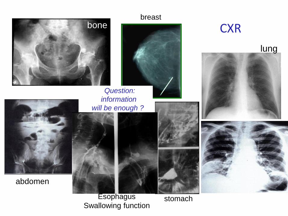
CXRbone
tápcsatorna
lung
abdomen
breast
Question:
information
will be enough ??
stomachEsophagus
Swallowing function
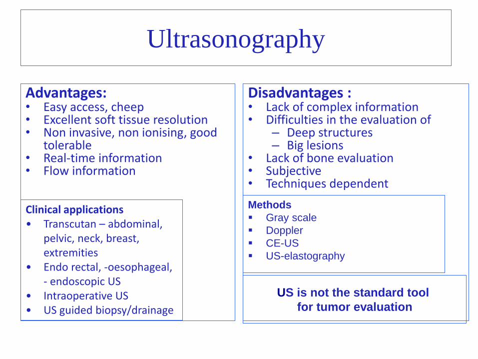
Ultrasonography
Advantages:• Easy access, cheep• Excellent soft tissue resolution• Non invasive, non ionising, good
tolerable• Real-time information• Flow information
Disadvantages :• Lack of complex information• Difficulties in the evaluation of
– Deep structures– Big lesions
• Lack of bone evaluation• Subjective• Techniques dependent
US is not the standard tool
for tumor evaluation
Clinical applications• Transcutan – abdominal,
pelvic, neck, breast, extremities
• Endo rectal, -oesophageal, - endoscopic US
• Intraoperative US• US guided biopsy/drainage
Methods
Gray scale
Doppler
CE-US
US-elastography
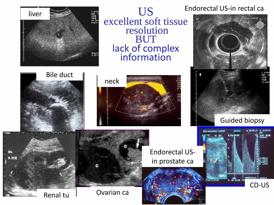
USexcellent soft tissue
resolutionBUT
lack of complex information
liver
Guided biopsy
Renal tu
Bile duct
Ovarian ca CD-US
Endorectal US-in rectal ca
neck
Endorectal US-in prostate ca
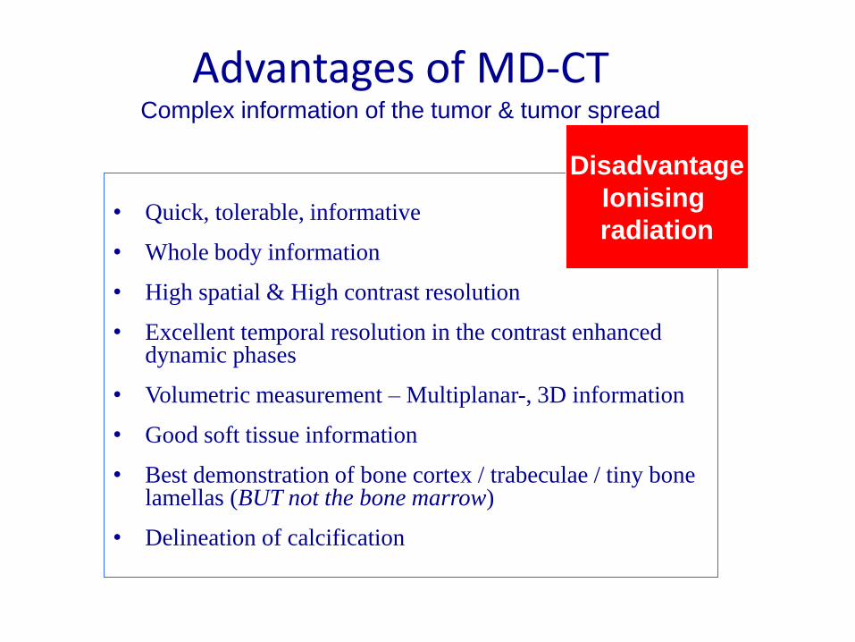
Advantages of MD-CTComplex information of the tumor & tumor spread
• Quick, tolerable, informative
• Whole body information
• High spatial & High contrast resolution
• Excellent temporal resolution in the contrast enhanceddynamic phases
• Volumetric measurement – Multiplanar-, 3D information
• Good soft tissue information
• Best demonstration of bone cortex / trabeculae / tiny bonelamellas (BUT not the bone marrow)
• Delineation of calcification
Disadvantage
Ionising
radiation
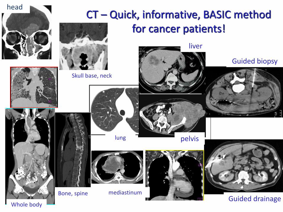
CT – Quick, informative, BASIC methodfor cancer patients!
Vezérelt biopszia
drenázs
liver
head
Skull base, neck
lung pelvis
Guided biopsy
Guided drainageWhole body
mediastinumBone, spine
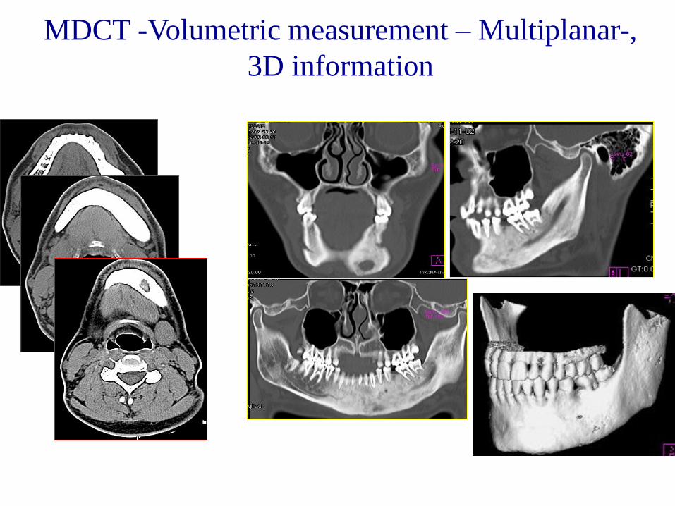
MDCT -Volumetric measurement – Multiplanar-,
3D information
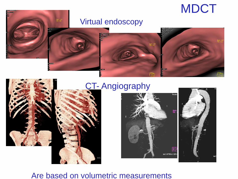
CT- Angiography
Virtual endoscopy
MDCT
Are based on volumetric measurements
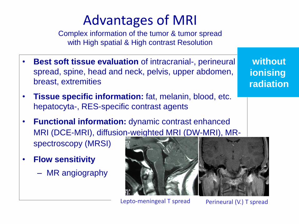
Advantages of MRIComplex information of the tumor & tumor spread
with High spatial & High contrast Resolution
• Best soft tissue evaluation of intracranial-, perineural
spread, spine, head and neck, pelvis, upper abdomen,
breast, extremities
• Tissue specific information: fat, melanin, blood, etc.
hepatocyta-, RES-specific contrast agents
• Functional information: dynamic contrast enhanced
MRI (DCE-MRI), diffusion-weighted MRI (DW-MRI), MR-
spectroscopy (MRSI)
• Flow sensitivity
– MR angiography
Lepto-meningeal T spread Perineural (V.) T spread
without
ionising
radiation

Advantages of MRI
•
• Brain tu– CT+MRI= 80% improvement in assessment of Tu
volume
• KHOO VS, British J. of Rad, 2006
• H&N (tu spread, perineural, lgl)
– Nasopharyngeal ca – CT+MR=50% better staging
» MANAVIS J, Clin. Imaging, 2005
• Pelvis
– Prostate ca–CT+MR=52% improvement in staging
– JOON DL, Int J Radiat Oncol Biol Phys,2005
– Gynecological tu’s– MR Acc 90%
– Rectal ca – MR Acc : 80 - 90% J.Husband, R. Reznek, 2004

MEDULLOBLASTOMA in the IV. ventricle
MRI- CE-T1-w imagesBest evaluation in intracranial tumors
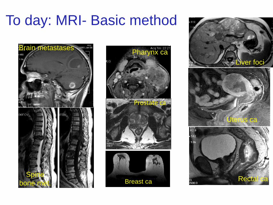
Spine
bone met.
To day: MRI- Basic method
Brain metastases
Liver foci
Pharynx ca
Breast ca
Uterus ca
Rectal ca
Prostate ca
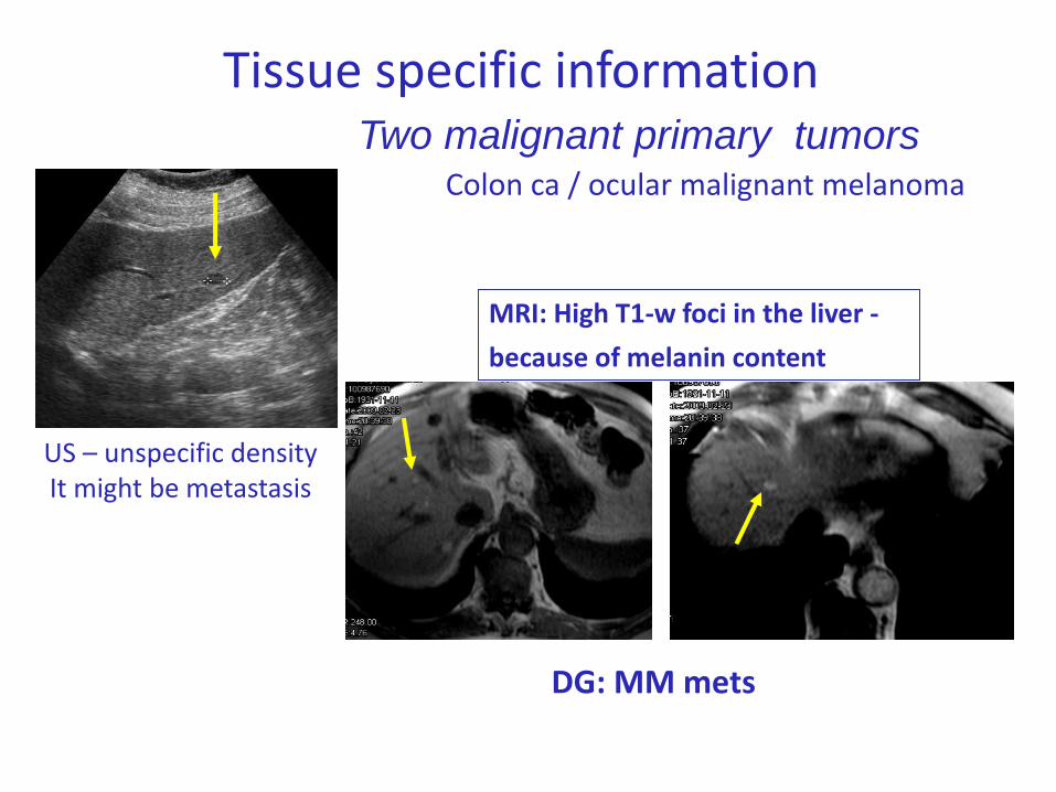
Tissue specific information
Colon ca / ocular malignant melanoma
MRI: High T1-w foci in the liver -
because of melanin content
Two malignant primary tumors
US – unspecific densityIt might be metastasis
DG: MM mets
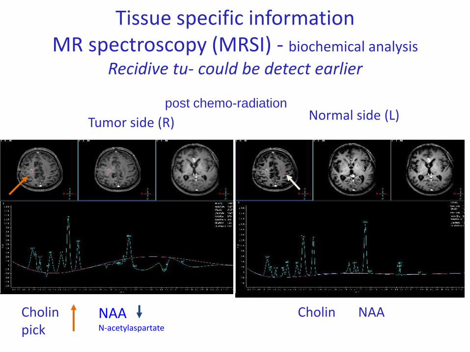
Tissue specific informationMR spectroscopy (MRSI) - biochemical analysis
Recidive tu- could be detect earlier
• post chemo-radiation
Cholinpick
NAACholin
Tumor side (R) Normal side (L)
NAAN-acetylaspartate
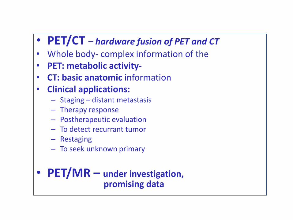
• PET/CT – hardware fusion of PET and CT
• Whole body- complex information of the• PET: metabolic activity-• CT: basic anatomic information• Clinical applications:
– Staging – distant metastasis– Therapy response– Postherapeutic evaluation– To detect recurrant tumor– Restaging– To seek unknown primary
• PET/MR – under investigation, promising data
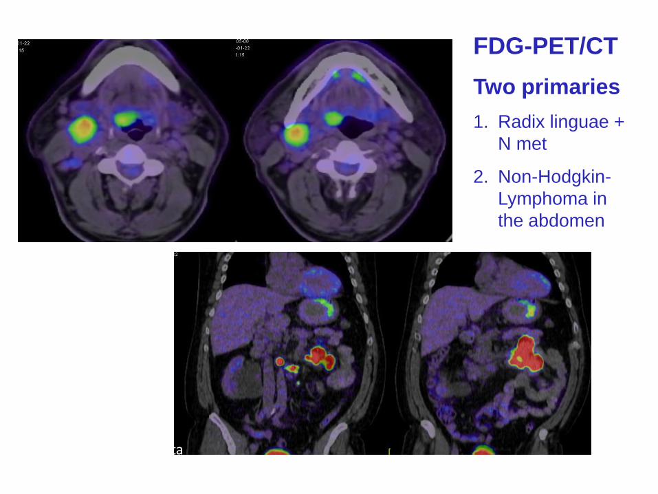
FDG-PET/CT
Two primaries
1. Radix linguae +
N met
2. Non-Hodgkin-
Lymphoma in
the abdomen
Pozitron KFT, dr Lengyel Zsolt vizsgálata
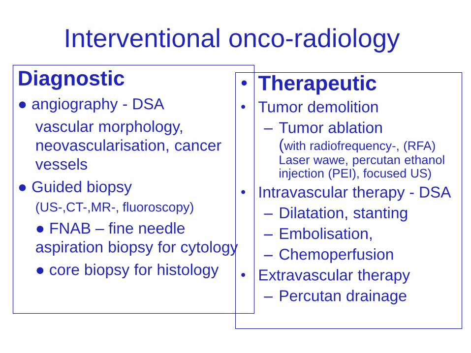
Interventional onco-radiology
Diagnostic● angiography - DSA
vascular morphology,
neovascularisation, cancer
vessels
● Guided biopsy
(US-,CT-,MR-, fluoroscopy)
● FNAB – fine needle
aspiration biopsy for cytology
● core biopsy for histology
• Therapeutic• Tumor demolition
– Tumor ablation (with radiofrequency-, (RFA) Laser wawe, percutan ethanol injection (PEI), focused US)
• Intravascular therapy - DSA
– Dilatation, stanting
– Embolisation,
– Chemoperfusion
• Extravascular therapy
– Percutan drainage
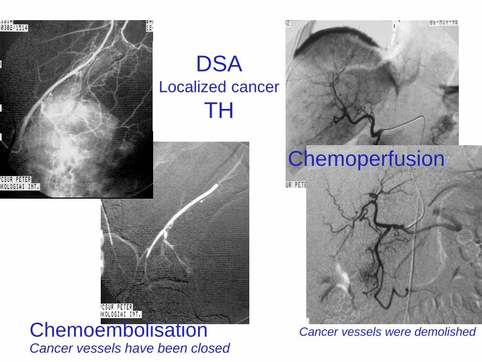
ChemoembolisationCancer vessels have been closed
Chemoperfusion
DSALocalized cancer
TH
Cancer vessels were demolished
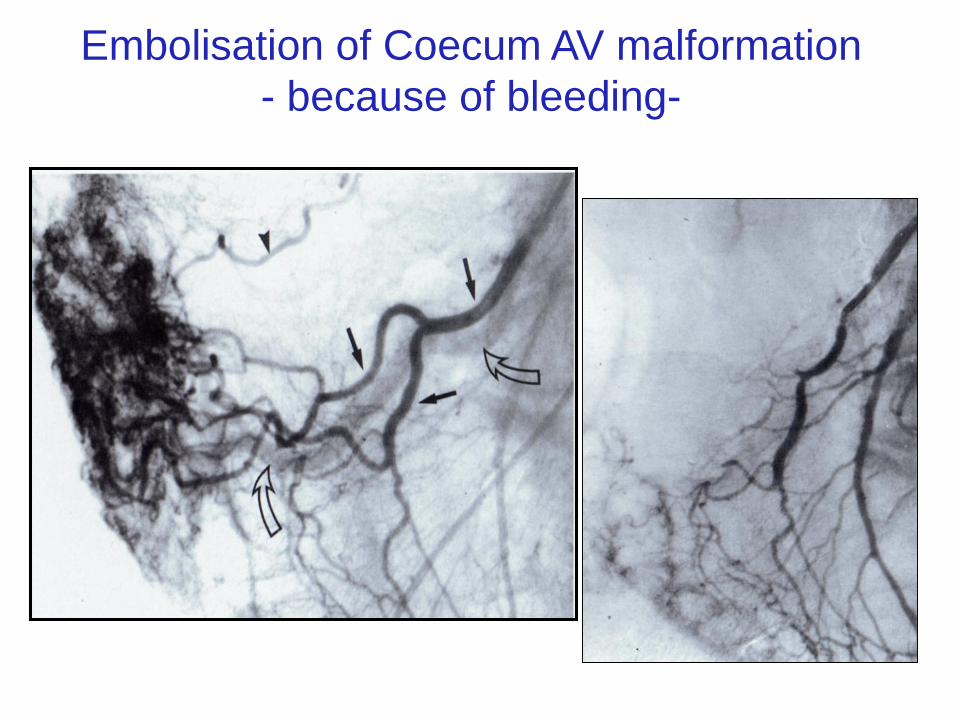
Embolisation of Coecum AV malformation
- because of bleeding-
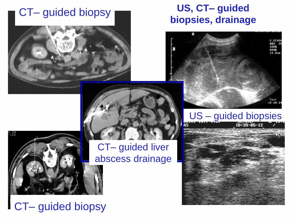
US – guided biopsies
CT– guided biopsy
CT– guided biopsy
CT– guided liver
abscess drainage
US, CT– guided
biopsies, drainage
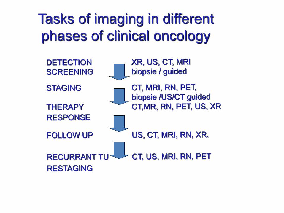
Tasks of imaging in different
phases of clinical oncology

RATIONALITY OF SCREENING
• Early diagnosis in preclinical stages
• To find high risk asymptomatic individuals
• To achieve higher cure rate
• 90% of all breast cancer cases could be cured if
diagnosed early and treated accurately
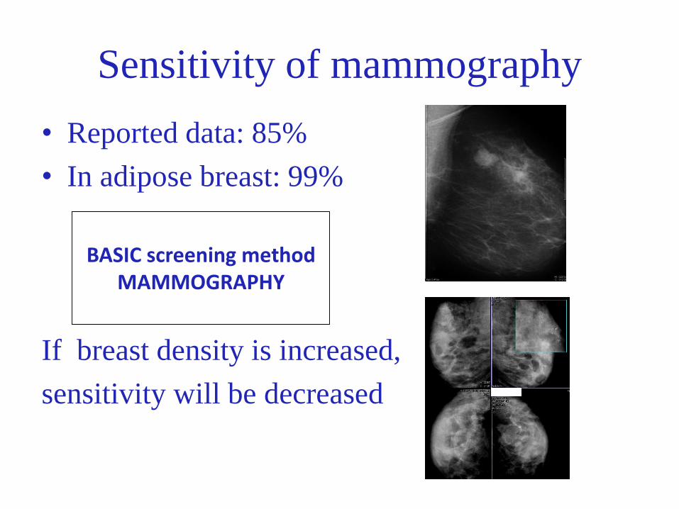
Sensitivity of mammography
• Reported data: 85%
• In adipose breast: 99%
If breast density is increased,
sensitivity will be decreased
BASIC screening methodMAMMOGRAPHY

Diagnostic procedures in BREAST CANCER
a) Mammography - Analog / Digital
b) US
c) Guided biopsy (FNA, core, vacuum assisted)
guided by US / mammography (stereotactic biopsy)
d) Multiparametric MRI (MP-MRI, DCE-MRI, DW-MRI)
e) Localization before op.(ROLL+SLNB, Hookwire)
f) specimen mammography /US
g) PET-CT
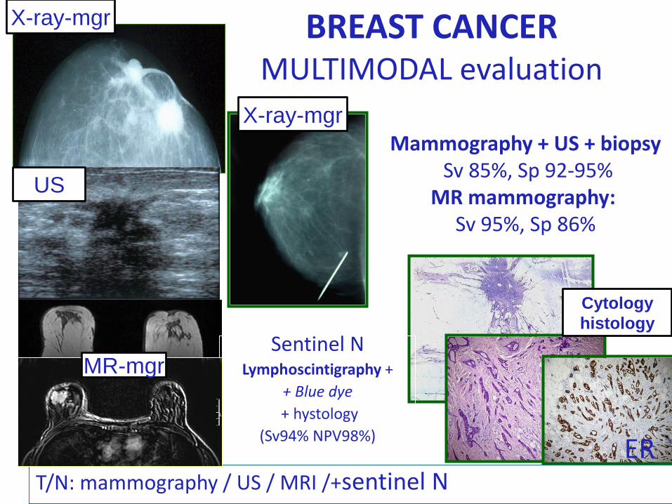
BREAST CANCERMULTIMODAL evaluation
ERT/N: mammography / US / MRI /+sentinel N
Mammography + US + biopsySv 85%, Sp 92-95%
MR mammography: Sv 95%, Sp 86%
Sentinel NLymphoscintigraphy +
+ Blue dye
+ hystology
(Sv94% NPV98%)
X-ray-mgr
MR-mgr
US
X-ray-mgr
Cytology
histology
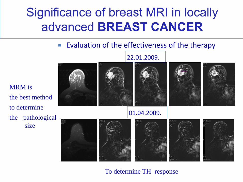
Evaluation of the effectiveness of the therapy
01.04.2009.
22.01.2009.
MRM is
the best method
to determine
the pathological
size
To determine TH response
LUNG CANCER
• Leading cause of death from malignancy
– 1.3 million deaths / year worldwide
– U.S. >/60,000 deaths – 2010
– Approximately 70% of cases have
incurable disease at presentation,
metastatic or locally advanced
– 14% overall 5 year survival
Theresa C. McLoud, MD
Massachusetts General Hospital, Harvard Medical School
LUNG CANCER mortality calls for screening
• CT highly sensitive for nodules
• CT detects more cancers than CXR
• CT screening for lung cancer has meaningful mortality benefit
NSCLC Stage IA > 65% survival
Small < 1 cm Stage IA > 80% survival
• Low dose CT minus 20-25% of standard dose
Annual control low dose CT
• Noninvasive management-follow up for growth
– CAD
– Volumetric measures
• High risk group > 30 pack years of smoking
> 45 age
36-53% survival increasing in the low dose CT group
(Henschke study, 2011)
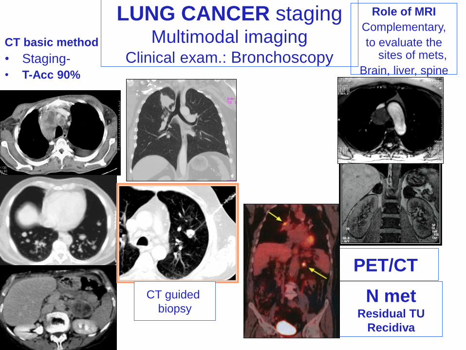
LUNG CANCER stagingMultimodal imaging
Clinical exam.: BronchoscopyCT basic method
• Staging-
• T-Acc 90%
Role of MRI
Complementary,
to evaluate thesites of mets,
Brain, liver, spine
PET/CT
N metResidual TU
Recidiva
CT guided
biopsy
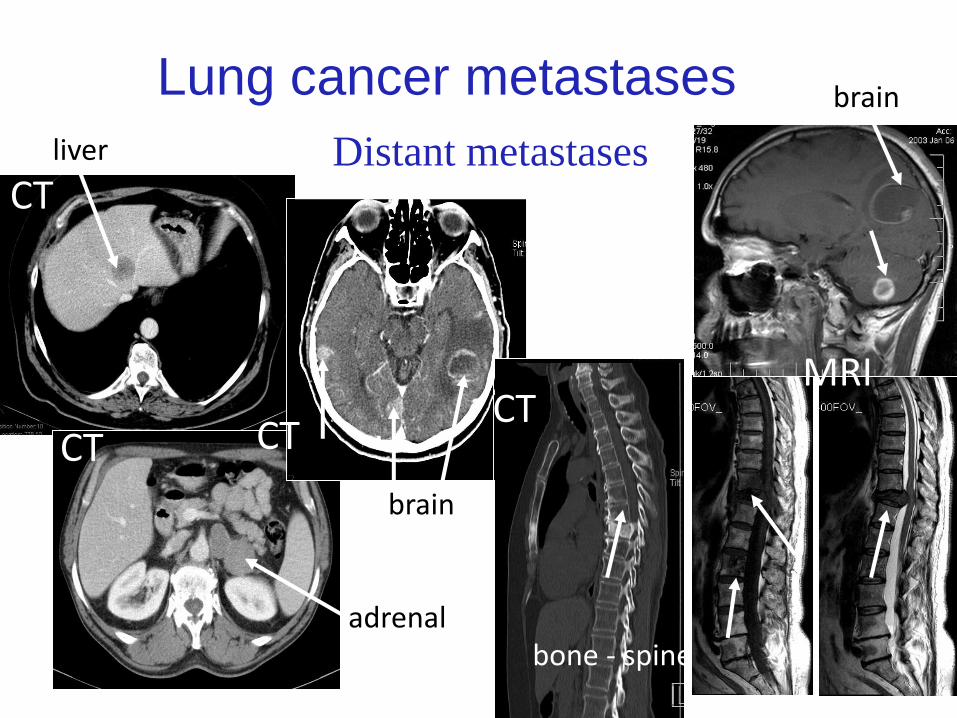
Lung cancer metastases
Distant metastasesliver
brain
adrenal
brain
bone - spine
CT
MRICT
CT
CT
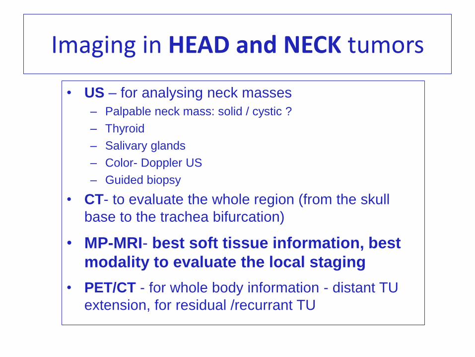
• US – for analysing neck masses
– Palpable neck mass: solid / cystic ?
– Thyroid
– Salivary glands
– Color- Doppler US
– Guided biopsy
• CT- to evaluate the whole region (from the skull
base to the trachea bifurcation)
• MP-MRI- best soft tissue information, best
modality to evaluate the local staging
• PET/CT - for whole body information - distant TU
extension, for residual /recurrant TU
Imaging in HEAD and NECK tumors
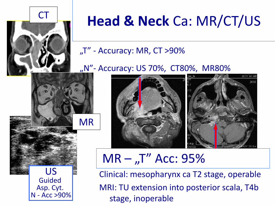
Head & Neck Ca: MR/CT/US
MR – „T” Acc: 95%Clinical: mesopharynx ca T2 stage, operable
MRI: TU extension into posterior scala, T4b stage, inoperable
„T” - Accuracy: MR, CT >90%
„N”- Accuracy: US 70%, CT80%, MR80%
CT
USGuided
Asp. Cyt. N - Acc >90%
MR
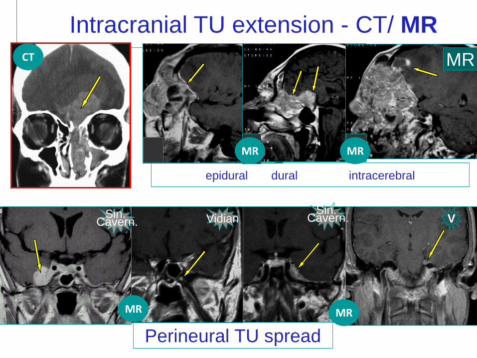
Intracranial TU extension - CT/ MR
epidural dural intracerebral
Perineural TU spread
Vidian
MR
VSin. Cavern.
Sin. Cavern.
MR
MR
MR
CT
MR
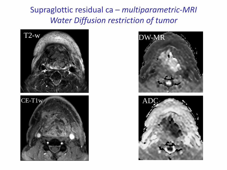
Supraglottic residual ca – multiparametric-MRIWater Diffusion restriction of tumor
T2-w
CE-T1w
DW-MR
ADC

• US – for general abdominal information
– Transabdominal US
– Endorectal US – intramural TU extension
• MP-MRI- best evaluation for tumor extension beyond
the wall, to determine resection margin, complex pelvic -,
and best liver information
• CT- to evaluate advanced TU extension
• UH/CT guided biopsy (liver)
• PET/CT - for whole body information - distant TU
extension, for recurrant TU
Multimodal Imaging in RECTAL TUMOR
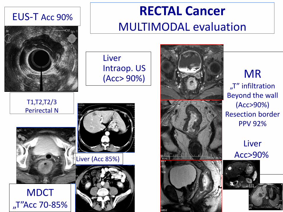
RECTAL CancerMULTIMODAL evaluation
T1,T2,T2/3Perirectal N
MR„T” infiltration
Beyond the wall(Acc>90%)
Resection borderPPV 92%
LiverAcc>90%
MDCT„T”Acc 70-85%
LiverIntraop. US (Acc> 90%)
EUS-T Acc 90%
Liver (Acc 85%)
• US – for the first information
– Transabdominal US – general
– Endorectal US – prostate
• Color- Doppler US
• MP-MRI - for the accurate prostate and pelvic
information, staging, recurrant ca, restaging
• Bone scan – bone metastasis
• CT- for evaluated advanced TU extension
• PET/CT – for recidive cancer, for whole body
information
Imaging in PROSTATE cancer

PROSTATE cancer
• Screening - PSA (prostate specific antigen) NOT reliable
• Diagnosis: Transrectal EUS - colour Doppler -
TRUS-guided biopsies
• Staging, MRI : for capsular penetration,for vesicula-,
bladder-,other pelvic invasion,
nodal status
Color Doppler may increase detecting prostate cancer
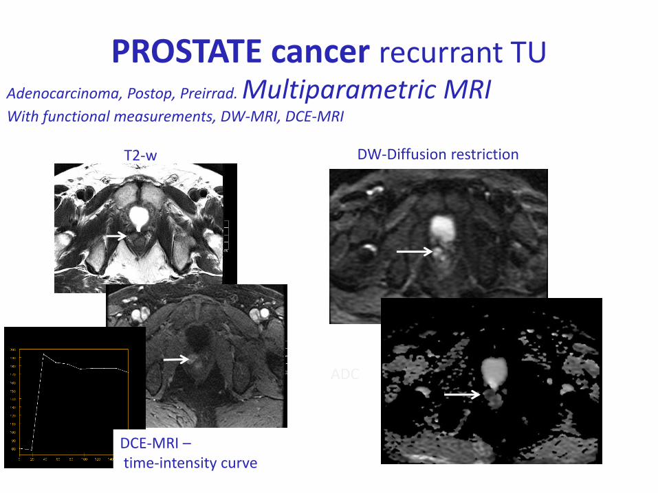
PROSTATE cancer recurrant TUAdenocarcinoma, Postop, Preirrad. Multiparametric MRIWith functional measurements, DW-MRI, DCE-MRI
T2-w DW-Diffusion restriction
ADC
DCE-MRI –time-intensity curve
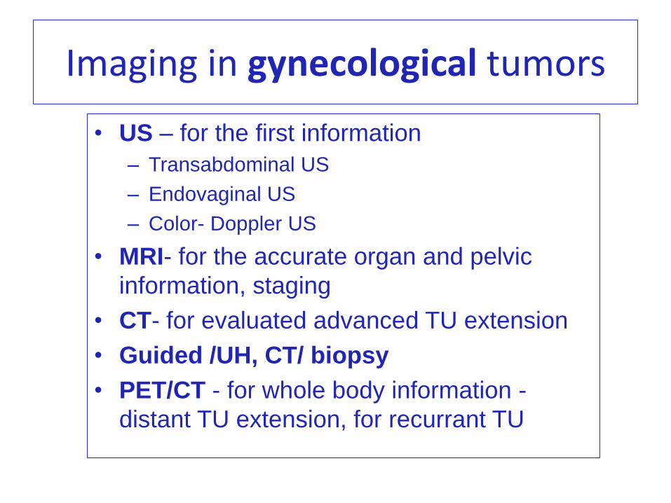
• US – for the first information
– Transabdominal US
– Endovaginal US
– Color- Doppler US
• MRI- for the accurate organ and pelvic
information, staging
• CT- for evaluated advanced TU extension
• Guided /UH, CT/ biopsy
• PET/CT - for whole body information -
distant TU extension, for recurrant TU
Imaging in gynecological tumors
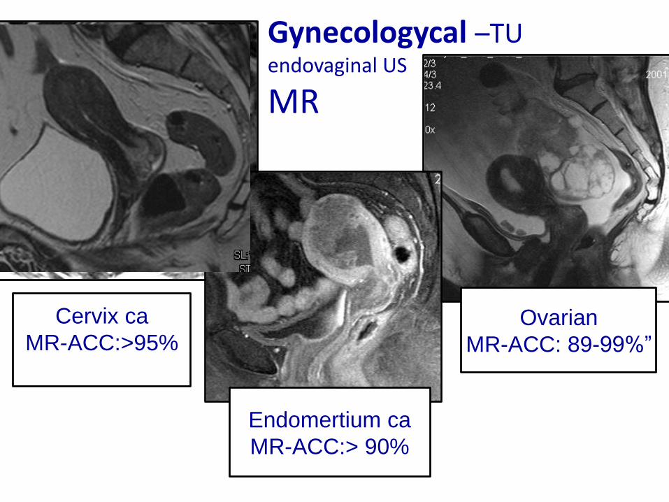
Gynecologycal –TUendovaginal US
MR
Ovarian
MR-ACC: 89-99%”
Cervix ca
MR-ACC:>95%
Endomertium ca
MR-ACC:> 90%
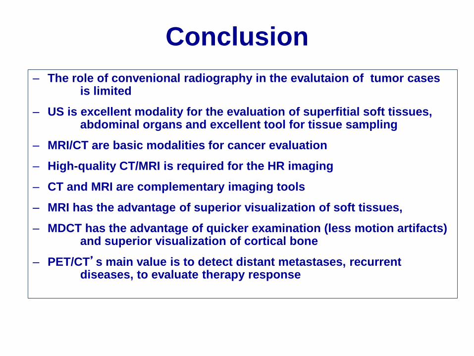
Conclusion
– The role of convenional radiography in the evalutaion of tumor casesis limited
– US is excellent modality for the evaluation of superfitial soft tissues, abdominal organs and excellent tool for tissue sampling
– MRI/CT are basic modalities for cancer evaluation
– High-quality CT/MRI is required for the HR imaging
– CT and MRI are complementary imaging tools
– MRI has the advantage of superior visualization of soft tissues,
– MDCT has the advantage of quicker examination (less motion artifacts) and superior visualization of cortical bone
– PET/CT’s main value is to detect distant metastases, recurrentdiseases, to evaluate therapy response
• Optimal treatment is based on multidisciplinary decision
• In the Oncologic Decision Process:
– the diagnostic radiologists,
– the surgical oncologists,
– the clinical oncologists and
– the radiotherapeutics need to strengthen the process from the
diagnostic imaging to the therapeutic imaging, in success of
• Image-guided oncologic treatment
Conclusion
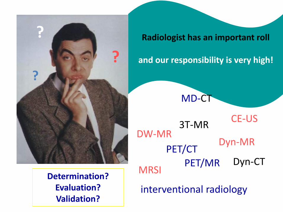
Radiologist has an important roll
and our responsibility is very high!
?
?
Dyn-CT
CE-US
CA-PET/CTUH
DW-MRDyn-MR
?
PETPET/MR
interventional radiology
SMD-CT
3T-MR
MRSIPETDetermination?
Evaluation?Validation?