Embed Size (px)
Citation preview

ARTICLE
Site-specific labeling of nucleotides for making RNA for highresolution NMR studies using an E. coli strain disabledin the oxidative pentose phosphate pathway
T. Kwaku Dayie • Chandar S. Thakur
Received: 14 December 2009 / Accepted: 26 February 2010 / Published online: 23 March 2010
� The Author(s) 2010. This article is published with open access at Springerlink.com
Abstract Escherichia coli (E. coli) is a versatile organism
for making nucleotides labeled with stable isotopes
(13C, 15N, and/or 2H) for structural and molecular dynamics
characterizations. Growth of a mutant E. coli strain deficient
in the pentose phosphate pathway enzyme glucose-6-phos-
phate dehydrogenase (K10-1516) on 2-13C-glycerol and15N-ammonium sulfate in Studier minimal medium enables
labeling at sites useful for NMR spectroscopy. However,13C-sodium formate combined with 13C-2-glycerol in the
growth media adds labels to new positions. In the absence of
labeled formate, both C5 and C6 positions of the pyrimidine
rings are labeled with minimal multiplet splitting due to1JC5C6 scalar coupling. However, the C2/C8 sites within
purine rings and the C10/C30/C50 positions within the ribose
rings have reduced labeling. Addition of 13C-labeled formate
leads to increased labeling at the base C2/C8 and the ribose
C10/C30/C50 positions; these new specific labels result in two-
to three-fold increase in the number of resolved resonances.
This use of formate and 15N-ammonium sulfate promises to
extend further the utility of these alternate site specific labels
to make labeled RNA for downstream biophysical applica-
tions such as structural, dynamics and functional studies of
interesting biologically relevant RNAs.
Keywords Alternate-site specific labeling �Formate enhanced isotope enrichment �Ribose and nucleobase � RNA � Structure and dynamics
Abbreviations
AMP Adenosine 50-monophosphate
CMP Cytidine 50-monophosphate
DHAP Dihydroxyacetone phosphate
FBP Fructose-6-bisphosphate
F6P Fructose-6-phosphate
G6PDH Glucose-6-phosphate dehydrogenase
GA3P Glyceraldehyde-3-phosphate
Gly Glycine
GMP Guanosine 50-monophosphate
noPPP Non-oxidative pentose phosphate pathway
OAA Oxaloacetate
oPPP Oxidative pentose phosphate pathway
R5P Ribose-5-phosphate
rNMPs Ribonucleoside monophosphates
rNTPs Ribonucleoside triphosphates
Ser Serine
TIM Triosephosphate isomerase
UMP Uridine 50-monophosphate
Introduction
Nucleic acids and proteins can be labeled with stable iso-
topes for structural and dynamics studies (Dayie 2008) using
E. coli as a common bacterial host (Ponchon and Dardel
2007; Ponchon et al. 2009), using enzymes from the pentose
phosphate or de novo purine biosynthetic pathways (Gross
et al. 1983; Parkin et al. 1984; Tolbert and Williamson 1996,
1997; Scott et al. 2000; Schultheisz et al. 2008), or using
chemical synthesis (Milecki 2002).
Of these three methods, use of different E. coli bacteria
grown on minimal media is attractive for a number of
reasons. E. coli grown on chemically defined minimal
T. K. Dayie (&) � C. S. Thakur
Department of Chemistry and Biochemistry, Center
for Biomolecular Structure and Organization, University
of Maryland, 1115 Biomolecular Sciences Bldg (#296), College
Park, MD 20742-3360, USA
e-mail: [email protected]
123
J Biomol NMR (2010) 47:19–31
DOI 10.1007/s10858-010-9405-0

media supplemented with 15N-labeled nitrogen and13C-labeled carbon sources (Batey et al. 1995; Hoffman
and Holland 1995; Nikonowicz 2001; Latham et al. 2005;
Dayie 2008) produces isotopically labeled total cellular
RNA that can be enzymatically digested to nucleoside
monophosphates (NMPs). The NMPs can be phosphory-
lated to the corresponding nucleoside triphosphates (NTPs)
and these labeled NTPs become the building blocks for
making labeled RNA using T7 RNA polymerase. Of the
different E. coli strains that offer potential for uniform and
alternate site specific 13C isotopic labeling, the E. coli
strain with a knockout of the zwf gene is particularly
attractive. Deleting the zwf gene means this E. coli strain
K10-1516 (hereafter referred to as K10) cannot encode the
glucose-6-phosphate dehydrogenase (G6PDH) enzyme for
use in the pentose phosphate pathway(PPP; Fraenkel
1968). Growth of this strain on 2-13C-glycerol without
formate provides labeling mostly at the C20 and C40 ribose
positions and at both C5 and C6 base positions of the
cytidine monophosphate (CMP) with minimal multiplet
splitting due to 1JC5C6 scalar coupling (Johnson et al. 2006;
Hoogstraten and Johnson 2008).
These previous labeling methods offered the advantage
of enriching only a few specific sites such as C8 in purine
(Pur, adenine or Ade and guanine or Gua) and ribose C20
and C40. However, there are at least two disadvantages with
such schemes. First, many useful sites are only fractionally
enriched. For example, the ribose C10 and C50 atoms and
the purine C2 and C8 positions are labeled at a very low
level when using K10. Yet the C10 carbon atoms have the
most sugar chemical shift dispersion and C50 carbon atoms
along with their attached protons can provide valuable
c-torsion angle information (Wijmenga and van Buuren
1998). In addition, the use of 13C-formate and 12C-glucose
as the only carbon sources limits the incorporation of an
isotopic label to the C8 position of adenine and guanine
bases and precludes their use for other NMR studies.
Second, it is more time and cost efficient if the same
sample can be used for numerous applications such as the
analyses of the function, structure and dynamics of RNAs.
Limiting the labels to a few selected ribose and base atoms
greatly lessens the general usefulness of the resulting
nucleotides. Here we sought to extend the utility of these
labels while preserving the advantage of isolating most of
the spin systems of interest and boosting the isotopic
enrichment level of the other useful NMR sites.
We reasoned that by combining 13C-glycerol with 13C-
formate, all the protonated carbons of high interest for
RNA NMR spectroscopy will be labeled in both purine and
pyrimidine (Pyr, that is cytosine or Cyt, uracil or Ura, and
thymine or Thy) bases. In addition, the isotopic enrichment
at sites within the ribose ring will increase without intro-
ducing multiplet splitting at key atomic sites such as C10
and C50. We can augment the level of isotopic enrichment
at all the sites useful for proton-detected heteronuclear
NMR experiments by adding 13C-formate to the 13C-
glycerol growth media. Plus, the use of 15(NH4)2SO4 as the
sole source of nitrogen leads to uniform and complete 15N
labeling of all nitrogen positions in all nucleotides and it
provides an alternative method for estimating the level of
isotopic enrichment. This labeling method appears to be
general and flexible, and affords a wide variety of purine
and pyrimidine isotope labeling patterns useful for struc-
tural, functional and dynamics studies.
Materials and methods
Bacterial strains
The mutant strain K10-15-16 (CGSC # 4858 Hfr fhuA22,
zwf-2, relA1, T2R, pfk-10) used in this work was obtained
from the Yale Coli Genetic Stock Center.
Isotopes
All labeled compounds were bought from Cambridge Iso-
tope Laboratory (Andover, MA) and Isotec-Sigma–Aldrich
(Miamisburg, OH) with the following isotopic enrich-
ments: [13C] sodium formate (99%), [2-13C] glycerol
(99%) and [15N]-(NH4)2SO4 (99%).
Stock solutions
The stock solutions required for bacterial growth were
prepared using distilled and deionized heat sterilized
water. The Studier medium (SPG) contained the follow-
ing (Studier 2005): 25 mM�(NH4)2SO4, 50 mM�KH2PO4,
50 mM�Na2HPO4, 2 mM�MgSO4 and trace metal solution;
SPG was then supplemented with labeled glycerol or
labeled formate and glycerol as needed.
Media for bacterial growth
Luria–Bertani (LB) and SPG minimal media were prepared as
described (Sambrook and Russell 2001; Studier 2005). Each
media was supplemented with the amount of the carbon
source (sodium formate and glycerol) and nitrogen source
(ammonium sulfate) that gives optimal growth conditions.
Protocol for growth optimization
The growth of each bacterial strain was optimized for the
highest production of biomass per input gram of carbon
source. Briefly, the first evening (Day 1) a glycerol stock of
the K10 strain was plated on fresh LB agar plates with no
20 J Biomol NMR (2010) 47:19–31
123

antibiotic and incubated overnight at 37�C. The following
evening (Day 2), a 5 ml starter culture in SPG medium
supplemented with unlabeled carbon sources was inocu-
lated with a single colony and incubated overnight at 37�C
(i.e., 12–16 h). At the end of this incubation period (Day
3), the solution was pelleted at 3000 rpm for 5 min; the
pellet was washed two times in 1x PBS and centrifuged at
3000 rpm for 5 min. This cell pellet was resuspended in
5 ml of fresh SPG medium without any carbon source;
1 ml of this resuspension was added to a 50 ml culture in
SPG medium with unlabeled carbon source and incubated
at 37�C for 12–14 h with shaking at 270 rpm, making sure
the OD600 did not saturate. At the end of this incubation,
the solution was centrifuged, the resultant pellet washed
twice in 1x PBS and the pellet was resuspended in 50 ml
fresh SPG medium without any carbon source. Then 5 ml
was added to two 500 ml SPG medium containing labeled
carbon source. About 10 ml of labeled media was saved for
resuspension of the 500 ml culture pellet. The 500 ml
labeled culture was incubated at 37�C for 12 h.
Nucleic acid digestion and cis diol affinity column
purification
The isotopically enriched ribonucleotides were isolated
from E. coli as described earlier (Batey et al. 1995). The
cell pellet was resuspended in 20 ml STE buffer (0.1 M
NaCl, 10 mM Tris, 1 mM EDTA, pH 8) for two grams of
frozen cells and lysed in STE with 0.5% SDS. The cellular
proteins were removed with 25:24:1 Phenol:Chloro-
form:Isoamyl mix. The upper aqueous layer, pooled from
multiple extractions of the organic and inclusion layers,
was back-extracted with an equal volume of 24:1 Chloro-
form:Isoamyl alcohol. The residual nucleic acids were
precipitated overnight with ethanol and acetate buffer (pH
5.2) at -20�C. The cellular nucleic acids were digested
with nuclease P1 in 15 mM sodium acetate, 0.1 mM�ZnSO4, pH 5.2. The deoxyribonucleotides were separated
from the ribonucleotides using a cis-diol boronate affinity
chromatography in a 20 9 2.5 cm glass column. The
digested nucleic acid solution was filtered and loaded onto
a boronate affinity resin (10 g of Affigel 601 from Biorad)
pre-equilibrated with 1 M TEABC at pH 9.5 at 4�C. The
deoxyribonucleotides, salts, and other impurities were
washed from the boronate column using five column vol-
umes of 1 M TEABC. The ribonucleotides were eluted
with water acidified with CO2 and the purity of the rNMPs
was verified by NMR.
NMR experiments
NMR experiments were run on a four channel Bruker
Avance 600 MHz spectrometer equipped with actively
shielded z-axis gradient triple resonance probe. NMR data
sets were processed and the peak positions and intensities
were analyzed with Bruker’s TOPSPIN 2.1. The rNMP
fractions were analyzed by 1H and 13C NMR experiments.
One dimensional (1D) 13C spectra were collected with a
45� and 90� tip angles, and GARP (Shaka et al. 1985) 1H
decoupling was applied only during acquisition. 1D 1H
spectra were also collected without 13C decoupling during
acquisition. All spectra, unless indicated, were collected
at 25�C. Two-dimensional non-constant-time (1H, 13C)
HSQC (Mueller 1979; Bodenhausen and Ruben 1980; Bax
et al. 1990) were recorded to resolve ambiguities arising
from overlap in the 1D spectra. Spectra requiring quanti-
tative analysis were acquired with a long recycle delay
(5 s) to ensure sufficient recovery of magnetization.
For the base region, the two-dimensional (2D) experi-
ments were acquired with sweep widths of 8013 Hz in the1H acquisition dimension and 3322 Hz in the 13C dimen-
sion. For each data set, 8 scans and 1024 complex points
were collected in t2 and 256 complex points were collected
in t1 using the Echo-Anti echo method (Palmer et al. 1991;
Kay et al. 1992) for quadrature detection. 13C-GARP
(Shaka et al. 1985) decoupling was applied during the
acquisition period or omitted to obtain residual labeling
information. Proton and carbon carrier was placed at
4.7 ppm and 142.5 ppm respectively. An INEPT delay of
2.5 ms (optimized for 1JHC = 200 Hz for the base region
and corresponding to 1/(2*JHC)) was used for magnetiza-
tion transfer. For the ribose region 2D experiments were
acquired with sweep widths of 8013 Hz in the 1H dimen-
sion and 5058 Hz in the 13C dimension. For each data set, 8
scans and 1024 complex points were collected in t2 and 256
complex points were collected in t1 using the Echo-Anti
echo method (Palmer et al. 1991; Kay et al. 1992) for
quadrature detection. 13C-GARP decoupling was applied
during the acquisition period. Proton and carbon carrier
was placed at 4.7 ppm and 76.5 ppm respectively. An
INEPT delay of 3.29 ms (optimized for 1JHC = 152 Hz for
the ribose region and corresponding to 1/(2*JHC)) was used
for magnetization transfer. The time domain data was zero
filled in t1 and t2 before Fourier transformation to give a
final real matrix size of 2048 9 1024 points.
Two- and three-bond 2D (1H, 15N) HSQC experiments
were acquired as follows. For the 1H acquisition period (t2),
sweep widths of 2761 Hz (4.6 ppm) were used, and for the15N evolution period (t1), sweep widths of 4563.8 or
6815.2 Hz (75 or 112 ppm) were used. For each data set,
16 or 32 scans and 1024 complex points were collected in
t2 and 128 complex points were collected in t1 using the
Echo-Anti echo method for quadrature detection. Proton,
carbon and nitrogen carrier was placed respectively at
4.7 ppm, 120.0 ppm and 202.5 (or 192) ppm. 15N-GARP
decoupling (Shaka et al. 1985) was applied during the
J Biomol NMR (2010) 47:19–31 21
123

acquisition period, and 13C-GARP decoupling (Shaka et al.
1985) was also applied or omitted during the acquisition
period. An INEPT delay of 15.6 or 25 ms (optimized for2JHN = 16 or 20 or 25 Hz for the two bond coupling to the
purine nitrogen and corresponding to 1/(2*JHN)) was used
for magnetization transfer. These delays did not affect the
% level labeling prediction (see below).
Relative peak intensities were determined by integrating
peaks observed with proton decoupling during acquisition
only and a long recycle delay (5 s) to allow sufficient
magnetization recovery for the direct carbon experiments.
For the 2D experiments, all data were plotted to the same
base level, level of increment and number of contour levels
before peak picking and peak integration.
To simulate the effect of a medium-sized RNA, we
ran non-constant time 2D 13C HSQC and 13C TROSY
(Meissner et al. 1998; Pervushin et al. 1998; Czisch and
Boelens 1998; Weigelt 1998; Rance et al. 1999; Zhu et al.
1999; Schulte-Herbruggen and Sorensen 2000) on a mix-
ture of all four nucleotides dissolved in 95% w/w per-
deuterated glycerol (Cambridge Isotope Labs, Andover,
MA) at 30�C.
Results and discussion
The ability to transcribe RNA (or DNA) labeled with
various isotopes, such as 13C, 15N and 2H, has enabled
the application of heteronuclear multi-dimensional NMR
techniques to characterize the structure and dynamics of
interesting biological RNA molecules (D’Souza et al.
2004; Gumbs et al. 2006; Lu et al. 2010). Unsurprisingly a
number of research groups have developed and continue to
develop techniques for the biosynthetic production of iso-
topically labeled nucleotides. Some produce uniformly
labeled nucleotides from E. coli (Nikonowicz et al. 1992;
Michnicka et al. 1993), M. methylotrophus (Batey et al.
1992), or Methylophilus extorquens (Hines et al. 1994).
Others produce site specifically labeled nucleotides from
E.coli (Latham et al. 2005; Johnson et al. 2006), and still
others produce these labels using the pentose phosphate
and de novo purine synthetic pathways (Schultheisz et al.
2008). Similar methods for the production of uniform
isotopically labeled deoxynucleosides have been proposed
(Zimmer and Crothers 1995; Louis et al. 1998; Masse et al.
1998; Werner et al. 2001; Nelissen et al. 2009). Previously,13C-formate added to an unlabeled glucose minimal med-
ium enabled selective labeling of purine C8 positions
(Latham et al. 2005), and use of E. coli deficient in the
G6PDH gene enabled the site-labeling of pyrimidine C5 or
C6 positions as well as various ribose carbon positions
(Johnson et al. 2006). To our knowledge, no published
reports have shown the combined advantages of both:
Grow E. coli strain K10 on 13C-labeled glycerol with13C-formate to overcome the limitations of each sepa-
rate method. Using 2 g of 13C-2-glycerol and 0.7 g of
NH4(SO4)2 as carbon and nitrogen sources, we obtained
4 g of wet cell pellet for a liter of E. coli K10 cell culture.
Addition of 0.2 g of 13C formate to this medium was
sufficient to label sites not otherwise labeled. Yields of
up to 66 mg of rNMPs and 13 mg of dNMPs obtained are
comparable to previous reports of labeling (Batey et al.
1992). To obtain optimal yields it is important to use
minimal media other than M9. As reported by others, M9 is
inferior to other buffers (Paliy and Gunasekera 2007; Dayie
unpublished).
Incorporation of 13C into ribose ring of nucleotides
using the pentose phosphate pathway
To place our results within the context of the metabolic
pathways in E. coli for nucleotide metabolism, we present a
brief overview of some of the steps in glycolysis, gluco-
neogenesis and the tricarboxylic acid (TCA) cycle. Details
can be found in standard textbooks (Voet et al. 2008;
Nelson and Cox 2008).
Glycerol enters the metabolic cycle as dihydroxyacetone
phosphate (DHAP) and equilibrates rapidly with glycer-
aldehyde-3-phosphate (GA3P) by the action of triose-
phosphate isomerase (TIM). From this point in the cycle,
GA3P can be converted to ribose-5-phosphate (R5P) via
gluconeogenesis using the oxidative pentose phosphate
pathway (oPPP; Fig. 1). Alternatively GA3P and fructose-
6-phosphate (F6P) can also be converted to R5P via the
action of transketolase and transaldolase in the reverse of
the non-oxidative PPP (noPPP; Fig. 1). In wild type E. coli
both the oPPP and noPPP can be operative and the parti-
tioning of the carbon flux through these two pathways leads
to scrambling of labels in the ribose ring. However, for
strains defective in the oPPP such as the K10 E. coli strain,
most of the carbon flux is re-routed through the reverse
noPPP via the action of transketolase and transaldolase
(Edwards and Palsson 2000; Zhao et al. 2004; Nicolas et al.
2007). Assuming auxiliary biosynthetic pathways contrib-
ute negligibly to the central pathway, a 13C-label at the
central C-2 carbon of glycerol would lead to isotopic
enrichment for [2, 4-13C2]ribose and [4-13C]ribose in a 2:1
ratio (Fig. 1), and no label is expected at the 1, 3 or 5 ribose
carbon positions (Johnson et al. 2006).
Exogenous formate can enter the metabolic cycle by
exchanging the carboxyl group of pyruvate by consuming
acetyl-CoA (Thauer et al. 1972; Knappe et al. 1974) by the
reversible action of pyruvate formate lyase (Kirkpatrick
et al. 2001). This modified pyruvate may populate gluco-
neogenesis intermediates such as GA3P and F6P for use in
the reverse of the noPPP. At the moment, the effect of
22 J Biomol NMR (2010) 47:19–31
123

exogenous formate on E. coli growth remains poorly
characterized and poorly understood. Nonetheless, as we
show later, addition of formate has an unexpected effect of
increasing the level of enrichment at the ribose carbon
positions predicted to have no label using the central
metabolic pathway.
Fig. 1 Major metabolic
pathways involved in the
production of nucleic acid
nucleotides, including key steps
in glycolysis, gluconeogenesis
and one pass through the
tricarboxylic (TCA) cycle. For
E. coli carrying the zwfgenotype (glucose 6-phosphate
dehydrogenase (G6PDH)
mutant), the oxidative branch of
the pentose phosphate pathway
is disabled (indicated by an Xthrough the orange arrow) such
that most of the carbon fluxes
are shunted through the reverse
non-oxidative pentose
phosphate pathway (noPPP).
Atom labels for the terminal
(1, 3) carbons (magenta and thincircle) and central (2) carbon
(cyan and thick circle) of
glycerol are highlighted.
Positions that are enriched due
to the presence of 13CO2 (as
bicarbonate) in the growth
medium are shown with an
encircled X, but this is lost
through the first and subsequent
pass through the TCA cycle.
Pyrimidine bases derived from
oxaloacetate (OAA) produced
by carboxylation of
phosphoenolpyruvate (PEP) is
shown via the aspartate
intermediate. This OAA is used
as a substrate in the first and
subsequent rounds of the TCA
cycle to produce OAA with a
pair of different labeling
schemes as products due to the
symmetric nature of the TCA
cycle intermediate succinate. If
[2-13C]glycerol is used Ca or Cb
or Cc or Cb and Cc but not all
three positions are labeled
simultaneously. Similarly the
labeling pattern of purines from
glycine (Gly) derived from 3-
phosphoglycerate (3PG) are
shown such that if
[2-13C]glycerol is used only the
Ca position of Gly and therefore
C5 position of the purine ring is
labeled. The use of GA3P and
F6P in the reverse of the non-
oxidative PPP produces ribose
labeled at the 2,4 and 4
positions if [2-13C]glycerol is
used
J Biomol NMR (2010) 47:19–31 23
123

Incorporation of 13C into base ring of nucleotides
via the tricarboxylic acid cycle, glycolysis,
and gluconeogenesis
Various metabolic precursors make amino acids from
which nucleotide bases are synthesized (Fig. 1). 3-phos-
phoglycerate (3PG) gives rise to serine (Ser) and glycine
(Gly), and oxaloacetate (OAA) gives rise to aspartic acid
(Asp). In turn, the six-membered Pyr ring is constructed
from four atoms of Asp such that the NH amide group, the
Ca-, Cb- and Cc-carbon positions of Asp becomes the N1,
C6, C5 and C4 ring atoms respectively of Pyr (Fig. 1). The
N3 and C2 positions are derived from glutamine amide and
bicarbonate pools respectively. The bicarbonate single
carbon pool is diluted by 12C carbons such that labeling at
the Pyr C2 position is random at low levels unless this
carbon pool is augmented with 13C-bicarbonate (Lundstrom
et al. 2007). The larger Pur ring atoms C2 and C8 also derive
from the formate pool. Thus, adding 13C-formate to the
growth media is again expected to increase the level of 13C
isotopic enrichment at the C2 and C8 sites. The amide
group, the Ca- and carbonyl (CO)-carbon positions of
Fig. 2 Increased level of labeling in K10 without (red) and with
(blue) 13C-formate in a 13C-2-glycerol background. The experiments
were performed on mixtures of the four rNMPs isolated from the K10
bacterial culture. a Direct carbon detection 1D spectrum showing all
the carbon positions for nucleotides labeled with glycerol and no
formate (bottom, red) or glycerol with formate (top, blue). A long
recycle delay of 5 s were used to allow for sufficient magnetization
recovery and proton decoupling was limited to the acquisition period
only. The level of enrichment at the adenine (Ade) and guanine (Gua)
C8 positions increases by spiking with 13C-labeled formate. The C50
region has an impurity that resonates in a distinct region in the 2D
spectrum. b 2D non-constant time HSQC spectrum of a mixture all
four labeled rNMPs showing the protonated base region. For ease of
comparison the spectrum obtained without labeled formate (redcontours) are displaced vertically relative to the formate labeled
spectrum (blue contours). c 2D non-constant time HSQC spectrum of
a mixture of all four labeled nucleotides showing the ribose region.
The cytosine (Cyt) and Uracil (Ura) C5 resonances at 96.67 ppm and
102.69 ppm respectively are folded into the spectrum. The boxed
resonances highlight the increased labeling level seen for C10, C30 and
C50 with spiking the growth medium with 13C-labeled formate
24 J Biomol NMR (2010) 47:19–31
123
glycine (Gly) become the N7, C5 and C4 ring atoms
respectively (Fig. 1). We use Fig. 1 as a framework for
interpreting some of our results with E. coli strain K10.
Label incorporation by E. coli strain K10 in the absence
of 13C-labeled formate
E. coli strain K10 grown in 13C-2-glycerol media in the
absence of labeled formate has varied labeling patterns in
both ribose and base moieties (Fig. 2; Table 1). The
ribose ring is labeled exclusively at the C20 and C40
positions ([80% label) as expected for the metabolic
carbon flux going mostly through the transketolase/trans-
aldolase branch of the noPPP. Little labeling is observed
at C30, and the negligible carbon–carbon splitting at C20
and C40 positions (Fig. 2a–c) further bears out the pre-
diction from the analysis of the metabolic pathway.
However, some residual labeling is observed at the C10
(*1%) and C50 (*1%) positions. The isotopic enrich-
ment level at C10 and C50 increases in the presence of
formate (as discussed further below). This residual
labeling suggests gluconeogenesis might be significant in
this mutant when grown on glycerol. Alternatively, a
fraction (*7%) of serine molecules is predicted to be
produced by a bypass of the disabled G6PDH in the zwf
mutant (Fischer and Sauer 2003). Further studies such as
metabolic flux analysis using gas chromatography–mass
spectrometry (GC–MS) and NMR spectroscopy are nee-
ded to address the origin of these residual labels fully
(Fischer and Sauer 2003).
For the base atoms, both the protonated C5 and C6
carbon positions of Pyr are substantially labeled at *45%
close to the expected 50% level, whereas the protonated
C2 and C8 carbon positions of Pur are labeled at a lower
level (*10–14%; Fig. 2a–b). The C5 and C6 pyrimidine
sites are constructed entirely from Asp which in turn is
generated from OAA either by direct carboxylation of
PEP or from the TCA cycle. Using [2-13C]-glycerol as the
sole carbon source, Asp formed from carboxylation of
PEP (using cellular bicarbonate breakdown to CO2 by
pyruvate carboxylase) is expected to be 100% enriched
exclusively at the Ca position or equivalently the C6
position of Pyr. A single pass through the TCA cycle
leads, because the TCA cycle metabolite succinate is
symmetric, to an equal probability of labeling either the
Ca or the Cb position. But both positions cannot be labeled
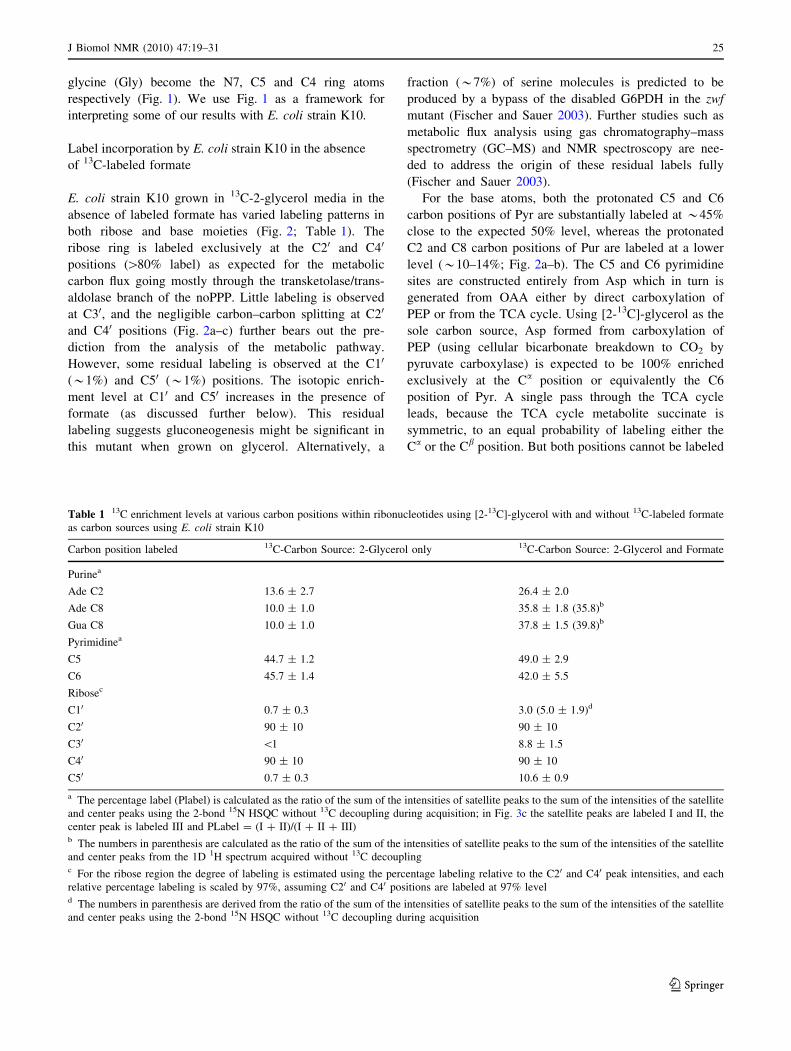
Table 1 13C enrichment levels at various carbon positions within ribonucleotides using [2-13C]-glycerol with and without 13C-labeled formate
as carbon sources using E. coli strain K10
Carbon position labeled 13C-Carbon Source: 2-Glycerol only 13C-Carbon Source: 2-Glycerol and Formate
Purinea
Ade C2 13.6 ± 2.7 26.4 ± 2.0
Ade C8 10.0 ± 1.0 35.8 ± 1.8 (35.8)b
Gua C8 10.0 ± 1.0 37.8 ± 1.5 (39.8)b
Pyrimidinea
C5 44.7 ± 1.2 49.0 ± 2.9
C6 45.7 ± 1.4 42.0 ± 5.5
Ribosec
C10 0.7 ± 0.3 3.0 (5.0 ± 1.9)d
C20 90 ± 10 90 ± 10
C30 \1 8.8 ± 1.5
C40 90 ± 10 90 ± 10
C50 0.7 ± 0.3 10.6 ± 0.9
a The percentage label (Plabel) is calculated as the ratio of the sum of the intensities of satellite peaks to the sum of the intensities of the satellite
and center peaks using the 2-bond 15N HSQC without 13C decoupling during acquisition; in Fig. 3c the satellite peaks are labeled I and II, the
center peak is labeled III and PLabel = (I ? II)/(I ? II ? III)b The numbers in parenthesis are calculated as the ratio of the sum of the intensities of satellite peaks to the sum of the intensities of the satellite
and center peaks from the 1D 1H spectrum acquired without 13C decouplingc For the ribose region the degree of labeling is estimated using the percentage labeling relative to the C20 and C40 peak intensities, and each
relative percentage labeling is scaled by 97%, assuming C20 and C40 positions are labeled at 97% leveld The numbers in parenthesis are derived from the ratio of the sum of the intensities of satellite peaks to the sum of the intensities of the satellite
and center peaks using the 2-bond 15N HSQC without 13C decoupling during acquisition
J Biomol NMR (2010) 47:19–31 25
123
simultaneously in the same molecule. Thus either the C5
or the C6 position of Pyr is labeled at 50% maximum
enrichment with no undesired C5-C6 labeled pair. In the
second pass through the TCA cycle, the C4 carbon is also
labeled to a maximum value of 25%; subsequent passes
through the cycle will reduce even further this level of
labeling at C4. Those molecules labeled at C4 are pre-
dicted to have no label at either the C5 or the C6 position.
Thus there should be no coupling between C4 and C5 or
C4 and C6.
The Pur C2 and C8 positions arise from metabolic
breakdown product of formate and the Pur C6 and Pyr C2
atomic positions arise from bicarbonate byproduct. As a
result these sites are expected to be randomly labeled at
very low levels in the absence of spiking the growth media
with 13C-labeled formate or bicarbonate.
Label incorporation by E. coli strain K10
in the presence of 13C-labeled formate
Addition of 13C-formate leads to increased labeling in
both ribose and base moieties (Fig. 2; Table 1). In the
ribose ring, labeling increases for the C10 (3–5%), C30
(*9%) and C50 (*11%) positions without introducing
significant carbon–carbon coupling at these positions (C10,C20, C40 and C50; Fig. 2). These labeling efficiencies can
be estimated from a comparative analysis of the 1D car-
bon spectra of uniformly labeled rNMPs and the site
specific labeled rNMPs derived from the K10 bacteria
culture. As discussed below a different method using two-
bond 15N HSQC gives comparable results. Nonetheless it
is unexpected that in the face of[80% labeling of C20 and
C40, C40–C50 and C10–C20 splittings are not observed.
Analysis of the reverse noPPP suggests oxaloacetate
generated by several passes through the TCA cycle will
populate a pyruvate intermediate that could ultimately
label R5P at the C10 and C50 positions with exclusion of
labels at C20 and C40 positions in the same molecule. This
is in addition to the expected labels at C20 and C40 without
adjacent labels at C10 and C50 in the same molecule.
Alternately a bypass of the disabled G6PDH in the zwf
mutant catalyzed by the perisplasmic glucose dehydroge-
nase (Fischer and Sauer 2003) could potentially produce a
label at the C10 and C50 without any coupled adjacent
labels. Further study using GC–MS and NMR are needed
to resolve this empirical observation of label at the C10
and C50 positions.
A similar increase in the labeling level is observed in the
base region on addition of labeled formate to the 13C-2-
glycerol media. Significant isotopic enrichment of the C8
(*40%) and C2 (*26%) carbon positions of the Pur ring
are observed, but those at the C6 and C5 Pyr positions
remain unchanged (Fig. 2b; Table 1).
Estimation of the degree of 13C isotope incorporation
using two- and three-bond 15N HSQC
Finally, addition of labeled 15N-ammonium sulfate enables
high level labeling of the aromatic nitrogens and estimation
of the degree of 13C isotope incorporation. The level of 13C
labeling efficiency is usually estimated using 1D 1H or
natural abundance 13C carbon spectra. Lack of a central
singlet peak and the presence of doublet satellite peaks
indicate close to 100% labeling efficiency. Absence of the
doublet satellite peaks and the presence of a dominant
central peak are then taken as lack of 13C incorporation.
Thus by comparing the intensity of each 13C satellite peak
to the intensity of the center peak, the labeling efficiency is
readily estimated. This 1D approach works well for single
nucleotides that have no spectral overlap. For a mixture of
the four rNMPs extracted from the K10 bacteria culture,
there is significant overlap in both the base and ribose
regions. For example Ade H10 (6.02 ppm) overlaps com-
pletely with Cyt H5 (6.02 ppm) in the proton chemical shift
region, and Ura H10 (5.90 ppm) overlaps with Cyt H10
(Fig. 2c). This overlap problem limits the usefulness of the
1D method. Long range (two- and three-bond) proton–
nitrogen correlations in 15N-HSQC spectra make it possible
to estimate the labeling efficiency of the C2 and C8 carbon
sites within the Pur aromatic ring, the C5 and C6 carbon
sites within the Pyr aromatic ring and the Pur C10 carbon
site (Fig. 3). Relaxation properties and transfer efficiencies
are different for long range and one-bond magnetization
transfers, and so it is important to validate the use of the
long range 15N-HSQC method to estimate the level of 13C
incorporation. The 1D slices from the 2D 2JHN HSQC
spectra (Fig. 3d) overlay completely with the 1D 1H
spectrum (Fig. 3c), suggesting the percentage label can be
estimated using either the 2D or 1D experiment, but the 2D
is preferable in case of overlap. With this experiment one
can correlate the H2 proton resonances to the N1 and N3
nitrogen positions in the adenine (Ade) ring, and also the
H8 proton resonances to the N7 and N9 nitrogen positions
in the Pur ring (Fig. 3b). By omitting the carbon decou-
pling field during the proton acquisition period, the proton
resonances are split by the directly attached 13C atom (C2
or C8) into a doublet (Fig. 3a–b). Using this method, the1JCH coupling constants measured for uniformly labeled
AMP, CMP, UMP, and GMP are in excellent agree-
ment with previous reported measurements. For uniformly13C/15N-labeled AMP and GMP, the 2D method, in
excellent agreement with the 1D 1H method, gives 98.9%13C isotopic enrichment at the C8 positions. As expected,
each of the H2 and H8 proton resonance is split into a
doublet with little central peak in the acquisition dimension
(Fig. 3a). As the level of 13C isotopic enrichment decreases
from 100 to 0%, each doublet gives rise to a central singlet.
26 J Biomol NMR (2010) 47:19–31
123

Analyses of the multiplet pattern of the four labeled
nucleotides derived from the K10 bacteria cultures facili-
tated the estimation of the degree of isotopic incorporation.
In the absence of formate, the level of enrichment was
*10% for the Pur C8 and *14.0% for the Pur C2. In the
presence of formate the level of enrichment increases to
*38% for Pur C8 and *28% for Ade C2 (Table 1).
Applications of selective labels for NMR study
of nucleic acids
High levels of isotopic enrichment lead to considerable
direct one-bond scalar couplings and residual dipolar
couplings from adjacent carbons yielding complex spectra
for macromolecules. These deleterious consequences can
Fig. 3 Estimation of C2 and C8-13C labeling efficiency using two-
and three-bond 15N-HSQC experiment without carbon decoupling
during acquisition. The 2D 1H-15N HSQC spectra depict H8-N7/N9
crosspeaks for Ade and Gua and H2-N1/N3 correlations for Ade. At
each N1 and N3 nitrogen position a singlet is observed for the H2
proton at 8.14 ppm if the C2 carbon is unlabeled and a doublet if C2
carbon is 13C-labeled due to the large one bond 1H-13C coupling of
*202 Hz. Similarly at each N7 and N9 nitrogen position a singlet is
observed for the H8 proton at 8.5 ppm (for Ade) and 8.08 ppm (for
Gua) if C8 is unlabeled and a doublet if C8 is13C-labeled due to the
large one bond 1H-13C coupling of *215 Hz. Thus the ratio of each
satellite peak to the central peak gives a good estimate of the degree
of 13C- labeling. a The 2D 2JHN HSQC spectra for uniformly labeled
NMPs (AMP, red; GMP, blue) are superimposed. The inset shows the
observable long range 1H-15N correlations in the purine ring. b 2D2JHN HSQC spectra for the mixture of four rNMPs obtained from the
K10 bacterial culture are superimposed (the spectrum obtained
without labeled formate, red contours and upper; formate labeled
spectrum, blue contours and lower). The H2 protons and N1 and N3
nitrogen atoms and H8 protons and the N7 atoms in nucleotides
labeled using K10 with formate in a 13C-2-glycerol background are
depicted. The carbon decoupling field is turned off during acquisition.
c The aromatic region of all 4 rNMPs extracted from K10 cultures.
The 1H spectrum with no 13C-decoulpling during acquisition (blue) is
superimposed on 1D slices of the rows corresponding to the nitrogen
chemical shifts of Ade N7 (green) and Gua N7 (red; see Fig. 3b). The
1D slices from the 2D 2JHN HSQC spectra overlay completely with
the proton spectrum, suggesting the percentage label can be estimated
using either the 2D or 1D experiment, but the 2D is preferable in case
of overlap. d 1D section of the Pur N7 position (see Fig. 3b) is
depicted for labeled rNMPs without formate (red) and with formate
(blue). The satellite peaks are labeled I and II, and the center peak is
labeled III
J Biomol NMR (2010) 47:19–31 27
123

negate the benefits of uniform labeling for monitoring
RNA-ligand interactions, assignment of resonances and
structural characterizations, to name only a few. For
example, spectral resolution is degraded and transfer of
magnetization through multiple pathways can attenuate the
resultant signal. Preparation of samples lacking 13C–13C
one-bond coupled spin pairs is thus critical for reducing
spectral complexity and improving spectral resolution for
multidimensional NMR experiments for assignment and
structure determination of RNAs. Figure 4 illustrates the
negative effect of coupling in a uniformly labeled sample,
even in the ideal case of four nucleotides with minimum
overlap. For example, in the uniformly labeled nucleotides,
the C20 and C40 positions form a doublet of a doublet
arising from the splitting of C20 by C10 and C30 and the
splitting of C40 by C30 and C50 (Fig. 4a–b). These cou-
plings give rise to a triplet at both positions instead of the
singlet obtained using the site specific labeling (Fig. 4).
These C20 and C40 regions of the HSQC spectra demon-
strate the nearly three-fold increase in the number of
resolved resonances due to the site specific labeling.
Similarly C10 and C50 positions form a doublet arising from
the splitting of C10 by C20 and the splitting of C50 by C40
(Fig. 4c–d). The new site specific labels again result in a
nearly two-fold increase in the number of resolved reso-
nances in the C10 and C50 regions using non-constant time
HQSC experiments.
Even though these unwanted splittings can be removed
using constant time (Bax et al. 1979; Bax and Freeman
1981; Grzesiek and Bax 1992; van de Ven and Philippens
1992) or adiabatic band selective decoupling during
the carbon evolution period (Kupce and Wagner 1996;
Brutscher et al. 2001; Dayie 2005), both solutions to the
splitting problem are unsatisfactory. Use of constant time
evolution limits considerably the acquisition time that can
be used to obtain adequate resolution, and the long con-
stant-time delay needed for improved resolution typically
leads to significant signal loss for medium-sized to large
RNA molecules (Dayie 2005). Similarly, use of band
selective decoupling means the sites decoupled are not
available for analysis. For example, selectively decoupling
C20 during carbon evolution precludes its observation. The
selective labeling presented here removes both of these
complications. A very important problem in NMR of
nucleic acids is monitoring how nucleic acids interact site
specifically with their ligands. High quality uncluttered
Fig. 4 2D non-constant time HSQC spectra of all four labeled
nucleotides showing the increased level of labeling in K10 with
formate in a 2-glycerol background without introducing significant
multiplet splitting in the ribose ring carbons atoms which contrasts
with the uniformly labeled nucleotides. The spectra of uniformly
labeled nucleotides are shown to the right of the site specific
labeled rNMPs. For the uniformly labeled nucleotide AMP = blue,
GMP = red, CMP = blue and UMP = purple. Note how the
uniformly labeled rNMPs suffer from multiplet splitting absent in
the new labels. a Ribose C40, b Ribose C20, c Ribose C10 and dRibose C50. The resonances from each of the four nucleotides are
annotated for adenine (Ade), cytosine (Cyt), guanine (Gua), and uracil
(Ura). Not shown is C30 that has doublet splitting instead of triplet
seen in the uniformly labeled NMP sample
28 J Biomol NMR (2010) 47:19–31
123

spectra is important for such studies and for efforts
in monitoring RNA-drug interactions (e.g., Thomas and
Hergenrother 2008).
While exceptional resolution is obtained with this new
label, the fully enzymatic methods can yield[95% label at
the C10 position compared to the 3–5% obtained here.
However, the fully enzymatic method is limited to piece-
meal labeling of each ribose position using site specifically
labeled glucose at increased cost. The enzymatic method
also requires the coupling of the base moiety to the labeled
sugar component. Unfortunately the selectively labeled
bases required for coupling are not commercially available
in useful forms and those available are quite pricey.
In addition to cost considerations, it is important to
ascertain the usefulness of site specific labels under
conditions of broadened resonances that accompany RNA
of increased size. By dissolving the labeled nucleotides in
95% w/w per deuterated glycerol, we can take advantage
of the increased viscosity of the glycerol as a function of
temperature. At 30�C the viscosity of glycerol is about
240 times that of water and at this temperature most of
the base resonances are reduced in intensity in the non-
constant time 13C HSQC spectrum such that the reso-
nances for Cyt C5 and Pur C8 are barely visible in the
spectrum (Fig. 5a). The reduction in intensity is consistent
with increased overall correlation time and rapid signal
decay. Use of the non-constant time 13C TROSY exper-
iment, as expected, rescues these signals (Fig. 5b). It
is clear that these and other new experiments can be
designed to probe RNA-ligand interactions at very high
resolution.
Additionally a number of important spin relaxation
applications benefit significantly from the selective 13C
labeling strategy. These include obtaining accurate relax-
ation parameters such as 13C-CPMG based relaxation dis-
persion rates for quantifying millisecond (ms) time-scale
processes, as well as longitudinal relaxation rate (R1) and
proton–carbon heteronuclear Overhauser effect (NOE;
Yamazaki et al. 1994; Dayie and Wagner 1997) important
for quantifying ns-ps time-scale motions in RNA (Johnson
et al. 2006; Johnson and Hoogstraten 2008).
Finally, conventional proton detected experiments are
deemed more valuable because of higher sensitivity com-
pared to carbon-detected ones. However, advances in
cryoprobe technology and higher fields have made carbon-
detected experiments entirely feasible and, in the case of
extracting residual dipolar coupling, an excellent alterna-
tive to proton-detected methods that suffer from 1H–1H
dipolar interactions (Fiala and Sklenar 2007). Thus selec-
tively enriching the non-protonated carbon position is
potentially valuable for obtaining additional structural
information other than that associated with protonated
sites.
Conclusion
We have taken advantage of the versatility of growing an
E. coli strain deficient in the glucose-6-phosphate dehy-
drogenase enzyme (K10) of the pentose phosphate pathway
in chemically defined minimal media to synthesize nucle-
otides labeled with stable isotopes of 13C and 15N for
structural and molecular dynamics characterizations. By
combining 13C-labeled glycerol with 13C-sodium formate,
the enrichment of 13C label is increased for all the pro-
tonated carbon sites that are of considerable interest for
RNA NMR spectroscopy without introducing significant
multiplet splitting at C5/C6 in the Pyr ring and C10/C20/C40/C50 in the ribose ring. Introduction of 15N labeling
provides another method for estimating the degree of 13C
label incorporation using a long range 15N HSQC with the
Fig. 5 Comparison of non-constant time sensitivity-enhanced aHSQC and b TROSY of selective 13C-enriched nucleotides dissolved
in 95% w/v d8-glycerol at 30�C for all 4 rNMPs derived from K10
bacterial culture. Base correlations are depicted. The ribose C20
resonances that normally resonate between 73.7 and 74.7 ppm and
Cyt and Ura C5 resonances at 96.67 ppm and 102.69 ppm respec-
tively (in a decoupled HSQC) are folded in. Identical acquisition and
processing parameters were used. The time domain matrices were
processed without apodization functions. As expected the TROSY
peaks are right and down shifted from the decoupled HSQC peaks.
Two resonances that are either very weak or absent in the HSQC
spectrum are boxed
J Biomol NMR (2010) 47:19–31 29
123

carbon decoupling field turned off during acquisition. This
efficient and inexpensive method for preparing ribonu-
cleotides with these distributions of 13C enrichment will
likely minimize not just scalar couplings but also splittings
from long-range dipolar couplings, thereby providing
greater spectral quality than normally obtained with fully
labeled nucleotides. Use of these nucleotides should
therefore allow high resolution probing of RNA-ligand
interactions, the measurement of structurally useful
parameters such as chemical shift anisotropy-offsets, and
the accurate extraction of relaxation parameters such as
chemical exchange lifetimes from power dependence of
R1q on the strength of the spinlock fields.
Acknowledgments This work was supported in part by the Nano-
Biotechnology Award and the National Institutes of Health grant
GM077326 to T.K.D. The authors thank Profs. David Fushman and
Vitali Tugarinov (University of Maryland) for useful discussions and
Jacob Sama for assistance with the initial stages of the research.
Open Access This article is distributed under the terms of the
Creative Commons Attribution Noncommercial License which per-
mits any noncommercial use, distribution, and reproduction in any
medium, provided the original author(s) and source are credited.
References
Batey RT, Inada M, Kujawinski E, Puglisi JD, Williamson JR (1992)
Preparation of isotopically labeled ribonucleotides for multidi-
mensional NMR spectroscopy of RNA. Nucleic Acids Res
20:4515–4523
Batey RT, Battiste JL, Williamson JR (1995) Preparation of
isotopically enriched RNAs for heteronuclear NMR. Methods
Enzymol 261:300–322
Bax A, Freeman R (1981) Investigation of complex networks of
spin-spin coupling by two-dimensional NMR. J Magn Reson
44:542–561
Bax A, Mehlkopf AF, Smidt J (1979) Homonuclear broad-band
decoupled absorption spectra. J Magn Reson 35:167–169
Bax A, Ikura M, Kay LE, Torchia DA, Tschudin R (1990)
Comparison of different modes of two-dimensional reverse
correlation NMR for the study of proteins. J Magn Reson 86:
304–318
Bodenhausen G, Ruben DJ (1980) Natural abundance nitrogen-15
NMR by enhanced heteronuclear spectroscopy. Chem Phys Lett
69:185–189
Brutscher B, Boisbouvier J, Kupce E, Tisne C, Dardel F, Marion D,
Simorre JP (2001) Base-type-selective high-resolution 13C edited
NOESY for sequential assignment of large RNAs. J Biomol
NMR 19:141–151
Czisch M, Boelens R (1998) Sensitivity enhancement in the TROSY
experiment. J Magn Reson 134:158–160
D’Souza V, Dey A, Habib D, Summers MF (2004) NMR structure of
the 101-nucleotide core encapsidation signal of the Moloney
murine leukemia virus. J Mol Biol 337:427–442
Dayie KT (2005) Resolution enhanced homonuclear carbon decou-
pled triple resonance experiments for unambiguous RNA
structural characterization. J Biomol NMR 32:129–139
Dayie KT (2008) Key labeling technologies to tackle sizeable
problems in RNA structural biology. Int J Mol Sci 9:1214–
1240
Dayie KT, Wagner G (1997) Carbonyl carbon probe of local mobility
in 13C, 15N-enriched proteins using high resolution NMR. J Am
Chem Soc 119:7797–7806
Edwards JS, Palsson BO (2000) The Escherichia coli MG1655 in
silico metabolic genotype: its definition, characteristics, and
capabilities. Proc Natl Acad Sci 97:5528–5533
Fiala R, Sklenar V (2007) 13C-detected NMR experiments for
measuring chemical shifts and coupling constants in nucleic acid
bases. J Biomol NMR 39:153–163
Fischer E, Sauer U (2003) Metabolic flux profiling of Escherichia coli
mutants in central metabolism using GC-MS. Eur J Biochem
270:880–891
Fraenkel DG (1968) Selection of Escherichia coli mutants lacking
glucose-6-phosphate dehydrogenase or gluconate-6-phosphate
dehydrogenase. J Bacteriol 95:1267–1271
Gross A, Abril O, Lewis JM, Geresh S, Whitesides GM (1983)
Practical synthesis of 5-phospho-D-ribosyl. alpha.-1-pyrophos-
phate (PRPP): enzymatic routes from ribose 5-phosphate or
ribose. J Am Chem Soc 105:7428–7435
Grzesiek S, Bax A (1992) Improved 3D triple resonance NMR
techniques applied to a 31 kDa protein. J Magn Reson 96:
432–440
Gumbs OH, Padgett RA, Dayie KT (2006) Fluorescence and solution
NMR study of the active site of a 160-kDa group II intron
ribozyme. RNA 12:1693–1707
Hines JV, Landry SM, Varani G, Tinoco I (1994) Carbon-proton
scalar couplings in RNA—3D heteronuclear and 2D isotope—
edited NMR of A C-13 labeled extra-stable hairpin. J Am Chem
Soc 116:5823–5831
Hoffman DW, Holland JA (1995) Preparation of C-13-labeled
ribonucleotides using acetate as an isotope source. Nucleic
Acids Res 23:3361–3362
Hoogstraten CG, Johnson JE (2008) Metabolic labeling: taking
advantage of bacterial pathways to prepare spectroscopically
useful isotope patterns in proteins and nucleic acids. Concepts
Magn Reson Part A 32:34–55
Johnson JE Jr, Hoogstraten CG (2008) Extensive backbone dynamics
in the GCAA RNA tetraloop analyzed using 13C NMR spin
relaxation and specific isotope labeling. J Am Chem Soc
130:16757–16769
Johnson JE, Julien KR, Hoogstraten CG (2006) Alternate-site
isotopic labeling of ribonucleotides for NMR studies of
ribose conformational dynamics in RNA. J Biomol NMR 35:
261–274
Kay LE, Keifer P, Saarinen T (1992) Pure absorption gradient
enhanced heteronuclear single quantum correlation spectroscopy
with improved sensitivity. J Am Chem Soc 114:10663–10665
Kirkpatrick C, Maurer LM, Oyelakin NE, Yoncheva YN, Maurer R,
Slonczewski JL (2001) Acetate and formate stress: opposite
responses in the proteome of Escherichia coli. J Bacteriol
183:6466–6477
Knappe J, Blaschkowski HP, Grobner P, Schmitt T (1974) Pyruvate
formate-lyase of Escherichia coli: the acetyl-enzyme intermedi-
ate. Eur J Biochem 50:253–263
Kupce E, Wagner G (1996) Multisite band-selective decoupling in
proteins. J Magn Reson B 110:309–312
Latham MR, Brown DJ, McCallum SA, Pardi A (2005) NMR
methods for studying the structure and dynamics of RNA.
Chembiochem 6:1492–1505
Louis JM, Martin RG, Clore GM, Gronenborn AM (1998) Preparation
of uniformly isotope-labeled DNA oligonucleotides for NMR
spectroscopy. J Biol Chem 273:2374–2378
Lu K, Miyazaki Y, Summers MF (2010) Isotope labeling strategies
for NMR studies of RNA. J Biomol NMR 46:113–125
Lundstrom P, Teilum K, Carstensen T, Bezsonova I, Wiesner S,
Hansen DF, Religa TL, Akke M, Kay LE (2007) Fractional 13C
30 J Biomol NMR (2010) 47:19–31
123

enrichment of isolated carbons using [1–13C]- or [2–13C]-
glucose facilitates the accurate measurement of dynamics at
backbone Calpha and side-chain methyl positions in proteins.
J Biomol NMR 38:199–212
Masse JE, Bortmann P, Dieckmann T, Feigon J (1998) Simple,
efficient protocol for enzymatic synthesis of uniformly 13C,15N-labeled DNA for heteronuclear NMR studies. Nucleic Acids
Res 26:2618–2624
Meissner A, Schulte-Herbrueggen T, Briand J, Sorensen OW (1998)
Double spin-state-selective coherence transfer. Application for
two-dimensional selection of multiplet components with long
transverse relaxation times. Mol Phys 96:1137–1142
Michnicka MJ, Harper JW, King GC (1993) Selective isotopic
enrichment of synthetic RNA: application to the HIV-1 TAR
element. Biochemistry 32:395–400
Milecki J (2002) Specific labelling of nucleosides and nucleotides
with 13C and 15N. J Label Comp Radiopharm 45:307–337
Mueller L (1979) Sensitivity enhanced detection of weak nuclei using
heteronuclear multiple quantum coherence. J Am Chem Soc
101:4481–4484
Nelissen FH, Girard FC, Tessari M, Heus HA, Wijmenga SS (2009)
Preparation of selective and segmentally labeled single-stranded
DNA for NMR by self-primed PCR and asymmetrical endonu-
clease double digestion. Nucleic Acids Res 37:e114
Nelson DL, Cox MM (2008) Lehninger principles of biochemistry.
W.H. Freeman, New York
Nicolas C, Kiefer P, Letisse F, Kromer J, Massou S, Soucaille P,
Wittmann C, Lindley ND, Portais JC (2007) Response of the
central metabolism of Escherichia coli to modified expression of
the gene encoding the glucose-6-phosphate dehydrogenase.
FEBS Lett 581:3771–3776
Nikonowicz EP (2001) Preparation and use of 2H-labeled RNA
oligonucleotides in nuclear magnetic resonance studies. Methods
Enzymol 338:320–341
Nikonowicz EP, Sirr A, Legault P, Jucker FM, Baer LM, Pardi A
(1992) Preparation of 13C and 15N labelled RNAs for heteronu-
clear multi-dimensional NMR studies. Nucleic Acids Res
20:4507–4513
Paliy O, Gunasekera TS (2007) Growth of E-coli BL21 in minimal
media with different gluconeogenic carbon sources and salt
contents. Appl Microbiol Biotechnol 73:1169–1172
Palmer AG, Cavanagh J, Wright PE, Rance M (1991) Sensitivity
improvement in proton-detected two-dimensional heteronuclear
correlation NMR spectroscopy. J Magn Reson 93:151–170
Parkin DW, Leung HB, Schramm VL (1984) Synthesis of nucleotides
with specific radiolabels in ribose. Primary 14C and secondary
3H kinetic isotope effects on acid-catalyzed glycosidic bond
hydrolysis of AMP, dAMP, and inosine. J Biol Chem 259:9411–
9417
Pervushin K, Wider G, Wuthrich K (1998) Single transition-to-single
transition polarization transfer (ST2-PT) in [15 N, 1H]-TROSY.
J Biomol NMR 12:345–348
Ponchon L, Dardel F (2007) Recombinant RNA technology: the
tRNA scaffold. Nat Methods 4:571–576
Ponchon L, Beauvais G, Nonin-Lecomte S, Dardel F (2009) A
generic protocol for the expression and purification of recombi-
nant RNA in Escherichia coli using a tRNA scaffold. Nat Prot
4:947–959
Rance M, Loria JP, Palmer AG III (1999) Sensitivity improvement of
transverse relaxation-optimized spectroscopy. J Magn Reson
136:92–101
Sambrook J, Russell DW (2001) Molecular cloning: a laboratory
manual. Cold Spring Harbor Laboratory, Cold Spring Harbor,
NY
Schulte-Herbruggen T, Sorensen OW (2000) Clean TROSY: com-
pensation for relaxation-induced artifacts. J Magn Reson 144:
123–128
Schultheisz HL, Szymczyna BR, Scott LG, Williamson JR (2008)
Pathway engineered enzymatic de novo purine nucleotide
synthesis. ACS Chem Biol 3:499–511
Scott LG, Tolbert TJ, Williamson JR (2000) Preparation of specif-
ically 2H- and 13C-labeled ribonucleotides. Methods Enzymol
317:18–38
Shaka AJ, Barker PB, Freeman R (1985) Computer-optimized
decoupling scheme for wideband applications and low-level
operation. J Magn Reson 64:547–552
Studier FW (2005) Protein production by auto-induction in high-
density shaking cultures. Protein Expr Purif 41:207–234
Thauer RK, Kirchniawy FH, Jungermann KA (1972) Properties and
function of the pyruvate-formate-lyase reaction in clostridiae.
Eur J Biochem 27:282–290
Thomas JR, Hergenrother PJ (2008) Targeting RNA with small
molecules. Chem Rev 108:1171–1224
Tolbert TJ, Williamson JR (1996) Preparation of specifically deuter-
ated RNA for NMR studies using a combination of chemical and
enzymatic synthesis. J Am Chem Soc 118:7929–7940
Tolbert TJ, Williamson JR (1997) Preparation of specifically deuter-
ated and 13C-Labeled RNA for NMR studies using enzymatic
synthesis. J Am Chem Soc 119:12100–12108
van de Ven FJM, Philippens MEP (1992) Optimization of constant-
time evolution in multidimensional experiments. J Magn Reson
97:637–644
Voet D, Voet JG, Pratt CW (2008) Fundamentals of biochemistry, 3rd
edn. Wiley, New York
Weigelt J (1998) Single scan, sensitivity- and gradient-enhanced
TROSY for multidimensional NMR experiments. J Am Chem
Soc 120:10778–10779
Werner MH, Gupta V, Lambert LJ, Nagata T (2001) Uniform13C/15N-labeling of DNA by tandem repeat amplification.
Methods Enzymol 338:283–304
Wijmenga SS, van Buuren BNM (1998) The use of NMR methods for
conformational studies of nucleic acids. Prog NMR Spectros-
copy 32:287–387
Yamazaki T, Muhandiram R, Kay LE (1994) NMR experiments for
the measurement of carbon relaxation properties in highly
enriched, uniformly 13C, 15 N-labeled proteins: application to
13Ca carbons. J Am Chem Soc 114:8266–8278
Zhao J, Baba T, Mori H, Shimizu K (2004) Effect of zwf gene
knockout on the metabolism of Escherichia coli grown on
glucose or acetate. Metab Eng 6:164–174
Zhu G, Xia Y, Kong XM, Sze KH (1999) 2D and 3D TROSY-
enhanced NOESY of 15N labeled proteins. J Biomol NMR
13:77–81
Zimmer DP, Crothers DM (1995) NMR of enzymatically synthesized
uniformly 13C, 15 N-labeled DNA oligonucleotides. Proc Natl
Acad Sci 92:3091–3095
J Biomol NMR (2010) 47:19–31 31
123