Embed Size (px)
Citation preview

Statewide Campus SystemCollege of Osteopathic MedicineCollege of
Osteopathic Medicine
Downloaded from: http://smrj.msu.edu/
Title:
Laparoscopic Paraesophageal Hernia Reduc-tion with Two Point Fixation via Ponsky PEG Tube in a Patient in their early 90sAuthors:Catherine T. Petzinger DO, John Parmely DO, FACOS
Citation:PETZINGER CT, PARMELY JD. Laparoscopic Paraesophageal Hernia Reduction with Two Point Fixation via Ponsky PEG Tube in a Patient in their early 90s. Spartan Med. Res. J. Vol. 2, No.1, pp. 74-85, 2017
Keywords:paraesophageal hernia reduction, gastropexy, percutaneous endoscopic gastrostomy (PEG) tube
Volume 2 Number 1 Summer, 2017 Pages 74-85
Research at Michigan State UniversityC o l l e g e o f O s t e o p a t h i c M e d i c i n e
Spartan MedicalResearch Journal

Case Report
Laparoscopic Paraesophageal Hernia Reduction with Two Point Fixation via Ponsky PEG Tube in a
Patient in their early 90s Catherine T. Petzinger DO,1 John D. Parmely DO, FACOS 2
1 Beaumont Health General Surgery Resident, PGY 4, Farmington Hills, MI 2 Beaumont Health Program Director of General Surgery, Farmington Hills, MI
Corresponding Author: CT Petzinger DO, [email protected]
ABSTRACT PETZINGER CT, PARMELY JD. Laparoscopic Paraesophageal Hernia Reduction with Two Point Fixation via Ponsky PEG Tube in a Patient in their early 90s. Spartan Med. Res. J. Vol. 2, No.1, pp. 74-85, 2017. CONTEXT: Paraesphageal hernia (PEH) repairs have been historically controversial due to widely variable clinician opinions. However, there is little research regarding the use of PEH reduction and gastropexy via a percutaneous endoscopic gastrostomy (PEG) tube. Guidelines by the Society of American Gastrointestinal and Endoscopic Surgeons do advise that the use of gastropexy alone is a valid option in patients with high risk of morbidity and mortality, but is associated with high hernia recurrence rates. CASE REPORT: A male in his early 90s presented with a six-week history of dysphagia, regurgitation and a 30-pound weight loss. Imaging revealed a large PEH and the entire stomach within the thoracic cavity. Despite the patient's age and significant risk factors, it was determined that he required surgical intervention due to the severity of his symptoms. The safest course of action was reduction of PEH with two-point gastric fixation, rather than a prolonged repair of the hiatus or mesh implant. RESULTS: Due to the patient’s significant surgical risks, it was determined that the safest surgical approach would be laparoscopic reduction with dual gastropexy via PEG tube gastropexy. This approach was quick, without encroachment into the mediastinum and avoided any complications that mesh implantation could have posed. CONCLUSIONS: Gastropexy is a relatively simple technique with minimal tissue dissection that is tolerated well in elderly patients or those with decreased cardiac and pulmonary status. Regardless of the surgical PEH approach, there are inherent hernia recurrence rates. Keywords: paraesophageal hernia reduction, gastropexy, percutaneous endoscopic gastrostomy (PEG) tube
________________________________________________________
INTRODUCTION Hiatal hernias are defects in the diaphragm that can allow for aberrant organs
to migrate into the chest, and generally categorized into four types. The first type, a
Type I hernia, is a sliding hernia in which the gastroesophageal (GE junction) migrates
above the diaphragm, accounts for 90% of all hiatal hernias.1 Type II-through-IV
hernias are categorized as paraesophageal hernias (PEH). Type II hernias have a fixed
GE junction below the diaphragm and the superior portion of the stomach (the fundus)
is found above the diaphragm. Type III hernias are the most common defects, with
74

both the GE junction and a portion of the stomach displaced above the diaphragm.
Type IV hernias entail all the components of Type III defects with another organ (small
bowel, colon, etc.) herniated above the diaphragm.1
A recent study examining outcomes in “giant” PEH demonstrated that
presenting symptoms can be extremely variable.2 In this prior study with over 500
patients, the most common presenting symptoms included heartburn (59%),
postprandial chest pain (40%), cough (16%), shortness of breath (53%), early satiety
(54%), dysphasia (47%), and anemia (37%).2 PEH are also more frequently seen in
women and the elderly population who may have tolerated their symptoms for years.3
PEH defects are usually asymptomatic, especially Type I. However, a study by
Shihvo et al. examined the possible fatal complications associated with PEH.4 Major
complications included: intrathoracic incarceration of the stomach, gastric volvulus,
bleeding, perforation, or decreased pulmonary function.4 Based on these risks, this
group of earlier authors advocated for repair of PEH in symptomatic patients unless
estimated mortality risks of greater than 10%.4 Generally, PEHs tend to increase in size
over time, and the annual incidence of acute symptoms requiring emergency surgery
is estimated to be between 0.7 and 7.0%.5 Since a larger-sized hernia can make
surgery more technically difficult, patients requiring emergent surgery for
complications of PEH have a much higher morbidity and mortality rate than those
receiving elective procedures.5
Historically, debates concerning different preferred techniques for PEH repairs
have endured.6 Techniques ranging from open versus laparoscopic to with or without
mesh implantation, as well as interventions for symptomatic PEH have evolved greatly
over the years.6 However, the morbidity and mortality associated with surgical PEH
repair has also been decreasing throughout the years.7 The latest 2013 Society of
American Gastrointestinal and Endoscopic Surgeons (SAGES) guidelines suggest
laparoscopic hiatal hernia repair is now as effective as open trans-abdominal repair.7
The principles of repair consists of the reduction of the hernia into the abdominal cavity
and fixation so that contents cannot rotate within the peritoneal cavity or herniate back
into the thorax.
Laproscopic Paraesophageal Hernia Reduction with Two Point Fixation via Ponsky PEG Tube
Vol. 2 No.1 Summer, 2017 75
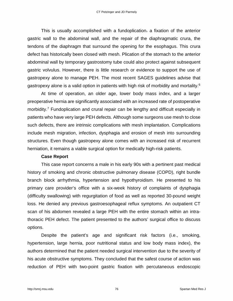
This is usually accomplished with a fundoplication, a fixation of the anterior
gastric wall to the abdominal wall, and the repair of the diaphragmatic crura, the
tendons of the diaphragm that surround the opening for the esophagus. This crura
defect has historically been closed with mesh. Plication of the stomach to the anterior
abdominal wall by temporary gastrostomy tube could also protect against subsequent
gastric volvulus. However, there is little research or evidence to support the use of
gastropexy alone to manage PEH. The most recent SAGES guidelines advise that
gastropexy alone is a valid option in patients with high risk of morbidity and mortality.6
At time of operation, an older age, lower body mass index, and a larger
preoperative hernia are significantly associated with an increased rate of postoperative
morbidity.7 Fundoplication and crural repair can be lengthy and difficult especially in
patients who have very large PEH defects. Although some surgeons use mesh to close
such defects, there are intrinsic complications with mesh implantation. Complications
include mesh migration, infection, dysphagia and erosion of mesh into surrounding
structures. Even though gastropexy alone comes with an increased risk of recurrent
herniation, it remains a viable surgical option for medically high-risk patients.
Case Report This case report concerns a male in his early 90s with a pertinent past medical
history of smoking and chronic obstructive pulmonary disease (COPD), right bundle
branch block arrhythmia, hypertension and hypothyroidism. He presented to his
primary care provider’s office with a six-week history of complaints of dysphagia
(difficulty swallowing) with regurgitation of food as well as reported 30-pound weight
loss. He denied any previous gastroesophageal reflux symptoms. An outpatient CT
scan of his abdomen revealed a large PEH with the entire stomach within an intra-
thoracic PEH defect. The patient presented to the authors’ surgical office to discuss
options.
Despite the patient’s age and significant risk factors (i.e., smoking,
hypertension, large hernia, poor nutritional status and low body mass index), the
authors determined that the patient needed surgical intervention due to the severity of
his acute obstructive symptoms. They concluded that the safest course of action was
reduction of PEH with two-point gastric fixation with percutaneous endoscopic
CT Petzinger and JD Parmely
http://smrj.msu.edu 76 Spartan Med Res J

gastrostomy (PEG) tubes, rather than subject the patient to a longer PEH repair with
reinforcement mesh or fundoplication. The authors also had concerns about the
patients’ nutritional status due to his significant weight-loss and its effects on his
postoperative healing, making this shorter less invasive alternative ideal for the patient.
Surgical Intervention and Hospital Course Intraoperatively, the patient was found to have a large hiatal defect with the
majority of the stomach within the paraesophageal defect and a gastric volvulus. The
stomach was easily reduced into the abdominal cavity without any dissection into the
mediastinum. The gastric volvulus was also reduced. Subsequently, the gastric outlet
was noted to be patent on evaluation with a gastroscope. There was also no evidence
of gastritis or esophagitis on EGD. Two Ponsky PEG tubes were then placed using
endoscopic/percutaneous technique with laparoscopic visualization for positioning of
the PEG tubes. The PEG tubes were placed at the proximal and distal stomach,
performing a pexy (fixation) of the anterior wall of the stomach to the abdominal wall.
Postoperatively, the patient was admitted to the hospital for monitoring and
postoperative care. Although he was at first hesitant to resume eating, he resumed a
regular diet after some encouragement without signs of dysphagia. An Upper GI Study
was performed on postoperative day #1 showing the cardia of the stomach in the left
hemithorax. However, the remainder of the stomach was within the abdomen and there
was no gastric outlet obstruction or volvulus. The patient was discharged home on
postoperative day #2 without any signs of dysphagia. The patient was followed closely
on an outpatient basis for one postoperative month and continued to tolerate a regular
diet, gaining weight without enteral supplementation. After the post-op month, both
Ponsky PEG tubes were removed without any recurrence of symptoms.
DISCUSSION Paraesophageal hernias, Types II-IV should be repaired in a timely manner due
to the significant comorbidities associated with these defects. These hernias will
continue to increase in size over time, making the non-emergent repair of these hernias
necessary in younger patients. Earlier repairs result in fewer intra-thoracic adhesions,
making the dissection of the hernia from the thoracic cavity less complicated. Dilation
Laproscopic Paraesophageal Hernia Reduction with Two Point Fixation via Ponsky PEG Tube
Vol. 2 No.1 Summer, 2017 77

of the hiatus and stretching of the cura also increases with time, which can also make
surgical repair more difficult. The approach and type of surgery performed should be
chosen based on the patient factors and fit the patients’ surgical profile. Elective repairs
avoid future complications and improve patients’ function.3
Traditional management of PEH calls for a lengthy and risky surgical approach
that is not always appropriate for patients with advanced age and multiple co-
morbidities. In a 2010 article by Luketich et al., 662 patients who underwent
laparoscopic giant PEH repairs were followed for 10 years.9 These patients underwent
screening esophagrams which showed hiatal hernia recurrence of up to 15.7%
although only 3.2% had return of their symptoms. In this 2010 study, those patients,
who underwent complete dissection of the intra-thoracic hernia sac, crural repair and
mesh application still had a 15.7% recurrence rate.9 In a similar study from Antonoff
and colleagues, their group of patients underwent similar steps for their PEH repairs
with morbidity rates of 8.2% and recurrence rates of 5.5%.10 These authors attributed
their success rates to observing a minimally invasive approach while still using the
fundamentals of open repair.10
Some surgeons are proponents of adding a wrap of the fundus of the stomach
after the hernia is reduced and mesh is fixed to prevent the occurrence of GERD. One
German randomized controlled pilot study with 40 patients compared Laparoscopic
mesh-augmented hiatoplasty with simple cardiophrenicopexy (LMAH-C) versus
Laparoscopic mesh-augmented hiatoplasty with fundoplication.11 Their data showed
that recurrence rates were similar between the two (33% and 21%, respectively).
However they proposed that fundoplication be added to all repairs due to the risk of
new onset GERD that occurred in a significant amount of patients who underwent
LMAH-C (53%) in comparison to those who underwent LMAH-F (17%). Moreover,
adding a fundoplication also added half an hour to surgery (Mean 153 minutes; range
90-250 minutes) in comparison to LMAH–C (Mean 124 minutes: range 85-210
minutes).11
Different surgical approaches, techniques and modalities for PEH repair add an
increased amount of time under anesthesia for patients who are elderly and frail,
increasing their morbidity and possible mortality risks. Mortality rates after emergent
CT Petzinger and JD Parmely
http://smrj.msu.edu 78 Spartan Med Res J

PEH repair are estimated at 5.4-8% and 0.8-1.4% after elective PEH, and frail patients
have higher mortality risks.5
Our approach in this case was not only unique in the use of dual PEG tube
insertion to prevent gastric volvulus, but also allowed a chronically ill patient to undergo
surgical intervention for which he otherwise would not have been a candidate. Due to
the significant risks attributed with this patient in their early 90s, the authors determined
that the safest approach to his large PEH would be laparoscopic reduction of the PEH
with gastropexy. This approach was relatively quick (76 minutes), without
encroachment into the mediastinum and avoided any complications mesh implantation
could pose. SAGES guidelines do report that morbidity and mortality of gastropexy is
significantly lower than other more invasive PEH repairs.7 However, gastropexy alone
has not demonstrated the same efficacy as formal repair of a PEH and should be
reserved for patients with significant comorbidities.7
Gastropexy is a relatively simple technique with minimal tissue dissection that
is generally well tolerated in the elderly population and those with decreased cardiac
and pulmonary status.13 Regardless of the approach to PEH repair, there are inherent
chances of recurrence. In a study from Daigle and associates, laparoscopic PEH
repairs using a modified Boerema anterior gastropexy (i.e., fixation to the abdominal
wall with nonabsorbable sutures putting the esophagus under some tension to form an
“angle of His”) was completed without fundoplication at multiple centers.14 Out of 101
patients, 70% had no postoperative reflux with recurrence rates of 16.8% when
followed on endoscopy or barium swallow evaluation.14 Surgical intervention and
approach should therefore be tailored to the patient, with risks and benefits carefully
weighed. In this case, the patient tolerated dual gastropexy alone with complete
resolution of symptoms without the increased accompanying risks of crural repair and
mesh implantation.
The authors report no external funding source for this study.
The authors declare no conflict of interest.
Submitted for publication February 2017 Accepted for publication July 2017
Laproscopic Paraesophageal Hernia Reduction with Two Point Fixation via Ponsky PEG Tube
Vol. 2 No.1 Summer, 2017 79

ACKNOWLEDGEMENTS 1. Surgical colleagues at Beaumont Hospital- Farmington Hills.2. Diane Piskorowski, MSLS, the Medical Library Coordinator at Beaumont-Farmington
Hills.3. Elsevier Copyright Clearance Center for use of their image for this publication.
CT Petzinger and JD Parmely
http://smrj.msu.edu 80 Spartan Med Res J

REFERENCES 1. Fischer, JE; Jones, DB; Pomposelli, FB. Fischer’s Mastery of Surgery. 6th Edition;
20112. El Lakis, MA., Kaplan, SJ., Hubka, M, Mohiuddin K, Low DE. Importance of age and
short term outcomes associated with repair of giant paraesophageal hernias. AnnThorac Surg. 2017;103(6):1700-1709.
3. Carrott PW, Hong J, Kuppusamy M, Koehler RP, Low DE. Clinical ramifications ofgiant paraesophageal hernias are underappreciated: Making the case for routinesurgical repair. Ann Thorac Surg. 2012;94:421-8.
4. Sihvo E, Salo JA, Rasanen JV, Rantanen TK. Fatal complications of adultparaesophageal hernia: Population the study. J Thorac Cardiovasc Surg. 2009;137:419-24.
5. Chimukangara M, Frelich MJ, Bosler ME, Rein LE, Azabo A, Gould JC. The impact offrailty on outcomes of paraesophageal hernia repair. J Surg Res. 15 May 2016;202(2): 259-266.
6. Lebenthal A, Waterford SD, Fisichella PM. Treatment and Controversies inParaesophageal Hernia Repair, Front Surg. 2:13 April 2015
7. Kohn G, Price RR, DeMeester SR, Zehetner J, Muensterer OJ, Awad Z, et.al.Guidelines for The Management of Hiatal Hernia. Surg Endo. 2013;27(12):4409-28.
8. Ballian N, Luketich JD, Levy RM, Awais O, Winger D, Weksler B, et.al. A ClinicalPrediction Rule for Perioperative Mortality and Major Morbidity after LaparoscopicGiant Paraesophageal Hernia Repair. J Thorac Cardiovasc Surg. 2013;145(3):721-729.
9. Lutetich, JD, Nason KS, Christie NA, Pennathur A, Jobe BA, Landreneau RJ, et al.Outcomes after a decade of laparoscopic giant paraesophageal hernia repair. JThorac Cardiovasc Surg. 2010; 139: 395-404; 404.e391
10. Antonoff MB, D’Cunha J, Andrade RS, Maddaus MA. Giant paraesophageal herniarepair: Technical Pearls. J Thorac Cardiovasc Surg. 2011; 144(3):S67-S70.
11. Muller-Stich BP, Achtstatter V, Diener MK, Gondan M, Warschkow R, Marra F, et al.Repair of paraesophageal hiatal hernias–is fundoplication needed? A randomizedcontrolled pilot trial. J Am Coll Surg. 2015. Volume to 221(2):602-610.
12. Agwunobi AO, Bancewicz J, Attwood SE. Simple Laparoscopic Gastropexy as theInitial Treatment of Paraoesophageal Hiatal Hernia. Brit J Surg. 1998;85(5):604-606.
13. Lukman MR, Sangar P, Sukumar N. Obstructed Paraesophageal Hernia in aNonagenerian Treated by Laparoscopic Anterior Gastropexy. Med J Malaysia.2006;62(1):83-84.
14. Daigle CR, Funch-Jensen P, Calatayud D, Rask P, Jacobsen B, Grantcharov TPLaparoscopic Repair of Paraesophageal Hernia with Anterior Gastropexy: AMulticenter Study. Surg Endosc. 2015;29(7):1856-61.
Laproscopic Paraesophageal Hernia Reduction with Two Point Fixation via Ponsky PEG Tube
Vol. 2 No.1 Summer, 2017 81

TABLES AND FIGURES
Figure 1: Anatomy of the Esophagus, Gastroesophageal (GE) Junction, and Stomach
Displaying Normal Anatomy and the Four Types of Hiatal Hernias
A. Normal.
B. Sliding or type I hiatal hernia.
C. Paraesophageal or type II hiatal hernia.
D. Type III hiatal hernia
E. Type IV hiatal hernia (note colon to the left of the herniated gastric fundus).
(Illustrations by Lindsay Agema and Christian B. Rodriguez.)
Used with permission from Elsevier.
CT Petzinger and JD Parmely
http://smrj.msu.edu 82 Spartan Med Res J
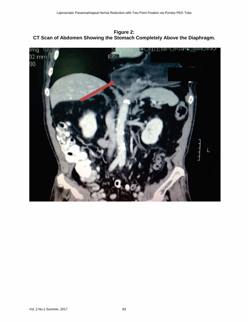
Figure 2: CT Scan of Abdomen Showing the Stomach Completely Above the Diaphragm.
Laproscopic Paraesophageal Hernia Reduction with Two Point Fixation via Ponsky PEG Tube
Vol. 2 No.1 Summer, 2017 83
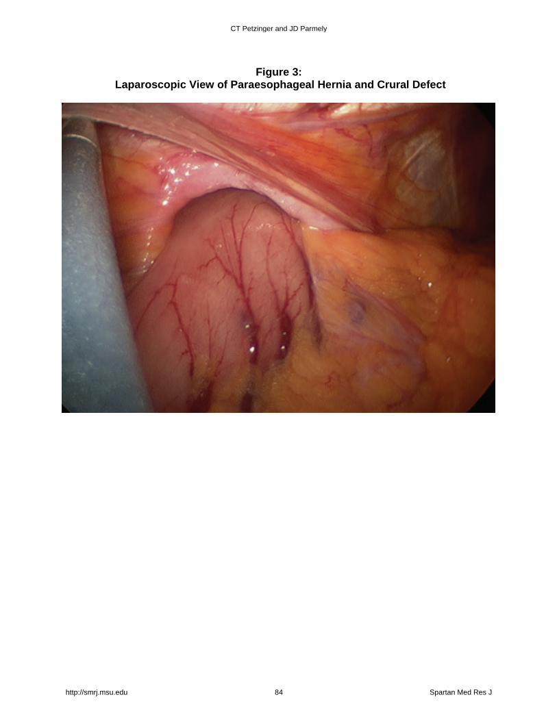
Figure 3: Laparoscopic View of Paraesophageal Hernia and Crural Defect
CT Petzinger and JD Parmely
http://smrj.msu.edu 84 Spartan Med Res J

Figure 4: Laparoscopic View After Reduction and Two-Point Ponsky Fixation to the
Anterior Abdominal Wall.
Laproscopic Paraesophageal Hernia Reduction with Two Point Fixation via Ponsky PEG Tube
Vol. 2 No.1 Summer, 2017 85