Embed Size (px)
DESCRIPTION
Spinal Epidural Abscess SEA. www.medkaau.com/vb. Done by Dr.Wala’a Gholam KAAU 2007 4 th year medical student. contents. - Pathogenesis - Causative agents - Mechanism of injury - Clinical features (symptoms. Physical exam) - Investigation (lab. Imaging) - DD - Diagnosis - PowerPoint PPT Presentation
Citation preview
Spinal Epidural Abscess
SEA
Done by
Dr.Wala’a Gholam
KAAU 2007
4th year medical student
www.medkaau.com/vb
contents
- Pathogenesis
- Causative agents
- Mechanism of injury
- Clinical features (symptoms. Physical exam)
- Investigation (lab. Imaging)
- DD
- Diagnosis
- Treatment (surgical , medical)
- Prognosis
- complications
EBM
Pathogenesis 1,2,3
-Underlying disease:
diabetes mellitus
alcoholism
infection with HIV
-A spinal abnormality or intervention:
degenerative joint disease
Trauma
Surgery
drug injection
placement of stimulators or catheters

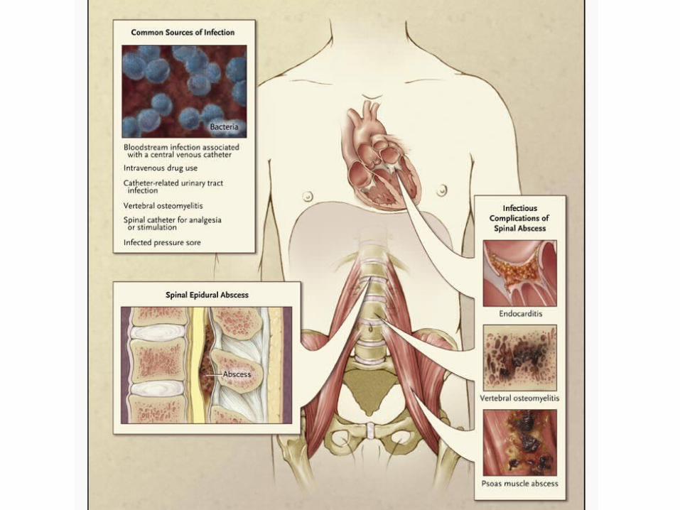
-a potential local or systemic source of infection: Skin and soft-tissue infections
Osteomyelitis Pott's disease (spinal TB)
UTI, URTI Sepsis
Dermal sinus tract , Dental abscess Retropharyngeal abscess
Lemierre’s syndrome (1 case report) !! 4
Indwelling vascular access, Intravenous drug use Nerve acupuncture, Tattooing
Epidural analgesia (in cancer pt) or nerve block hemodialysis patients (12 case report) 5
1 -Chowfin A, Potti A, Paul A, Carson P. Spinal epidural abscess after tattooing. Clin Infect Dis 19992 -Sillevis Smitt P, Tsafka A, van den Bent M, et al. Spinal epidural abscess complicating chronic epidural analgesia in 11 cancer patients: clinical findings and magnetic resonance
imaging. J Neurol 19993 -Grewal S, Hocking G, Wildsmith JA. Epidural abscesses. Br J Anaesth 2006
4 -Royal National Orthopaedic Hospital, Stanmore HA7 4LP, UK 5 -Department of Medicine, Boston University Medical Center, MA, USA
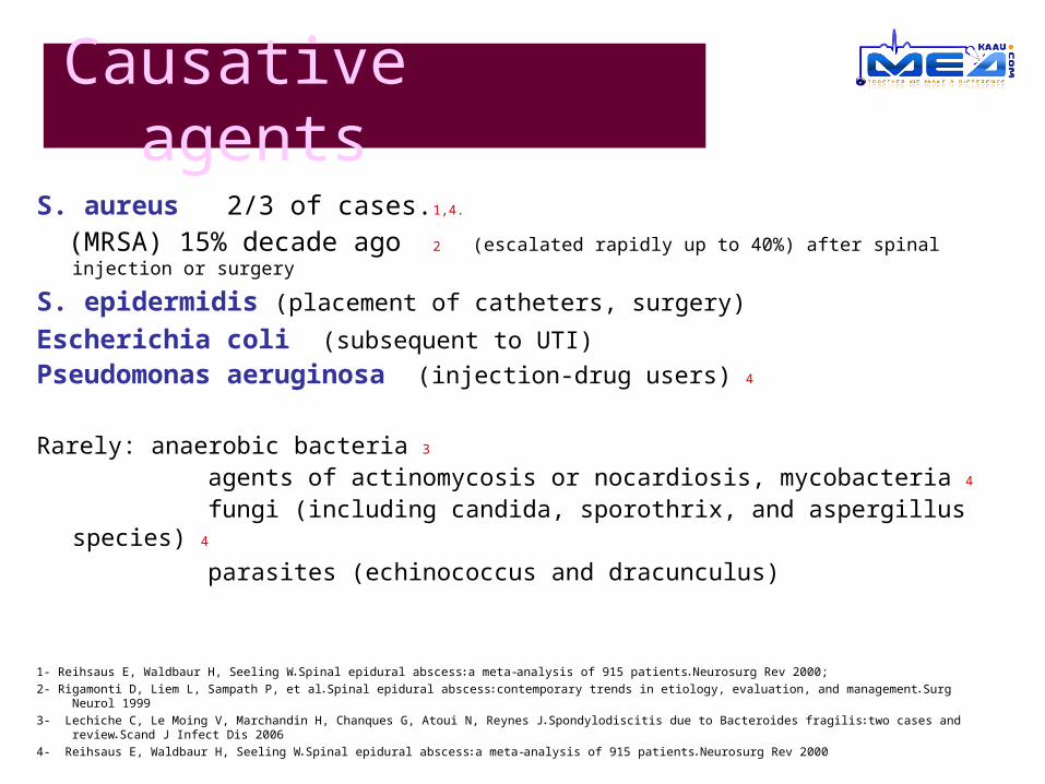
S. aureus 2/3 of cases.1,4.
(MRSA) 15% decade ago 2 (escalated rapidly up to 40%) after spinal injection or surgery
S. epidermidis (placement of catheters, surgery)
Escherichia coli (subsequent to UTI)
Pseudomonas aeruginosa (injection-drug users) 4
Rarely: anaerobic bacteria 3
agents of actinomycosis or nocardiosis, mycobacteria 4
fungi (including candida, sporothrix, and aspergillus species) 4
parasites (echinococcus and dracunculus)
1- Reihsaus E, Waldbaur H, Seeling W. Spinal epidural abscess: a meta-analysis of 915 patients. Neurosurg Rev 2000;
2- Rigamonti D, Liem L, Sampath P, et al. Spinal epidural abscess: contemporary trends in etiology, evaluation, and management. Surg Neurol 1999
3- Lechiche C, Le Moing V, Marchandin H, Chanques G, Atoui N, Reynes J. Spondylodiscitis due to Bacteroides fragilis: two cases and review. Scand J Infect Dis 2006
4- Reihsaus E, Waldbaur H, Seeling W. Spinal epidural abscess: a meta-analysis of 915 patients. Neurosurg Rev 2000
Causative agents
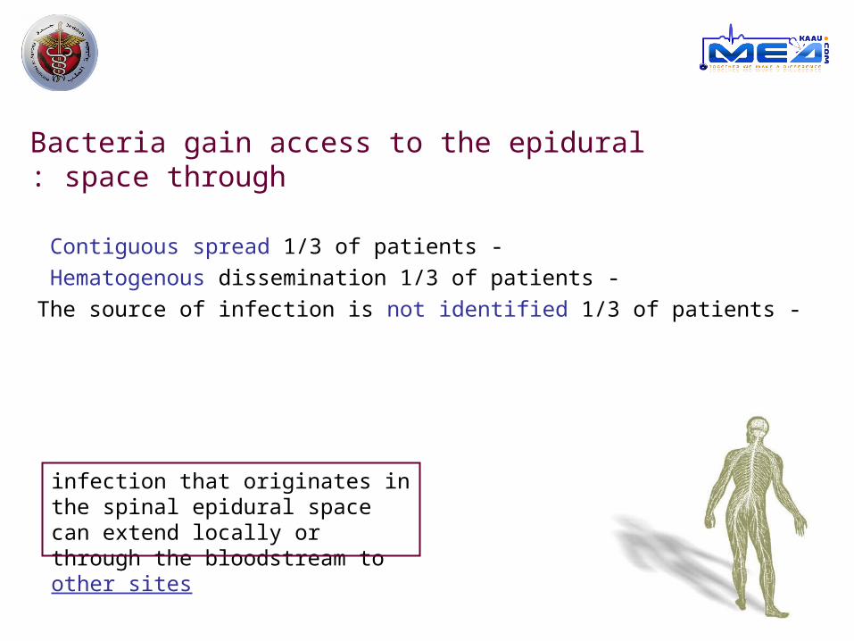
Bacteria gain access to the epidural space through:
- Contiguous spread 1/3 of patients
- Hematogenous dissemination 1/3 of patients
-The source of infection is not identified 1/3 of patients
infection that originates in the spinal epidural space can extend locally or through the bloodstream to other sites
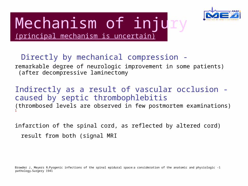
Mechanism of injury(principal mechanism is uncertain)
-Directly by mechanical compression )remarkable degree of neurologic improvement in some patients after
decompressive laminectomy (
-Indirectly as a result of vascular occlusion caused by septic thrombophlebitis
)thrombosed levels are observed in few postmortem examinations (1
)infarction of the spinal cord, as reflected by altered cord signal MRI (result from
both
1 -Browder J, Meyers R. Pyogenic infections of the spinal epidural space: a consideration of the anatomic and physiologic pathology. Surgery 1941
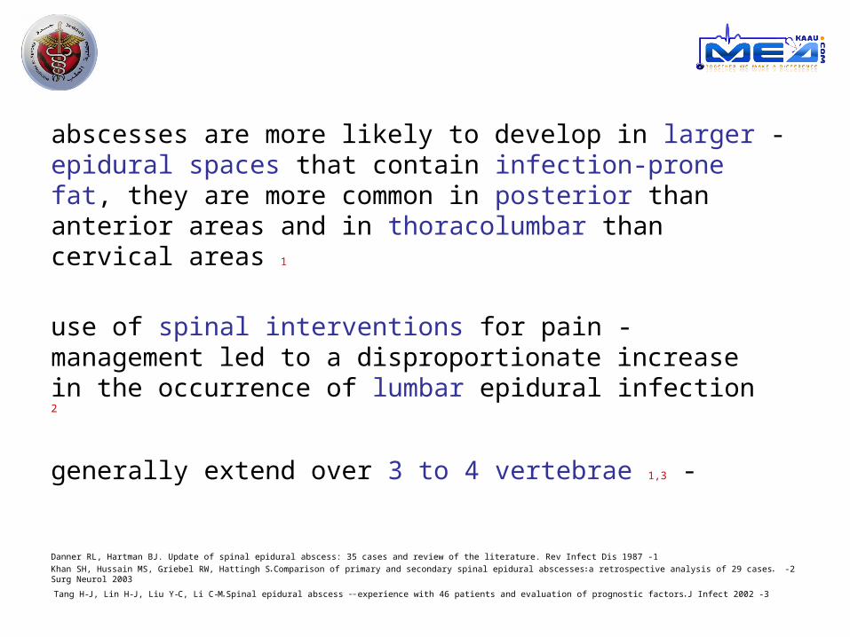
-abscesses are more likely to develop in larger epidural spaces that contain infection-prone fat, they are more common in posterior than anterior areas and in thoracolumbar than cervical areas 1
-use of spinal interventions for pain management led to a disproportionate increase in the occurrence of lumbar epidural infection 2
-generally extend over 3 to 4 vertebrae 1,3
1 -Danner RL, Hartman BJ. Update of spinal epidural abscess: 35 cases and review of the literature. Rev Infect Dis 1987
2 -Khan SH, Hussain MS, Griebel RW, Hattingh S. Comparison of primary and secondary spinal epidural abscesses: a retrospective analysis of 29 cases. Surg Neurol 2003
3 -Tang H-J, Lin H-J, Liu Y-C, Li C-M. Spinal epidural abscess -- experience with 46 patients and evaluation of prognostic factors. J Infect 2002
Paraspinal Infection
In rare cases they involve the whole spine,
resulting in so-called panspinal infection 1,2
1 -Rigamonti D, Liem L, Sampath P, et al. Spinal epidural abscess: contemporary trends in etiology, evaluation, and management. Surg Neurol 1999
2 -Solomou E, Maragkos M, Kotsarini C, Konstantinou D, Maraziotis T. Multiple spinal epidural abscesses extending to the whole spinal canal. Magn Reson
Imaging 2004
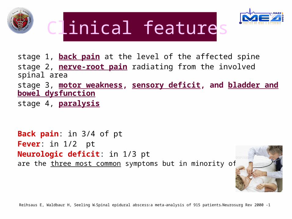
Clinical features
stage 1, back pain at the level of the affected spinestage 2, nerve-root pain radiating from the involved spinal areastage 3, motor weakness, sensory deficit, and bladder and bowel dysfunctionstage 4, paralysis
Back pain: in 3/4 of ptFever: in 1/2 ptNeurologic deficit: in 1/3 ptare the three most common symptoms but in minority of pts 1
1 -Reihsaus E, Waldbaur H, Seeling W. Spinal epidural abscess: a meta-analysis of 915 patients. Neurosurg Rev 2000
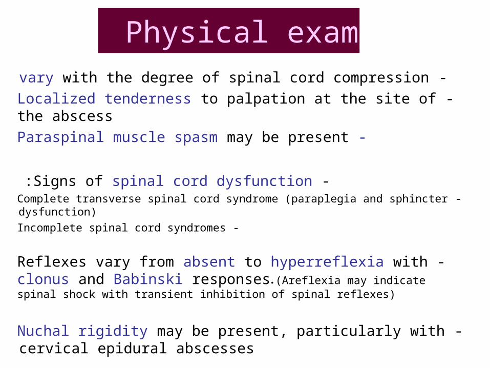
Physical exam -vary with the degree of spinal cord compression
-Localized tenderness to palpation at the site of the abscess
-Paraspinal muscle spasm may be present
-Signs of spinal cord dysfunction: - Complete transverse spinal cord syndrome (paraplegia and sphincter dysfunction)
- Incomplete spinal cord syndromes
-Reflexes vary from absent to hyperreflexia with clonus and Babinski responses. (Areflexia may indicate spinal shock with transient inhibition of spinal reflexes)
-Nuchal rigidity may be present, particularly with cervical epidural abscesses
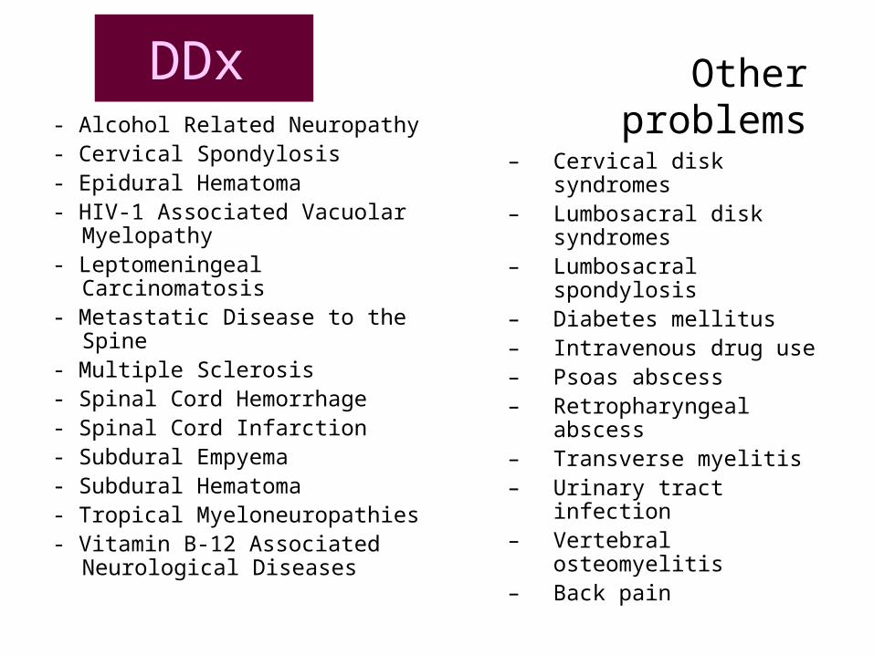
DDx
– Cervical disk syndromes– Lumbosacral disk
syndromes– Lumbosacral spondylosis– Diabetes mellitus– Intravenous drug use– Psoas abscess– Retropharyngeal
abscess– Transverse myelitis– Urinary tract infection– Vertebral osteomyelitis– Back pain
- Alcohol Related Neuropathy - Cervical Spondylosis- Epidural Hematoma - HIV-1 Associated Vacuolar
Myelopathy - Leptomeningeal Carcinomatosis - Metastatic Disease to the Spine- Multiple Sclerosis - Spinal Cord Hemorrhage - Spinal Cord Infarction - Subdural Empyema- Subdural Hematoma - Tropical Myeloneuropathies- Vitamin B-12 Associated
Neurological Diseases
Other problems
Diagnosis suspected on the basis of clinical findingssupported by laboratory data and imaging studiesbut can be confirmed only by drainage
Although leukocytosis detected in about 2/3 of pt 1,2 and inflammatory markers (ESR and C-reactive protein) are almost uniformly elevated, they are not specific
Bacteremia causing or arising from spinal epidural abscess is detected in about 60% of patients 3
1- Darouiche RO, Hamill RJ, Greenberg SB, Weathers SW, Musher DM. Bacterial spinal epidural abscess: review of 43 cases and literature survey. Medicine (Baltimore) 1992 2- Soehle M, Wallenfang T. Spinal epidural abscesses: clinical manifestations, prognostic factors, and outcomes. Neurosurgery 2002 3- Curry WT Jr, Hoh BL, Amin-Hanjani S, Eskandar EN. Spinal epidural abscess: clinical presentation, management, and outcome. Surg Neurol 2005
DiagnosisCSF
CSF analysis shows a high level of protein and pleocytosis (with either a polymorphonuclear or a mononuclear predominance) parameningeal inflammation, but are not specific for epidural infection 1
Gram staining of CSF is usually –veCSF cultures are +ve in less than 25% of pt.
However, blood cultures yield the infecting pathogen in almost all patients with a positive CSF culture 1
1- Darouiche RO, Hamill RJ, Greenberg SB, Weathers SW, Musher DM. Bacterial spinal epidural abscess: review of 43 cases and literature survey. Medicine (Baltimore) 1992
But!!
although rare, there is a risk of meningitis or subdural infection if the needle traverses the epidural abscess
Because lumbar puncture affords meager information and is associated with a slight potential risk
it should not be done routinely
CSF should be analyzed only if myelography is performed .
Radiology
Both MRI with intravenous administration of gadolinium and myelography followed by CT of the spine are highly sensitive (more than 90%) in diagnosing spinal epidural abscess 1, 2
MRI better :longitudinal and paraspinal extensionabscess or cancer !! 3
1 -Rigamonti D, Liem L, Sampath P, et al. Spinal epidural abscess: contemporary trends in etiology, evaluation, and management. Surg Neurol 19992 -Hlavin ML, Kaminski HJ, Ross JS, Ganz E. Spinal epidural abscess: a ten-year perspective. Neurosurgery 1990
3 -Parkinson JF, Sekhon LH. Spinal epidural abscess: appearance on magnetic resonance imaging as a guide to surgical management. Neurosurg Focus 2004
-plain roentgenograph or CT: narrowing of the disk and bone lysis to indicate the presence of diskitis and osteomyelitis (which coexist with SEA in up to 80% of patients) 1
-radionuclide scanning (with technetium, gallium, or indium) may show increased uptake
)the findings of these tests are neither sensitive nor specific for SEA and should not take the place of MRI(
-The presence of pulmonary infiltrates on the chest radiography, is evidence of immunodeficiency
1 -Khan SH, Hussain MS, Griebel RW, Hattingh S. Comparison of primary and secondary spinal epidural abscesses: a retrospective analysis of 29 cases. Surg Neurol 2003
Radiology
-direct smear or culture sputum positive for acid-fast bacilli, is suggestive of TB .
Diagnosis cont .
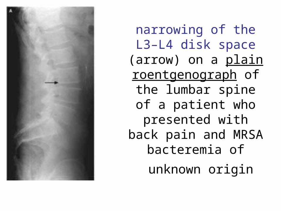
narrowing of the L3–L4 disk space (arrow) on a plain roentgenograph of
the lumbar spine of a patient who presented
with back pain and MRSA bacteremia of
unknown origin
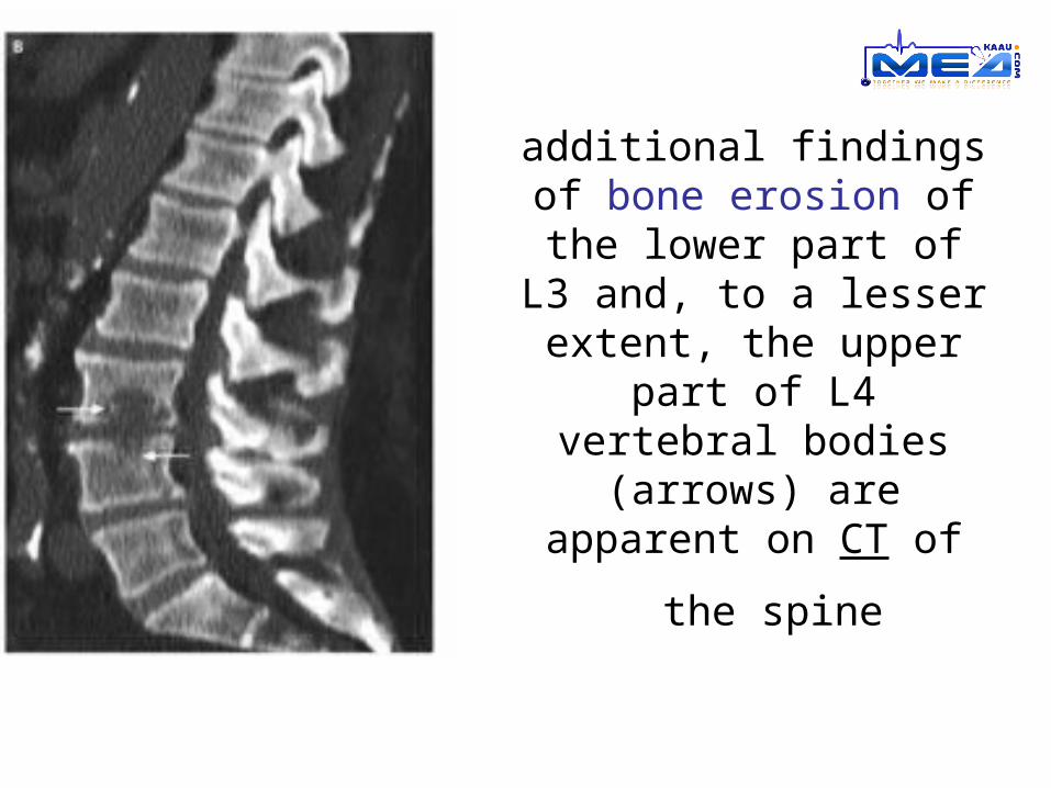
additional findings of bone erosion of the lower part of L3 and, to a lesser extent, the upper part of
L4 vertebral bodies (arrows) are apparent on
CT of the spine
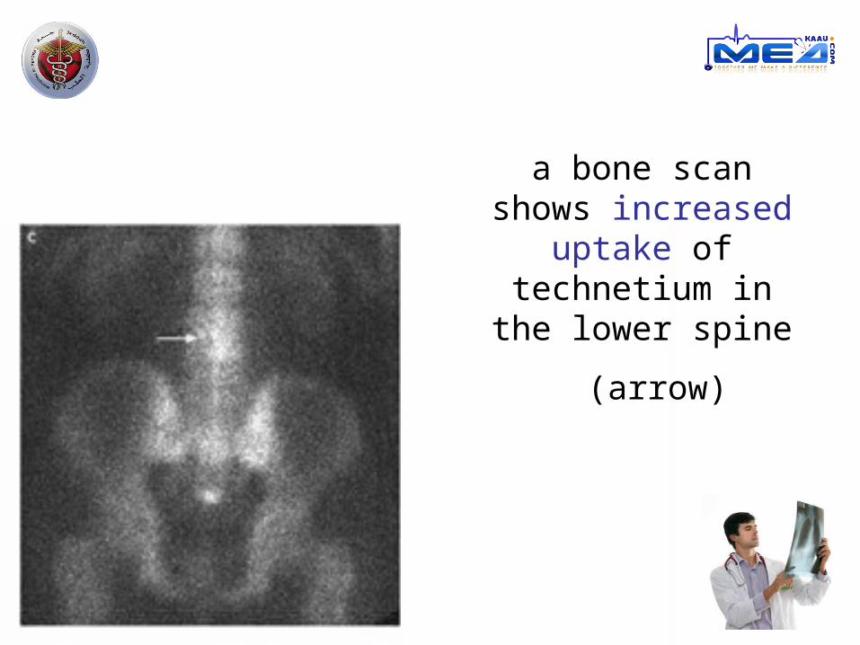
a bone scan shows increased uptake of
technetium in the
lower spine (arrow)
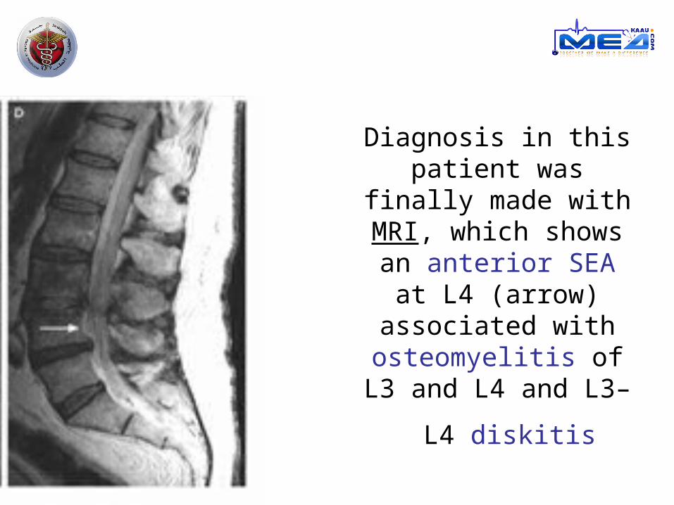
Diagnosis in this patient was finally
made with MRI, which shows an anterior SEA
at L4 (arrow) associated with
osteomyelitis of L3 and
L4 and L3–L4 diskitis
Diagnosis cont.
- Rare condition
- Non specific finding (fever, back pain, leukocytosis, ↑ ESR, ↑ C-reactive protien misdiagnosed
particularly in neurologically intact patients (those in stage 1 or stage 2) 1.2
- Other diagnosis !! infectious conditions (osteomyelitis, diskitis, meningitis, UTI, sepsis, and
endocarditis)
noninfectious conditions (intervertebral-disk prolapse, degenerative joint disease, spinal tumor, demyelinating illness, transverse myelitis, and spinal hematoma)
1- Davis DP, Wold RM, Patel RJ, et al. The clinical presentation and impact of diagnostic delays on emergency department patients with spinal epidural abscess . J Emerg Med 2004
2- Tang H-J, Lin H-J, Liu Y-C, Li C-M. Spinal epidural abscess -- experience with 46 patients and evaluation of prognostic factors. J Infect 2002
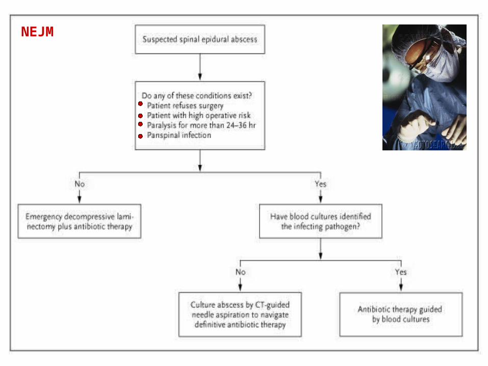
Treatment
Surgical
Medical

Treatment
-prospective, randomized clinical trials to determine the optimal treatment (DIFFECULT)
-Butmajority of retrospective studies provide support for:
surgical drainage together with systemic antibiotics is the treatment of choice 1, 2, 3
- decompressive laminectomy and débridement of infected tissues should be done ASAP 1
1 -Lu C-H, Chang W-N, Lui C-C, Lee P-Y, Chang HW. Adult spinal epidural abscess: clinical features and prognostic factors. Clin Neurol Neurosurg 20022 -Curry WT Jr, Hoh BL, Amin-Hanjani S, Eskandar EN. Spinal epidural abscess: clinical presentation, management, and outcome. Surg Neurol 2005
3 -Pereira CE, Lynch JC. Spinal epidural abscess: an analysis of 24 cases. Surg Neurol 2005
NEJM
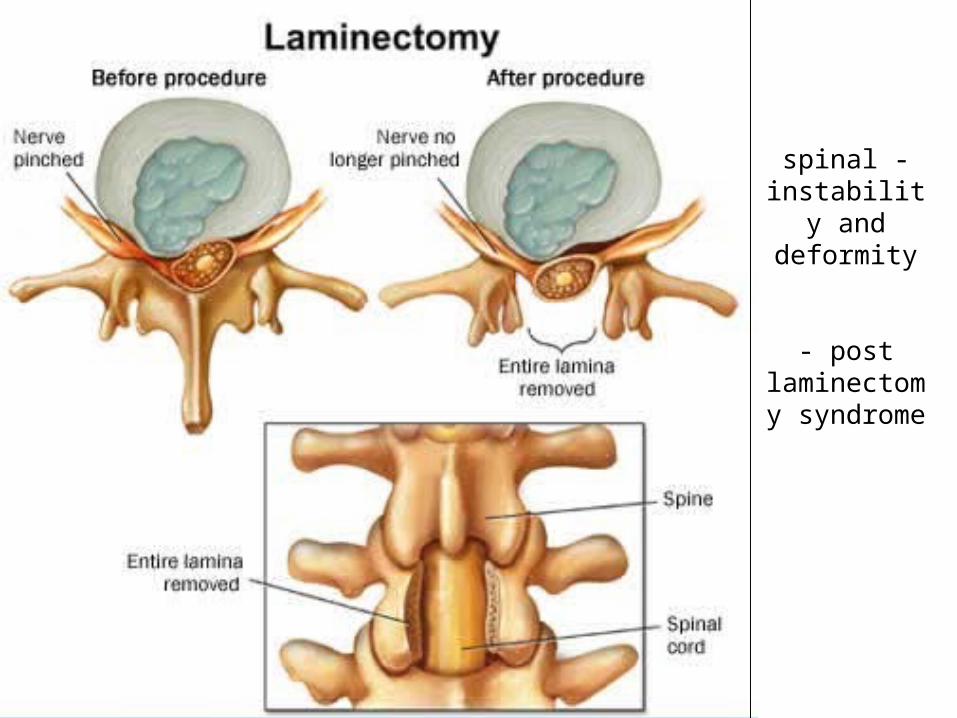
-spinal instability and
deformity
- post laminectomy
syndrome
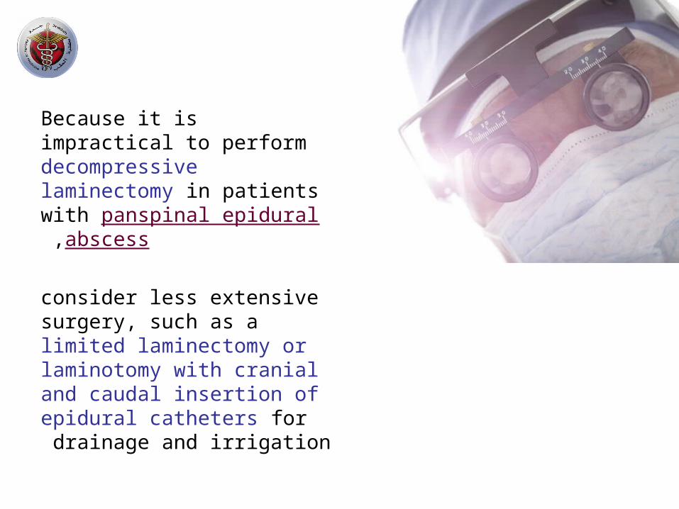
Because it is impractical to perform decompressive laminectomy in patients with
panspinal epidural abscess ,
consider less extensive surgery, such as a limited laminectomy or laminotomy with cranial and caudal insertion of epidural catheters for drainage and irrigation
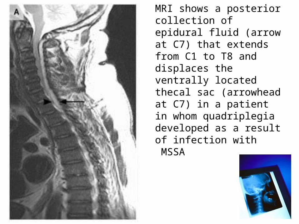
MRI shows a posterior collection of epidural fluid (arrow at C7) that extends from C1 to T8 and displaces the ventrally located thecal sac (arrowhead at C7) in a patient in whom quadriplegia developed as a result of infection with MSSA
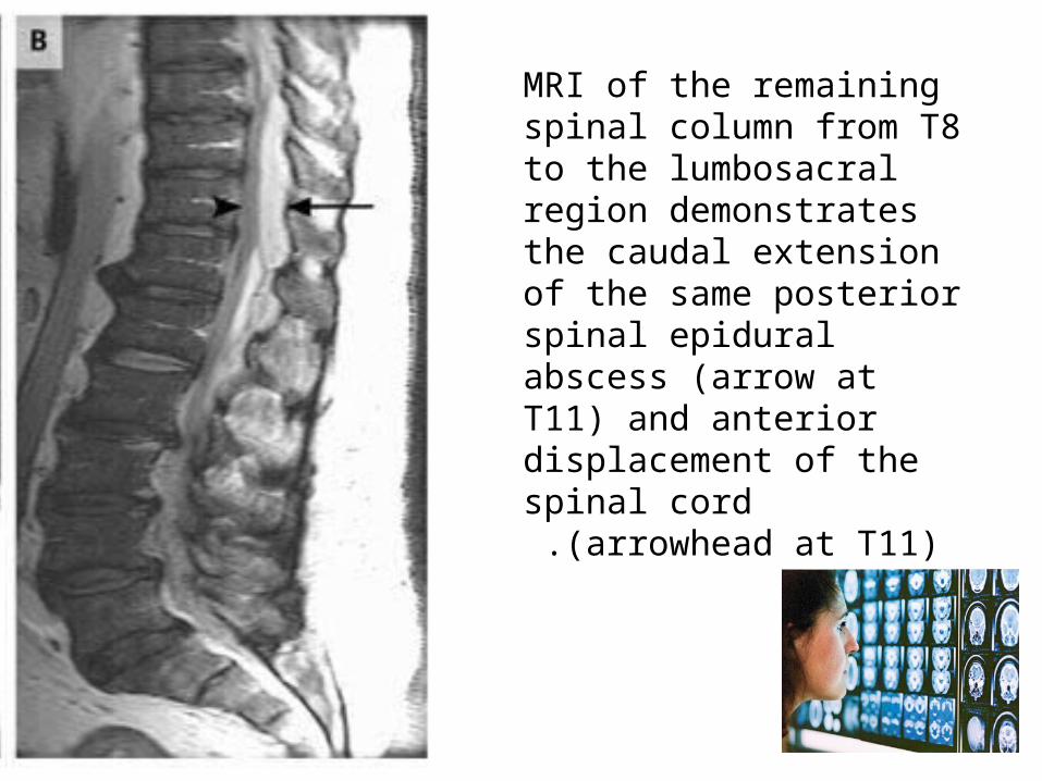
MRI of the remaining spinal column from T8 to the lumbosacral region demonstrates the caudal extension of the same posterior spinal epidural abscess (arrow at T11) and anterior displacement of the spinal cord (arrowhead at
T11) .
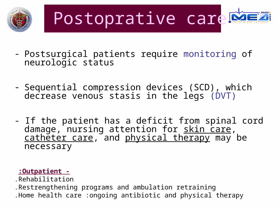
Postoprative care
- Postsurgical patients require monitoring of neurologic status
- Sequential compression devices (SCD), which decrease venous stasis in the legs (DVT)
- If the patient has a deficit from spinal cord damage, nursing attention for skin care, catheter care, and physical therapy may be necessary
-Outpatient: Rehabilitation.Restrengthening programs and ambulation retraining.Home health care :ongoing antibiotic and physical therapy.

Antibiotics
At least 6 weeks because vertebral osteomyelitis exists in most patients
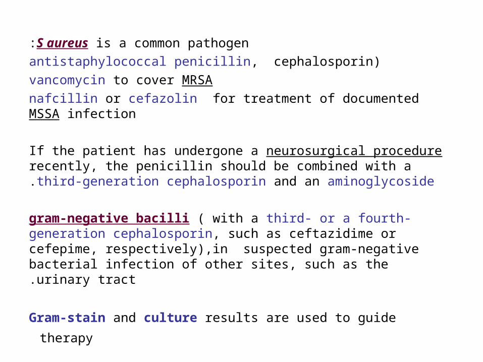
S aureus is a common pathogen:
)antistaphylococcal penicillin, cephalosporin
vancomycin to cover MRSA
nafcillin or cefazolin for treatment of documented MSSA infection
If the patient has undergone a neurosurgical procedure recently, the penicillin should be combined with a third-generation cephalosporin and an aminoglycoside.
gram-negative bacilli ( with a third- or a fourth-generation cephalosporin, such as ceftazidime or cefepime, respectively),in suspected gram-negative bacterial infection of other sites, such as the urinary tract.
Gram-stain and culture results are used to guide therapy
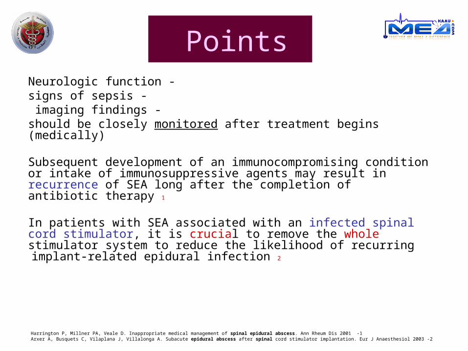
Points -Neurologic function
-signs of sepsis -imaging findings
should be closely monitored after treatment begins (medically)
Subsequent development of an immunocompromising condition or intake of immunosuppressive agents may result in recurrence of SEA long after the completion of antibiotic therapy 1
In patients with SEA associated with an infected spinal cord stimulator, it is crucial to remove the whole stimulator system to reduce the likelihood of recurring implant-related epidural infection 2
1 -Harrington P, Millner PA, Veale D. Inappropriate medical management of spinal epidural abscess. Ann Rheum Dis 2001 2 -Arxer A, Busquets C, Vilaplana J, Villalonga A. Subacute epidural abscess after spinal cord stimulator implantation. Eur J Anaesthesiol 2003
unexplained persistent or recurrent epidural infection may be assessed for rare sources of infection :esophageal tear (in the case of cervical epidural abscess) intestinal–spinal fistula (in the case of thoracolumbar abscess) .
Although there have been sporadic reports in which glucocorticoid therapy has been associated with an adverse outcome in patients who already had a severe case of spinal epidural abscess,1 it may help to reduce swelling in patients with progressive neurologic compromise who are awaiting surgical decompression .
1 -Danner RL, Hartman BJ. Update of spinal epidural abscess: 35 cases and review of the literature. Rev Infect Dis 1987
Points
Prognosis
No studies have been done to assist in predicting prognosis.
Prognosis in general is related to the duration of spinal cord dysfunction and the degree of cord impairment at the time of diagnosis
Complications of SEA
1 -Irreversible paralysis (4 to 22% of pt) 1,2
2 -bladder dysfunction
3 -decubiti (decubitus ulcer, pressure sore)
4 -supine hypertension
5 -recurrent sepsis
6 -UTI7 -Pneumonia (in cervical abscess) 3
8 -death 5% (uncontrolled sepsis, meningitis, others)
1 -Khanna RK, Malik GM, Rock JP, Rosenblum ML. Spinal epidural abscess: evaluation of factors influencing outcome. Neurosurgery 1996
2 -Danner RL, Hartman BJ. Update of spinal epidural abscess: 35 cases and review of the literature. Rev Infect Dis 1987
3 -Soehle M, Wallenfang T. Spinal epidural abscesses: clinical manifestations, prognostic factors, and outcomes. Neurosurgery 2002
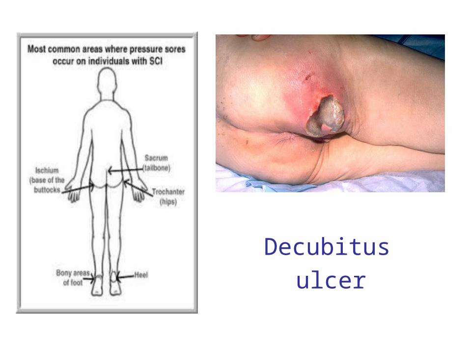
Decubitus
ulcer

Clinical Trails Done

Delay diagnosis in ER department
Department of Emergency Medicine, University of California, San Diego, San Diego, California USA.
Residual motor weakness was present in 45% of these patients vs. only 13% of patients without diagnostic delays
residual motor weakness
0
10
20
30
40
50
delay diagnosis no delay diagnosis
residual motor weakness
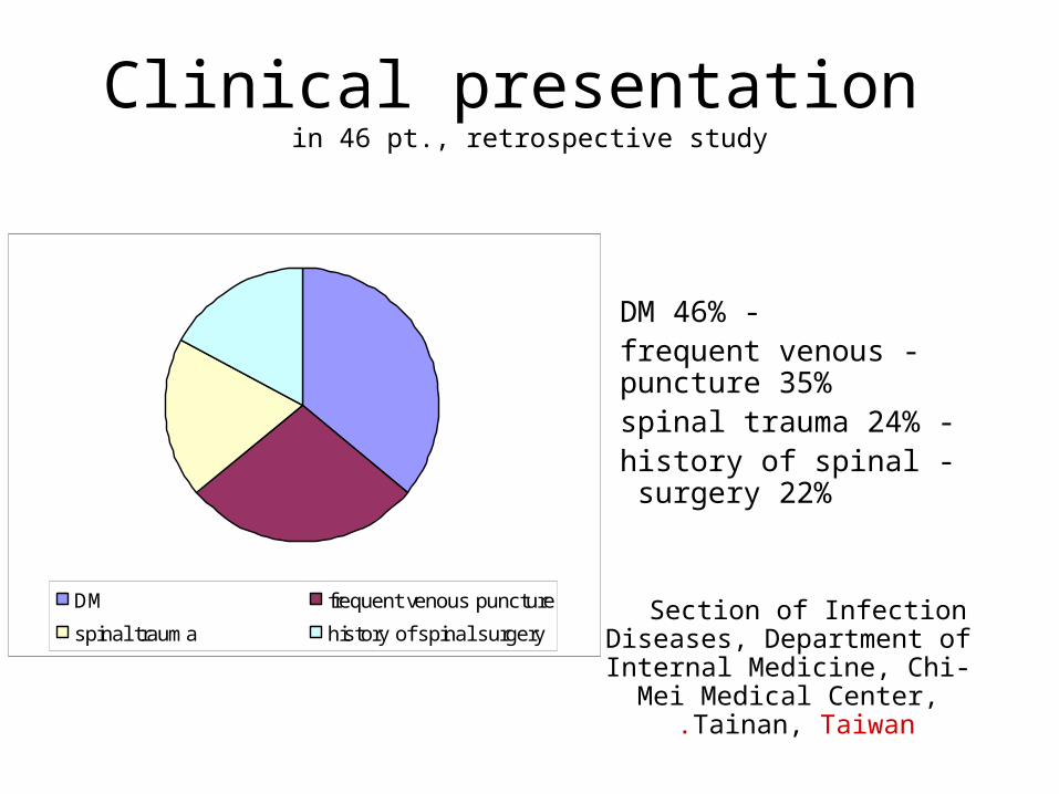
Clinical presentation in 46 pt., retrospective study
Section of Infection Diseases, Department of Internal
Medicine, Chi-Mei Medical Center, Tainan, Taiwan.
-DM 46% -frequent venous puncture 35%
-spinal trauma 24% -history of spinal surgery 22%
DM frequent venous puncture
spinal trauma history of spinal surgery
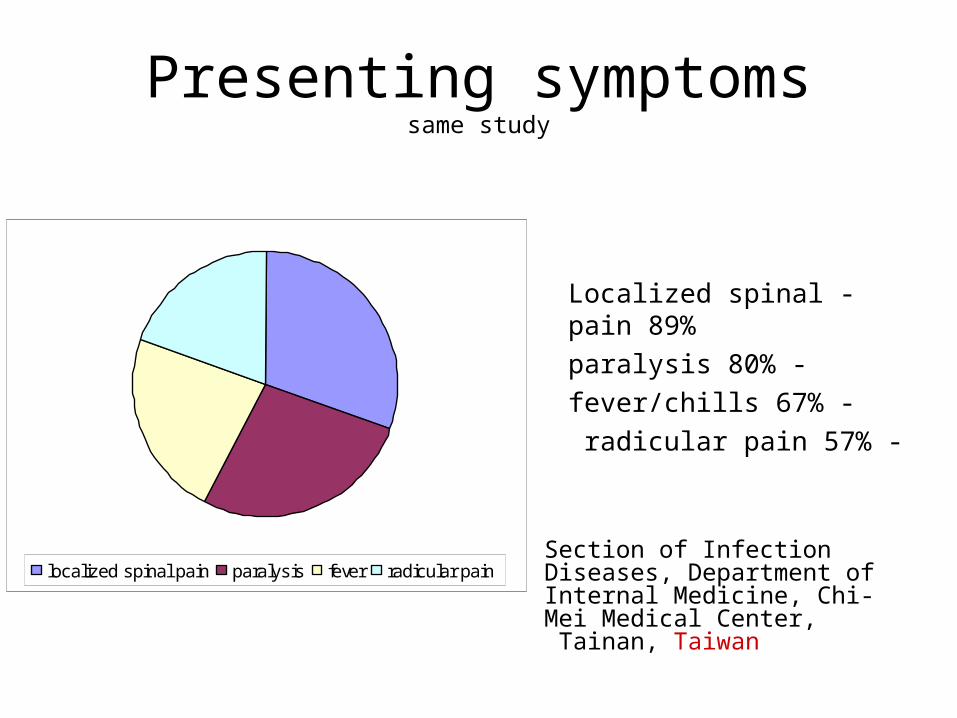
Presenting symptomssame study
Section of Infection Diseases, Department of Internal Medicine, Chi-Mei Medical Center, Tainan, Taiwan
-Localized spinal pain 89%
-paralysis 80%
-fever/chills 67%
-radicular pain 57%
localized spinal pain paralysis fever radicular pain
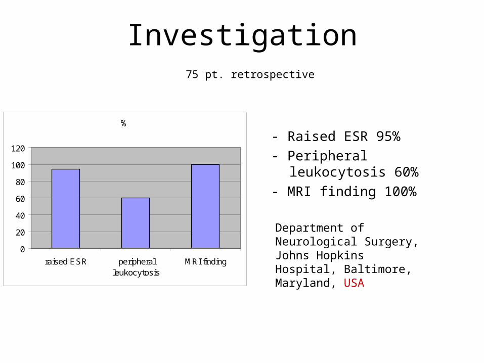
Investigation75 pt. retrospective
Department of Neurological Surgery, Johns Hopkins Hospital, Baltimore, Maryland, USA
- Raised ESR 95%
- Peripheral leukocytosis 60%
- MRI finding 100%
%
0
20
40
60
80
100
120
raised ESR peripheralleukocytosis
MRI finding

Thank you for your time
Dr.Wala'a Gholam
KAAU 2007
4th year medical student
www.medkaau.com/vb
Source used:
- The new England journal o f medicine, review article 2006
- E-medicine website


![Donald H. Lambert Boston, Massachusetts Spinal - Epidural - [Combined Spinal Epidural]](https://img.pdfslide.net/doc/110x75/5517e537550346d5568b46b6/donald-h-lambert-boston-massachusetts-httpwwwdebunk-itorg-spinal-epidural-combined-spinal-epidural.jpg)