Embed Size (px)
Citation preview
1
Archiv für Mikrobiologie, 21, p178‐201 (1954)
German original: “Untersuchungen über die Morphologie und Vermehrung der
pleuropneumonie‐ähnlichen Organismen und der L‐Phase der Bakterien.
I. Lichtmikroskopische Untersuchungen.“
http://link.springer.com/article/10.1007%2FBF01816378
Studies on morphology and multiplication (proliferation) of
pleuropneumonia‐like organisms (PPLOs, now classified as
mycoplasmas) and on bacterial L‐phase (L‐form bacteria)
I. Light microscopy1
Gertraud Kandler and Otto Kandler
Staatliche Bakteriologische Untersuchungsanstalt, Munich, Germany
Institute of Botany, University of Munich
(Submitted August 31st, 1954; translated by Kathrin Schirner and Gertraud Kandler in 2009,
corrected in 2015)
Since the discovery that bacteria can enter a state where they, morphologically, resemble
the pathogen causing bovine pleuropneumonia, a group of organisms, the so‐called
pleuropneumonia‐like organisms (PPLOs, now mycoplasmas), have become a major subject
of interest for microbiologists. Klieneberger (1935) found small atypical colonies in cultures
of Streptobacillus moniliformis, consisting of irregular spherical bodies with a diameter from
0.3 µm up to several µm. They closely resembled pathogens causing bovine
pleuropneumonia (Nocard and Roux, 1898) or agalactia of sheep and goats (Bridré and
Donatien (1923). Both pathogens can be cultivated in cell‐free media, but they can pass
bacteria‐tight filters, therefore they have also been classified as viruses (Tang et al., 1935).
Klieneberger (1935) first assumed the colonies she had discovered were related to
PPLOs and lived in a symbiontic association with Str. moniliformis, and denominated them
L1. She was able to isolate and cultivate this strain (Klieneberger, 1936), but failed to
completely separate it from Str. moniliformis. Later it was shown that L1 was not a
symbiontic organism but instead a growth form of Str. moniliformis (Dienes, 1939a; Dawson
and Hobby, 1939). In the following, similar forms were found to be present in many other
bacteria, often organisms that had been known as pleomorphic previously (Dienes, 1939b;
1939c; 1941; 1944; 1946a; 1946b). Since then they are referred to as L‐forms, L‐variants or L‐
phases. The conversion from the bacterial phase into L‐form phase and reversion of this
process have recently been observed and photographed (Dienes and Smith, 1944; Stempen
and Hutchinson, 1951; von Prittwitz and Gaffron, 1953).
1 Gratefully dedicated to Professor Dr. Gustav Seiffert on the occasion of his 70th birthday.
2
Whilst the first L‐forms, mainly from organisms in the Parvo‐ and Enterobacteriaceae, had
appeared spontaneously, several conditions leading to the conversion to the L‐form growth of many
bacteria could be described later on (Dienes, 1946b; 1949; Dienes et al., 1950a; Dienes and
Zamecnick, 1952). Such conditions were co‐culture of young motile organisms of different strains,
cold shock, substances like carboxylamin, amino acids or various salts in unfavourable
concentrations, or the influence of bacteriophages or antibodies. Already in 1942 Pierce found that
the Str. moniliformis L‐form was resistant against penicillin, but only Dienes (1949) was able to show
that penicillin was likely to be responsible for the conversion of many bacteria to L‐forms. After
cultivation on penicillin‐containing medium for a short time, L‐forms revert to the original bacterial
growth when plated on penicillin‐free medium. However, after cultivation on penicillin‐containing
medium for a long time, the L‐form is stable also when grown in the absence of penicillin. Bacteria
lose their pathogenicity when converting to L‐forms, but can regain it after reversion to normal
bacterial growth (Tulasne, 1953). Recently, the high reproducibility of conversion to L‐forms with the
help of penicillin allowed comprehensive experimental investigation of this astonishing
phenomenon, which also might have been known to Löhnis (1922) when he developed his
symplasma theory.
There are many views regarding the significance of L‐forms. Oerskov (1942) and Heilman
(1941a; 1941b) regard them as forms representing involution / degeneration; however, this is
contradicting the good growth of many L‐forms. Dienes and Weinberger (1951) consider them to
have a genetic function and describe the spontaneous conversion as a regeneration process. The
conversion is even thought to be a primitive sexual process (Klieneberger‐Nobel, 1949; 1951; Smith,
1944), or it has been compared to the haplo‐form of yeast (Dienes, 1946a). Considering their high
vitality, Tulasne (1951) rejects the idea of L‐forms being a degenerative version of bacteria and,
because of their high resistance to bactericides, regards them as resistant forms of bacteria against
different chemical and physical influences.
Shortly after the discovery of L1, a number of PPLOs were identified, which so far
have to be considered independent organisms, because they could not be traced back to any
known bacteria.
PPLOs were isolated from infected lungs of rats (Klieneberger and Steabben, 1937), from the
respiratory tract of mice (Sabin, 1941a), from a dog with distemper (Shoetensack, 1934) and from the
inflamed genital tract from infertile cattle (Edwards et al., 1947). The first PPLOs in humans were
found in sputum (Seiffert, 1937b), later also in the female genital tract (Dienes, 1940), where they
often occur as commensals, and in the human oral cavity (Morton et al., 1951). Only one pathogenic
strain was found, apart from the germ causing bovine pleuropneumonia and agalactia, which causes
polyarthritis in rats. Many PPLOs have been found in rodents (Sabin, 1941a).
In general, the strains isolated from mammals are opposed to the so‐called
saprophytic strains, which are found in decaying organic material without obvious
association to other organisms.
Seiffert (1937a; 1937b) was able to isolate PPLOs by filtration of a compost suspension after
leaving it incubating with starch for a few days, and Laidlaw and Elford (1936) found similar
organisms in London waste water. At present, no more information is available on free‐living PPLOs;
soil samples only yielded PPLOs when previously contaminated with urine or faeces (Seiffert,
personal communication).
PPLOs and L‐forms are clearly very similar in morphology, and originally Klieneberger
thought her L1 strain to be a PPLO.
3
Tulasne (1951; 1953) on the other hand does not regard PPLOs as independent organisms,
but considers them stable L‐forms of yet unknown bacteria, that have converted to L‐form growth in
their natural habitat; only the conditions for the reversion of these strains to normal growth have not
been identified. However, so far no serological relationship to known bacterial species has been
shown. In contrast, this is possible for L‐forms (Dienes et al., 1950b; Weinberger et al., 1950). Dienes
and Weinberger (1951) claim that it is impossible to morphologically distinguish between L‐forms
and PPLOs, and that the definition for the pleuropneumonia group (Sabin, 1941a) is equally
applicable to L‐forms. They regard the PPLOs in analogy to the fungi imperfecti, of which only one
growth form is known whilst the other –important for the final classification‐ is missing.
On the other hand, Oerskov (1942) and Freundt (1950) reject the idea of L‐forms and
PPLOs being identical.
Similarly, Edward (1942) suggests to avoid emphasising the similarities between the two
groups too strongly and points out significant differences between their respective colonies. This
opinion is shared by Klieneberger‐Nobel (1954). A relationship between PPLOs and L‐forms might be
analogous to a relationship between bacteria and the big viruses of the psittakosis‐lymphogranuloma
group, considering their similarities with PPLOs (Seiffert, 1937a; Ruska and Poppe, 1947;
Liebermeister, 1953).
Over the past years, it has been repeatedly tried to systematically categorise the
PPLOs.
Tang et al. (1935) write about the pleuropneumonia‐causing agent: “...if it is considered as an
organism it is more a fungus than a bacterium...” On the other hand Sabin (1941a; 1941b) regards
the PPLOs as fundamentally different and assigns them their own systematic group, the
Paramycetes. Ledingham (1933) suggests to put the pleuropnomonia pathogen into the group of
Actinomycetes, and Turner (1935) proposed the creation of a new order, the Borrelomycetales (after
Borrel, the first to describe the pleuropneumonia causing pathogen). Dienes (1945) sees in the PPLOs
an intermediate between small Gram‐negative bacteria and the viruses of the psittakosis‐
lymphogranuloma group. He considers them to be bacteria capable of binary fission and belonging in
a genus close to Pasteurella and Hemophilus. Recently, Dienes and Weinberger (1951) argue against
grouping together all PPLOs, because they regard them not as one entity, but just as stable L‐forms
of various bacterial species. In contrast, Ruska and Poppe (1947) combine the large viruses and the
PPLO in one group, the so‐called Cysticetes, of which the viruses are only the parasitic version of the
free living organisms. Edward (1954) regards PPLO as a group of very primitive organisms from which
the bacteria originate and which still exist at present. Nevertheless, he asks for the exact relationship
between L‐forms and PPLOs to be clarified before finally assigning them their systematic class.
In spite of the doubtlessly important theoretical role of the PPLOs due to their
classification between bacteria and large viruses, their morphology and mode of
proliferation are still unclear today.
So far, various shapes such as spheres, vesicles, strings, cysts and rods have been seen by
various preparation techniques, but their genesis is unknown. Several, usually complicated
proliferation cycles have been described (Ledingham, 1933; Tang et al., 1935; Klieneberger and
Smiles, 1942; Dienes, 1945; Freundt, 1952a). Different forms of proliferations have been reported,
including segmentation and granulation (Klieneberger and Smiles, 1942), budding (Tang et al., 1935;
Ledingham, 1933), binary fission and the formation of rod‐shaped organisms in cyst‐like bodies
(Dienes, 1945) and the formation of a mycelium (Freundt, 1952a). Even sexual reproduction has been
described (Wroblewski, 1931). But all these observations have been made on single usually stained

4
preparations instead on time‐lapse micrographs of growing organisms. Therefore, more than one
way of proliferation is considered possible in the latest reviews (Liebermeister, 1953; Borel, 1951;
Poetschke, 1954).
Because of the confusion resulting from the attempt to clarify the proliferation of PPLOs on
the basis of their different shapes, Gerber (1953), in a recent study, does not use microscopic
observations at all, but prefers indirect methods such as viable count or optical density
measurements. Since he finds a step‐wise increase in optimal density, he comes to the conclusion
that they propagate by disintegration into multiple cells, similar to speculations about virus
proliferation. But these postulations need to be confirmed by microscopic observations, as suggested
by various authors (Edward, 1954; Poetschke, 1954; Pulvertaft, 1952). It seems of particular
importance to include L‐forms in these studies.
It was the aim of the present paper to contribute to the elucidation of the
relationship between the different morphological elements described so far for PPLO‐strains
and L‐forms, and, in particular, to contribute to the clarification of the way of proliferation of
these forms of life. It was aimed to study strains isolated from waste water / compost or
humans / animals by microscopy and, above all, by microphotography of microcultures
supplemented by schematic sketches.
Additionally, electron microscopic studies were carried out on organisms grown in
liquid culture, in order to distinguish different developmental stages of PPLOs and appear in
a separate paper.
Material and Methods
Several PPLO (mycoplasma) strains were used: Laidlaw A and Laidlaw B (isolated from waste water);
strain L (from compost, Seiffert, 1937a); strain “Findlay” (from mice, isolated by Findlay); a strain
isolated from urine (isolated by Seiffert). Furthermore, several strains isolated by Seiffert not by
filtration as before, but by passaging on penicillin plates were analysed, but only for comparison to
the above mentioned PPLOs. Thus they are possibly L‐forms, although the colonies initially appeared
to have typical PPLO morphology instead of the “rough” L‐form appearance. Additionally, one strain
causing bovine pleuropneumonia was used.
The L‐forms in this study were one stable L‐form strain originating from Bacterium proteus
(strain Dienes 52) and a L‐form strain of Vibrio cholerae (gift from Dr. Minck, Strassbourg).
Media composition: 1% Meat extract‐peptone agar with 10‐20% horse serum or ascites liquid
(pH 7.4‐7.8); alternatively 0.5% peptone (tryptic digest from meat, Merck), 0.2% glucose, 0.5% yeast
extract, 0.3% NaCl, 0.2% K2HPO4, 10‐20% horse serum or ascites liquid, 0.7‐1% agar (S‐agar / S‐
bouillon; developed by Seiffert). Cultures were usually grown in absence of penicillin.
Microcultures for phase‐contrast microscopy were set up based on a method by Poetschke
(1952): a thin layer of agar was poured into a thin‐walled petri dish, inoculated with a spatula and
covered by a cover slip. The remaining surface was covered by paraffin to avoid drying out. PPLOs
grow both aerobically and anaerobically, therefore no precautions concerning oxygen supply needed
to be taken. To achieve a thinner agar layer as required for using the Zeiss‐W‐stand, a thin layer with
inoculums was inserted between two cover slips (for phase‐contrast microscopy techniques see
original German paper).
Preparations were made using the agar‐fixation method (Klieneberger and Smiles, 1942) to
provide the best possible representation of the labile organisms. However, 2% chromic acid or

5
osmium vapour was used for fixation (instead of Bouin mixture). After fixation, preparations were
washed and stained with Giemsa solution for 30 min at 35°C.
Morphology of colonies and individual organisms
Already in the first description of the pathogen causing bovine pleuropneumonia (Bordet, 1910), the
unusual polymorphic appearance of these organisms was described. According to Sabin (1941a), the
presence of spheres, rings, strings and small bodies are characteristic for the group of PPLOs. The
smallest reproductive units have a diameter of 0.125‐0.175 µm, both in the germs causing bovine
pleuropneumonia and in the waste water strains (Laidlaw and Elford, 1936; Elford, 1938). Similarly a
minimal diameter of 0.15 µm was determined for strains isolated from compost (Seiffert, 1937b).
All studies agree on the fact that PPLOs are extraordinary pleomorphic organisms.
However, different shapes have been suggested to be important for different developmental
stages by various authors.
Dienes (1945), who mainly investigated bacterial L‐forms, sees more or less clear bacterial
structures in the individual shapes. He calls them rod‐like bodies, originating in bigger vesicles (large
bodies). The presence of threads with branches (referred to as mycelium) is emphasized by Freundt
(1952a). In contrast, Klieneberger and Smiles (1942) consider the threads to be preparation artefacts,
whilst large granulous bodies are regarded as especially important. These however are considered
degenerated forms due to unfavourable growth conditions, e.g. acidification by glucose
fermentation, by Freundt (1952b).
These contradictions made it necessary to characterise all our PPLO and L‐form
strains by careful morphological analysis. This work focuses on the influence of media
conditions on morphological structures. Whilst it was previously almost impossible to study
unstained organisms, the recent development of the phase‐contrast technique allowed the
examination of live cells even of these smallest visible organisms. Therefore, our
investigations were performed using phase‐contrast microscopy, stained preparations and
electron microscopy (see Paper II).
The morphology of a PPLO colony grown on 1% agar containing 20% horse serum is
considered typical by all authors (Fig. 1). It is round with a diameter of 0.1 – 0.6 mm, has a
dense centre and a translucent areola. By changing microscope setting it became clear that
the centre of the colony is elevated in a dome‐shaped manner, and at the same time it is
also sunk into the agar. The areola, however, is only a thin flat layer on the agar surface. This
colony morphology is particularly clear when light is coming from the side (Fig. 1).

Occasionally,
neighbouring
colonies coalesce into one (Fig. 1). When pouring water on the cultures, the translucent
colony boundaries loosen and often float on the surface, whilst the centres stick to the agar.
Often giant colonies appear on older plates; their centres are hardly different from normal
colonies, but the boundary is strongly developed, leading to a significant increase in
diameter.
Fig. 1. “Findlay” (mycoplasma) on 1% agar, Fig. 2. “Findlay” on 2% agar, light from the side, magnification 110x light from the side, magnification 110x
It had originally been thought that it was different strains leading to the different colonies.
Therefore, it was repeatedly attempted to separate the strains by re‐streaking from both colony
types; however, in both cases typical colonies appeared, mixed with a few giant colonies. Thus, we
suspect that the differences are due to changes in the nutrient conditions, which is supported by the
observation that the giant colonies mainly appear at the edge of the plates.
Colony size depends in general on the medium.
Fig. 3. “Findlay” (mycoplasma) on 0.5% agar, Fig. 4. “Findlay” on 0.3% agar, light from the side, magnification 110x phase-contrast microphotograph, magnification 110x
6
It had been found that some strains formed bigger and other strains smaller colonies, so we
hoped to use this observation to at least partially distinguish between different strains. The available
strains were therefore repeatedly plated on plates with identical medium and the diameter of
several hundred colonies was measured after two days. However, it was not possible to define a
specific order of colony sizes for the different strains due to high fluctuations between the individual
experiments.
The only strain consistently smaller than all others was the strain isolated from urine.
Additionally, we observed that the colony shape (as represented in Fig. 1) changed according
to the amount of serum in the medium. But closer investigations showed that this was not due to the
serum concentration, but instead a result of the dilution of the agar.
Increasing the agar concentration to 2% results in more convex colonies with a small
centre (Fig. 2); in extreme cases the centre is even missing. However, growth on media with
such high agar concentrations is much slower. Accordingly, on plates with a low agar
concentration (0.5%), the translucent boundaries disappear and instead the colonies consist
only of the centre and appear small, irregular and rough (Fig. 3). Similar colonies can be
found on plates with 1% agar when heavily inoculated. Decreasing the agar concentration to
0.3% leads to an almost liquid texture, and on this medium only finely granulated bodies are
found instead of real colonies (Fig. 4). This can be considered the intermediate state to
growth in liquid culture, where a homogeneous turbidity is seen after 20 h of growth.
Fig. 6. Net-like surface texture of colonies of the L-form of B. proteus, light from the side, magnification 110x
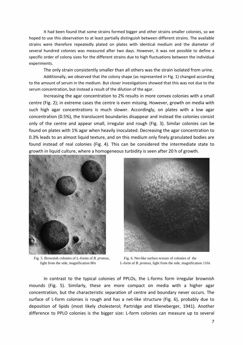
Fig. 5. Brownish colonies of L-forms of B. proteus, light from the side, magnification 80x
In contrast to the typical colonies of PPLOs, the L‐forms form irregular brownish
mounds (Fig. 5). Similarly, these are more compact on media with a higher agar
concentration, but the characteristic separation of centre and boundary never occurs. The
surface of L‐form colonies is rough and has a net‐like structure (Fig. 6), probably due to
deposition of lipids (most likely cholesterol; Partridge and Klieneberger, 1941). Another
difference to PPLO colonies is the bigger size: L‐form colonies can measure up to several
7
millimetres, thus clearly visible by eye whilst the total of PPLO colonies –apart from the giant
colonies –mainly appears as an opalescent surface layer.
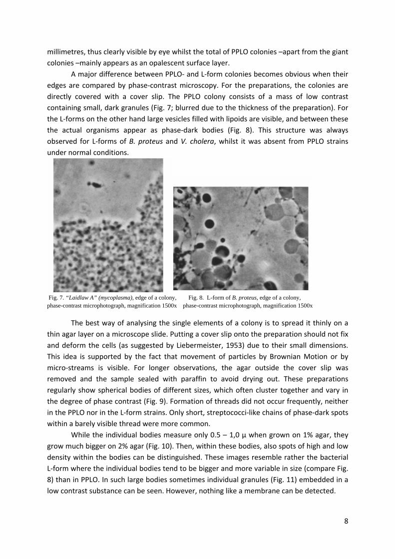
A major difference between PPLO‐ and L‐form colonies becomes obvious when their
edges are compared by phase‐contrast microscopy. For the preparations, the colonies are
directly covered with a cover slip. The PPLO colony consists of a mass of low contrast
containing small, dark granules (Fig. 7; blurred due to the thickness of the preparation). For
the L‐forms on the other hand large vesicles filled with lipoids are visible, and between these
the actual organisms appear as phase‐dark bodies (Fig. 8). This structure was always
observed for L‐forms of B. proteus and V. cholera, whilst it was absent from PPLO strains
under normal conditions.
Fig. 7. “Laidlaw A” (mycoplasma), edge of a colony, Fig. 8. L-form of B. proteus, edge of a colony, phase-contrast microphotograph, magnification 1500x phase-contrast microphotograph, magnification 1500x
The best way of analysing the single elements of a colony is to spread it thinly on a
thin agar layer on a microscope slide. Putting a cover slip onto the preparation should not fix
and deform the cells (as suggested by Liebermeister, 1953) due to their small dimensions.
This idea is supported by the fact that movement of particles by Brownian Motion or by
micro‐streams is visible. For longer observations, the agar outside the cover slip was
removed and the sample sealed with paraffin to avoid drying out. These preparations
regularly show spherical bodies of different sizes, which often cluster together and vary in
the degree of phase contrast (Fig. 9). Formation of threads did not occur frequently, neither
in the PPLO nor in the L‐form strains. Only short, streptococci‐like chains of phase‐dark spots
within a barely visible thread were more common.
While the individual bodies measure only 0.5 – 1,0 µ when grown on 1% agar, they
grow much bigger on 2% agar (Fig. 10). Then, within these bodies, also spots of high and low
density within the bodies can be distinguished. These images resemble rather the bacterial
L‐form where the individual bodies tend to be bigger and more variable in size (compare Fig.
8) than in PPLO. In such large bodies sometimes individual granules (Fig. 11) embedded in a
low contrast substance can be seen. However, nothing like a membrane can be detected.
8

Fig. 9. “Laidlaw A” (mycoplasma) on 1% agar, phase-contrast Fig. 10. “Laidlaw A” on 2% agar, phase-contrast microphotograph, magnification 1500x microphotograph, magnification 1500x
Fig. 11. L-form of B. proteus, phase-contrast Fig. 12. “Findlay” (mycoplasma), 2 days on 1% agar, microphotograph, magnification 3400x Giemsa preparation, magnification 1400x
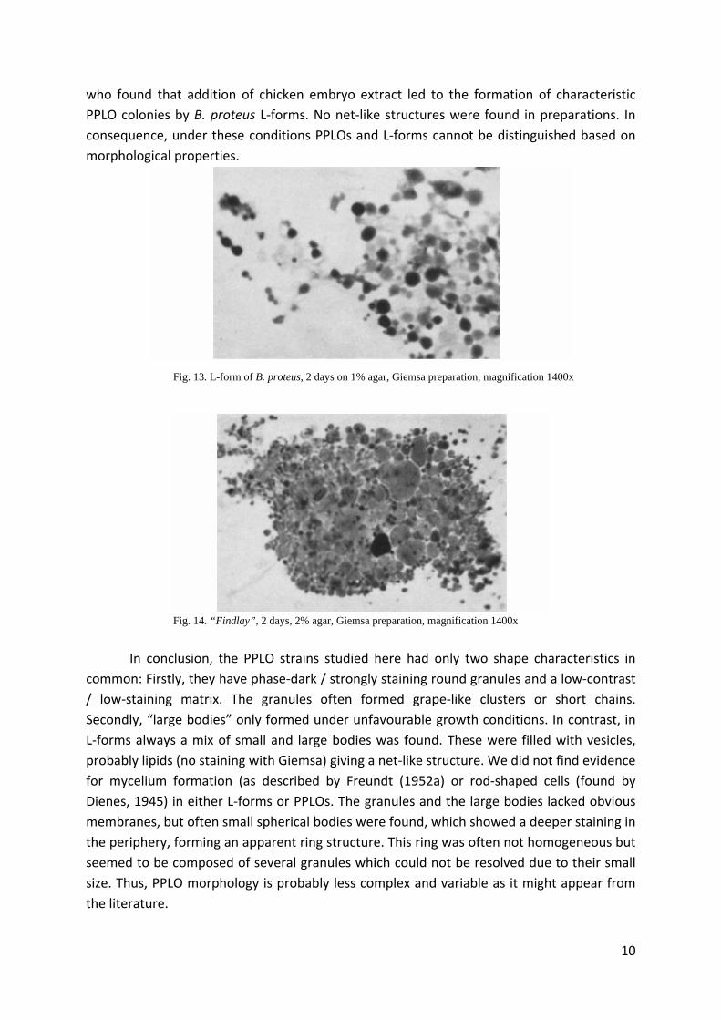
Similar results were obtained with chromic acid‐fixed and Giemsa‐stained
preparations: Similarly, cultivation on 1% agar gives rise to small spherical particles which
are sometimes arranged in chains (Fig. 12). Here, a young colony shows nicely the small
granulation within the barely stained matrix (fuzzy due to technical limitations). It is obvious
that the strongly stained areas are equivalent to the phase‐dark regions seen by phase‐
contrast microscopy. Fig. 13 shows the L‐forms grown under the same conditions. The
individual bodies tend to be bigger, but the size is highly variable.
Also PPLO‐strains can grow larger on 2% agar (Fig. 14) and form so‐called “large
bodies”. The same effect was observed when 3% maltose or 2% glucose were added to 1%
agar. Addition of 1% sugar was not enough to lead to the formation of “large bodies”. These
results show that the “large bodies” claimed to be important by Klieneberger and Smiles
(1942) were nothing but abnormal developmental stages which occur during unfavourable
growth conditions and which are comparable to bacterial involution‐forms. If the conditions
become favourable, they are able to propagate. If not, the cells often die by vacuolisation.
However, L‐forms usually show the large bodies, independent of the medium, which could
also be interpreted as a form of involution. It is possible that the media used are not ideal for
L‐forms and that changes of the conditions might lead to the complete convergence of L‐
forms and PPLOs. This is supported by observations by Seiffert (personal communication),
9
who found that addition of chicken embryo extract led to the formation of characteristic
PPLO colonies by B. proteus L‐forms. No net‐like structures were found in preparations. In
consequence, under these conditions PPLOs and L‐forms cannot be distinguished based on
morphological properties.
Fig. 13. L-form of B. proteus, 2 days on 1% agar, Giemsa preparation, magnification 1400x
Fig. 14. “Findlay”, 2 days, 2% agar, Giemsa preparation, magnification 1400x
In conclusion, the PPLO strains studied here had only two shape characteristics in
common: Firstly, they have phase‐dark / strongly staining round granules and a low‐contrast
/ low‐staining matrix. The granules often formed grape‐like clusters or short chains.
Secondly, “large bodies” only formed under unfavourable growth conditions. In contrast, in
L‐forms always a mix of small and large bodies was found. These were filled with vesicles,
probably lipids (no staining with Giemsa) giving a net‐like structure. We did not find evidence
for mycelium formation (as described by Freundt (1952a) or rod‐shaped cells (found by
Dienes, 1945) in either L‐forms or PPLOs. The granules and the large bodies lacked obvious
membranes, but often small spherical bodies were found, which showed a deeper staining in
the periphery, forming an apparent ring structure. This ring was often not homogeneous but
seemed to be composed of several granules which could not be resolved due to their small
size. Thus, PPLO morphology is probably less complex and variable as it might appear from
the literature.
10
Mode of proliferation of PPLOs and L‐forms
Studying the mode of growth was likely to result in insights about how grape‐like cell
clusters and chains were formed.
Micro‐cultures were prepared as described above. The small size of the object made the
photographic image acquisition difficult, therefore only cultures grown on high‐percentage agar were
used because the bodies became larger under these conditions. Images were taken every 20‐30 min,
and photographs showing a recognizable growth were selected. The following figures therefore
represent one and the same body during development. The objects were continually monitored by
in‐between acquisition of photographs, which allowed often a clearer view than technically possible
by the camera. If required, sketches of the developmental stages of interest are shown in addition to
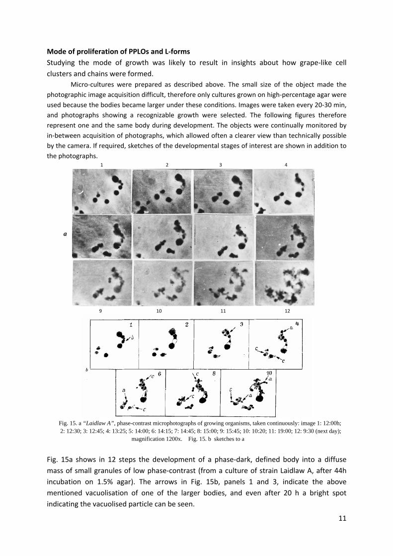
the photographs. 1 2 3 4
a
9 10 11 12
Fig. 15. a “Laidlaw A”, phase-contrast microphotographs of growing organisms, taken continuously: image 1: 12:00h; 2: 12:30; 3: 12:45; 4: 13:25; 5: 14:00; 6: 14:15; 7: 14:45; 8: 15:00; 9: 15:45; 10: 10:20; 11: 19:00; 12: 9:30 (next day); magnification 1200x. Fig. 15. b sketches to a
Fig. 15a shows in 12 steps the development of a phase‐dark, defined body into a diffuse
mass of small granules of low phase‐contrast (from a culture of strain Laidlaw A, after 44h
incubation on 1.5% agar). The arrows in Fig. 15b, panels 1 and 3, indicate the above
mentioned vacuolisation of one of the larger bodies, and even after 20 h a bright spot
indicating the vacuolised particle can be seen.
11
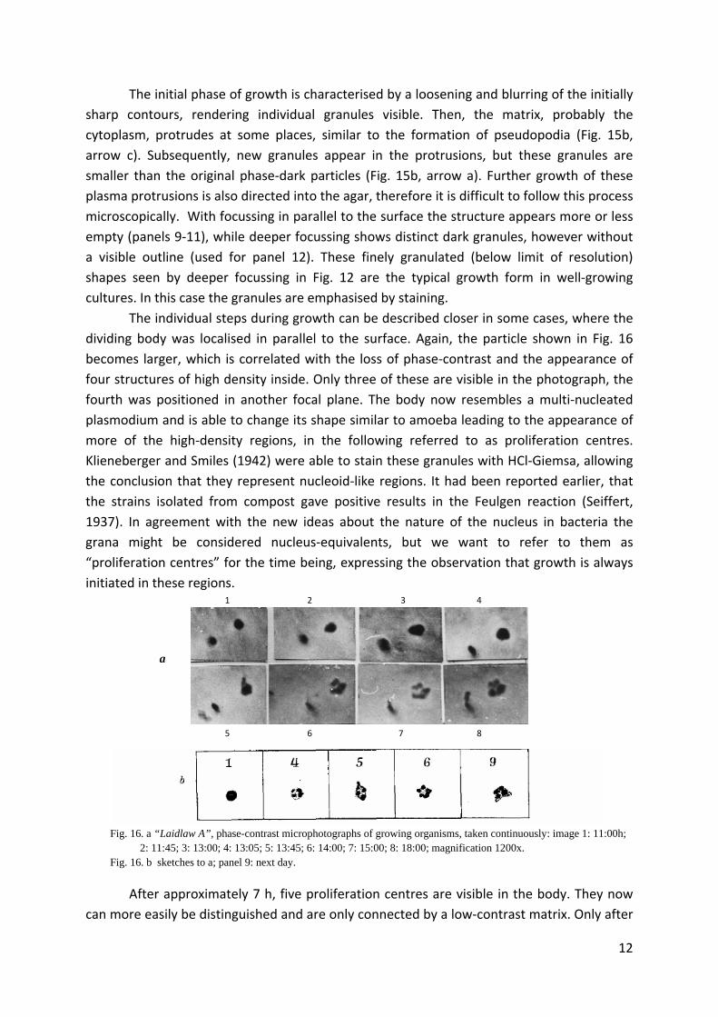
The initial phase of growth is characterised by a loosening and blurring of the initially
sharp contours, rendering individual granules visible. Then, the matrix, probably the
cytoplasm, protrudes at some places, similar to the formation of pseudopodia (Fig. 15b,
arrow c). Subsequently, new granules appear in the protrusions, but these granules are
smaller than the original phase‐dark particles (Fig. 15b, arrow a). Further growth of these
plasma protrusions is also directed into the agar, therefore it is difficult to follow this process
microscopically. With focussing in parallel to the surface the structure appears more or less
empty (panels 9‐11), while deeper focussing shows distinct dark granules, however without
a visible outline (used for panel 12). These finely granulated (below limit of resolution)
shapes seen by deeper focussing in Fig. 12 are the typical growth form in well‐growing
cultures. In this case the granules are emphasised by staining.
The individual steps during growth can be described closer in some cases, where the
dividing body was localised in parallel to the surface. Again, the particle shown in Fig. 16
becomes larger, which is correlated with the loss of phase‐contrast and the appearance of
four structures of high density inside. Only three of these are visible in the photograph, the
fourth was positioned in another focal plane. The body now resembles a multi‐nucleated
plasmodium and is able to change its shape similar to amoeba leading to the appearance of
more of the high‐density regions, in the following referred to as proliferation centres.
Klieneberger and Smiles (1942) were able to stain these granules with HCl‐Giemsa, allowing
the conclusion that they represent nucleoid‐like regions. It had been reported earlier, that
the strains isolated from compost gave positive results in the Feulgen reaction (Seiffert,
1937). In agreement with the new ideas about the nature of the nucleus in bacteria the
grana might be considered nucleus‐equivalents, but we want to refer to them as
“proliferation centres” for the time being, expressing the observation that growth is always
initiated in these regions. 1 2 3 4
a
5 6 7 8
Fig. 16. a “Laidlaw A”, phase-contrast microphotographs of growing organisms, taken continuously: image 1: 11:00h; 2: 11:45; 3: 13:00; 4: 13:05; 5: 13:45; 6: 14:00; 7: 15:00; 8: 18:00; magnification 1200x. Fig. 16. b sketches to a; panel 9: next day.
After approximately 7 h, five proliferation centres are visible in the body. They now
can more easily be distinguished and are only connected by a low‐contrast matrix. Only after
12

30 h, some sort of new budding was seen, which, however, could not be captured on
photographs (Fig. 16b, panel 9). There is a clear difference in the growth of the particles
shown in Fig. 15 and Fig. 16 during the first seven hours of growth: the body shown in Fig.
16a has a lower growth rate, and the proliferation centre enlarges significantly. In contrast,
the rapidly growing particles (Fig. 15) show considerably finer inner structures. We will come
back to this phenomenon in various other examples.
The budding of a particle similar to the final stages of the time‐lapse series shown in
Fig. 16 is presented in Fig. 17. Whilst it is difficult to distinguish internal structures, it is easy
to follow the appearance of a plasma thread, within which new proliferation centres form.
The arrow in panel 8 indicates the appearance of a small dot, seemingly unconnected to the
main body, which represents the tip of a thin “pseudopodium”. The thread grows from the
newly formed apical proliferation centre sideways. Similar processes occur on all sides of the
body, and budding at the proliferation centres leads to the above mentioned finely
granulated internal structures. Also a small particle in the lower left corner shows the
characteristic loss of contrast in connection with spreading of the plasma. In this case
however, the proliferation centres moved downwards into the agar and are therefore
undetectable on the later images.
1 2 3 4 5
a
6 7 8 9 10
Fig. 17. a “Laidlaw A”, phase-contrast microphotographs of growing organisms, taken continuously: image 1: 8:00h; 2: 8:30; 3: 9:35; 4: 10:00; 5: 11:00; 6: 11:45; 7: 12:00; 8: 12:20; 9: 14:00; 10: 8:00 (next day); magnification 1200x. Fig. 17. b sketches to a
Inoculation of rapidly growing cultures in the above described state of finely granulated
internal structures results in micro‐cultures shown in Fig. 18. Growth continues by the
development of threads, but no large bodies are formed. These seem to appear only under
13
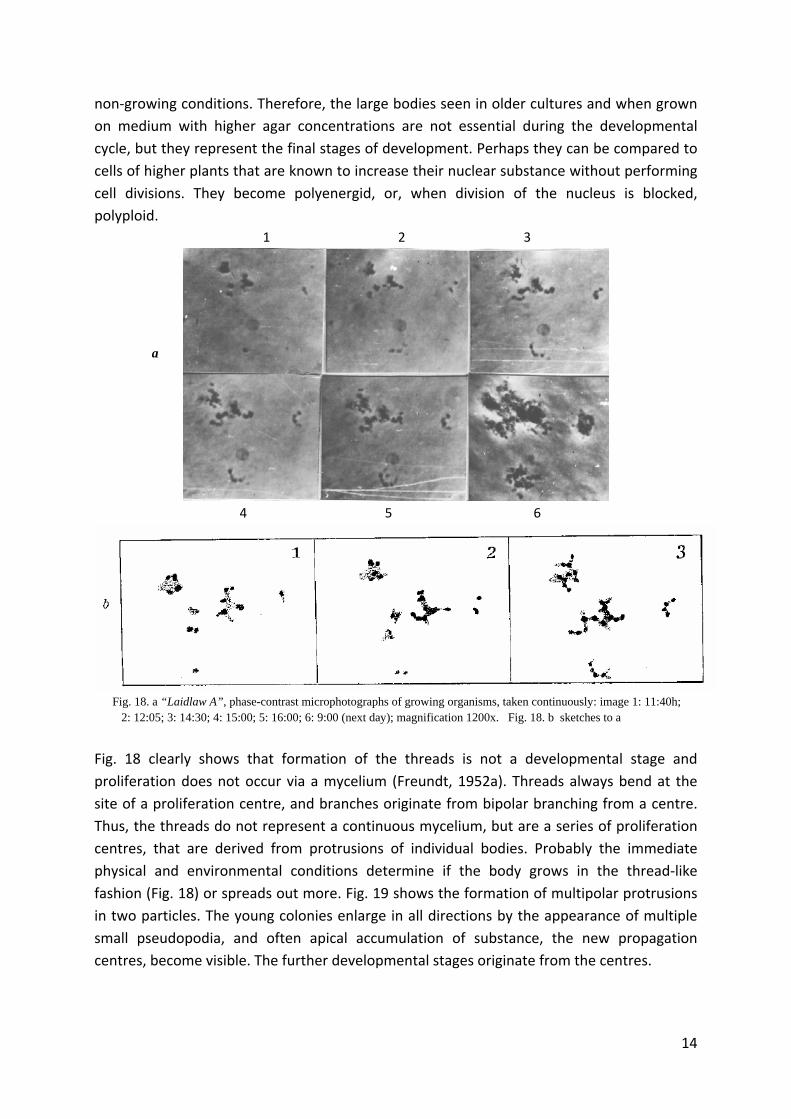
non‐growing conditions. Therefore, the large bodies seen in older cultures and when grown
on medium with higher agar concentrations are not essential during the developmental
cycle, but they represent the final stages of development. Perhaps they can be compared to
cells of higher plants that are known to increase their nuclear substance without performing
cell divisions. They become polyenergid, or, when division of the nucleus is blocked,
polyploid.
1 2 3
a
4 5 6
Fig. 18. a “Laidlaw A”, phase-contrast microphotographs of growing organisms, taken continuously: image 1: 11:40h; 2: 12:05; 3: 14:30; 4: 15:00; 5: 16:00; 6: 9:00 (next day); magnification 1200x. Fig. 18. b sketches to a
Fig. 18 clearly shows that formation of the threads is not a developmental stage and
proliferation does not occur via a mycelium (Freundt, 1952a). Threads always bend at the
site of a proliferation centre, and branches originate from bipolar branching from a centre.
Thus, the threads do not represent a continuous mycelium, but are a series of proliferation
centres, that are derived from protrusions of individual bodies. Probably the immediate
physical and environmental conditions determine if the body grows in the thread‐like
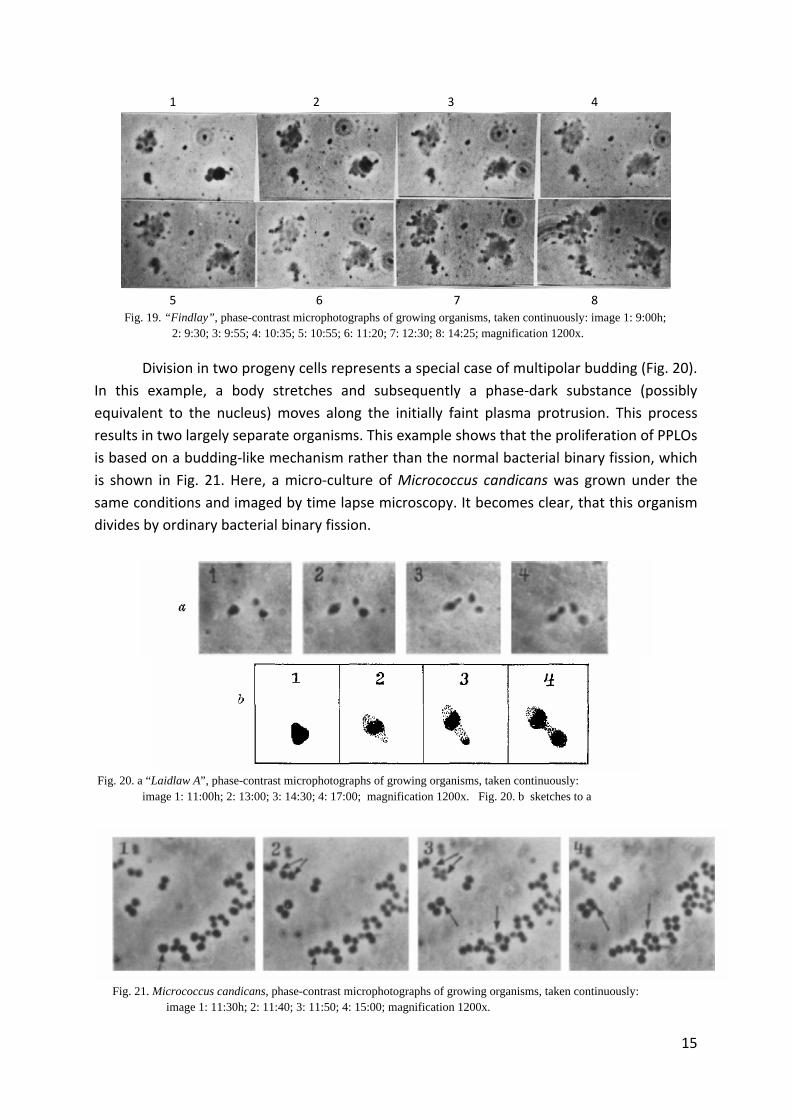
fashion (Fig. 18) or spreads out more. Fig. 19 shows the formation of multipolar protrusions
in two particles. The young colonies enlarge in all directions by the appearance of multiple
small pseudopodia, and often apical accumulation of substance, the new propagation
centres, become visible. The further developmental stages originate from the centres.
14
1 2 3 4
5 6 7 8
Fig. 19. “Findlay”, phase-contrast microphotographs of growing organisms, taken continuously: image 1: 9:00h; 2: 9:30; 3: 9:55; 4: 10:35; 5: 10:55; 6: 11:20; 7: 12:30; 8: 14:25; magnification 1200x.
Division in two progeny cells represents a special case of multipolar budding (Fig. 20).
In this example, a body stretches and subsequently a phase‐dark substance (possibly
equivalent to the nucleus) moves along the initially faint plasma protrusion. This process
results in two largely separate organisms. This example shows that the proliferation of PPLOs
is based on a budding‐like mechanism rather than the normal bacterial binary fission, which
is shown in Fig. 21. Here, a micro‐culture of Micrococcus candicans was grown under the
same conditions and imaged by time lapse microscopy. It becomes clear, that this organism
divides by ordinary bacterial binary fission.
Fig. 20. a “Laidlaw A”, phase-contrast microphotographs of growing organisms, taken continuously: image 1: 11:00h; 2: 13:00; 3: 14:30; 4: 17:00; magnification 1200x. Fig. 20. b sketches to a
Fig. 21. Micrococcus candicans, phase-contrast microphotographs of growing organisms, taken continuously: image 1: 11:30h; 2: 11:40; 3: 11:50; 4: 15:00; magnification 1200x.
15
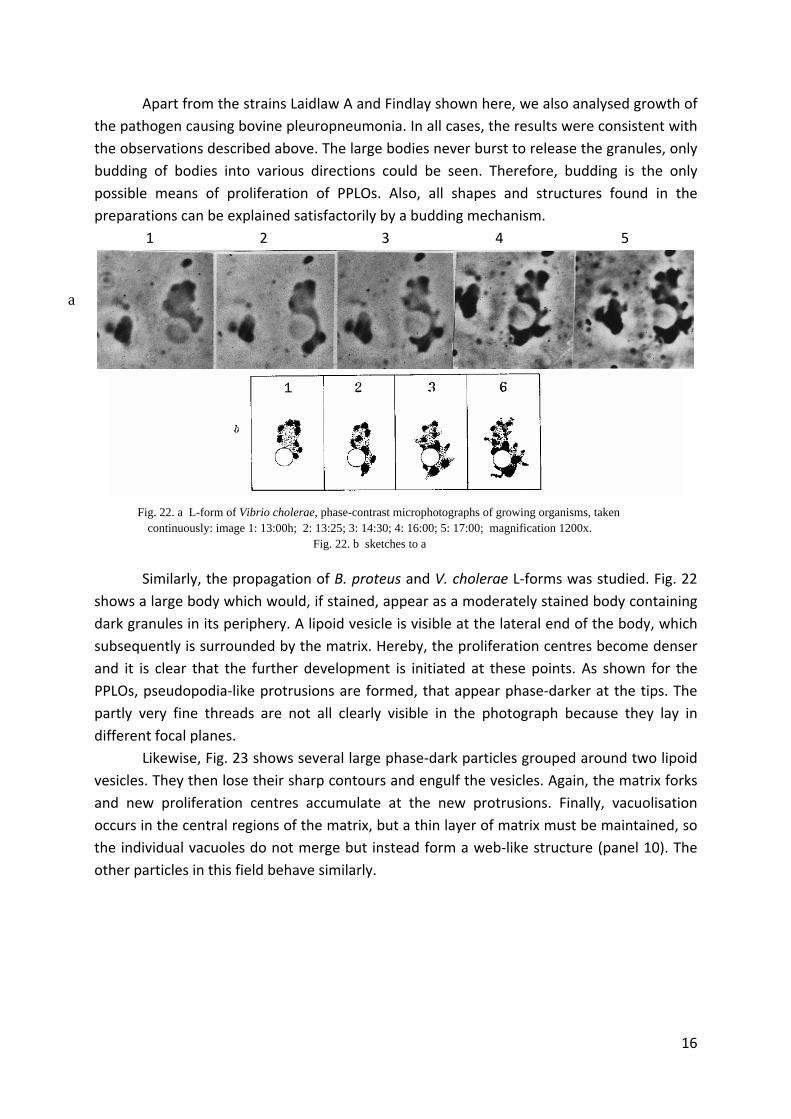
Apart from the strains Laidlaw A and Findlay shown here, we also analysed growth of
the pathogen causing bovine pleuropneumonia. In all cases, the results were consistent with
the observations described above. The large bodies never burst to release the granules, only
budding of bodies into various directions could be seen. Therefore, budding is the only
possible means of proliferation of PPLOs. Also, all shapes and structures found in the
preparations can be explained satisfactorily by a budding mechanism.
1 2 3 4 5
a
Fig. 22. a L-form of Vibrio cholerae, phase-contrast microphotographs of growing organisms, taken continuously: image 1: 13:00h; 2: 13:25; 3: 14:30; 4: 16:00; 5: 17:00; magnification 1200x.
Fig. 22. b sketches to a
Similarly, the propagation of B. proteus and V. cholerae L‐forms was studied. Fig. 22
shows a large body which would, if stained, appear as a moderately stained body containing
dark granules in its periphery. A lipoid vesicle is visible at the lateral end of the body, which
subsequently is surrounded by the matrix. Hereby, the proliferation centres become denser
and it is clear that the further development is initiated at these points. As shown for the
PPLOs, pseudopodia‐like protrusions are formed, that appear phase‐darker at the tips. The
partly very fine threads are not all clearly visible in the photograph because they lay in
different focal planes.
Likewise, Fig. 23 shows several large phase‐dark particles grouped around two lipoid
vesicles. They then lose their sharp contours and engulf the vesicles. Again, the matrix forks
and new proliferation centres accumulate at the new protrusions. Finally, vacuolisation
occurs in the central regions of the matrix, but a thin layer of matrix must be maintained, so
the individual vacuoles do not merge but instead form a web‐like structure (panel 10). The
other particles in this field behave similarly.
16
1 2 3 4 5
6 7 8 9 10
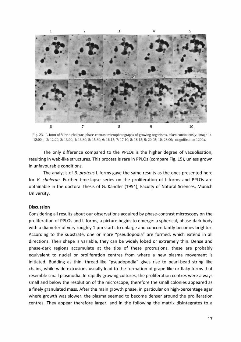
Fig. 23. L-form of Vibrio cholerae, phase-contrast microphotographs of growing organisms, taken continuously: image 1: 12:00h; 2: 12:20; 3: 13:00; 4: 13:30; 5: 15:30; 6: 16:15; 7: 17:10; 8: 18:15; 9: 20:05; 10: 23:00; magnification 1200x.
The only difference compared to the PPLOs is the higher degree of vacuolisation,
resulting in web‐like structures. This process is rare in PPLOs (compare Fig. 15), unless grown
in unfavourable conditions.
The analysis of B. proteus L‐forms gave the same results as the ones presented here
for V. cholerae. Further time‐lapse series on the proliferation of L‐forms and PPLOs are
obtainable in the doctoral thesis of G. Kandler (1954), Faculty of Natural Sciences, Munich
University.
Discussion
Considering all results about our observations acquired by phase‐contrast microscopy on the
proliferation of PPLOs and L‐forms, a picture begins to emerge: a spherical, phase‐dark body
with a diameter of very roughly 1 µm starts to enlarge and concomitantly becomes brighter.
According to the substrate, one or more “pseudopodia” are formed, which extend in all
directions. Their shape is variable, they can be widely lobed or extremely thin. Dense and
phase‐dark regions accumulate at the tips of these protrusions, these are probably
equivalent to nuclei or proliferation centres from where a new plasma movement is
initiated. Budding as thin, thread‐like “pseudopodia” gives rise to pearl‐bead string like
chains, while wide extrusions usually lead to the formation of grape‐like or flaky forms that
resemble small plasmodia. In rapidly growing cultures, the proliferation centres were always
small and below the resolution of the microscope, therefore the small colonies appeared as
a finely granulated mass. After the main growth phase, in particular on high‐percentage agar
where growth was slower, the plasma seemed to become denser around the proliferation
centres. They appear therefore larger, and in the following the matrix disintegrates to a
17
18
higher or lower extent in‐between them. Inoculating from this stage results in the roughly 1
µm sized particles we used as the starting point for our proliferation studies.
A detailed understanding of the structure of the particles is not possible by light
microscopic analyses. Also the smallest reproductive units found after the filtration
experiments mentioned at the beginning are with 0.15 µm diameter below the resolution of
the light microscope. Therefore, an electron microscopic approach was required to analyse
these details. The present light microscopic data show that the proliferation cycles of PPLOs
and L‐forms are essentially identical. They propagate by a uni‐ or multi‐polar budding
mechanism resulting in the variety of shapes found in stained preparations. The formation of
large bodies with relatively big granules can be considered an inhibited type of budding,
equivalent to bacterial involution forms.
The grape‐like associations discovered here are highly reminiscent of what Herzberg
(1936; 1953) found when studying the proliferation of large viruses. Additionally, our
observations resemble the proliferation stages during bronchial‐pneumonia in mice, where
also darkly stained elemental bodies could be distinguished from a weakly stained matrix
(Gönnert, 1953). The proliferation centres we observed would then be equivalent to the
elemental bodies of the large viruses, and the contents of an inclusion body would
correspond to a colony. The breakdown into multiple subunits, as assumed for the viral and
also the PPLO proliferation (Gerber, 1953), would then have to be considered the equivalent
to the disintegration of the associations in the grape‐like associations that had formed by
budding. The only difference between large viruses and PPLOs would be, that the former
have lost the ability of independent life and completely depend on a host cell. The PPLOs in
contrast are still capable of the saprophytic life style. This view has repeatedly been
discussed as the most likely option by several groups (Edward, 1954; Dienes, 1945; Gönnert,
1952) and is supported by recent results on “incomplete viruses” (Schäfer et al., 1954).
The question about the relationship between PPLOs and L‐forms can not be
answered by the present study on their proliferation. Both groups propagate in a similar way
by a budding mechanism, which does not rule out either the possibility that PPLOs are a
separate phylogenetic group or that PPLOs are only stable L‐form bacteria. Because due to
their small size it is unlikely to discover significant morphological differences, it seems more
promising to focus on physiological studies of both groups. Work in this direction is in
progress in our institute, and preliminary results hint to fundamental metabolic differences
between L‐forms and PPLOs.
Summary
Light microscopic studies of PPLOs and L‐forms of B. proteus and V. cholerae showed the
presence of 0.5 – 1 µm big spherical bodies that often assembled to grape‐like structures or
chains. In contrast to PPLOs, which are regularly sized, the L‐forms often occur in large
bodies of several µm diameter and frequently show vacuolisations. Bodies of a comparable
size were found in PPLOs only under unfavourable growth conditions. Also colony
morphology of PPLOs varied with the media and agar concentration.
Proliferation of growing PPLOs and L‐forms was analysed by phase‐contrast
microscopy in a time series. The only way of proliferation observed was a uni‐ or multi‐polar
budding process. Often, the particles were arranged in chains or threads, however, these can
not be considered a mycelium, as previously suggested by various authors.
We emphasize the high degree of similarity between PPLOs and large viruses.
References:
19