Embed Size (px)
Citation preview
www.sciencemag.org/cgi/content/full/science.aaf5036/DC1
Supplementary Materials for
Zika virus in the Americas: Early epidemiological and genetic findings
Nuno Rodrigues Faria, Raimunda do Socorro da Silva Azevedo, Moritz U. G. Kraemer,
Renato Souza, Mariana Sequetin Cunha, Sarah C. Hill, Julien Thézé, Michael B. Bonsall,
Thomas A. Bowden, Ilona Rissanen, Iray Maria Rocco, Juliana Silva Nogueira, Adriana
Yurika Maeda, Fernanda Giseli da Silva Vasami, Fernando Luiz de Lima Macedo,
Akemi Suzuki, Sueli Guerreiro Rodrigues, Ana Cecilia Ribeiro Cruz, Bruno Tardeli
Nunes, Daniele Barbosa de Almeida Medeiros, Daniela Sueli Guerreiro Rodrigues, Alice
Louize Nunes Queiroz, Eliana Vieira Pinto da Silva, Daniele Freitas Henriques, Elisabeth
Salbe Travassos da Rosa, Consuelo Silva de Oliveira, Livia Caricio Martins, Helena
Baldez Vasconcelos, Livia Medeiros Neves Casseb, Darlene de Brito Simith, Jane P.
Messina, Leandro Abade, José Lourenço, Luiz Carlos Junior Alcantara, Maricélia Maia
de Lima, Marta Giovanetti, Simon I. Hay, Rodrigo Santos de Oliveira, Poliana da Silva
Lemos, Layanna Freitas de Oliveira, Clayton Pereira Silva de Lima, Sandro Patroca da
Silva, Janaina Mota de Vasconcelos, Luciano Franco, Jedson Ferreira Cardoso, João
Lídio da Silva Gonçalves Vianez-Júnior, Daiana Mir, Gonzalo Bello, Edson Delatorre,
Kamran Khan, Marisa Creatore, Giovanini Evelim Coelho, Wanderson Kleber de
Oliveira, Robert Tesh, Oliver G. Pybus,* Marcio R. T. Nunes,* Pedro F. C. Vasconcelos*
*Correspondening author. E-mail: [email protected] (O.G.P.); [email protected]
(M.R.T.N.); [email protected] (P.F.C.V.)
Published 24 March 2016 on Science First Release
DOI: 10.1126/science.aaf5036
This PDF file includes:
Materials and Methods
Supplementary Text
Figs. S1 to S8
Tables S1 to S5
Full Reference List

2
This PDF file includes:
Section 1. Materials and Methods 3
Supplementary Tables S1 and S2
Supplementary Fig. S1
Section 2. Patient data associated with new ZIKV genetic sequences 9
Section 3. Maximum likelihood phylogenetics and root-to-tip analyses 10
Supplementary Figs. S2 – S5
Section 4. Evolutionary rate estimates under different models 14
Supplementary Table S3
Section 5. Dating estimates under different models 15
Supplementary Tables S4 and S5
Section 6. Number of passengers flying to Brazil per country (2012 to 2014) 17
Supplementary Fig. S6
Section 7. Mapping of variant sites onto existing protein structures 18
Supplementary Fig. S7 and S8
Other Supplementary Materials for this manuscript includes the following:
Nucleotide alignments, BEAST XML files, epidemiological data available from Dryad, at
DOI: 10.5061/dryad.6kn23
3
Section 1. Materials and Methods
1.1. Patients, samples and diagnostic confirmation
We tested blood and serum samples of (i) four symptomatic patients with suspected ZIKV
infection, (ii) blood from one blood donor, and (iii) viscera from two subjects who died due to
complications that were hypothesised to have been associated with ZIKV infection. Demographic
data including patient age, gender, municipality, brief case description, date of onset of symptoms,
date of sample collection, date of isolation, municipality of residence, isolate passage history, and
diagnostic results are provided in Section 2. Samples were sent to the Evandro Chagas Institute as
part of the National Program for Dengue, Chikungunya, Zika and Yellow Fever Control,
Department of Surveillance in Health, Brazilian Ministry of Health, for differential diagnostic by
IgM ELISA, real-time PCR, and C6/36 cell culture for DENV, ZIKV and CHIKV. All samples
were obtained from persons visiting local clinics or hospitalized by the Ministry of Health
personnel as part of dengue, Chikungunya, and Zika fever surveillance activities. In these cases,
patient consent is oral and not recorded. The study was authorized by the Coordination of the
National Program for Dengue, Chikungunya and Zika Control coordinated by Brazil’s Ministry of
Health.
1.2. Virus isolation, RNA and IgM detection
Virus isolation attempts were performed in monolayer cell cultures of Aedes albopictus C6/36 (46),
harvested between days 6 to 10 after infection and tested for evident positive reaction in an
immunofluorescence assay using a polyclonal antibody against ZIKV antigen as previously
described (47). Suspensions of confirmed infected cell supernatants were first centrifuged and then
treated with 50% polyethylene glycol 8000 and 23% NaCl for precipitation of viral particles. After
centrifugation, virus pellets were eluted in 250 uL RNase-free water and used for RNA extraction
using an initial step of virus particle lysis followed by RNA isolation using the Qiamp Viral RNA
Minikit (Qiagen) according to the manufacturer’s instructions. Detection of ZIKV genomes was
performed using the RT-qPCR method as described in (3, 48).
1.3. Genome sequencing
Each ZIKV genome was obtained using the Ion PGMTM
Next-Generation Sequencing (Life
Technologies) and the GS-FLX/GS-Junior 454 pyrosequencing (Roche, Life Sciences) platforms.
For samples BeH818995, BeH819015, BeH819966 and BeH815744, amplicon-based deep
sequencing was used to close sequencing gaps. Regardless of the method, cDNA synthesis was
performed using random amplification, followed by an emulsion PCR and sequencing steps as
previously described (49). Raw reads were combined and assembled using Mira v4.0 (50) using the
KJ776791 genome as the reference strain. Sequences were inspected for quality and coverage in
Geneious v.7 (51). Coverage was low for isolate SPH2015 but was sufficient to generate a
consensus genome for molecular epidemiological analysis. This isolate was independently
sequenced in a second lab (52); coverage was not improved but the same sequence was replicated
(KU321639). Further sequencing details are provided in Table S1 below. The new ZIKV genomes
reported in this study are deposited in GenBank under the accession numbers KU321639,
KU365777-KU365780, KU729217 and KU729218.
4
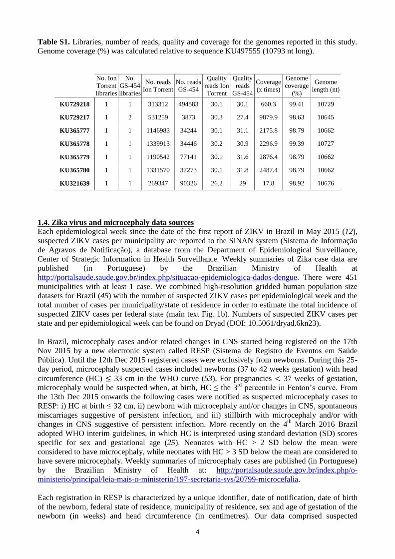
Table S1. Libraries, number of reads, quality and coverage for the genomes reported in this study.
Genome coverage (%) was calculated relative to sequence KU497555 (10793 nt long).
No. Ion
Torrent
libraries
No.
GS-454
libraries
No. reads
Ion Torrent
No. reads
GS-454
Quality
reads Ion
Torrent
Quality
reads
GS-454
Coverage
(x times)
Genome
coverage
(%)
Genome
length (nt)
KU729218 1 1 313312 494583 30.1 30.1 660.3 99.41 10729
KU729217 1 2 531259 3873 30.3 27.4 9879.9 98.63 10645
KU365777 1 1 1146983 34244 30.1 31.1 2175.8 98.79 10662
KU365778 1 1 1339913 34446 30.2 30.9 2296.9 99.39 10727
KU365779 1 1 1190542 77141 30.1 31.6 2876.4 98.79 10662
KU365780 1 1 1331570 37273 30.1 31.8 2487.4 98.79 10662
KU321639 1 1 269347 90326 26.2 29 17.8 98.92 10676
1.4. Zika virus and microcephaly data sources
Each epidemiological week since the date of the first report of ZIKV in Brazil in May 2015 (12),
suspected ZIKV cases per municipality are reported to the SINAN system (Sistema de Informação
de Agravos de Notificação), a database from the Department of Epidemiological Surveillance,
Center of Strategic Information in Health Surveillance. Weekly summaries of Zika case data are
published (in Portuguese) by the Brazilian Ministry of Health at
http://portalsaude.saude.gov.br/index.php/situacao-epidemiologica-dados-dengue. There were 451
municipalities with at least 1 case. We combined high-resolution gridded human population size
datasets for Brazil (45) with the number of suspected ZIKV cases per epidemiological week and the
total number of cases per municipality/state of residence in order to estimate the total incidence of
suspected ZIKV cases per federal state (main text Fig. 1b). Numbers of suspected ZIKV cases per
state and per epidemiological week can be found on Dryad (DOI: 10.5061/dryad.6kn23).
In Brazil, microcephaly cases and/or related changes in CNS started being registered on the 17th
Nov 2015 by a new electronic system called RESP (Sistema de Registro de Eventos em Saúde
Pública). Until the 12th Dec 2015 registered cases were exclusively from newborns. During this 25-
day period, microcephaly suspected cases included newborns (37 to 42 weeks gestation) with head
circumference (HC) ≤ 33 cm in the WHO curve (53). For pregnancies < 37 weeks of gestation,
microcephaly would be suspected when, at birth, HC ≤ the 3rd
percentile in Fenton’s curve. From
the 13th Dec 2015 onwards the following cases were notified as suspected microcephaly cases to
RESP: i) HC at birth ≤ 32 cm, ii) newborn with microcephaly and/or changes in CNS, spontaneous
miscarriages suggestive of persistent infection, and iii) stillbirth with microcephaly and/or with
changes in CNS suggestive of persistent infection. More recently on the 4th
March 2016 Brazil
adopted WHO interim guidelines, in which HC is interpreted using standard deviation (SD) scores
specific for sex and gestational age (25). Neonates with HC > 2 SD below the mean were
considered to have microcephaly, while neonates with HC > 3 SD below the mean are considered to
have severe microcephaly. Weekly summaries of microcephaly cases are published (in Portuguese)
by the Brazilian Ministry of Health at: http://portalsaude.saude.gov.br/index.php/o-
ministerio/principal/leia-mais-o-ministerio/197-secretaria-svs/20799-microcefalia.
Each registration in RESP is characterized by a unique identifier, date of notification, date of birth
of the newborn, federal state of residence, municipality of residence, sex and age of gestation of the
newborn (in weeks) and head circumference (in centimetres). Our data comprised suspected
5
microcephaly cases recorded up to the 9th
January 2016. In order to avoid the impact of different
reporting guidelines over time, we analysed RESP registrations using the most recent guidelines
from WHO, outlined above (25). This resulted in 1118 microcephaly cases, of which 605 (54%)
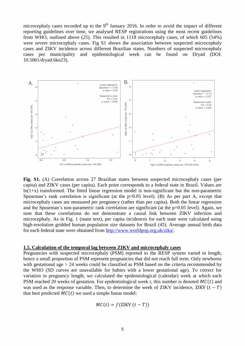
were severe microcephaly cases. Fig S1 shows the association between suspected microcephaly
cases and ZIKV incidence across different Brazilian states. Numbers of suspected microcephaly
cases per municipality and epidemiological week can be found on Dryad (DOI:
10.5061/dryad.6kn23).
Fig. S1. (A) Correlation across 27 Brazilian states between suspected microcephaly cases (per
capita) and ZIKV cases (per capita). Each point corresponds to a federal state in Brazil. Values are
ln(1+x) transformed. The fitted linear regression model is non-significant but the non-parametric
Spearman’s rank correlation is significant (at the p=0.05 level). (B) As per part A, except that
microcephaly cases are measured per pregnancy (rather than per capita). Both the linear regression
and the Spearman’s non-parametric rank correlation are significant (at the p=0.05 level). Again, we
note that these correlations do not demonstrate a causal link between ZIKV infection and
microcephaly. As in Fig. 1 (main text), per capita incidences for each state were calculated using
high-resolution gridded human population size datasets for Brazil (45). Average annual birth data
for each federal state were obtained from http://www.worldpop.org.uk/zika/.
1.5. Calculation of the temporal lag between ZIKV and microcephaly cases
Pregnancies with suspected microcephaly (PSM) reported to the RESP system varied in length,
hence a small proportion of PSM represent pregnancies that did not reach full term. Only newborns
with gestational age > 24 weeks could be classified as PSM based on the criteria recommended by
the WHO (SD curves are unavailable for babies with a lower gestational age). To correct for
variation in pregnancy length, we calculated the epidemiological (calendar) week at which each
PSM reached 20 weeks of gestation. For epidemiological week t, this number is denoted 𝑀𝐶(𝑡) and
was used as the response variable. Then, to determine the week of ZIKV incidence, ZIKV (𝑡 − 𝑇)
that best predicted 𝑀𝐶(𝑡) we used a simple linear model:
𝑀𝐶(𝑡) = 𝑓(ZIKV (𝑡 − 𝑇))
6
We fitted this model across all epidemiological weeks using maximum likelihood with a Gaussian
error distribution, which enables estimation of the time lag parameter T. In short, this approach
finds the time lag T that provides the best correlation between reported PSM cases and ZIKV
incidence.
When the model was applied to all PSM cases (n=1118), we found that the best fitting temporal lag
between ZIKV incidence and PSM corresponded to week 17 of pregnancy on average (95%
confidence interval, CI, of mean = +/-0.11 weeks). When applied to severe microcephaly cases
(n=605) the result was week 14 of pregnancy (95% CI of mean = +/-0.08 weeks). In addition, for
those municipalities with >100 reported ZIKV cases and >1 microcephaly case (n=16), we also
fitted the model using ZIKV incidence specific to each location. Under this model the best fitting
temporal lag between ZIKV incidence and PSM cases corresponded to week 16 of pregnancy (95%
CI of mean = +/-1.58 weeks). R scripts and datasets for both analyses are available on Dryad (DOI:
10.5061/dryad.6kn23).
1.6. Phylogenetic analyses
All published near-complete ZIKV genomes as well longer sub-genomic regions and partial E and
NS5 gene sequences were retrieved from Genbank on the 7th
of March 2016 (54) through a
graphical user interface for ACNUC databases (55). Sequences were aligned using MAFFT (56)
and manually curated using MR_766 as a guiding sequence. Maximum likelihood (ML)
phylogenies were reconstructed using the heuristic tree search algorithm implemented in PhyML
(57). ML bootstrapping was performed with 100 replicates to assess the robustness of tree
topologies. We used the general time reversible (GTR) nucleotide substitution model with a
proportion of invariant sites, which was identified as the best fitting model for ML inference by
jModelTest v.1.6 (58). Trees were mid-point rooted.
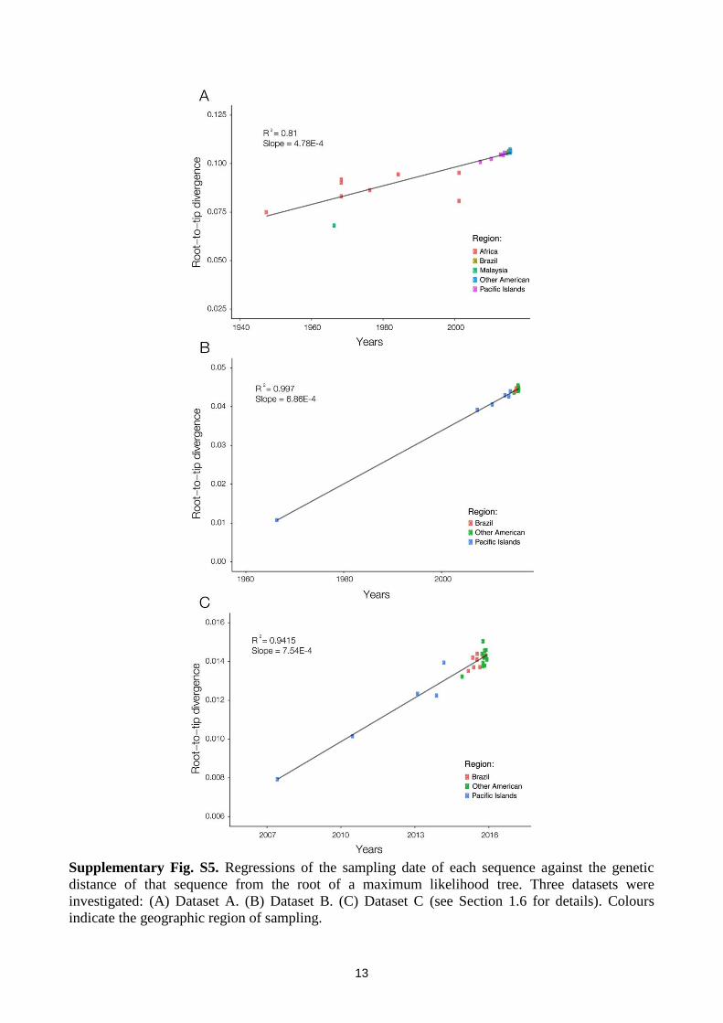
Next, in order to determine temporal signal, analyses of the correlation between root-to-tip genetic
divergence and date of sampling were conducted in Path-O-Gen v.1.4 (59). We analysed 3 datasets
(A-C) that contained published sequences, as well as unpublished sequences available in Genbank
for which we were able to obtain permission from sequence depositors: (A) complete coding
sequences (cds) and long partial cds (>2380bp) from both African and Asian genotypes (n=34,
sampling range: 1947 to Dec 2015). (B) Complete coding region and long partial cds (>2380bp)
sequences from the Asian genotype (n=26, sampling range: 1966 to Dec 2015), (C) complete
coding region and long partial cds (>2380bp) sequences from recent (2007 onwards) Asian
genotype samples (n=25, sampling range: 2007 to Dec 2015). For each dataset, Fig S5 shows a
regression of the sampling date of each sequence against the genetic distance of that sequence to the
root of a maximum likelihood tree. Dataset A is less likely to represent the evolution of the current
outbreak lineage because (i) it contains pre-1970s isolates that may be unsuitable for molecular
clock analysis (e.g. HQ234498 has been passaged 147 times in 2 different cell lines), (ii) there may
be differences between the evolutionary dynamics of endemic and epidemic ZIKV, as previously
proposed for Chikungunya virus (60), and (iii) it may contain recombinant sequences (EC Holmes,
pers. comm.). Indeed, evidence of recombination using the phi-test in Splitstree (61) was found for
dataset A (p-value < 0.0001) but not for the other datasets (p-values > 0.5). Therefore subsequent
molecular clock phylogenetic analyses were conducted using all complete and partial coding region
sequences belonging to the Asian genotype. Full sequence details and alignments can be found in
Dryad (DOI: 10.5061/dryad.6kn23).
Bayesian phylogenies were estimated in BEASTv.1.8.2 (62) using strict and relaxed uncorrelated
molecular clock (UCLN) models (63) under the GTR+I nucleotide substitution model (64). To
account for uncertainty in the day of sampling, precision values of 1 year and 1 month were given
to sequences for which sampling dates were known only to the nearest year or month, respectively.
7
A non-informative CTMC reference prior (65) was used for the molecular clock rate. Equivalent
results were obtained using a gamma prior distribution (shape parameter = 0.001; scale parameter =
1000) and a unit uniform prior distribution. Multiple combinations of molecular clock and
coalescent models were explored, and to select the best fitting model we used path-sampling and
stepping-stone model selection approaches. The results of this model selection procedure are shown
in Table S2. We estimated the time of the most common recent ancestor (TMRCA) of a clade
containing isolates sampled in the Americas (clade B), as well as the clade containing both the
current outbreak and the 2013 French Polynesia outbreak (clade A). Bayesian MCMC analyses
were run for 50 million steps using a PhyML tree (57) as a starting tree and MCMC convergence
was explored using Tracer. All runs had an effective sample size >1000. Maximum clade credibility
(MCC) trees were generated using TreeAnnotator after discarding 10% as burn-in, and summary
phylogenies were visualised in FigTree v.1.4.2 (59). XML files for the BEAST analyses can be
found in Dryad (DOI: 10.5061/dryad.6kn23).
Table S2. Log marginal likelihood estimates for different molecular clock and coalescent model
combinations. The best fitting model combination is ranked 1 (bold) and the worst is ranked 6.
Coalescent models used were the parametric Constant and Exponential models and the non-
parametric Bayesian skyline (BSKY) model. The molecular clock models used were the strict
molecular clock (Strict) and the uncorrelated relaxed lognormal molecular clock (UCLN). PS =
path sampling model selection, SS = stepping-stone model selection.
Model Combination PS Ranking SS Ranking
Strict, Constant -19558.064 5 -19558.311 5
Strict, Exponential -19556.921 2 -19557.077 4
Strict, BSKY -19554.282 1 -19554.439 1
UCLN, Constant -19556.989 4 -19557.108 2
UCLN, Exponential -19558.761 6 -19558.881 6
UCLN, BSKY -19556.986 3 -19557.028 3
1.7. Investigation of the Brazilian ZIKV outbreak source
A literature search was undertaken to determine possible sources of the outbreak in the Americas.
Specifically, we identified countries in which ZIKV infections were reported between 2012 and the
end of 2014. Multiple cases were confirmed in Thailand (March 2012 to July 2014) (66), the Cook
Islands (October 2013 to June 2014) (67, 68), French Polynesia (October 2013 to March 2014) (7,
8, 10, 22, 37, 69, 70), Easter Island (Chile) (January to May 2014) (11) and New Caledonia
(November 2013 to August 2014) (67). Single cases were confirmed in the Philippines (May 2012,
no recent travel history) (71) and Haiti (December 2014, no recent travel history; Lednicky JA pers.
comm.). Isolated cases were also diagnosed in tourists who had returned from visits to Indonesia
(2012) (72), Malaysia (August 2014) (73), Tonga and Vanuatu (both during April to June 2014)
(74), though the latter three cases were not confirmed by presence of the virus. It is notable that
autochthonous infections were identified in Thailand retrospectively, following confirmation of a
case in a returning Canadian traveller (66, 75). It is possible that ZIKV infections may have
remained unidentified in other countries in South East Asia and the Pacific during 2012-2014, in
part because symptoms in adults are generally mild and/or lack of routine testing.
1.8. Monthly airline passenger numbers to Brazil
Patterns of passengers arriving in Brazil via commercial airlines were analysed to identify the
global origins of travellers from countries where ZIKV has been reported in the recent past.
Specifically, we obtained and analysed monthly anonymized flight itinerary data from the
8
International Air Transport Association (IATA) (www.iata.org/) between January 2012 to
December 2014. IATA data captures an estimated 90% of all passenger trips on commercial flights
worldwide, including the full route of each trip from the initial point of origin to the final
destination, including all connecting flights where applicable.
9
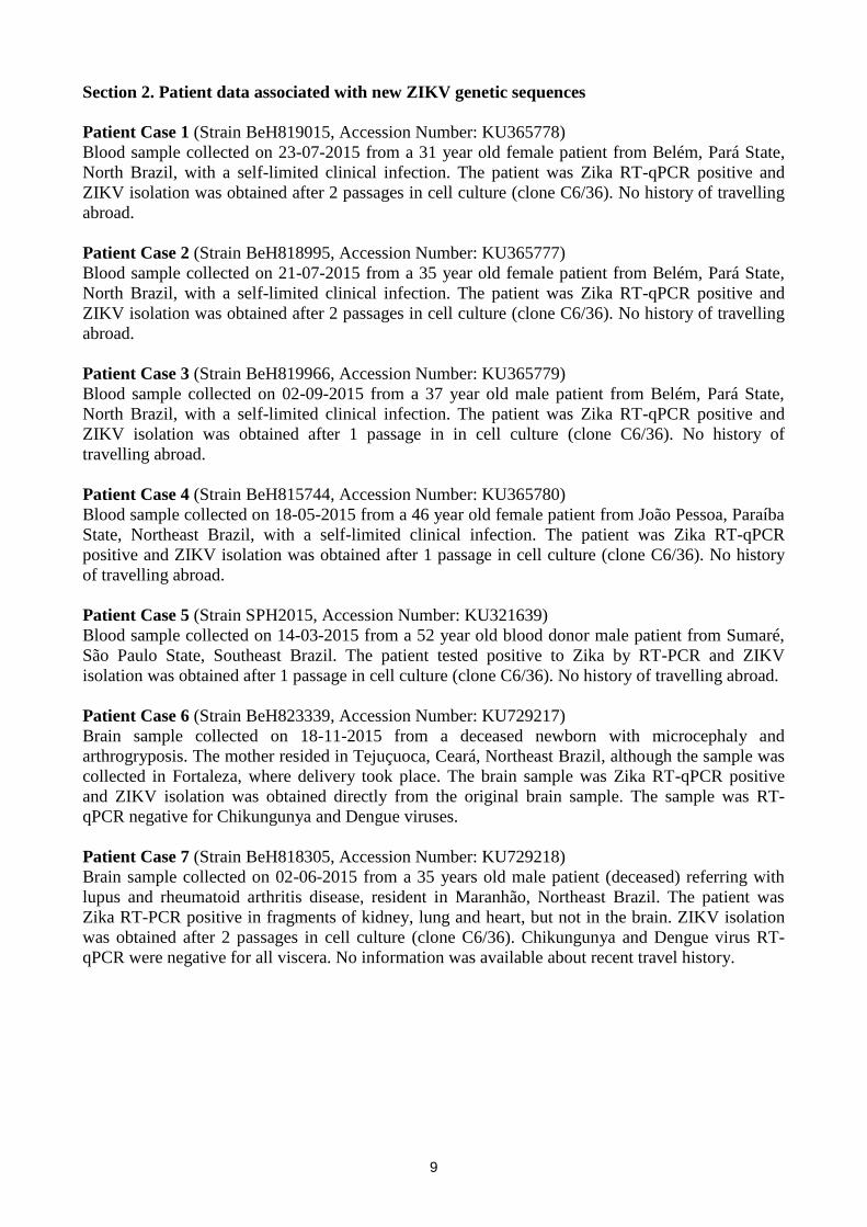
Section 2. Patient data associated with new ZIKV genetic sequences
Patient Case 1 (Strain BeH819015, Accession Number: KU365778)
Blood sample collected on 23-07-2015 from a 31 year old female patient from Belém, Pará State,
North Brazil, with a self-limited clinical infection. The patient was Zika RT-qPCR positive and
ZIKV isolation was obtained after 2 passages in cell culture (clone C6/36). No history of travelling
abroad.
Patient Case 2 (Strain BeH818995, Accession Number: KU365777)
Blood sample collected on 21-07-2015 from a 35 year old female patient from Belém, Pará State,
North Brazil, with a self-limited clinical infection. The patient was Zika RT-qPCR positive and
ZIKV isolation was obtained after 2 passages in cell culture (clone C6/36). No history of travelling
abroad.
Patient Case 3 (Strain BeH819966, Accession Number: KU365779)
Blood sample collected on 02-09-2015 from a 37 year old male patient from Belém, Pará State,
North Brazil, with a self-limited clinical infection. The patient was Zika RT-qPCR positive and
ZIKV isolation was obtained after 1 passage in in cell culture (clone C6/36). No history of
travelling abroad.
Patient Case 4 (Strain BeH815744, Accession Number: KU365780)
Blood sample collected on 18-05-2015 from a 46 year old female patient from João Pessoa, Paraíba
State, Northeast Brazil, with a self-limited clinical infection. The patient was Zika RT-qPCR
positive and ZIKV isolation was obtained after 1 passage in cell culture (clone C6/36). No history
of travelling abroad.
Patient Case 5 (Strain SPH2015, Accession Number: KU321639)
Blood sample collected on 14-03-2015 from a 52 year old blood donor male patient from Sumaré,
São Paulo State, Southeast Brazil. The patient tested positive to Zika by RT-PCR and ZIKV
isolation was obtained after 1 passage in cell culture (clone C6/36). No history of travelling abroad.
Patient Case 6 (Strain BeH823339, Accession Number: KU729217)
Brain sample collected on 18-11-2015 from a deceased newborn with microcephaly and
arthrogryposis. The mother resided in Tejuçuoca, Ceará, Northeast Brazil, although the sample was
collected in Fortaleza, where delivery took place. The brain sample was Zika RT-qPCR positive
and ZIKV isolation was obtained directly from the original brain sample. The sample was RT-
qPCR negative for Chikungunya and Dengue viruses.
Patient Case 7 (Strain BeH818305, Accession Number: KU729218)
Brain sample collected on 02-06-2015 from a 35 years old male patient (deceased) referring with
lupus and rheumatoid arthritis disease, resident in Maranhão, Northeast Brazil. The patient was
Zika RT-PCR positive in fragments of kidney, lung and heart, but not in the brain. ZIKV isolation
was obtained after 2 passages in cell culture (clone C6/36). Chikungunya and Dengue virus RT-
qPCR were negative for all viscera. No information was available about recent travel history.
10
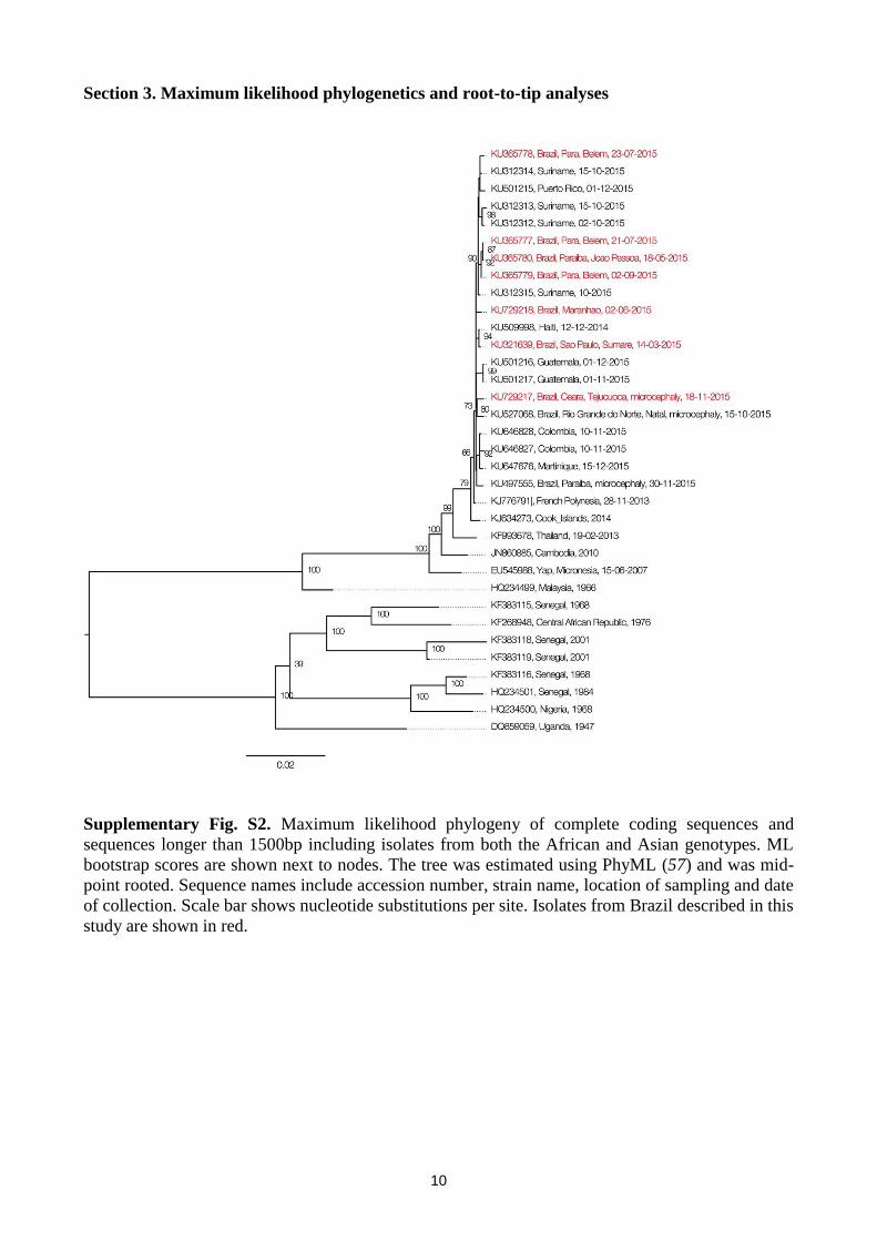
Section 3. Maximum likelihood phylogenetics and root-to-tip analyses
Supplementary Fig. S2. Maximum likelihood phylogeny of complete coding sequences and
sequences longer than 1500bp including isolates from both the African and Asian genotypes. ML
bootstrap scores are shown next to nodes. The tree was estimated using PhyML (57) and was mid-
point rooted. Sequence names include accession number, strain name, location of sampling and date
of collection. Scale bar shows nucleotide substitutions per site. Isolates from Brazil described in this
study are shown in red.
11
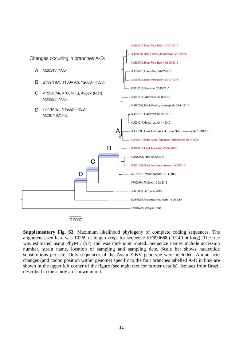
Supplementary Fig. S3. Maximum likelihood phylogeny of complete coding sequences. The
alignment used here was 10269 nt long, except for sequence KF993668 (10140 nt long). The tree
was estimated using PhyML (57) and was mid-point rooted. Sequence names include accession
number, strain name, location of sampling and sampling date. Scale bar shows nucleotide
substitutions per site. Only sequences of the Asian ZIKV genotype were included. Amino acid
changes (and codon position within genome) specific to the four branches labelled A-D in blue are
shown in the upper left corner of the figure (see main text for further details). Isolates from Brazil
described in this study are shown in red.
12
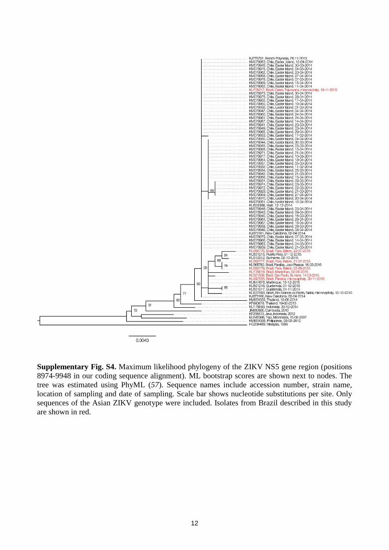
Supplementary Fig. S4. Maximum likelihood phylogeny of the ZIKV NS5 gene region (positions
8974-9948 in our coding sequence alignment). ML bootstrap scores are shown next to nodes. The
tree was estimated using PhyML (57). Sequence names include accession number, strain name,
location of sampling and date of sampling. Scale bar shows nucleotide substitutions per site. Only
sequences of the Asian ZIKV genotype were included. Isolates from Brazil described in this study
are shown in red.
13
Supplementary Fig. S5. Regressions of the sampling date of each sequence against the genetic
distance of that sequence from the root of a maximum likelihood tree. Three datasets were
investigated: (A) Dataset A. (B) Dataset B. (C) Dataset C (see Section 1.6 for details). Colours
indicate the geographic region of sampling.
14
Section 4. Evolutionary rate estimates under different models
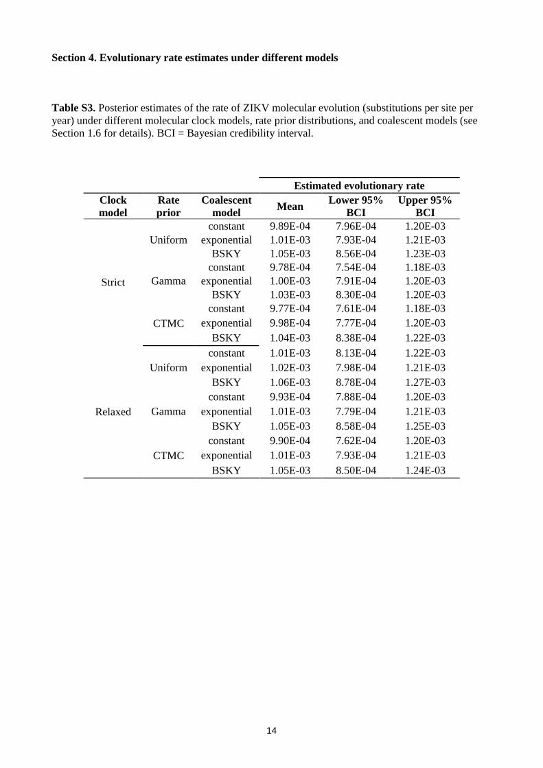
Table S3. Posterior estimates of the rate of ZIKV molecular evolution (substitutions per site per
year) under different molecular clock models, rate prior distributions, and coalescent models (see
Section 1.6 for details). BCI = Bayesian credibility interval.
Estimated evolutionary rate
Clock
model
Rate
prior
Coalescent
model Mean
Lower 95%
BCI
Upper 95%
BCI
Strict
Uniform
constant 9.89E-04 7.96E-04 1.20E-03
exponential 1.01E-03 7.93E-04 1.21E-03
BSKY 1.05E-03 8.56E-04 1.23E-03
Gamma
constant 9.78E-04 7.54E-04 1.18E-03
exponential 1.00E-03 7.91E-04 1.20E-03
BSKY 1.03E-03 8.30E-04 1.20E-03
CTMC
constant 9.77E-04 7.61E-04 1.18E-03
exponential 9.98E-04 7.77E-04 1.20E-03
BSKY 1.04E-03 8.38E-04 1.22E-03
Relaxed
Uniform
constant 1.01E-03 8.13E-04 1.22E-03
exponential 1.02E-03 7.98E-04 1.21E-03
BSKY 1.06E-03 8.78E-04 1.27E-03
Gamma
constant 9.93E-04 7.88E-04 1.20E-03
exponential 1.01E-03 7.79E-04 1.21E-03
BSKY 1.05E-03 8.58E-04 1.25E-03
CTMC
constant 9.90E-04 7.62E-04 1.20E-03
exponential 1.01E-03 7.93E-04 1.21E-03
BSKY 1.05E-03 8.50E-04 1.24E-03
15
Section 5. Dating estimates under different models
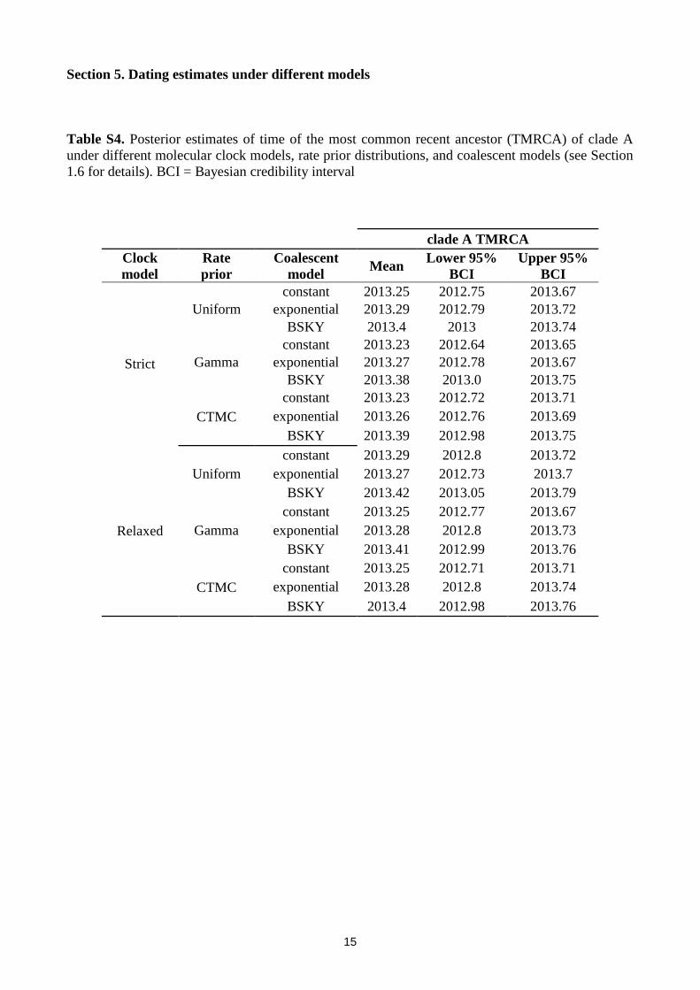
Table S4. Posterior estimates of time of the most common recent ancestor (TMRCA) of clade A
under different molecular clock models, rate prior distributions, and coalescent models (see Section
1.6 for details). BCI = Bayesian credibility interval
clade A TMRCA
Clock
model
Rate
prior
Coalescent
model Mean
Lower 95%
BCI
Upper 95%
BCI
Strict
Uniform
constant 2013.25 2012.75 2013.67
exponential 2013.29 2012.79 2013.72
BSKY 2013.4 2013 2013.74
Gamma
constant 2013.23 2012.64 2013.65
exponential 2013.27 2012.78 2013.67
BSKY 2013.38 2013.0 2013.75
CTMC
constant 2013.23 2012.72 2013.71
exponential 2013.26 2012.76 2013.69
BSKY 2013.39 2012.98 2013.75
Relaxed
Uniform
constant 2013.29 2012.8 2013.72
exponential 2013.27 2012.73 2013.7
BSKY 2013.42 2013.05 2013.79
Gamma
constant 2013.25 2012.77 2013.67
exponential 2013.28 2012.8 2013.73
BSKY 2013.41 2012.99 2013.76
CTMC
constant 2013.25 2012.71 2013.71
exponential 2013.28 2012.8 2013.74
BSKY 2013.4 2012.98 2013.76
16
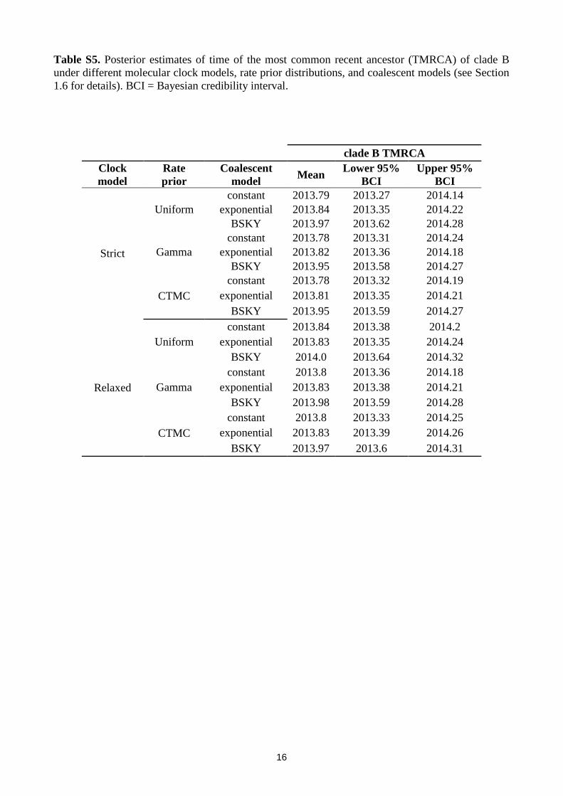
Table S5. Posterior estimates of time of the most common recent ancestor (TMRCA) of clade B
under different molecular clock models, rate prior distributions, and coalescent models (see Section
1.6 for details). BCI = Bayesian credibility interval.
clade B TMRCA
Clock
model
Rate
prior
Coalescent
model Mean
Lower 95%
BCI
Upper 95%
BCI
Strict
Uniform
constant 2013.79 2013.27 2014.14
exponential 2013.84 2013.35 2014.22
BSKY 2013.97 2013.62 2014.28
Gamma
constant 2013.78 2013.31 2014.24
exponential 2013.82 2013.36 2014.18
BSKY 2013.95 2013.58 2014.27
CTMC
constant 2013.78 2013.32 2014.19
exponential 2013.81 2013.35 2014.21
BSKY 2013.95 2013.59 2014.27
Relaxed
Uniform
constant 2013.84 2013.38 2014.2
exponential 2013.83 2013.35 2014.24
BSKY 2014.0 2013.64 2014.32
Gamma
constant 2013.8 2013.36 2014.18
exponential 2013.83 2013.38 2014.21
BSKY 2013.98 2013.59 2014.28
CTMC
constant 2013.8 2013.33 2014.25
exponential 2013.83 2013.39 2014.26
BSKY 2013.97 2013.6 2014.31
17
Section 6. Number of passengers flying to Brazil per country
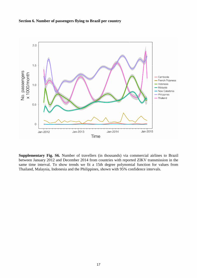
Supplementary Fig. S6. Number of travellers (in thousands) via commercial airlines to Brazil
between January 2012 and December 2014 from countries with reported ZIKV transmission in the
same time interval. To show trends we fit a 15th degree polynomial function for values from
Thailand, Malaysia, Indonesia and the Philippines, shown with 95% confidence intervals.
18
Section 7. Mapping of variant sites onto existing protein structures
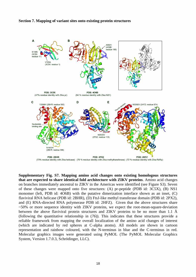
Supplementary Fig. S7. Mapping amino acid changes onto existing homologous structures
that are expected to share identical fold architecture with ZIKV proteins. Amino acid changes
on branches immediately ancestral to ZIKV in the Americas were identified (see Figure S3). Seven
of these changes were mapped onto five structures: (A) pr-peptide (PDB id: 3C5X), (B) NS1
monomer (left, PDB id: 4O6B) with the putative dimerization interface shown as an inset, (C)
flaviviral RNA helicase (PDB id: 2BHR), (D) FtsJ-like methyl transferase domain (PDB id: 2PX2),
and (E) RNA-directed RNA polymerase PDB id: 2HFZ). Given that the above structures share
~50% or more sequence identity with ZIKV proteins, we expect the root-mean-square-deviation
between the above flaviviral protein structures and ZIKV proteins to be no more than 1.1 Å
(following the quantitative relationship in (76)). This indicates that these structures provide a
reliable framework from mapping the overall localization of the amino acid changes of interest
(which are indicated by red spheres at C-alpha atoms). All models are shown in cartoon
representation and rainbow coloured, with the N-terminus in blue and the C-terminus in red.
Molecular graphics images were generated using PyMOL (The PyMOL Molecular Graphics
System, Version 1.7.0.3, Schrödinger, LLC).
19
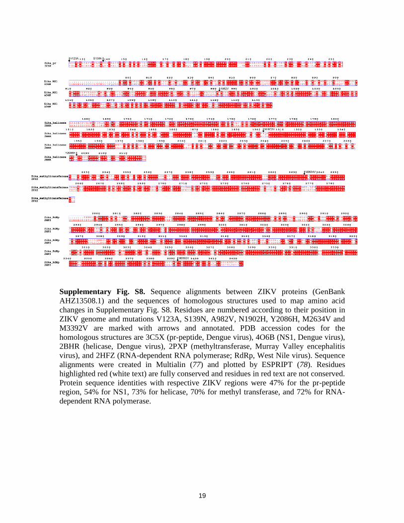
Supplementary Fig. S8. Sequence alignments between ZIKV proteins (GenBank
AHZ13508.1) and the sequences of homologous structures used to map amino acid
changes in Supplementary Fig. S8. Residues are numbered according to their position in
ZIKV genome and mutations V123A, S139N, A982V, N1902H, Y2086H, M2634V and
M3392V are marked with arrows and annotated. PDB accession codes for the
homologous structures are 3C5X (pr-peptide, Dengue virus), 4O6B (NS1, Dengue virus),
2BHR (helicase, Dengue virus), 2PXP (methyltransferase, Murray Valley encephalitis
virus), and 2HFZ (RNA-dependent RNA polymerase; RdRp, West Nile virus). Sequence
alignments were created in Multialin (77) and plotted by ESPRIPT (78). Residues
highlighted red (white text) are fully conserved and residues in red text are not conserved.
Protein sequence identities with respective ZIKV regions were 47% for the pr-peptide
region, 54% for NS1, 73% for helicase, 70% for methyl transferase, and 72% for RNA-
dependent RNA polymerase.
References and Notes
1. B. D. Lindenbach, C. M. Rice, Molecular biology of flaviviruses. Adv. Virus Res. 59, 23–61
(2003). Medline doi:10.1016/S0065-3527(03)59002-9
2. G. W. Dick, S. F. Kitchen, A. J. Haddow, Zika virus. I. Isolations and serological specificity.
Trans. R. Soc. Trop. Med. Hyg. 46, 509–520 (1952). Medline doi:10.1016/0035-
9203(52)90042-4
3. O. Faye, O. Faye, D. Diallo, M. Diallo, M. Weidmann, A. A. Sall, Quantitative real-time PCR
detection of Zika virus and evaluation with field-caught mosquitoes. Virol. J. 10, 311
(2013). Medline doi:10.1186/1743-422X-10-311
4. S. Ioos, H. P. Mallet, I. Leparc Goffart, V. Gauthier, T. Cardoso, M. Herida, Current Zika
virus epidemiology and recent epidemics. Med. Mal. Infect. 44, 302–307 (2014). Medline
doi:10.1016/j.medmal.2014.04.008
5. M. R. Duffy, T. H. Chen, W. T. Hancock, A. M. Powers, J. L. Kool, R. S. Lanciotti, M.
Pretrick, M. Marfel, S. Holzbauer, C. Dubray, L. Guillaumot, A. Griggs, M. Bel, A. J.
Lambert, J. Laven, O. Kosoy, A. Panella, B. J. Biggerstaff, M. Fischer, E. B. Hayes, Zika
virus outbreak on Yap Island, Federated States of Micronesia. N. Engl. J. Med. 360,
2536–2543 (2009). Medline doi:10.1056/NEJMoa0805715
6. A. D. Haddow, A. J. Schuh, C. Y. Yasuda, M. R. Kasper, V. Heang, R. Huy, H. Guzman, R.
B. Tesh, S. C. Weaver, Genetic characterization of Zika virus strains: Geographic
expansion of the Asian lineage. PLOS Negl. Trop. Dis. 6, e1477 (2012). Medline
doi:10.1371/journal.pntd.0001477
7. V. M. Cao-Lormeau, C. Roche, A. Teissier, E. Robin, A. L. Berry, H. P. Mallet, A. A. Sall, D.
Musso, Zika virus, French polynesia, South Pacific, 2013. Emerg. Infect. Dis. 20, 1085–
1086 (2014). Medline doi:10.3201/eid2006.140138
8. M. Dupont-Rouzeyrol, O. O’Connor, E. Calvez, M. Daurès, M. John, J. P. Grangeon, A. C.
Gourinat, Co-infection with Zika and dengue viruses in 2 patients, New Caledonia, 2014.
Emerg. Infect. Dis. 21, 381–382 (2015). Medline doi:10.3201/eid2102.141553
9. A. T. Pyke, M. T. Daly, J. N. Cameron, P. R. Moore, C. T. Taylor, G. R. Hewitson, J. L.
Humphreys, R. Gair, Imported Zika virus infection from the Cook Islands into Australia,
2014. PLOS Curr. 10.1371/currents.outbreaks.4635a54dbffba2156fb2fd76dc49f65e
(2014). Medline
10. T. Wæhre, A. Maagard, D. Tappe, D. Cadar, J. Schmidt-Chanasit, Zika virus infection after
travel to Tahiti, December 2013. Emerg. Infect. Dis. 20, 1412–1414 (2014). Medline
doi:10.3201/eid2008.140302
11. J. Tognarelli, S. Ulloa, E. Villagra, J. Lagos, C. Aguayo, R. Fasce, B. Parra, J. Mora, N.
Becerra, N. Lagos, L. Vera, B. Olivares, M. Vilches, J. Fernández, A report on the
outbreak of Zika virus on Easter Island, South Pacific, 2014. Arch. Virol. 161, 665–668
(2016). Medline
12. M. Hennessey, M. Fischer, J. E. Staples, Zika virus spreads to new areas – region of the
Americas, May 2015–January 2016. MMWR Morb. Mortal. Wkly. Rep. 65, 55–58 (2016).
Medline doi:10.15585/mmwr.mm6503e1
13. M. M. O. Silva, M. S. Rodrigues, I. A. D. Paploski, M. Kikuti, A. M. Kasper, J. S. Cruz, T.
L. Queiroz, A. S. Tavares, P. M. Santana, J. M. G. Araújo, A. I. Ko, M. G. Reis, G. S.
Ribeiro, Accuracy of dengue reporting by national surveillance system, Brazil. Emerg.
Infect. Dis. 22, 336–339 (2016). Medline doi:10.3201/eid2202.150495
14. Ministério da Saúde do Brasil, “Boletim Epidemiológico 47:7: Semana epidemiológica (SE)
04 (30/01/2016)” Secretaria de Vigilância em Saúde (2016) (in Portuguese). Available at:
http://portalsaude.saude.gov.br/index.php/situacao-epidemiologica-dados-dengue.
15. ECDC, Microcephaly in Brazil potentially linked to the Zika virus epidemic. ECDC (2015).
16. J. Mlakar, M. Korva, N. Tul, M. Popović, M. Poljšak-Prijatelj, J. Mraz, M. Kolenc, K.
Resman Rus, T. Vesnaver Vipotnik, V. Fabjan Vodušek, A. Vizjak, J. Pižem, M.
Petrovec, T. Avšič Županc, Zika virus associated with microcephaly. N. Engl. J. Med.
374, 951–958 (2016). Medline doi:10.1056/NEJMoa1600651
17. L. Schuler-Faccini, E. M. Ribeiro, I. M. Feitosa, D. D. Horovitz, D. P. Cavalcanti, A. Pessoa,
M. J. Doriqui, J. I. Neri, J. M. Neto, H. Y. Wanderley, M. Cernach, A. S. El-Husny, M.
V. Pone, C. L. Serao, M. T. Sanseverino; Brazilian Medical Genetics Society–Zika
Embryopathy Task Force, Possible association between Zika virus infection and
microcephaly - Brazil, 2015. MMWR Morb. Mortal. Wkly. Rep. 65, 59–62 (2016).
Medline doi:10.15585/mmwr.mm6503e2
18. C. V. Ventura, M. Maia, B. V. Ventura, V. V. Linden, E. B. Araújo, R. C. Ramos, M. A.
Rocha, M. D. Carvalho, R. Belfort Jr., L. O. Ventura, Ophthalmological findings in
infants with microcephaly and presumable intra-uterus Zika virus infection. Arq. Bras.
Oftalmol. 79, 1–3 (2016). Medline doi:10.5935/0004-2749.20160002
19. R. B. Martines, J. Bhatnagar, M. K. Keating, L. Silva-Flannery, A. Muehlenbachs, J. Gary,
C. Goldsmith, G. Hale, J. Ritter, D. Rollin, W. J. Shieh, K. G. Luz, A. M. Ramos, H. P.
Davi, W. Kleber de Oliveria, R. Lanciotti, A. Lambert, S. Zaki, Notes from the field:
Evidence of Zika virus infection in brain and placental tissues from two congenitally
infected newborns and two fetal losses - Brazil, 2015. MMWR Morb. Mortal. Wkly. Rep.
65, 159–160 (2016). Medline doi:10.15585/mmwr.mm6506e1
20. PAHO, “Neurological syndrome, congenital malformations, and Zika virus infection.
Implications for public health in the Americas” (PAHO/WHO, 2015).
21. G. Calvet, R. S. Aguiar, A. S. O. Melo, S. A. Sampaio, I. Filippis, A. Fabri, E. S. M. Araujo,
P. C. Sequeira, M. C. L. Mendonca, L. Oliveira, D. A. Tschoeke, C. G. Schrago, F. L.
Thompson, P. Brasil, F. B. Santos, R. M. R. Nogueira, A. Tanuri, A. M. B. Filippis,
Detection and sequencing of Zika virus from amniotic fluid of fetuses with microcephaly
in Brazil: A case study. Lancet Infect. Dis. 10.1016/S1473-3099(16)00095-5 (2016).
doi:10.1016/S1473-3099(16)00095-5
22. M. Besnard, S. Lastere, A. Teissier, V. Cao-Lormeau, D. Musso, Evidence of perinatal
transmission of Zika virus, French Polynesia, December 2013 and February 2014. Euro
Surveill. 19, 20751 (2014). Medline doi:10.2807/1560-7917.ES2014.19.13.20751
23. H. Tang, C. Hammack, S. C. Ogden, Z. Wen, X. Qian, Y. Li, B. Yao, J. Shin, F. Zhang, E.
M. Lee, K. M. Christian, R. A. Didier, P. Jin, H. Song, G. L. Ming, Zika virus infects
human cortical neural progenitors and attenuates their growth. Cell Stem Cell
10.1016/j.stem.2016.02.016 (2016). Medline doi:10.1016/j.stem.2016.02.016
24. C. G. Victora, L. Schuler-Faccini, A. Matijasevich, E. Ribeiro, A. Pessoa, F. C. Barros,
Comment: Microcephaly in Brazil: How to interpret reported numbers. Lancet 387, 621–
624 (2016). doi:10.1016/S0140-6736(16)00273-7
25. WHO Interim Report, “Assessment of infants with microcephaly in the context of Zika
virus” (2016).
26. C. V. Ventura, M. Maia, V. Bravo-Filho, A. L. Góis, R. Belfort Jr., Zika virus in Brazil and
macular atrophy in a child with microcephaly. Lancet 387, 228 (2016). Medline
doi:10.1016/S0140-6736(16)00006-4
27. M. R. Nunes, N. R. Faria, H. B. Vasconcelos, D. B. Medeiros, C. P. Silva de Lima, V. L.
Carvalho, E. V. Pinto da Silva, J. F. Cardoso, E. C. Sousa Jr., K. N. Nunes, S. G.
Rodrigues, A. B. Abecasis, M. A. Suchard, P. Lemey, P. F. Vasconcelos,
Phylogeography of dengue virus serotype 4, Brazil, 2010-2011. Emerg. Infect. Dis. 18,
1858–1864 (2012). Medline doi:10.3201/eid1811.120217
28. O. G. Pybus, M. A. Suchard, P. Lemey, F. J. Bernardin, A. Rambaut, F. W. Crawford, R. R.
Gray, N. Arinaminpathy, S. L. Stramer, M. P. Busch, E. L. Delwart, Unifying the spatial
epidemiology and molecular evolution of emerging epidemics. Proc. Natl. Acad. Sci.
U.S.A. 109, 15066–15071 (2012). Medline doi:10.1073/pnas.1206598109
29. C. Zanluca, V. C. Melo, A. L. Mosimann, G. I. Santos, C. N. Santos, K. Luz, First report of
autochthonous transmission of Zika virus in Brazil. Mem. Inst. Oswaldo Cruz 110, 569–
572 (2015). Medline doi:10.1590/0074-02760150192
30. A. G. Meyer, S. J. Spielman, T. Bedford, C. O. Wilke, Time dependence of evolutionary
metrics during the 2009 pandemic influenza virus outbreak. Virus Evol. 1, vev006 (2015).
10.1093/ve/vev006 Medline doi:10.1093/ve/vev006
31. D. J. Park, G. Dudas, S. Wohl, A. Goba, S. L. Whitmer, K. G. Andersen, R. S. Sealfon, J. T.
Ladner, J. R. Kugelman, C. B. Matranga, S. M. Winnicki, J. Qu, S. K. Gire, A. Gladden-
Young, S. Jalloh, D. Nosamiefan, N. L. Yozwiak, L. M. Moses, P. P. Jiang, A. E. Lin, S.
F. Schaffner, B. Bird, J. Towner, M. Mamoh, M. Gbakie, L. Kanneh, D. Kargbo, J. L.
Massally, F. K. Kamara, E. Konuwa, J. Sellu, A. A. Jalloh, I. Mustapha, M. Foday, M.
Yillah, B. R. Erickson, T. Sealy, D. Blau, C. Paddock, A. Brault, B. Amman, J. Basile, S.
Bearden, J. Belser, E. Bergeron, S. Campbell, A. Chakrabarti, K. Dodd, M. Flint, A.
Gibbons, C. Goodman, J. Klena, L. McMullan, L. Morgan, B. Russell, J. Salzer, A.
Sanchez, D. Wang, I. Jungreis, C. Tomkins-Tinch, A. Kislyuk, M. F. Lin, S. Chapman,
B. MacInnis, A. Matthews, J. Bochicchio, L. E. Hensley, J. H. Kuhn, C. Nusbaum, J. S.
Schieffelin, B. W. Birren, M. Forget, S. T. Nichol, G. F. Palacios, D. Ndiaye, C. Happi,
S. M. Gevao, M. A. Vandi, B. Kargbo, E. C. Holmes, T. Bedford, A. Gnirke, U. Ströher,
A. Rambaut, R. F. Garry, P. C. Sabeti, Ebola Virus epidemiology, transmission, and
evolution during seven months in Sierra Leone. Cell 161, 1516–1526 (2015). Medline
doi:10.1016/j.cell.2015.06.007
32. M. R. Nunes, N. R. Faria, J. M. de Vasconcelos, N. Golding, M. U. Kraemer, L. F. de
Oliveira, R. S. Azevedo, D. E. da Silva, E. V. da Silva, S. P. da Silva, V. L. Carvalho, G.
E. Coelho, A. C. Cruz, S. G. Rodrigues, J. L. Vianez Jr., B. T. Nunes, J. F. Cardoso, R. B.
Tesh, S. I. Hay, O. G. Pybus, P. F. Vasconcelos, Emergence and potential for spread of
Chikungunya virus in Brazil. BMC Med. 13, 102 (2015). Medline doi:10.1186/s12916-
015-0348-x
33. M. R. Nunes, G. Palacios, N. R. Faria, E. C. Sousa Jr., J. A. Pantoja, S. G. Rodrigues, V. L.
Carvalho, D. B. Medeiros, N. Savji, G. Baele, M. A. Suchard, P. Lemey, P. F.
Vasconcelos, W. I. Lipkin, Air travel is associated with intracontinental spread of dengue
virus serotypes 1-3 in Brazil. PLOS Negl. Trop. Dis. 8, e2769 (2014). Medline
doi:10.1371/journal.pntd.0002769
34. D. Musso, Zika virus transmission from French Polynesia to Brazil. Emerg. Infect. Dis. 21,
1887 (2015). Medline doi:10.3201/eid2110.151125
35. P. Lemey, A. Rambaut, T. Bedford, N. Faria, F. Bielejec, G. Baele, C. A. Russell, D. J.
Smith, O. G. Pybus, D. Brockmann, M. A. Suchard, Unifying viral genetics and human
transportation data to predict the global transmission dynamics of human influenza
H3N2. PLOS Pathog. 10, e1003932 (2014). Medline doi:10.1371/journal.ppat.1003932
36. O. G. Pybus, A. J. Tatem, P. Lemey, Virus evolution and transmission in an ever more
connected world. Proc. Biol. Sci. 282, 20142878 (2015). Medline
doi:10.1098/rspb.2014.2878
37. V. M. Cao-Lormeau, A. Blake, S. Mons, S. Lastère, C. Roche, J. Vanhomwegen, T. Dub, L.
Baudouin, A. Teissier, P. Larre, A. L. Vial, C. Decam, V. Choumet, S. K. Halstead, H. J.
Willison, L. Musset, J. C. Manuguerra, P. Despres, E. Fournier, H. P. Mallet, D. Musso,
A. Fontanet, J. Neil, F. Ghawché, Guillain-Barré Syndrome outbreak associated with
Zika virus infection in French Polynesia: A case-control study. Lancet 10.1016/S0140-
6736(16)00562-6 (2016). Medline doi:10.1016/S0140-6736(16)00562-6
38. K. A. Dowd, T. C. Pierson, Antibody-mediated neutralization of flaviviruses: A reductionist
view. Virology 411, 306–315 (2011). Medline doi:10.1016/j.virol.2010.12.020
39. J. T. Roehrig, Antigenic structure of flavivirus proteins. Adv. Virus Res. 59, 141–175 (2003).
Medline doi:10.1016/S0065-3527(03)59005-4
40. P. Gérardin, S. Sampériz, D. Ramful, B. Boumahni, M. Bintner, J. L. Alessandri, M.
Carbonnier, I. Tiran-Rajaoefera, G. Beullier, I. Boya, T. Noormahomed, J. Okoï, O.
Rollot, L. Cotte, M. C. Jaffar-Bandjee, A. Michault, F. Favier, M. Kaminski, A.
Fourmaintraux, X. Fritel, Neurocognitive outcome of children exposed to perinatal
mother-to-child Chikungunya virus infection: The CHIMERE cohort study on Reunion
Island. PLOS Negl. Trop. Dis. 8, e2996 (2014). Medline
doi:10.1371/journal.pntd.0002996
41. A. Fagbami, S. B. Halstead, N. Marchette, K. Larsen, Heterologous flavivirus infection-
enhancing antibodies in sera of Nigerians. Am. J. Trop. Med. Hyg. 38, 205–207 (1988).
Medline
42. B. D. Foy, K. C. Kobylinski, J. L. Chilson Foy, B. J. Blitvich, A. Travassos da Rosa, A. D.
Haddow, R. S. Lanciotti, R. B. Tesh, Probable non-vector-borne transmission of Zika
virus, Colorado, USA. Emerg. Infect. Dis. 17, 880–882 (2011). Medline
doi:10.3201/eid1705.101939
43. D. Musso, C. Roche, E. Robin, T. Nhan, A. Teissier, V. M. Cao-Lormeau, Potential sexual
transmission of Zika virus. Emerg. Infect. Dis. 21, 359–361 (2015). Medline
doi:10.3201/eid2102.141363
44. D. Musso, T. Nhan, E. Robin, C. Roche, D. Bierlaire, K. Zisou, A. Shan Yan, V. M. Cao-
Lormeau, J. Broult, Potential for Zika virus transmission through blood transfusion
demonstrated during an outbreak in French Polynesia, November 2013 to February 2014.
Euro Surveill. 19, 20761 (2014). Medline doi:10.2807/1560-7917.ES2014.19.14.20761
45. A. Sorichetta, G. M. Hornby, F. R. Stevens, A. E. Gaughan, C. Linard, A. J. Tatem, High-
resolution gridded population datasets for Latin America and the Caribbean in 2010,
2015, and 2020. Sci. Data 2, 150045 (2015). Medline doi:10.1038/sdata.2015.45
46. I. Zamree, N. Drakes, A. Rohani, H. L. Lee, Sensitivity of Aedes albopictus C6/36 cells line
for the detection and infectivity titration of dengue virus. Trop. Biomed. 22, 217–219
(2005). Medline
47. E. H. Lennette, N. J. Schmidt, Diagnostic Procedures for Viral, Rickttsial and Chlamydial
Infections 5th Edition, E. H. Lennette, N. J. Schmidt, Eds. (American of Public Health
Association, Washington, 1974).
48. R. S. Lanciotti, O. L. Kosoy, J. J. Laven, J. O. Velez, A. J. Lambert, A. J. Johnson, S. M.
Stanfield, M. R. Duffy, Genetic and serologic properties of Zika virus associated with an
epidemic, Yap State, Micronesia, 2007. Emerg. Infect. Dis. 14, 1232–1239 (2008).
Medline doi:10.3201/eid1408.080287
49. M. Margulies, M. Egholm, W. E. Altman, S. Attiya, J. S. Bader, L. A. Bemben, J. Berka, M.
S. Braverman, Y. J. Chen, Z. Chen, S. B. Dewell, A. de Winter, J. Drake, L. Du, J. M.
Fierro, R. Forte, X. V. Gomes, B. C. Godwin, W. He, S. Helgesen, C. H. Ho, S. K.
Hutchison, G. P. Irzyk, S. C. Jando, M. L. Alenquer, T. P. Jarvie, K. B. Jirage, J. B. Kim,
J. R. Knight, J. R. Lanza, J. H. Leamon, W. L. Lee, S. M. Lefkowitz, M. Lei, J. Li, K. L.
Lohman, H. Lu, V. B. Makhijani, K. E. McDade, M. P. McKenna, E. W. Myers, E.
Nickerson, J. R. Nobile, R. Plant, B. P. Puc, M. Reifler, M. T. Ronan, G. T. Roth, G. J.
Sarkis, J. F. Simons, J. W. Simpson, M. Srinivasan, K. R. Tartaro, A. Tomasz, K. A.
Vogt, G. A. Volkmer, S. H. Wang, Y. Wang, M. P. Weiner, D. A. Willoughby, P. Yu, R.
F. Begley, J. M. Rothberg, Genome sequencing in microfabricated high-density picolitre
reactors. Nature 437, 376–380 (2005). Medline
50. B. Chevreux, T. Pfisterer, B. Drescher, A. J. Driesel, W. E. Müller, T. Wetter, S. Suhai,
Using the miraEST assembler for reliable and automated mRNA transcript assembly and
SNP detection in sequenced ESTs. Genome Res. 14, 1147–1159 (2004). Medline
doi:10.1101/gr.1917404
51. M. Kearse, R. Moir, A. Wilson, S. Stones-Havas, M. Cheung, S. Sturrock, S. Buxton, A.
Cooper, S. Markowitz, C. Duran, T. Thierer, B. Ashton, P. Meintjes, A. Drummond,
Geneious Basic: An integrated and extendable desktop software platform for the
organization and analysis of sequence data. Bioinformatics 28, 1647–1649 (2012).
Medline doi:10.1093/bioinformatics/bts199
52. M. S. Cunha, D. L. Esposito, I. M. Rocco, A. Y. Maeda, F. G. Vasami, J. S. Nogueira, R. P.
de Souza, A. Suzuki, M. Addas-Carvalho, M. L. Barjas-Castro, M. R. Resende, R. S.
Stucchi, I. F. Boin, G. Katz, R. N. Angerami, B. A. da Fonseca, First complete genome
sequence of Zika virus (Flaviviridae, Flavivirus) from an autochthonous transmission in
Brazil. Genome Announc. 4, e00032-16 (2016). Medline doi:10.1128/genomeA.00032-16
53. WHO, The WHO Child Growth Standards. Child growth standards (2016).
54. K. Clark, I. Karsch-Mizrachi, D. J. Lipman, J. Ostell, E. W. Sayers, GenBank. Nucleic Acids
Res. 44, D67–D72 (2016). Medline doi:10.1093/nar/gkv1276
55. M. Gouy, S. Delmotte, Remote access to ACNUC nucleotide and protein sequence databases
at PBIL. Biochimie 90, 555–562 (2008). Medline doi:10.1016/j.biochi.2007.07.003
56. K. Katoh, D. M. Standley, MAFFT: Iterative refinement and additional methods. Methods
Mol. Biol. 1079, 131–146 (2014). Medline doi:10.1007/978-1-62703-646-7_8
57. S. Guindon, F. Delsuc, J. F. Dufayard, O. Gascuel, Estimating maximum likelihood
phylogenies with PhyML. Methods Mol. Biol. 537, 113–137 (2009). Medline
doi:10.1007/978-1-59745-251-9_6
58. D. Darriba, G. L. Taboada, R. Doallo, D. Posada, jModelTest 2: More models, new heuristics
and parallel computing. Nat. Methods 9, 772 (2012). Medline doi:10.1038/nmeth.2109
59. A. Rambaut, (2014) available at http://tree.bio.ed.ac.uk/software/.
60. S. M. Volk, R. Chen, K. A. Tsetsarkin, A. P. Adams, T. I. Garcia, A. A. Sall, F. Nasar, A. J.
Schuh, E. C. Holmes, S. Higgs, P. D. Maharaj, A. C. Brault, S. C. Weaver, Genome-scale
phylogenetic analyses of chikungunya virus reveal independent emergences of recent
epidemics and various evolutionary rates. J. Virol. 84, 6497–6504 (2010). Medline
doi:10.1128/JVI.01603-09
61. T. C. Bruen, H. Philippe, D. Bryant, A simple and robust statistical test for detecting the
presence of recombination. Genetics 172, 2665–2681 (2006). Medline
doi:10.1534/genetics.105.048975
62. A. J. Drummond, M. A. Suchard, D. Xie, A. Rambaut, Bayesian phylogenetics with BEAUti
and the BEAST 1.7. Mol. Biol. Evol. 29, 1969–1973 (2012). Medline
doi:10.1093/molbev/mss075
63. A. J. Drummond, S. Y. Ho, M. J. Phillips, A. Rambaut, Relaxed phylogenetics and dating
with confidence. PLOS Biol. 4, e88 (2006). Medline doi:10.1371/journal.pbio.0040088
64. S. Tavaré, “Some probabilistic and statistical problems in the analysis of DNA sequences,” in
Some Mathematical Questions in Biology: DNA Sequence Analysis, M. S. Waterman, Ed.
(American Mathematical Society, Providence, RI, 1986), pp. 57–86.
65. M. A. R. Ferreira, M. A. Suchard, Bayesian analysis of elapsed times in continuous-time
Markov chains. Can. J. Stat. 36, 355–368 (2008). doi:10.1002/cjs.5550360302
66. R. Buathong, L. Hermann, B. Thaisomboonsuk, W. Rutvisuttinunt, C. Klungthong, P.
Chinnawirotpisan, W. Manasatienkij, A. Nisalak, S. Fernandez, I. K. Yoon, P. Akrasewi,
T. Plipat, Detection of Zika virus infection in Thailand, 2012-2014. Am. J. Trop. Med.
Hyg. 93, 380–383 (2015). Medline doi:10.4269/ajtmh.15-0022
67. A. Roth, A. Mercier, C. Lepers, D. Hoy, S. Duituturaga, E. Benyon, L. Guillaumot, Y.
Souares, Concurrent outbreaks of dengue, chikungunya and Zika virus infections - an
unprecedented epidemic wave of mosquito-borne viruses in the Pacific 2012-2014. Euro
Surveill. 19, 20929 (2014). Medline doi:10.2807/1560-7917.ES2014.19.41.20929
68. WHO, “Pacific syndromic surveillance report, Week 21, ending 25 May, 2014” WHO Wester
Pacific Region (2014).
69. C. Baronti, G. Piorkowski, R. N. Charrel, L. Boubis, I. Leparc-Goffart, X. de Lamballerie,
Complete coding sequence of Zika virus from a French Polynesia outbreak in 2013.
Genome Announc. 2, e00500-14 (2014). Medline doi:10.1128/genomeA.00500-14
70. S. Kutsuna, Y. Kato, T. Takasaki, M. Moi, A. Kotaki, H. Uemura, T. Matono, Y. Fujiya, M.
Mawatari, N. Takeshita, K. Hayakawa, S. Kanagawa, N. Ohmagari, Two cases of Zika
fever imported from French Polynesia to Japan, December 2013 to January 2014. Euro
Surveill. 19, 20683 (2014). Medline doi:10.2807/1560-7917.ES2014.19.4.20683
71. M. T. Alera, L. Hermann, I. A. Tac-An, C. Klungthong, W. Rutvisuttinunt, W.
Manasatienkij, D. Villa, B. Thaisomboonsuk, J. M. Velasco, P. Chinnawirotpisan, C. B.
Lago, V. G. Roque Jr., L. R. Macareo, A. Srikiatkhachorn, S. Fernandez, I. K. Yoon,
Zika virus infection, Philippines, 2012. Emerg. Infect. Dis. 21, 722–724 (2015). Medline
doi:10.3201/eid2104.141707
72. J. C. Kwong, J. D. Druce, K. Leder, Zika virus infection acquired during brief travel to
Indonesia. Am. J. Trop. Med. Hyg. 89, 516–517 (2013). Medline doi:10.4269/ajtmh.13-
0029
73. D. Tappe, S. Nachtigall, A. Kapaun, P. Schnitzler, S. Günther, J. Schmidt-Chanasit, Acute
Zika virus infection after travel to Malaysian Borneo, September 2014. Emerg. Infect.
Dis. 21, 911–913 (2015). Medline doi:10.3201/eid2105.141960
74. N. Z. P. Health, “New Zealand Public Health Surveillance Report: September 2014,” No. 3
(2014).
75. K. Fonseca, B. Meatherall, D. Zarra, M. Drebot, J. MacDonald, K. Pabbaraju, S. Wong, P.
Webster, R. Lindsay, R. Tellier, First case of Zika virus infection in a returning Canadian
traveler. Am. J. Trop. Med. Hyg. 91, 1035–1038 (2014). Medline doi:10.4269/ajtmh.14-
0151
76. C. Chothia, A. M. Lesk, The relation between the divergence of sequence and structure in
proteins. EMBO J. 5, 823–826 (1986). Medline
77. F. Corpet, Multiple sequence alignment with hierarchical clustering. Nucleic Acids Res. 16,
10881–10890 (1988). Medline doi:10.1093/nar/16.22.10881
78. X. Robert, P. Gouet, Deciphering key features in protein structures with the new ENDscript
server. Nucleic Acids Res. 42, W320–W324 (2014). Medline doi:10.1093/nar/gku316


![Supporting Online Material forscience.sciencemag.org/highwire/filestream/590781/field_highwire...Verde [Guanacaste] Biological Stations, 2006; Corcovado National Park [Puntarenas],](https://img.pdfslide.net/doc/110x75/5e215cb3bf01800aa4125a36/supporting-online-material-guanacaste-biological-stations-2006-corcovado-national.jpg)