Embed Size (px)
Citation preview

CHAPTER 3I
TECHNIQUES OF GASTROCNEMIUS RE,CESSION
Gerard V Yw, DPM, FACFAS
Theresa L. Schinke, DPMWissam E. Kbourlt, DPMShane IV. Mannirug, DPM
Recession of the gastrocnemius muscle is one of the oldest
orthopaedic procedures described in the literature. It is an
effective and predictable method for the treatment ofspastic and non-spastic gastrocnemius equinus deformities.Equinus is thought to be a major contributing factor for the
development of a number of pathologic conditionsincluding pediatric pes valgus, adult acquired flatfoot and
charcot arthropathy.'3 The focus of this paper is to describe
the distal gastrocnemius recession, with emphasis on pearls
of the surgical technique to diminish complications, enhance
outcomes, and facilitate execution of the procedure.
Equinus is defined as less than 10 degrees ofdorsiflexion ofthe foot to the leg and can be classified as
osseous, or muscular.t-:':'e (see Thble) It should be notedthat surgical procedures to correct the different types ofequinus are distinctly different, therefore it is imperativeto identi$. the specific rype of equinus present.
Muscular equinus involves contracture of the poste-
rior crural muscles which can be the result of spastic ornonspastic etiologies. Spastic equinus is usually found inpatients with neuromuscular diseases, such as cerebral
palsy; it can be acquired or congenital. In the past,
spastic equinus was thought to be the most common type
of equinus. These individuals are easy to recognize, as
they tend to present with classic 'toe-walker' gait, and are
Thble 1
TYPES OF ANKLE JOINT EQUINUS
Muscular Eouinus Osseous EquinusGastroc-soleus Equinus Ankle Joint exostosis
Spastic Talotibial exostosis
Non-spastic Tibio-fibular synostosis
Gastrocnemius Equinus Pseudoequinus
Spastic Anterior cavus
Non-spastic
more difficult to correct. Nonspastic equinus is beingrecognized with increasing frequency, and is probablymore common than spastic equinus. Nonspastic equinus
may be due to a congenitally shortened tendoachilles. Itmay also be an acquired deformity or iatrogenic in nature(ie. poor casting techniques).'''
Posterior muscular equinus can further be broken
down into gastrocnemius-soleus equinus or gastrocnemius
equinus and can be differentiated by performing the
Silverskioid test. To perform the test, the patient should be
Iying in the supine position. The examiner first determines
that an equinus deformity is present by attempting to
dorsiflex the foot beyond ninety degrees while the knee is
extended. \W4ren evaluating for gastrocnemius equinus, the
examiner flexes the patients knee to ninety degrees (to
remove the influence of the soleus muscle), and again
dorsiflexes the patientt ankle. Ifan increase in dorsiflexion
at the ankle joint is appreciated with the knee flexed, butnot with the knee extended, a gastrocnemius equinus is
felt to be present. This involves contracture of the
gastrocnemius and/or plantaris muscles only. t'z'r-z
Gastrocnemius-soleus equinus (gastrocsoleus
equinus) involves limitation of ankle joint dorsiflexion
both with the knee flexed, and with the knee extended
during the Silverskiold test. It involves contracture of all
or some of the structures passing posterior to the ankle
joint. It must, however, be differentiated from osseous
equinus, which produces similar findings with the
Silverskiold test.'rOsseous equinus involves limited ankle joint
dorsiflexion due to pathologic changes about the anklejoint. Thlotibial exostosis or other osseous bridgingbetween the tibia and fibula is most commonly the cause
of osseous equinus. The clinical examination of ankle
equinus gives a significantly different feel at end range ofdorsiflexory motion. In osseous equinus, the end range ofmotion in dorsiflexion is notably a hard or abrupt feel,
whereas in gastocsoleus or gastrocnemius equinus, the
end feel is soft or smooth. Conventional radiographs may
be taken and examined for possible osseous projections
CHAPTER31 169
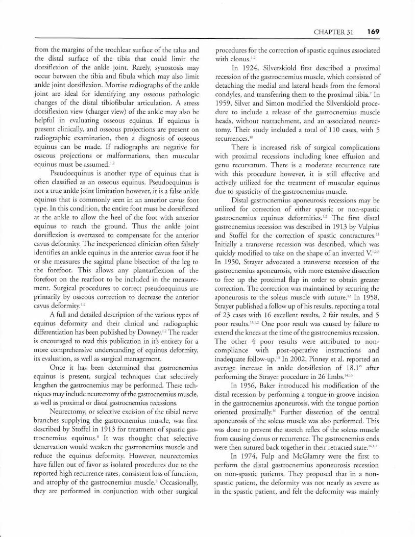
from the margins of the trochlear surface of the talus andthe distal surface of the tibia that could limit thedorsiflexion of the ankle joint. Rarely, synostosis mayoccur between the tibia and fibula which may also limitankle joint dorsiflexion. Mortise radiographs of the anklejoint are ideal for identifying any osseous pathologicchanges of the distal tibiofibular articulation. A stress
dorsiflexion view (charger view) of the ankle may also be
helpful in evaluating osseous equinus. If equinus is
present clinically, and osseous projections are present onradiographic examination, rhen a diagnosis of osseous
equinus can be made. If radiographs are negative forosseous projections or malformations, then muscularequinus must be assumed.'''
Pseudoequinus is another type of equinus that is
often classified as an osseous equinus. Pseudoequinus is
not a true ankle joint limitation however, it is a false ankleequinus that is commonly seen in an anterior cavus footrype. In this condition, the entire foor must be dorsiflexedat the ankle to allow the heel of the foot with anteriorequinus to reach the ground. Thus the ankle jointdorsiflexion is overtaxed to compensate for the anteriorcavus deformiry. The inexperienced clinician often falselyidentifies an ankle equinus in the anterior cavus foot if he
or she measures the sagittal plane bisection of the leg tothe forefoot. This allows any plantarflexion of theforefoot on the rearfoot to be included in the measure-ment. Surgical procedures to correct pseudoequinus are
primariiy by osseous correction to decrease the anteriorcavus deformiry.'''
A full and detailed description of the various rypes ofequinus deformity and their clinical and radiographicdifferentiation has been published by Downey." The reader
is encouraged to read this publication in it's enrirery for a
more comprehensive understanding of equinus deformity,its evaluation, as well as surgical management.
Once it has been determined that gastrocnemiusequinus is present, surgical techniques that selectivelylengthen the gastrocnemius may be performed. These tech-niques may include neurectomy of the gastrocnemius muscle,as well as proximal or distal gastrocnemius recessions.
Neurectomy, or selective excision of the tibial nervebranches supplying the gastrocnemius muscle, was firstdescribed by Stoffei in 1913 for treatment of spastic gas-
trocnemius equinus.t It was thought that selectivedenervation would weaken the gastronemius muscle andreduce the equinus deformity. However, neurectomieshave fallen out of favor as isolated procedures due to thereported high recurrence rates, consistent loss offunction,and atrophy of the gastrocnemius muscle.e Occasionally,they are performed in conjunction with other surgical
procedures for the correction ofspastic equinus associated
with clonus.'''In 1924, Silverskiold first described a proximal
recession of the gastrocnemius muscle, which consisted ofdetaching the medial and lateral heads from the femoralcondyles, and transferring them to the proximal tibia.'In1959, Silver and Simon modified the Silverskiold proce-dure to include a release of the gastrocnemius muscleheads, without reattachment, and an associated neurec-tomy. Their study included a total of 110 cases, with 5
recufrences.lo
There is increased risk of surgical complicationswith proximal recessions including knee effusion andgenu recurvatum. There is a moderate recurrence rate
with this procedure however, it is still effective andactively utilized for the treatment of muscular equinusdue to spasticity of the gastrocnemius muscle.
Distal gastrocnemius aponeurosis recessions may be
utilized for correction of either spastic or non-spasticgastrocnemius equinus deformities.''' The first distalgastrocnemius recession was described in 1913 by Vulpiusand Stoffel for the correction of spastic contractures.l'Initially a transverse recession was described, which was
quickly modified to take on the shape of an inverted V''16
In 1950, Strayer advocated a ransverse recession of thegastrocnemius aponeurosis, with more extensive dissectionto free up the proximal flap in order to obtain greater
correction. The correction was maintained by securing theaponeurosis to the soleus muscle with suture.t' In 1958,Strayer published a follow up ofhis results, reporting a totalof 23 cases with 16 excellent results,2 fair results, and 5
poor results.l3'1'2 One poor result was caused by failure toextend the knees at the time of the gastrocnemius recession.
The other 4 poor results were attributed to non-compliance with post-operative instructions andinadequate follow-up." In 2002, Pinney et al. reported an
average increase in ankle dorsiflexion of 18.1'afterperforming the Strayer procedure in 26 limbs.'a'"
In 1956, Baker introduced his modification of thedistal recession by performing a tongue-in-groove incisionin the gastrocnemius aponeurosis, with the tongue pofiionoriented proximally.'6 Further dissection of the centralaponeurosis of the soleus muscle was also performed. Thiswas done to prevent the stretch reflex of the soleus muscle
from causing clonus or recurrence. The gastrocnemius ends
were then sutured back together in their retracted state.'6''''
In 1974, Fulp and McGlamry were the first toperform the distal gastrocnemius aponeurosis recession
on non-spastic patients. They proposed that in a non-spastic patient, the deformity was not nearly as severe as
in the spastic patient, and felt the deformity was mainly

17O CHAPTER3l
due to the gastrocnemius muscle without concomitantspasticity or clonus from the soleus muscle. Theirmodification to the Baker procedure involved invertingthe incisions to orient the tongue distally and the grooveproximally, while eliminating dissection of the soleus
aponeurosis altogether.t''In a retrospective study, Downey and Banks
evaluated 20 patients with non-spastic equinus, whounderwent 36 gastrocnemius recessions, utilizing eitherthe traditional Baker or the inverted Baker procedure
with an average postoperative period of 5.29 years. Therewas an average increase of 9 degrees in ankle joint dorsi-flexion with both procedures. Mild atrophy of the medialhead of the gastrocnemius muscle was noted in those
patients that underwent the inverted Baker procedure.They concluded that the gastrocnemius recession
procedure does not result in any residual muscle weakness
provided the aponeurosis is not over lengthened and the
traditional Baker technique is used.o''''
Saxena introduced a new technique consisting of an
endoscopic release of the gastrocmenius aponeurosis in2002. He reported performing the procedure on 5
patients, a1l of which experienced uneventful post-operative recovery, with an average increase in ankle .iointdorsiflexion of 10". Saxena believed the endoscopic
technique had considerable advantages over thetraditional open procedures including the abiliry toperform the procedure either supine or prone, shortened
anesthesia time, diminished wound healing complica-tions, and improved cosmesis.'' Thshjian and DiGiovanniet al, subsequently reported their performance of the
endoscopic procedure on 15 fresh frozen cadaver limbs.They found that with a modified version of theendoscopic method, complete transection of thegastrocnemius aponeurosis in 15 of 15 (100%) of the
specimens and a mean improvement in ankle dorsiflexionof 20 degrees with the knee extended. The sural nerve was
injured in one specimen with easy visualization in 5 of 15
specimens.''
SURGICAL TECHNIQUE
Careful placement and positioning of the patient in a
prone position is important before execution of the
procedure. The extremity is bolstered so that neither
external nor internal rotation of the limb is present. Amalpositioned limb can result in placement of the incisionin an improper location making the procedure more
difficult to perform and increasing the likelihood of a
complication, especially entrapment of the sural nerve. Thecontralateral limb can be supported with a sandbag
beneath the patient's hip, underneath the mattress, to
minimize external limb rotation of the opposite side; this is
the most frequent problem (external rotated limb whileprone) and probably represents, in part, compensation forthe equinus deformity itself, Once the incision has been
marked, it is usually infiltrated with bupivicaine withepinephrine for local hemostasis; a thigh tourniquet can be
employed but is rarely utilized. Anatomic dissection withlocal anesthetic infiltration provides excellent hemostasis
for this procedure.
The skin incision is 1.0 - 2.0 centimeters medialto midline and well below the heads of the gastrocnemius
muscle. The gastrocnemius aponeurosis can be palpated
and appreciated when the foot is dorsiflexed at the ankle
with the knee extended while in the prone position. Theincision should not be placed midline or lateral to mid-line as this will increase the likelihood of encounteringthe sural nerve and its branches and making execution ofthe remainder of the procedure more difficult. Inaddition, entrapment neuropathy of the sural nerve
becomes a more likely complication.Dissection through the subcutaneous layer is
facilitated by elevation of the skin with skin hooks withapposing tension on the wound edges, suspending the
tissues. \7hile sharp dissection through the subcutaneous
tissue can be employed, blunt dissection withmetzenbaum scissors is usually preferred as this layer is
composed of adipose tissue. One should maintain a
watchful eye for the lesser saphenous vein and nerve. Ifencountered they should be retracted, preferably laterallyand carefully protected through the balance of the
procedure. Additional hemostasis is achieved withelectrocautery and rarely ligation if larger lumen vessels
are encountered. Dissection is carried directly downthrough the subcutaneous tissues (adipose tissue) to the
level ofthe deep fascia. (Figure 1)
Figure 1. Intraoperative appearance of the deep fascia and paratenon belore
rhey are incised and reflected. Notice the skin and subcutaneous tissue has been
reflected and retracted as a single layer lrom the underlving deep fascia layer.
(left leg)

CFIAPTER 31 I7I
Figure 2. Caclaveric dissection specimen depictingincision ofthe deep flscia and paratenon rvhich har.c beenreflected back from the urderlying gastro-soleusaponcurosis. A portion ofrhe fibers has been transected.
In some patients a pseudo-fascial layer will be
encountered making the dissection somewhat confusing. Ifone encounters additional adipose tissue beneath what is
thought to be the deep fascia, then the deep fascial layer has
probably not yet been correctly identified. In addition, ifone identifies the lesser saphenous vein and nerve beneath
this pseudo-fascial layer, it again confirms that the deep
fascia has not yet been reached. There is no adipose tissue
beneath the deep fascia; only the aponeurosis tissue (whichis very well defined as a white dense tendinous layer)
should be seen directly beneath the deep fascia.
Once the deep fascia is identified the subcutaneous
Iayer is separated both medially and laterally along thistissue plane. This can be accomplished with bluntdissection techniques. A very effective technique is to"peel" this layer back using a moistened saline sponge. Thisallows increased retraction of the skin and subcutaneous
tissues as a single layer with longer retractors, such as
Army Nary retractors, facilitating visualization of theaponeurosis; this distinct separation of the skin andsubcutaneous layer from the deep fascia also facilitatesclosure of the deeper tissues following tendon lengthening.
Next, the deep fascia and paratenon tissues are
incised as a single layer. These two layers are intimatelyarranged. The deep fascial layer is thicker and denser,
consistent with most deep fascial tissues rvithin the body,
while the paratenon layer is a loosely organize
Figure 3. Intraoperative appearance following incision ofthe deep fascia andpararenon rvhich have bcen reflected. l'he medial border ofthe tendon is seen.
The plantaris tendon is readily appreciated as a distinct entity. One can readilyappreciate the natural separation of the gastrocnemius aponeurosis fibers From
the underlving soleal fibers. The plantaris tendon is transected as part of thesurgical lcngthening
gossamer-like tissue with little structlrral strength ororganization. It is the interface between the deep fascia
itself and the actual aponeurosis layer of the muscle. Thetendon itself should not be incised. Once the fascia and
paratenon have been incised, they are separated from the
underlying aponeurosis by taking the blunt back end of the
scalpel handle and "sweeping" superiorly and theninferiorly both medially and laterally between theparatenon and the aponeurosis fibers. The technique is
analogous to sweeping your hands beneath the bed sheets
or blankets in search of a small lost ob.iect withoutsignificantly disturbing the arrangement of the sheets orblankets themselves. (Figure 2) The skin, subcutaneous
tissues, deep fascia and paratenon are now retracted
medially and laterally with the deep end of the Army Naryretractors. (Figure 3) Next is the actual technique of tendontransection; we will describe the most common techniques
employed by the senior author for over 20 years.
Modified Vulpius/Strayer Technique
Before performing tendon transection, the surgeon
should carefully observe the specific anatomy of the
gastrocnemius aponeurosis in each patient. The plantaris
tendon is usually identified at the medial border of the
aponeurosis and should be sharply transected. A careful,
detailed, focused examination of the edges of theaponeurosis will reveal whether the gastrocnemius and
soleal fibers can be visualized as separate, distinct layers
merging together or whether they are already merged
together as one single layer with no visual distinction.
172 CHAPTER3l
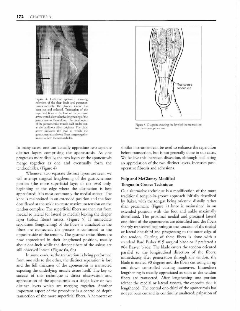
Figure 4. Cadaveric specimen showingreflection of the deep fmcia md paratenontissues medially. The plantaris tendon hmbeen cut md reflected. Trmsection of the
superdcial fibers at the level of the proximalrrowwou.ld allow selective lengthening of the
gutrocnemius fibers alone. The distal mpect
of the gastrocnemius mrscle itself can be seen
m the tendinous fibers originate. The distal
arrow indicates the level at which thegastrocnemius md soleal fibers merge togetheril one to form the tendoachilles.
In many cases, one can actually appreciate two separate
distinct layers comprising the aponeurosis. As one
progresses more distally, the two layers of the aponeurosis
merge together as one and eventually form thetendoachilles. (Figure 4)
\Thenever two separate distinct layers are seen, we
will attempt surgical lengthening of the gastrocnemius
portion (the more superficial layer of the two) only,beginning at the edge where the distinction is best
appreciated; it is most commonly the medial aspect. Theknee is maintained in an extended position and the footdorsiflexed at the ankle ro create maximum rension on the
tendon complex. The superficial fibers are then cut frommedial to lateral (or lateral to medial) leaving the deeper
Iayer (soleal fibers) intact. (Figure 5) If immediateseparation (lengthening) of the fibers is visualized as the
fibers are transected, the process is continued to theopposite side of the tendon. The gastrocnemius fibers are
now appreciated in their lengthened position, usuallyabout one-inch while the deeper fibers of the soleus are
still observed intact. (Figure 6a,6b)In some cases, as the transection is being performed
from one side to the other, the distinct separation is lostand the full thickness of the aponeurosis is transected
exposing the underlying muscle tissue itself. The key tosuccess of this technique is direct observation and
appreciation of the aponeurosis as a single layer or twodistinct layers which are merging together. Anotherimportant aspect of the procedure is a controlled depthtransection of the more superficial fibers. A hemostat or
Ilir -ir -#
'\ilI
l,litlt'tendon cut
Figure 5. Diagram showing the level ofthe transactionfor the strayer procedure.
similar instrument can be used to enhance the separation
before transection, but is not generally done in our cases.
We believe this increased dissection, although faciiitatingan appreciation of the rwo distinct layers, increases post-
operative fibrosis and adhesions.
Fulp and McGlamry ModifiedTongue-in-Groove Technique
Our alternative technique is a modification of the more
traditional tongue-in-groove approach initially described
by Bakea with the tongue being oriented distally rather
than proximally. (Figure 7) knee is maintained in an
extended position with the foot and ankle maximallydorsiflexed. The proximal medial and proximal lateral
one-third of the aponeurosis are identified and the fibers
sharpiy transected beginning at the junction of the medialor lateral one-third and progressing to the outer edge ofthe tendon, Cutting of these fibers is done with a
standard Bard Parker #15 surgical blade or if preferred a#64 Beaver blade. The blade enters the tendon orientedparallel to the longitudinal direction of the fibers;
immediately after penetration through the tendon, the
blade is rotated 90 degrees and the fibers cut using an up
and down controlled cutting maneuver. Immediatelengthening is usually appreciated as soon as the tendonfibers are transected. After lengthening one portion(either the medial or lateral aspect), the opposite side is
lengthened. The central one-third of the aponeurosis has
not yet been cut and its continuiry unaltered; palpation of

CHAPTER31 173
Figure 6a. This is the intraoperative appearance followingffmsverse transection of the gastrocnemius aponeurosis
fibers only. The underlying fibers represent the tendinousportion of the soleus muscle. The lateral fibers of theunderlying soleus have also been transected due to thetightness of the fibers following lengthening of thegmtrocnemius fibers. The decision to transect these fibersis an intraoperative one.
these central fibers will demonstrate their toughness. Care
is taken to avoid accidental cutting of muscle tissues as
these bleed quite readily when traumatized.The foot is then plantarflexed and the distal central
one-third fibers of the aponeurosis are identified, grasped
with forceps and transected using a sharp technique.Slight dorsiflexion of the ankle will cause the centralfibers to become taught and facilitate grasping them withforceps. These fibers are not cut from medial to lateral orvice verse, but rather, cut the outside edge to the center ofthe aponeurosis. The fibers should be maintained orbunched as a group until they have been completedtransected. Now, the distal central one-third fibers of theaponeurosis have been transected in effect creating thetongue portion of the lengthening technique. At thispoint the surgeon should confirm that the proximalmedial and proximal lateral fibers of the aponeurosis andthe distal central fibers of the aponeurosis have all been
cut. Several inches of distance should be present betweenthe proximal tendon cuts and the distal rendon cur.
Once confirmation of the segments lengthened is
confirmed, the foot is held in neutral position and the knee
maintained in an extended position; the ankle is dorsiflexedunder a controlled gradually increasing load until sufficienttendon lengthening is observed. The actual lengthening is
Figure 6b. Appearance of a pediatric patient lollowing a modified strayerprocedure in which the gastrocnemius aponeurosis fibers have been transectedand lengthened. Note the underlying preseruation of the soleal fibers. Thedistal portion of rhe transected plantaris tendon can also be seen.
Proximalmedial
113 libers
Proximallateral'l13 fibers
Distalcentral
1/3 fibers
5.0 clndistancebetweenproximal anddisiaitendon culs
Figure 7. Diagram showing the location of the 3
separate cuts for the Fulp & McGlamry ModifiedTongue-in-Groove procedure
quite impressive and is typically I - 1-,lz inches. If length-
ening is not observed, the site is inspected to ensure that themedial one-third, lateral one-third and central one-thirdfibers have indeed been cut.
'While most surgeons reinforce the lengthened tendonat various points where the slide lengthening overlaps, thesenior author does not perform any suturing of the newly
lengthened tendon and has not done so for the last 13 years.
No complications attributed to not suturing the lengthened
tendon have been encountered. Further lengthening does
not seem to occur as the limb is protected in a short leg cast

174 CHAPTER3l
with the foot at 90 degrees. Some surgeons do prefer to ciose
the defect distal to the end of the tongue portion L,y
suturing the medial and lateral edges together. The senior
author does not perform this maneuver and has notencountered any complications as a result of not doing so.
The wound is then irrigated with copious amounts ofnormal sterile saline. \Mith the knee extended and the footdorsiflexed, the areas oflengthening are palpated to check
for individual deeper strands of tendon which are stillintact and require transection. They are like individualspaghetti strands of taut tendon which can cause significantpostoperative discomfort if not released. Release of these
individual strands is analogous to performing a
percutaneous tenotomy.Once adequate lengthening of the tendon is
confirmed, the wound is irrigated and prepared forclosure. The deep fascia and paratenon layer are closed as
a single layer or unit with synthetic absorbablemultifilament 3-0 or 4-0 suture in a continuous runningmanner. (Figure 8) The foot is periodically dorsiflexedand plantarflexed to ensure that this layer is notaccidentally sutured to the underlying tendon itself. Theunderlying tendon should be observed moving freelywithout attachment at any point to the overlyingparatenon or deep fascia. Next, the subcutaneous tissue
layer is closed with a running stitch of syntheticabsorbable, multifilament 4-0 suture. The skin is repairedwith an intradermal or subcuticular stitch of 5-0absorbable or nonabsorbable suture ofchoice. Full lengthwound closure strips are applied to minimize tension onthe skin edges and help attain a fine line scar; they are leftintact and undisturbed for 4-6 weeks.
No additional local anesthetic is usually required since
long acting solutions were employed from the start of the
procedure. If the nerve is encountered it can be bathed withshort acting steroid to decrease inflammation and
postoperative scar tissue formation around the nerve. If a
tourniquet was empioyed, which we find unnecessary it is
released and additional hemostasis achieved with ligatures
or electrocautery. A dry sterile dressing is applied.
P OSTO PERAITVE MANAGEMENT
Postoperative management consists of cast immobilization fora minimum of four weeks and more commonly 5-6 weels.Other procedures will dictate longer periods of immobiiiza-tion in most patients. At the very minimum, the senior
author recommends a non-weightbearing status for the first 4
weeks, followed by progressing increase in weight bearing tofrrll weightbearing and ambulation by the sixth week.
The cast is usually removed six weeks postoperatively.
figure 8. Intraoperative photo showing partial closure ofthe deep fascia md underlving parateneon as a single
Iayer. The deep fmcia layer, rvhich is more super'{icial, hma whiter slightlv denscr appearmce although it is a very'
thin layer. The underli,ing paratenon fibers are a
gossamerJike tissue layer, which presewe the glidingftlnction of the underlying tendon.
The wound closure strips are also removed. Physical therapy
can be instituted to help restore full strength while main-taining good ankle dorsiflexion to at least 10 degrees.
Edema, which is rarely a significant issue with this
procedure, is easily controlled with compression stockings,
Iow dose diuretic therapy and physical therapy. By 3-4months, most patients will make a full functional recovery.
COMPLICAIIONS
\fith meticulous surgical technique and proper post-
operative management, complications following this rypeof surgery are rare. Like most surgical procedures,
wound infection and complication remain dreaded butrare occurrences.
Complications specific to tendon procedures
include over lengthening or under lengthening and again
are rare problems. Over lengthening could result in a
calcaneus deformity but is more common followingtendoachilles lengthening. Recurrence of the equinus
could also occur but is rarely reported. Its occurrence is
more likely when these procedures are performed inpatients with underlying neuromuscular disease,
especially those associated with spasticity.

CHAPTER31 175
A complication of particular concern is entrapmentneuropathy of the sural nerve within the incision site.
This complication can result in minimal symptomatologyand in other cases, severe neuritic symptoms whichmimick complex regional pain syndrome. A specifictrigger point can usually be identified. A selectivediagnostic nerve biock can be administered and willusuaily resolve all symptoms for a short period of timeconfirming the clinical suspicion of a post surgicalentrapment neuropathy. If patients fail to respond topharmacologic agents, physical therapy and tincture oftime, surgical exploration with release of the nerve is
likely to be required. In some patients resection of thesural nerve may be necessary.
REFERENCES
1. Downey MS. Ankle equinus. In: Banks AS, Downey MS, MartinDE, Miller SJ, editors. Comprehensive textbook of foot surgery.
Baltimore: Lippincott \i7i11iams & Vilkins; 2001. p. 715-760.2. Downey MS. Arkte equinus. In: McGlamry ED, Banks AS,
Downey MS, editors. Comprehensive textbook ol foot surgery.
Baltimore: \Williams &'Milkins; 1992. p. 687 -7 30.
3. Schoenhaus HD, ]ay R1vI. A modified gastrocnemius lengthening. /Am Podiatry Assoc 1978:68:31-7.
4. DiGiovanni C-i7, Kuo R, Tejwani N, Price R et al. Isolated
gastrocnemius tightness. J Bone Joint Surg Am 2002;84:962-9.
5. Fulp MJ, McGlamry ED. Gastrocnemius tendon recession: tongue
in groove procedure to lengthen gastrocnemius tendon. J AmPodiatry Asso c 197 4;64:1 63 -7 1.
6. Downey MS, Banks AS. Gastronemius recession in the treatment ofnonspastic ankle equinus: a retrospective srtdy. J Am Podiany MedAssoc l9B9 19:159-74.
7. Silverskiold N. Reduction of the uncrossed two-joints muscles of the
leg to one-joint muscles in spastic conditions. Acta Chir Scand
t924;55:3t5-30.B. Stoffel A. The treatment of spastic contractures. Ant J Orthop Surg
t913;10617-44.9. Martz CD. Talipes equinus Correction in cerebral palsy. J Bone Jt
Swrg 7950;42:769-76.
10. Silver CM. Simon SD. Gastrocnemius-muscle recession
(Silfverskiold operation) for spastic equines deformiry in cerebral
palsy. J Bone Jt Surg Am 1959; 4l:1021-8.ll.Vulpius O, Stoffel A. Orthopadische Operationslehre. Ferdinand
Enke, Stuttgart, 1913. Tenotomie der end schnen der mm.gastrocnemius e1 soleus mittels rutschenlassens nach vulpius; p 29-31.
12. Strayer LM. Recession of the gastrocnemius: an operation to relieve
spastic contracture of the calf muscles. J Bone Jt Surg Am 7950;
32:677-6.
l3.Strayer LM. Gastrocnemius recession: five-year report of cases.
J Bone Jt Surg Am.l958;40:1019-30.14. Pinney S], Hansen ST, Sangeorzan BJ. The effect on anlde dorsiflex-
ion of gastrocnemius recession. Foot Ankle Int 2002;23:26-9.
15.Donley BG, Pinney SJ, Holmes A. Gastronemius recession. Terlr
Fo ot An k le Surg 2003 ;2:3 5 -9.
16.Baker LD. A Rational approach to the surgical needs ofthe cerebral
palsy patient. / Bone Jt Surg Am 1916;38:313-23.
17.Sa-xena A. Endoscopic gastrocnemius tenotomy. J Foot Ankle Surg
2002;41.:57-8.l8.Tashjian RZ, Appel lA, Banerjee, DiGiovanni C\7. Endoscopic
gastrocnemius recession: evaluation in a cadaver model. Foot Ankle
Int 2003:24:607-12.
ADDITIONAL REFERENCES
Banks HH, Green \7T. The correction of equinus deformity in cerebral
palsey. J Bone Joint Surg Am 7958; 40:1.359-79.
Borton DC, Valker K, Pirpiris M, Nattrass GR, Graham HK. Isolated
calf lengthening in cerebral palsy. J Bone Jt Surg Br 2001,;83:364-70.
Craig JJ, Van Vuren J. The importance of gastrocnemius recession inthe correction ofequines deformity in cerebral palsy. J Bone Jt Surg
Br 1976:58:84-7.
Grady JF, Sa-rena A. Eflects of Stretching the gastrocnemius muscle.
J Foot Surg 1991;30:465-69.Green \flT, Calandriello B. The detachment of gastrocnemius muscles
in the treatment of spastic equinus foot. Bull Hosp Jt Dis1959;20:48-52.
Green'VT, McDermott LJ. Operative treatment of cerebral palsy of the
spastic rype. JAMA 19 42;1 18 :43 4-40.
Javors JR, Klarren HE. The vulpius procedure for correction of equinus
deformiry in cerebral palsy. J Pediatric Orthop 1987;7:191-3.
McGlamry ED, Kitting RrW. Equinus foot: An anlysis of the etiology,
pathology and treatment techniques. J Am Podiatry Assoc
1973;63:765-84.
Pollack GA. Lengthening of the gastrocnemius tendon in cases ofspastic equines deformity. J Bone Jt Surg Br 1953'35:148.
Sharrard \X!\7, Bernstein S. Equinus deformiry in cerebral palsy:
a comparison between elongation of the tendo calcaneus and
gastrocnem.ius recession. J Bone Jt Surg Br 1972; 54:272-6.Thomas F, Sands AK. Treatment of equinus contracture. Tech Foot
Ankle Surg 2003; 2;lB0-5.Takahashi S, Shrestha A. The Vulpius procedure for correction of
equinus deformiry in patients with hemiplegia. J Bone Jt Surg Br2002;84:979-80.
Yngve DA, Chambers C. Vulpius and ZJengthening. J Pediatr Orthop
1996;16759-64.