Embed Size (px)
Citation preview
286 | MAY 2012 | VOLUME 9 www.nature.com/nrgastro
Department of Anatomy and Neuroscience, University of Melbourne, Grattan Street, Parkville, VIC 3010, Australia. j.furness@ unimelb.edu.au
The enteric nervous system and neurogastroenterologyJohn B. Furness
Abstract | Neurogastroenterology is defined as neurology of the gastrointestinal tract, liver, gallbladder and pancreas and encompasses control of digestion through the enteric nervous system (ENS), the central nervous system (CNS) and integrative centers in sympathetic ganglia. This Review provides a broad overview of the field of neurogastroenterology, with a focus on the roles of the ENS in the control of the musculature of the gastrointestinal tract and transmucosal fluid movement. Digestion is controlled through the integration of multiple signals from the ENS and CNS; neural signals also pass between distinct gut regions to coordinate digestive activity. Moreover, neural and endocrine control of digestion is closely coordinated. Interestingly, the extent to which the ENS or CNS controls digestion differs considerably along the digestive tract. The importance of the ENS is emphasized by the life-threatening effects of certain ENS neuropathies, including Hirschsprung disease and Chagas disease. Other ENS disorders, such as esophageal achalasia and gastroparesis, cause varying degrees of dysfunction. The neurons in enteric reflex pathways use a wide range of chemical messengers that signal through an even wider range of receptors. These receptors provide many actual and potential targets for modifying digestive function.
Furness, J. B. Nat. Rev. Gastroenterol. Hepatol. 9, 286–294 (2012); published online 6 March 2012; doi:10.1038/nrgastro.2012.32
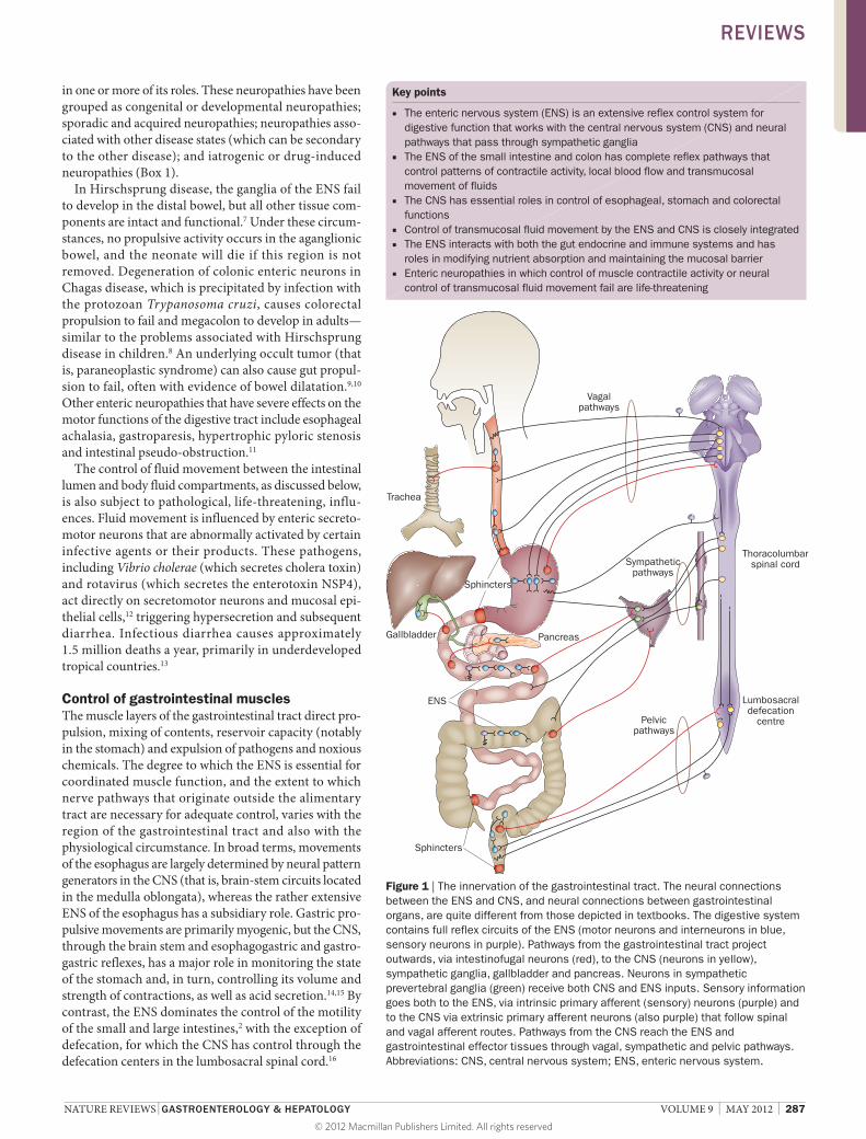
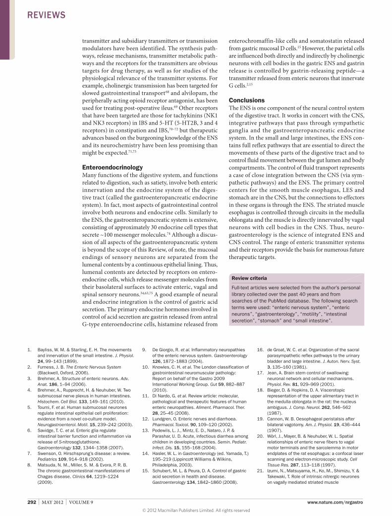
Introduction The gastrointestinal tract differs from all other peripheral organs in that it has an extensive intrinsic nervous system, termed the enteric nervous system (ENS) that can control functions of the intestine even when it is completely separated from the central nervous system (CNS).1 The ENS, however, is not autonomous. Indeed, neuronal control of gastrointestinal function is an integrated system involving interactions between local enteric reflexes, reflexes that pass through sympathetic ganglia and reflexes that pass from the gut and back through the CNS (Figure 1). Conventional textbook descriptions of the autonomic nervous system depict efferent pathways from the CNS as two neurons in series, a preganglionic and a postganglionic neuron, and depict sensory information flowing from the periphery to the CNS through spinal and cranial primary afferent neurons. The organization of the ENS and of neuronal pathways to and from the intestine does not, however, follow these conventional concepts of the organization of the nervous system (Figure 1). For example, axons of neurons with cell bodies in the ENS (called intestinofugal neurons) project to sympathetic ganglia, the pancreas, gallbladder and trachea, and to the spinal cord and brain stem (Figure 1).2
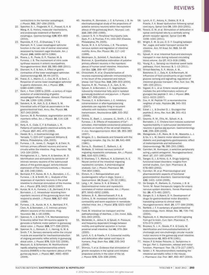
The ENS is composed of small aggregations of nerve cells, enteric ganglia, the neural connections between these ganglia, and nerve fibers that supply effector tissues, including the muscle of the gut wall, the epithelial lining, intrinsic blood vessels and gastroentero pancreatic
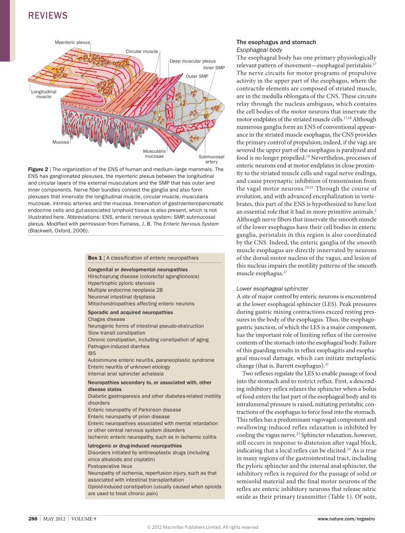
endocrine cells (Figure 2). The total number of enteric neurons in humans is 400–600 million, which is greater than the total of all sympathetic and parasympathetic ganglia combined and approximately equal to the number of neurons in the spinal cord.2 Figure 2 is representative of the ENS of the mammalian small intestine; the differences in structure and organization between regions of the gastrointestinal tract and species have been reviewed elsewhere.2–4 Differences include the absence of a ganglionated submucosal plexus in the esophagus and stomach (organs that lack the large fluid fluxes across the mucosal epithelium that occur in the small and large intestines), the presence of a diffuse network of ganglia in the pancreas and a single layer of ganglia in the intestinal submucosa of small mammals. This Review is confined to discussion of monogastric mammals, in which most investigations have been done and which are arguably most relevant to humans.
Importance of the ENS The ENS has multiple roles: determining the patterns of movement of the gastrointestinal tract; controlling gastric acid secretion; regulating movement of fluid across the lining epithelium; changing local blood flow; modifying nutrient handling; and interacting with the immune and endocrine systems of the gut.2 The ENS also contributes, along with glial cells, to maintaining the integrity of the epithelial barrier between the gut lumen and cells and tissues within the gut wall.5,6 The importance of the ENS is highlighted by the wide range of enteric neuropathies that are caused following a failure
Competing interestsThe author declares no competing interests.
REVIEWS
© 2012 Macmillan Publishers Limited. All rights reserved
NATURE REVIEWS | GASTROENTEROLOGY & HEPATOLOGY VOLUME 9 | MAY 2012 | 287
in one or more of its roles. These neuropathies have been grouped as congenital or developmental neuropathies; sporadic and acquired neuropathies; neuropathies associated with other disease states (which can be secondary to the other disease); and iatrogenic or druginduced neuropathies (Box 1).
In Hirschsprung disease, the ganglia of the ENS fail to develop in the distal bowel, but all other tissue components are intact and functional.7 Under these circumstances, no propulsive activity occurs in the aganglionic bowel, and the neonate will die if this region is not removed. Degeneration of colonic enteric neurons in Chagas disease, which is precipitated by infection with the protozoan Trypanosoma cruzi, causes colorectal propulsion to fail and megacolon to develop in adults—similar to the problems associated with Hirschsprung disease in children.8 An underlying occult tumor (that is, paraneoplastic syndrome) can also cause gut propulsion to fail, often with evidence of bowel dilatation.9,10 Other enteric neuropathies that have severe effects on the motor functions of the digestive tract include esophageal achalasia, gastroparesis, hypertrophic pyloric stenosis and intestinal pseudoobstruction.11
The control of fluid movement between the intestinal lumen and body fluid compartments, as discussed below, is also subject to pathological, lifethreatening, influences. Fluid movement is influenced by enteric secretomotor neurons that are abnormally activated by certain infective agents or their products. These pathogens, including Vibrio cholerae (which secretes cholera toxin) and rotavirus (which secretes the enterotoxin NSP4), act directly on secretomotor neurons and mucosal epithelial cells,12 triggering hypersecretion and subsequent diarrhea. Infectious diarrhea causes approximately 1.5 million deaths a year, primarily in underdeveloped tropical countries.13
Control of gastrointestinal muscles The muscle layers of the gastrointestinal tract direct propulsion, mixing of contents, reservoir capacity (notably in the stomach) and expulsion of pathogens and noxious chemicals. The degree to which the ENS is essential for coordinated muscle function, and the extent to which nerve pathways that originate outside the alimentary tract are necessary for adequate control, varies with the region of the gastrointestinal tract and also with the physiological circumstance. In broad terms, movements of the esophagus are largely determined by neural pattern generators in the CNS (that is, brainstem circuits located in the medulla oblongata), whereas the rather extensive ENS of the esophagus has a subsidiary role. Gastric propulsive movements are primarily myogenic, but the CNS, through the brain stem and esophagogastric and gastrogastric reflexes, has a major role in monitoring the state of the stomach and, in turn, controlling its volume and strength of contractions, as well as acid secretion.14,15 By contrast, the ENS dominates the control of the motility of the small and large intestines,2 with the exception of defecation, for which the CNS has control through the defecation centers in the lumbosacral spinal cord.16
Key points
■ The enteric nervous system (ENS) is an extensive reflex control system for digestive function that works with the central nervous system (CNS) and neural pathways that pass through sympathetic ganglia
■ The ENS of the small intestine and colon has complete reflex pathways that control patterns of contractile activity, local blood flow and transmucosal movement of fluids
■ The CNS has essential roles in control of esophageal, stomach and colorectal functions
■ Control of transmucosal fluid movement by the ENS and CNS is closely integrated ■ The ENS interacts with both the gut endocrine and immune systems and has
roles in modifying nutrient absorption and maintaining the mucosal barrier ■ Enteric neuropathies in which control of muscle contractile activity or neural
control of transmucosal fluid movement fail are life-threatening
Sphincters
Pelvicpathways
Sympatheticpathways
Vagalpathways
Lumbosacraldefecation
centre
Sphincters
Trachea
Gallbladder Pancreas
ENS
Thoracolumbarspinal cord
Figure 1 | The innervation of the gastrointestinal tract. The neural connections between the ENS and CNS, and neural connections between gastrointestinal organs, are quite different from those depicted in textbooks. The digestive system contains full reflex circuits of the ENS (motor neurons and interneurons in blue, sensory neurons in purple). Pathways from the gastrointestinal tract project outwards, via intestinofugal neurons (red), to the CNS (neurons in yellow), sympathetic ganglia, gallbladder and pancreas. Neurons in sympathetic prevertebral ganglia (green) receive both CNS and ENS inputs. Sensory information goes both to the ENS, via intrinsic primary afferent (sensory) neurons (purple) and to the CNS via extrinsic primary afferent neurons (also purple) that follow spinal and vagal afferent routes. Pathways from the CNS reach the ENS and gastrointestinal effector tissues through vagal, sympathetic and pelvic pathways. Abbreviations: CNS, central nervous system; ENS, enteric nervous system.
REVIEWS
© 2012 Macmillan Publishers Limited. All rights reserved
288 | MAY 2012 | VOLUME 9 www.nature.com/nrgastro
The esophagus and stomach Esophageal body The esophageal body has one primary physiologically relevant pattern of movement—esophageal peristalsis.17 The nerve circuits for motor programs of propulsive activity in the upper part of the esophagus, where the contractile elements are composed of striated muscle, are in the medulla oblongata of the CNS. These circuits relay through the nucleus ambiguus, which contains the cell bodies of the motor neurons that innervate the motor endplates of the striated muscle cells.17,18 Although numerous ganglia form an ENS of conventional appearance in the striated muscle esophagus, the CNS provides the primary control of propulsion; indeed, if the vagi are severed the upper part of the esophagus is paralyzed and food is no longer propelled.19 Nevertheless, processes of enteric neurons end at motor endplates in close proximity to the striated muscle cells and vagal nerve endings, and cause pre synaptic inhibition of transmission from the vagal motor neurons.20,21 Through the course of evolution, and with advanced encephalization in vertebrates, this part of the ENS is hypothesized to have lost an essential role that it had in more primitive animals.2 Although nerve fibers that innervate the smooth muscle of the lower esophagus have their cell bodies in enteric ganglia, peristalsis in this region is also coordinated by the CNS. Indeed, the enteric ganglia of the smooth muscle esophagus are directly innervated by neurons of the dorsal motor nucleus of the vagus, and lesion of this nucleus impairs the motility patterns of the smooth muscle esophagus.17
Lower esophageal sphincter A site of major control by enteric neurons is encountered at the lower esophageal sphincter (LES). Peak pressures during gastric mixing contractions exceed resting pressures in the body of the esophagus. Thus, the esophagogastric junction, of which the LES is a major component, has the important role of limiting reflux of the corrosive contents of the stomach into the esophageal body. Failure of this guarding results in reflux esophagitis and esophageal mucosal damage, which can initiate metaplastic change (that is, Barrett esophagus).22
Two reflexes regulate the LES to enable passage of food into the stomach and to restrict reflux. First, a descending inhibitory reflex relaxes the sphincter when a bolus of food enters the last part of the esophageal body and its intralumenal pressure is raised, initiating peristaltic contractions of the esophagus to force food into the stomach. This reflex has a predominant vagovagal component and swallowinginduced reflex relaxation is inhibited by cooling the vagus nerve.23 Sphincter relaxation, however, still occurs in response to distension after vagal block, indicating that a local reflex can be elicited.23 As is true in many regions of the gastrointestinal tract, including the pyloric sphincter and the internal anal sphincter, the inhibitory reflex is required for the passage of solid or semisolid material and the final motor neurons of the reflex are enteric inhibitory neurons that release nitric oxide as their primary transmitter (Table 1). Of note,
Myenteric plexus
Circular muscle
Deep muscular plexusInner SMP
Outer SMP
Submucosalartery
Muscularismucosae
Mucosa
Longitudinalmuscle
Figure 2 | The organization of the ENS of human and medium–large mammals. The ENS has ganglionated plexuses, the myenteric plexus between the longitudinal and circular layers of the external musculature and the SMP that has outer and inner components. Nerve fiber bundles connect the ganglia and also form plexuses that innervate the longitudinal muscle, circular muscle, muscularis mucosae, intrinsic arteries and the mucosa. Innervation of gastroenteropancreatic endocrine cells and gut-associated lymphoid tissue is also present, which is not illustrated here. Abbreviations: ENS, enteric nervous system; SMP, submucosal plexus. Modified with permission from Furness, J. B. The Enteric Nervous System (Blackwell, Oxford, 2006).
Box 1 | A classification of enteric neuropathies
Congenital or developmental neuropathiesHirschsprung disease (colorectal aganglionosis)Hypertrophic pyloric stenosisMultiple endocrine neoplasia 2BNeuronal intestinal dysplasiaMitochondriopathies affecting enteric neurons
Sporadic and acquired neuropathiesChagas diseaseNeurogenic forms of intestinal pseudo-obstructionSlow transit constipationChronic constipation, including constipation of agingPathogen-induced diarrheaIBSAutoimmune enteric neuritis, paraneoplastic syndromeEnteric neuritis of unknown etiologyInternal anal sphincter achalasia
Neuropathies secondary to, or associated with, other disease statesDiabetic gastroparesis and other diabetes-related motility disordersEnteric neuropathy of Parkinson diseaseEnteric neuropathy of prion diseaseEnteric neuropathies associated with mental retardation or other central nervous system disordersIschemic enteric neuropathy, such as in ischemic colitis
Iatrogenic or drug-induced neuropathiesDisorders initiated by antineoplastic drugs (including vinca alkaloids and cisplatin)Postoperative ileusNeuropathy of ischemia, reperfusion injury, such as that associated with intestinal transplantationOpioid-induced constipation (usually caused when opioids are used to treat chronic pain)
REVIEWS
© 2012 Macmillan Publishers Limited. All rights reserved
NATURE REVIEWS | GASTROENTEROLOGY & HEPATOLOGY VOLUME 9 | MAY 2012 | 289
although enteric neurons utilize multiple transmitters,2 each class of neuron has a primary transmitter, which, in this case, is nitric oxide. Nitric oxide is formed in neurons by neuronal nitric oxide synthase. A defect in nitric oxide production results in a failure of esophageal propulsion (termed esophageal achalasia).11 Moreover, failure of nitricoxidemediated transmission from enteric neurons in other parts of the gastrointestinal tract also causes defects in propulsion.24 For example, in the stomach this defect causes gastroparesis, at the pyloric sphincter it causes hypertrophic pyloric stenosis and in the final part of the colon, deficiency of nitric oxide transmission is involved in internal anal sphincter achalasia and Chagas disease.24 In addition, increased pressure in the stomach initiates a reflex constriction of the LES that limits reflux of gastric contents which, like the descending inhibitory reflex, is mediated by a vagovagal reflex pathway that passes through the brain stem.25,26 Failure of the maintained closure of the LES when intragastric pressure exceeds intraesophageal pressure results in GERD.27 In summary, although a structurally welldeveloped ENS exists in the esophageal wall, propulsive activity of the esophagus and the activity of the LES are
controlled through motor pattern generators in the brain stem and reflexes that depend on centers in the CNS.
StomachThe stomach is similar to the esophagus in that much of its neural control is dependent on vagovagal reflexes.2 However, the rhythmic contractile waves that pass caudally over the stomach to knead and propel the contents are generated in the muscle through the activity of interstitial cells of Cajal28 and coordinated gastric muscle activity that is dependent on the ENS is difficult to demon strate.2,29,30 By contrast, propulsive activity in the small intestine and colon is dependent on ENS reflexes.
Small intestine The ability of the small intestine to function when isolated from the CNS was demonstrated over a century ago.1 The small intestine is now known to be dependent on the ENS to direct its various patterns of movement: rapid orthograde propulsion of contents (peristalsis), mixing movements (segmentation), slow orthograde propulsion (the migrating myoelectric complex, MMC) and retropulsion (expulsion of noxious substances associated with
Table 1 | Multiple transmitters of neurons that control digestive function*
Type of neuron Primary transmitter
Secondary transmitters, modulators
Other neurochemical markers
Study
Enteric excitatory muscle motor neuron
ACh Tachykinin, enkephalin (presynaptic inhibition)
Calretinin, γ-aminobutyric acid
Brookes et al. (1991);76 Holzer & Holzer Petsche (1997);77 Grider (2003)78
Enteric inhibitory muscle motor neuron
Nitric oxide VIP, ATP or ATP-like compound, carbon monoxide
PACAP, opioids Fahrenkrug et al. (1978);79 Costa et al. (1992);80 Sanders & Ward (1992);81 Xue et al. (2000)82
Ascending interneuron ACh Tachykinin, ATP Calretinin, enkephalin Brookes et al. (1991)83
ChAT, NOS descending interneuron
ATP, ACh ND Nitric oxide, VIP Young et al. (1995);84 Brookes (2001)85
ChAT, 5-HT descending interneuron
ACh 5-HT, ATP ND Furness & Costa (1982);86 Monro et al. (2002);87 Gwynne & Bornstein (2007)88
ChAT, somatostatin descending interneuron
ACh ND Somatostatin Gwynne & Bornstein (2007);88 Portbury et al. (1995)89
Intrinsic sensory neuron ACh, CGRP, tachykinin
ND Calbindin, calretinin, IB4 binding
Grider (2003);78 Gwynne & Bornstein (2007);88 Li & Furness (1998);90 Johnson & Bornstein (2004)91
Interneurons supplying secretomotor neurons
ACh ATP, 5-HT ND Suprenant (1984);92 Monro et al. (2004)93
Noncholinergic secretomotor neuron
VIP PACAP NPY (in most species) Cassuto et al. (1981);94 Banks et al. (2005)95
Cholinergic secretomotor neuron ACh ND Calretinin Brookes et al. (1991);83 Keast et al. (1985)96
Motor neuron to gastrin cells GRP, ACh ND NPY Holst et al. (1987);97 Weiget et al. (1997)98
Motor neurons to parietal cells ACh Potentially VIP ND Nilsson et al. (1972);99 Feldman et al. (1979)100
Sympathetic neurons, motility inhibiting
Noradrenaline ND NPY in some species Finkleman (1930);101 Macrae et al. (1986)102
Sympathetic neurons, secretion inhibiting
Noradrenaline Somatostatin (in guinea pig)
ND Costa & Furness (1984)103
Sympathetic neurons, vasoconstrictor
Noradrenaline, ATP
Potentially NPY NPY Dresel & Wallentin (1966);104 Furness (1971);105 Furness et al. (1983)106
Intestinofugal neurons to sympathetic ganglia
ACh VIP Opioid peptides, CCK, GRP
Crowcroft et al. (1971);107 Dalsgaard et al. (1983);108 Love & Szurszewski (1987)109
*This field continues to advance as improved pharmacological and other tools are developed. Some of the information provided here will therefore undoubtedly be superseded in the future. Further details of postsynaptic receptors have been described elsewhere.2,88 Abbreviations: 5-HT, 5-hydroxytryptamine; ACh, acetylcholine; CCK, cholecystokinin; ChAT, choline acetyltransferase; CGRP, calcitonin gene-related peptide; GRP, gastrin releasing peptide; ND, not determined; NPY, neuropeptide Y; NOS, nitric oxide synthase; PACAP, pituitary adenylyl-cyclase activating peptide; VIP vasoactive intestinal peptide.
REVIEWS
© 2012 Macmillan Publishers Limited. All rights reserved
290 | MAY 2012 | VOLUME 9 www.nature.com/nrgastro
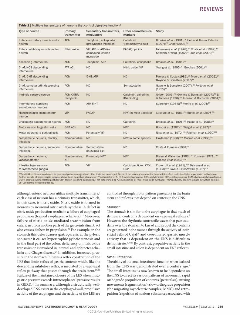
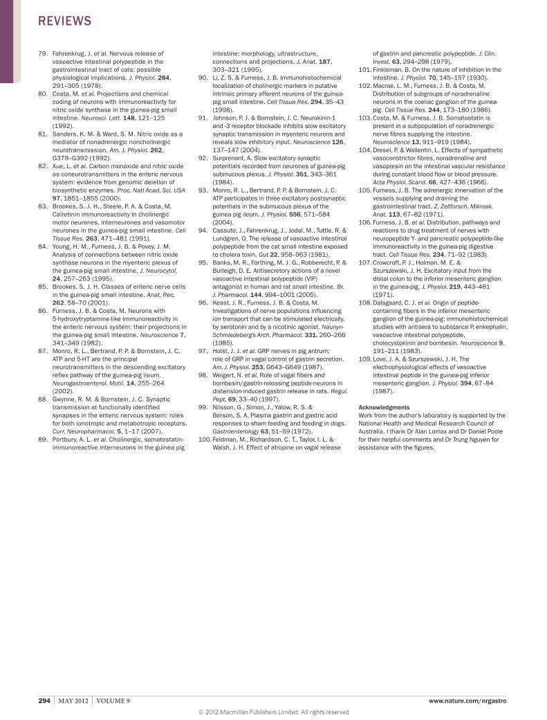
vomiting).31 To orchestrate these movement patterns, the state of the intestine is sensed and appropriate motor patterns are generated. The structural organization of the circuits that detect the state of the small intestine, integrate the information and direct the activities of motor neurons has now been determined (Figure 3).2
Original observations suggested that the ENS contains intrinsic sensory neurons (also referred to as intrinsic primary afferent neurons, IPANs), interneurons and both excitatory and inhibitory motor neurons that innervate the muscle.1 However, identification of these neurons by their shapes, projections to targets, neurochemistries and cell physiologies has only occurred in the past decade. IPANs were first identified as large neurons with multiple, usually 2–6, long processes (type II morphology) that respond to changes in lumenal chemistry, mechanical distortion of the mucosa and direct mechanical distortion of their processes in the external musculature.32–36 Since that time, mechanical distortion has also been shown to excite some uni polar neurons in the enteric nerve circuits,37–39 indicating that reflexes are not uniquely initiated through type II neurons. Cell bodies of multipolar IPANs are 10–30% of neurons in the submucosal and myenteric ganglia of the small and large intestines. The long axonlike processes of these neurons conduct action potentials. Moreover, action potentials that are initiated in one process traverse the cell bodies and are conducted along other processes to synaptic connections with IPANs, interneurons and motor neurons (Figure 3).36,40 Consistent with the motor functions of the esophagus being controlled from or through the brain stem, type II neurons are not found in the esophagus and are rare in the stomach.2
Enteric IPANs have many features in common with small diameter primary afferent neurons of the dorsal root ganglia (DRG), which connect in the CNS. These features include containing peptides that are typical
of smalldiameter afferent neurons (such as substance P and calcitonin generelated peptide), having tetrodotoxin resistant voltagedependent Na+ and highvoltageactivated Ca2+ currents, isolectin B4 binding and unmyelinated axons.32,41 As with smalldiameter DRG primary afferents, the enteric IPANs are polymodal and some, at least, are nociceptors.32,41 However, unlike DRG neurons, the multipolar IPANs connect with each other to form selfreinforcing networks (Figure 3).42 The similarities of IPANs and DRG primary afferent neurons that supply visceral organs suggest that primary afferent neurons of neural crest origin have similar phenotypes independent of the final location of their cell bodies. Thus, these characteristics can be used to identify IPANs in other species, for example in humans3,43 and large mammals44 where physiological characterization is not always feasible. Structural identification of these neurons has relevance for the investigation of both human and animal enteric neuropathies.
Although the neural circuits are now characterized, the mechanisms within the integrative circuitry through which one pattern of activity is converted to another are not yet defined. Nevertheless, signals that trigger changes in patterns of movement have been identified. For example, fatty acids added to the lumenal surface convert propulsive contractile activity to mixing movements, through a neural mechanism.45 Conversion from one pattern to another can also be achieved with drugs that target enteric neurons. For example, TRAM34mediated inhibition of the intermediate conductance, calciumactivated, potassium IKCa channel, which underlies the slow hyperpolarization that follows the action potential in IPANs, changes propulsive movements to mixing movements, possibly by altering the timing of action potential firing in IPANs.46,47
The MMC traverses the small intestine periodically between meals, in humans approximately every 90 min.48 Its passage is slow, ~1–4 cm per minute in humans, and its suggested role is to remove residual contents from the intestine after a meal has been digested (and thus it has been called the intestinal housekeeper), thereby reducing bacterial overgrowth in the small intestine.31 The MMC is dependent on the ENS for its progress along the intestine, as shown by its inhibition by the local infusion of tetrodotoxin49 or by the nicotinic blocker, hexamethonium.50
The detection of noxious agents (nociception) is important to initiate reactions that expel pathogens and potentially injurious chemicals. Intrinsic retropulsive reflexes in the small intestine force the contents back to the stomach in association with vomiting,51,52 and in the large intestine pathogens activate the ENS to cause large propulsive contractions and copious fluid secretion.53,54 Under nonpathological conditions fluid secretion is also coupled with contractions of the circular muscle of the bowel wall.55
Large intestine Colonic motility is dependent on the ENS for propulsion of contents. Indeed, colonic motility fails when the ENS of the distal colon and rectum is congenitally absent in
Oral Anal
Interneuron
InterneuronLongitudinal muscle
motor neuronCircular musclemotor neuron
Network of IPANs
Figure 3 | Nerve circuits for control of motility in the small intestine. Nerve circuits were identified from studies in the guinea pig small intestine and similar component neurons have been identified in the small intestine of other species, including humans, and in the large intestine. Nevertheless, this figure is a simplified circuit diagram showing the major circuit features that have been identified. Networks of interconnected intrinsic sensory neurons (IPANs; red) detect mechanical distortion and lumenal chemistry. These synapse with descending (yellow) and ascending (green) interneurons, excitatory muscle motor neurons (blue) and inhibitory muscle motor neurons (purple). Abbreviation: IPAN, intrinsic primary afferent neuron. Modified with permission from Furness, J. B. The Enteric Nervous System (Blackwell, Oxford, 2006).
REVIEWS
© 2012 Macmillan Publishers Limited. All rights reserved
NATURE REVIEWS | GASTROENTEROLOGY & HEPATOLOGY VOLUME 9 | MAY 2012 | 291
patients with Hirschsprung disease,7 when it degenerates in later life, as in Chagas disease8 or is compromised in other forms of enteric neuropathies. Nevertheless, the CNS directs ENS activity in the colorectum. In healthy individuals, the propulsive reflexes of the distal colon and rectum are regulated by central control centers that maintain fecal continence and, when it is appropriate, trigger defecation through central commands that are relayed through the defecation center in the lumbosacral spinal cord (Figure 1).16,56 Indeed, direct stimulation of the defecation center in the spinal cord causes coordinated emptying of the colon, via the ENS.57 Voluntary controls of defecation (both inhibition and facilitation) are lost if cortico–spinal connections to the defecation centers are severed by spinal injury.58 Nevertheless, if the defecation center remains intact after spinal injury it can be stimulated to command the ENS pathways for bowel emptying.59 Indeed, a ghrelin receptor agonist is now being developed for use in clinical practice in patients with spinal injuries.
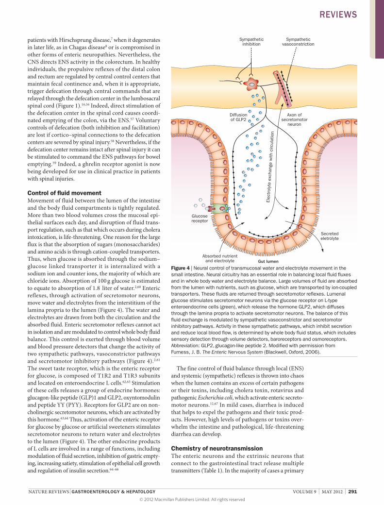
Control of fluid movement Movement of fluid between the lumen of the intestine and the body fluid compartments is tightly regulated. More than two blood volumes cross the mucosal epithelial surfaces each day, and disruption of fluid transport regulation, such as that which occurs during cholera intoxication, is lifethreatening. One reason for the large flux is that the absorption of sugars (mono saccharides) and amino acids is through cationcoupled transporters. Thus, when glucose is absorbed through the sodium–glucose linked transporter it is internalized with a sodium ion and counter ions, the majority of which are chloride ions. Absorption of 100 g glucose is estimated to equate to absorption of 1.8 liter of water.2,60 Enteric reflexes, through activation of secretomotor neurons, move water and electrolytes from the interstitium of the lamina propria to the lumen (Figure 4). The water and electrolytes are drawn from both the circulation and the absorbed fluid. Enteric secretomotor reflexes cannot act in isolation and are modulated to control whole body fluid balance. This control is exerted through blood volume and blood pressure detectors that change the activity of two sympathetic pathways, vasoconstrictor pathways and secretomotor inhibitory pathways (Figure 4).2,61 The sweet taste receptor, which is the enteric receptor for glucose, is composed of T1R2 and T1R3 subunits and located on enteroendocrine L cells.62,63 Stimulation of these cells releases a group of endocrine hormones: glucagonlike peptide (GLP)1 and GLP2, oxyntomodulin and peptide YY (PYY). Receptors for GLP2 are on noncholinergic secretomotor neurons, which are activated by this hormone.63,64 Thus, activation of the enteric receptor for glucose by glucose or artificial sweeteners stimulates secretomotor neurons to return water and electrolytes to the lumen (Figure 4). The other endocrine products of L cells are involved in a range of functions, including modulation of fluid secretion, inhibition of gastric emptying, increasing satiety, stimulation of epithelial cell growth and regulation of insulin secretion.64–66
The fine control of fluid balance through local (ENS) and systemic (sympathetic) reflexes is thrown into chaos when the lumen contains an excess of certain pathogens or their toxins, including cholera toxin, rotavirus and pathogenic Escherichia coli, which activate enteric secretomotor neurons.12,67 In mild cases, diarrhea is induced that helps to expel the pathogens and their toxic products. However, high levels of pathogens or toxins overwhelm the intestine and pathological, life threatening diarrhea can develop.
Chemistry of neurotransmission The enteric neurons and the extrinsic neurons that connect to the gastrointestinal tract release multiple transmitters (Table 1). In the majority of cases a primary
Gut lumen
Sympatheticinhibition
Sympatheticvasoconstriction
Diffusionof GLP2
Axon ofsecretomotor
neuron
Secretedeletrolyte
Absorbed nutrientand electrolyte
Glucosereceptor
Elec
trol
yte
exch
ange
with
circ
ulat
ion
Figure 4 | Neural control of transmucosal water and electrolyte movement in the small intestine. Neural circuitry has an essential role in balancing local fluid fluxes and in whole body water and electrolyte balance. Large volumes of fluid are absorbed from the lumen with nutrients, such as glucose, which are transported by ion-coupled transporters. These fluids are returned through secretomotor reflexes. Lumenal glucose stimulates secretomotor neurons via the glucose receptor on L-type enteroendocrine cells (green), which release the hormone GLP2, which diffuses through the lamina propria to activate secretomotor neurons. The balance of this fluid exchange is modulated by sympathetic vasoconstrictor and secretomotor inhibitory pathways. Activity in these sympathetic pathways, which inhibit secretion and reduce local blood flow, is determined by whole body fluid status, which includes sensory detection through volume detectors, baroreceptors and osmoreceptors. Abbreviation: GLP2, glucagon-like peptide 2. Modified with permission from Furness, J. B. The Enteric Nervous System (Blackwell, Oxford, 2006).
REVIEWS
© 2012 Macmillan Publishers Limited. All rights reserved
292 | MAY 2012 | VOLUME 9 www.nature.com/nrgastro
transmitter and subsidiary transmitters or transmission modulators have been identified. The synthesis pathways, release mechanisms, transmitter metabolic pathways and the receptors for the transmitters are obvious targets for drug therapy, as well as for studies of the physiological relevance of the transmitter systems. For example, cholinergic transmission has been targeted for slowed gastrointestinal transport68 and alvilopam, the peripherally acting opioid receptor antagonist, has been used for treating postoperative ileus.69 Other receptors that have been targeted are those for tachykinins (NK1 and NK3 receptors) in IBS and 5HT (5HT2B, 3 and 4 receptors) in constipation and IBS,70–72 but therapeutic advances based on the burgeoning knowledge of the ENS and its neuro chemistry have been less promising than might be expected.71,73
Enteroendocrinology Many functions of the digestive system, and functions related to digestion, such as satiety, involve both enteric innervation and the endocrine system of the digestive tract (called the gastroenteropancreatic endocrine system). In fact, most aspects of gastrointestinal control involve both neurons and endocrine cells. Similarly to the ENS, the gastroenteropancreatic system is extensive, consisting of approximately 30 endocrine cell types that secrete ~100 messenger molecules.74 Although a discussion of all aspects of the gastroenteropancreatic system is beyond the scope of this Review, of note, the mucosal endings of sensory neurons are separated from the lumenal contents by a continuous epithelial lining. Thus, lumenal contents are detected by receptors on enteroendocrine cells, which release messenger molecules from their basolateral surfaces to activate enteric, vagal and spinal sensory neurons.34,63,75 A good example of neural and endocrine integration is the control of gastric acid secretion. The primary endocrine hormones involved in control of acid secretion are gastrin released from antral Gtype enteroendocrine cells, histamine released from
enterochromaffinlike cells and somatostatin released from gastric mucosal D cells.15 However, the parietal cells are influenced both directly and indirectly by cholinergic neurons with cell bodies in the gastric ENS and gastrin release is controlled by gastrinreleasing peptide—a transmitter released from enteric neurons that innervate G cells.2,15
Conclusions The ENS is one component of the neural control system of the digestive tract. It works in concert with the CNS, integrative pathways that pass through sympathetic ganglia and the gastroenteropancreatic endocrine system. In the small and large intestines, the ENS contains full reflex pathways that are essential to direct the movements of these parts of the digestive tract and to control fluid movement between the gut lumen and body compartments. The control of fluid transport represents a case of close integration between the CNS (via sympathetic pathways) and the ENS. The primary control centers for the smooth muscle esophagus, LES and stomach are in the CNS, but the connections to effectors in these organs is through the ENS. The striated muscle esophagus is controlled through circuits in the medulla oblongata and the muscle is directly innervated by vagal neurons with cell bodies in the CNS. Thus, neurogastroenterology is the science of integrated ENS and CNS control. The range of enteric transmitter systems and their receptors provide the basis for numerous future therapeutic targets.
Review criteria
Full-text articles were selected from the author’s personal library collected over the past 40 years and from searches of the PubMed database. The following search terms were used: “enteric nervous system”, “enteric neurons”, “gastroenterology”, “motility”, “intestinal secretion”, “stomach” and “small intestine”.
1. Bayliss, W. M. & Starling, E. H. The movements and innervation of the small intestine. J. Physiol. 24, 99–143 (1899).
2. Furness, J. B. The Enteric Nervous System (Blackwell, Oxford, 2006).
3. Brehmer, A. Structure of enteric neurons. Adv. Anat. 186, 1–94 (2006).
4. Brehmer, A., Rupprecht, H. & Neuhuber, W. Two submucosal nerve plexus in human intestines. Histochem. Cell Biol. 133, 149–161 (2010).
5. Toumi, F. et al. Human submucosal neurones regulate intestinal epithelial cell proliferation: evidence from a novel co-culture model. Neurogastroenterol. Motil. 15, 239–242 (2003).
6. Savidge, T. C. et al. Enteric glia regulate intestinal barrier function and inflammation via release of S-nitrosoglutathione. Gastroenterology 132, 1344–1358 (2007).
7. Swenson, O. Hirschsprung’s disease: a review. Pediatrics 109, 914–918 (2002).
8. Matsuda, N. M., Miller, S. M. & Evora, P. R. B. The chronic gastrointestinal manifestations of Chagas disease. Clinics 64, 1219–1224 (2009).
9. De Giorgio, R. et al. Inflammatory neuropathies of the enteric nervous system. Gastroenterology 126, 1872–1883 (2004).
10. Knowles, C. H. et al. The London classification of gastrointestinal neuromuscular pathology: Report on behalf of the Gastro 2009 International Working Group. Gut 59, 882–887 (2010).
11. Di Nardo, G. et al. Review article: molecular, pathological and therapeutic features of human enteric neuropathies. Aliment. Pharmacol. Ther. 28, 25–45 (2008).
12. Lundgren, O. Enteric nerves and diarrhoea. Pharmacol. Toxicol. 90, 109–120 (2002).
13. Podewils, L. J., Mintz, E. D., Nataro, J. P. & Parashar, U. D. Acute, infectious diarrhea among children in developing countries. Semin. Pediatr. Infect. Dis. 15, 155–168 (2004).
14. Hasler, W. L. in Gastroenterology (ed. Yamada, T.) 195–219 (Lippincott Williams & Wilkins, Philadelphia, 2003).
15. Schubert, M. L. & Peura, D. A. Control of gastric acid secretion in health and disease. Gastroenterology 134, 1842–1860 (2008).
16. de Groat, W. C. et al. Organization of the sacral parasympathetic reflex pathways to the urinary bladder and large intestine. J. Auton. Nerv. Syst. 3, 135–160 (1981).
17. Jean, A. Brain stem control of swallowing: neuronal network and cellular mechanisms. Physiol. Rev. 81, 929–969 (2001).
18. Bieger, D. & Hopkins, D. A. Viscerotopic representation of the upper alimentary tract in the medulla oblongata in the rat: the nucleus ambiguus. J. Comp. Neurol. 262, 546–562 (1987).
19. Cannon, W. B. Oesophageal peristalsis after bilateral vagotomy. Am. J. Physiol. 19, 436–444 (1907).
20. Wörl, J., Mayer, B. & Neuhuber, W. L. Spatial relationships of enteric nerve fibers to vagal motor terminals and the sarcolemma in motor endplates of the rat esophagus: a confocal laser scanning and electron-microscopic study. Cell Tissue Res. 287, 113–118 (1997).
21. Izumi, N., Matsuyama, H., Ko, M., Shimizu, Y. & Takewaki, T. Role of intrinsic nitrergic neurones on vagally mediated striated muscle
REVIEWS
© 2012 Macmillan Publishers Limited. All rights reserved
NATURE REVIEWS | GASTROENTEROLOGY & HEPATOLOGY VOLUME 9 | MAY 2012 | 293
contractions in the hamster oesophagus. J. Physiol. 551, 287–294 (2003).
22. Spechler, S. J., Fitzgerald, R. C., Prasad, G. A. & Wang, K. G. History, molecular mechanisms, and endoscopic treatment of Barrett’s esophagus. Gastroenterology 138, 854–869 (2010).
23. Reynolds, R. P. E., El-Sharkawy, T. Y. & Diamant, N. E. Lower esophageal sphincter function in the cat: role of central innervation assessed by transient vagal blockade. Am. J. Physiol. 246, G666–G674 (1984).
24. Rivera, L. R., Poole, D. P., Thacker, M. & Furness, J. B. The involvement of nitric oxide synthase neurons in enteric neuropathies. Neurogastroenterol. Motil. 23, 980–988 (2011).
25. Diamant, N. E. & Akin, A. Effect of gastric contraction of the lower esophageal sphincter. Gastroenterology 63, 38–44 (1972).
26. Franzi, S. J., Martin, C. J., Cox, M. R. & Dent, J. Response of canine lower esophageal sphincter to gastric distension. Am. J. Physiol. 259, G380–G385 (1990).
27. Dent, J. From 1906 to 2006—a century of major evolution of understanding of gastro-oesophageal reflux disease. Aliment. Pharmacol. Ther. 24, 1269–1281 (2006).
28. Sanders, K. M., Koh, S. D. & Ward, S. M. Interstitial cells of Cajal as pacemakers in the gastrointestinal tract. Annu. Rev. Physiol. 68, 307–343 (2006).
29. Cannon, W. B. Peristalsis, segmentation and the myenteric reflex. Am. J. Physiol. 30, 114–128 (1912).
30. Kelly, K. A., Code, C. F. & Elveback, L. R. Patterns of canine gastric electrical activity. Am. J. Physiol. 217, 461–470 (1969).
31. Hasler, W. L. in Gastroenterology (ed. Yamada, T.) 220–247 (Lippincott Williams & Wilkins, Philadelphia, USA, 2003).
32. Furness, J. B., Jones, C., Nurgali, K. & Clerc, N. Intrinsic primary afferent neurons and nerve circuits within the intestine. Prog. Neurobiol. 72, 143–164 (2004).
33. Kirchgessner, A. L., Tamir, H. & Gershon, M. D. Identification and stimulation by serotonin of intrinsic sensory neurons of the submucosal plexus of the guinea pig gut: activity-induced expression of Fos immunoreactivity. J. Neurosci. 12, 235–248 (1992).
34. Bertrand, P. P., Kunze, W. A. A., Bornstein, J. C., Furness, J. B. & Smith, M. L. Analysis of the responses of myenteric neurons in the small intestine to chemical stimulation of the mucosa. Am. J. Physiol. 273, G422–G435 (1997).
35. Kunze, W. A. A., Furness, J. B., Bertrand, P. P. & Bornstein, J. C. Intracellular recording from myenteric neurons of the guinea-pig ileum that respond to stretch. J. Physiol. 506, 827–842 (1998).
36. Furness, J. B., Kunze, W. A. A., Bertrand, P. P., Clerc, N. & Bornstein, J. C. Intrinsic primary afferent neurons of the intestine. Prog. Neurobiol. 54, 1–18 (1998).
37. Spencer, N. J. & Smith, T. K. Mechanosensory S-neurons rather than AH-neurons appear to generate a rhythmic motor pattern in guinea-pig distal colon. J. Physiol. 558, 577–596 (2004).
38. Spencer, N. J., Dickson, E. J., Hennig, G. W. & Smith, T. K. Sensory elements within the circular muscle are essential for mechanotransduction of ongoing peristaltic reflex activity in guinea-pig distal colon. J. Physiol. 576, 519–531 (2006).
39. Mazzuoli, G. & Schemann, M. Multifunctional rapidly adapting mechanosensitive enteric neurons (RAMEN) in the myenteric plexus of the guinea pig ileum. J. Physiol. 587, 4681–4693 (2009).
40. Hendriks, R., Bornstein, J. C. & Furness, J. B. An electrophysiological study of the projections of putative sensory neurons within the myenteric plexus of the guinea-pig ileum. Neurosci. Lett. 110, 286–290 (1990).
41. Lawson S. N. in Peripheral Neuropathy (eds Dyck, P. J. & Thomas, P. K.) 163–202 (Elsevier, Philadelphia, PA, 2005).
42. Kunze, W. A. A. & Furness, J. B. The enteric nervous system and regulation of intestinal motility. Annu. Rev. Physiol. 61, 117–142 (1999).
43. Weidmann, S., Schrödl, F., Neuhuber, W. & Brehmer, A. Quantitative estimation of putative primary afferent neurons in the myenteric plexus of human small intestine. Histochem. Cell Biol. 128, 399–407 (2007).
44. Chiocchetti, R. et al. Characterisation of neurons expressing calbindin immunoreactivity in the ileum of the unweaned and mature sheep. Cell Tissue Res. 318, 289–303 (2004).
45. Gwynne, R. M., Thomas, E. A., Goh, S. M., Sjövall, H. & Bornstein, J. C. Segmentation induced by intraluminal fatty acid in isolated guinea-pig duodenum and jejunum. J. Physiol. 556, 557–569 (2004).
46. Thomas, E. A. & Bornstein, J. C. Inhibitory cotransmission or after-hyperpolarizing potentials can regulate firing in recurrent networks with excitatory metabotropic transmission. Neuroscience 120, 333–352 (2003).
47. Ferens, D., Baell, J., Lessene, G., Smith, J. E. & Furness, J. B. Effects of modulators of Ca2+-activated, intermediate-conductance potassium channels on motility of the rat small intestine, in vivo. Neurogastroenterol. Motil. 19, 383–389 (2007).
48. Wingate, D. L. Backwards and forwards with the migrating complex. Dig. Dis. Sci. 26, 641–666 (1981).
49. Sarna, S., Stoddard, C., Belbeck, L. & McWade, D. Intrinsic nervous control of migrating myoelectric complexes. Am. J. Physiol. 241, G16–G23 (1981).
50. El Sharkawy, T. Y., Markus, H. & Diamant, N. E. Neural control of the intestinal migrating myoelectric complex. A pharmacological analysis. Can. J. Physiol. Pharmacol. 60, 794–804 (1982).
51. Ehrlein, H. J. Retroperistaltism and duodenogastric reflux in dogs. Scand. J. Gastroenterol. 16 (Suppl.), 29–32 (1981).
52. Lang, I. M., Sarna, S. K. & Shaker, R. Gastrointestinal motor and myoelectric correlates of motion sickness. Am. J. Physiol. 277, G642–G652 (1999).
53. Vallance, B. A., Blennerhassett, P. A. & Collins, S. M. Increased intestinal muscle contractility and worm expulsion in nematode-infected mice. Am. J. Physiol. 272, G321–G327 (1997).
54. Field, M. Intestinal ion transport and the pathophysiology of diarrhea. J. Clin. Invest. 111, 931–943 (2003).
55. Mellander, A., Järbur, K. & Sjövall, H. Pressure and frequency dependent linkage between motility and epithelial secretion in human proximal small intestine. Gut 46, 376–384 (2000).
56. Lynch, A. C. & Frizelle, F. A. Colorectal motility and defecation after spinal cord injury in humans. Prog. Brain Res. 152, 335–343 (2006).
57. Shimizu, Y. et al. Evidence that stimulation of ghrelin receptors in the spinal cord initiates propulsive activity in the colon of the rat. J. Physiol. 576, 329–338 (2006).
58. Lynch, A. C., Antony, A., Dobbs, B. R. & Frizelle, F. A. Bowel dysfunction following spinal cord injury. Spinal Cord 39, 193–203 (2001).
59. Ferens, D. M. et al. Stimulation of defecation in spinal cord-injured rats by a centrally acting ghrelin receptor agonist. Spinal Cord 49, 1036–1041 (2011).
60. Wright, E. M. & Loo, D. D. F. Coupling between Na+, sugar, and water transport across the intestine. Ann. NY Acad. Sci. 915, 54–66 (2000).
61. Sjövall, H. et al. Intestinal fluid and electrolyte transport in man during reduced circulating blood volume. Gut 27, 913–918 (1986).
62. Young, R. L. Sensing via intestinal sweet taste pathways. Front. Neurosci. 5, 1–13 (2011).
63. Shirazi-Beechey, S. P., Moran, A. W., Batchelor, D. J., Daly, K. & Al-Rammahi, M. Influences of food constituents on gut health glucose sensing and signalling; regulation of intestinal glucose transport. Proc. Nutr. Soc. 70, 185–193 (2011).
64. Sigalet, D. L. et al. Enteric neural pathways mediate the anti-inflammatory actions of glucagon-like peptide 2. Am. J. Physiol. 293, G211–G221 (2007).
65. Cox, H. M. Peptide YY: a neuroendocrine neighbor of note. Peptides 28, 345–351 (2007).
66. Estall J. L. & Drucker D. J. Glucagon-like peptide-2. Annu. Rev. Nutr. 26, 391–411 (2006).
67. Gwynne, R. M., Ellis, M., Sjövall, H. & Bornstein, J. C. Cholera toxin induces sustained hyperexcitability in submucosal secretomotor neurons in guinea pig jejunum. Gastroenterology 136, 299–308 (2009).
68. Malagelada, J.-R., Rees, W. D. W., Mazzotta, L. J. & Go, V. L. W. Gastric motor abnormalities in diabetic and postvagotomy gastroparesis: effect of metoclopramide and bethanechol. Gastroenterology 78, 286–293 (1980).
69. Camilleri, M. Alvimopan, a selective peripherally acting μ-opioid antagonist. Neurogastroenterol. Motil. 17, 157–165 (2005).
70. Sanger, G. J. & Hicks, G. A. Drugs targeting functional bowel disorders: insights from animal studies. Curr. Opin. Pharmacol. 2, 678–683 (2002).
71. Camilleri, M. et al. Pharmacological and pharmacokinetic aspects of functional gastrointestinal disorders. Gastroenterology 130, 1421–1434 (2006).
72. De Giorgio, R., Barbara, G., Furness, J. B. & Tonini, M. Novel therapeutic targets for enteric nervous system disorders. Trends Pharmacol. Sci. 28, 473–481 (2007).
73. Sanger, G. J. & Alpers, D. H. Development of drugs for gastrointestinal motor disorders: translating science to clinical need. Neurogastroenterol. Motil. 20, 177–184 (2008).
74. Rehfeld, J. F. A centenary of gastrointestinal endocrinology. Horm. Metab. Res. 36, 735–741 (2004).
75. Raybould, H. E. Mechanisms of CCK signaling from gut to brain. Curr. Opin. Pharmacol. 7, 570–574 (2007).
76. Brookes, S. J. H., Steele, P. A. & Costa, M. Identification and immunohistochemistry of cholinergic and non-cholinergic circular muscle motor neurons in the guinea-pig small intestine. Neuroscience 42, 863–878 (1991).
77. Holzer, P. & Holzer Petsche, U. Tachykinins in the gut. Part 1. Expression, release and motor function. Pharmacol. Ther. 73, 173–217 (1997).
78. Grider, J. R. Neurotransmitters mediating the intestinal peristaltic reflex in the mouse. J. Pharmacol. Exp. Ther. 307, 460–467 (2003).
REVIEWS
© 2012 Macmillan Publishers Limited. All rights reserved
294 | MAY 2012 | VOLUME 9 www.nature.com/nrgastro
79. Fahrenkrug, J. et al. Nervous release of vasoactive intestinal polypeptide in the gastrointestinal tract of cats: possible physiological implications. J. Physiol. 284, 291–305 (1978).
80. Costa, M. et al. Projections and chemical coding of neurons with immunoreactivity for nitric oxide synthase in the guinea-pig small intestine. Neurosci. Lett. 148, 121–125 (1992).
81. Sanders, K. M. & Ward, S. M. Nitric oxide as a mediator of nonadrenergic noncholinergic neurotransmission. Am. J. Physiol. 262, G379–G392 (1992).
82. Xue, L. et al. Carbon monoxide and nitric oxide as coneurotransmitters in the enteric nervous system: evidence from genomic deletion of biosynthetic enzymes. Proc. Natl Acad. Sci. USA 97, 1851–1855 (2000).
83. Brookes, S. J. H., Steele, P. A. & Costa, M. Calretinin immunoreactivity in cholinergic motor neurones, interneurones and vasomotor neurones in the guinea-pig small intestine. Cell Tissue Res. 263, 471–481 (1991).
84. Young, H. M., Furness, J. B. & Povey, J. M. Analysis of connections between nitric oxide synthase neurons in the myenteric plexus of the guinea-pig small intestine. J. Neurocytol. 24, 257–263 (1995).
85. Brookes, S. J. H. Classes of enteric nerve cells in the guinea-pig small intestine. Anat. Rec. 262, 58–70 (2001).
86. Furness, J. B. & Costa, M. Neurons with 5-hydroxytryptamine-like immunoreactivity in the enteric nervous system: their projections in the guinea-pig small intestine. Neuroscience 7, 341–349 (1982).
87. Monro, R. L., Bertrand, P. P. & Bornstein, J. C. ATP and 5-HT are the principal neurotransmitters in the descending excitatory reflex pathway of the guinea-pig ileum. Neurogastroenterol. Motil. 14, 255–264 (2002).
88. Gwynne, R. M. & Bornstein, J. C. Synaptic transmission at functionally identified synapses in the enteric nervous system: roles for both ionotropic and metabotropic receptors. Curr. Neuropharmacol. 5, 1–17 (2007).
89. Portbury, A. L. et al. Cholinergic, somatostatin-immunoreactive interneurons in the guinea pig
intestine: morphology, ultrastructure, connections and projections. J. Anat. 187, 303–321 (1995).
90. Li, Z. S. & Furness, J. B. Immunohistochemical localization of cholinergic markers in putative intrinsic primary afferent neurons of the guinea-pig small intestine. Cell Tissue Res. 294, 35–43 (1998).
91. Johnson, P. J. & Bornstein, J. C. Neurokinin-1 and -3 receptor blockade inhibits slow excitatory synaptic transmission in myenteric neurons and reveals slow inhibitory input. Neuroscience 126, 137–147 (2004).
92. Surprenant, A. Slow excitatory synaptic potentials recorded from neurones of guinea-pig submucous plexus. J. Physiol. 351, 343–361 (1984).
93. Monro, R. L., Bertrand, P. P. & Bornstein, J. C. ATP participates in three excitatory postsynaptic potentials in the submucous plexus of the guinea pig ileum. J. Physiol. 556, 571–584 (2004).
94. Cassuto, J., Fahrenkrug, J., Jodal, M., Tuttle, R. & Lundgren, O. The release of vasoactive intestinal polypeptide from the cat small intestine exposed to cholera toxin. Gut 22, 958–963 (1981).
95. Banks, M. R., Farthing, M. J. G., Robberecht, P. & Burleigh, D. E. Antisecretory actions of a novel vasoactive intestinal polypeptide (VIP) antagonist in human and rat small intestine. Br. J. Pharmacol. 144, 994–1001 (2005).
96. Keast, J. R., Furness, J. B. & Costa, M. Investigations of nerve populations influencing ion transport that can be stimulated electrically, by serotonin and by a nicotinic agonist. Naunyn-Schmiedeberg’s Arch. Pharmacol. 331, 260–266 (1985).
97. Holst, J. J. et al. GRP nerves in pig antrum: role of GRP in vagal control of gastrin secretion. Am. J. Physiol. 253, G643–G649 (1987).
98. Weigert, N. et al. Role of vagal fibers and bombesin/gastrin-releasing peptide-neurons in distension-induced gastrin release in rats. Regul. Pept. 69, 33–40 (1997).
99. Nilsson, G., Simon, J., Yalow, R. S. & Berson, S. A. Plasma gastrin and gastric acid responses to sham feeding and feeding in dogs. Gastroenterology 63, 51–59 (1972).
100. Feldman, M., Richardson, C. T., Taylor, I. L. & Walsh, J. H. Effect of atropine on vagal release
of gastrin and pancreatic polypeptide. J. Clin. Invest. 63, 294–298 (1979).
101. Finkleman, B. On the nature of inhibition in the intestine. J. Physiol. 70, 145–157 (1930).
102. Macrae, L. M., Furness, J. B. & Costa, M. Distribution of subgroups of noradrenaline neurons in the coeliac ganglion of the guinea-pig. Cell Tissue Res. 244, 173–180 (1986).
103. Costa, M. & Furness, J. B. Somatostatin is present in a subpopulation of noradrenergic nerve fibres supplying the intestine. Neuroscience 13, 911–919 (1984).
104. Dresel, P. & Wallentin, L. Effects of sympathetic vasoconstrictor fibres, noradrenaline and vasopresin on the intestinal vascular resistance during constant blood flow or blood pressure. Acta Physiol. Scand. 66, 427–436 (1966).
105. Furness, J. B. The adrenergic innervation of the vessels supplying and draining the gastrointestinal tract. Z. Zellforsch. Mikrosk. Anat. 113, 67–82 (1971).
106. Furness, J. B. et al. Distribution, pathways and reactions to drug treatment of nerves with neuropeptide Y- and pancreatic polypeptide-like immunoreactivity in the guinea-pig digestive tract. Cell Tissue Res. 234, 71–92 (1983).
107. Crowcroft, P. J., Holman, M. E. & Szurszewski, J. H. Excitatory input from the distal colon to the inferior mesenteric ganglion in the guinea-pig. J. Physiol. 219, 443–461 (1971).
108. Dalsgaard, C. J. et al. Origin of peptide-containing fibers in the inferior mesenteric ganglion of the guinea-pig: immunohistochemical studies with antisera to substance P, enkephalin, vasoactive intestinal polypeptide, cholecystokinin and bombesin. Neuroscience 9, 191–211 (1983).
109. Love, J. A. & Szurszewski, J. H. The electrophysiological effects of vasoactive intestinal peptide in the guinea-pig inferior mesenteric ganglion. J. Physiol. 394, 67–84 (1987).
AcknowledgmentsWork from the author’s laboratory is supported by the National Health and Medical Research Council of Australia. I thank Dr Alan Lomax and Dr Daniel Poole for their helpful comments and Dr Trung Nguyen for assistance with the figures.
REVIEWS
© 2012 Macmillan Publishers Limited. All rights reserved