Embed Size (px)
Citation preview
Nova Southeastern UniversityNSUWorks
Biology Faculty Articles Department of Biological Sciences
11-7-2008
The Evolutionary Dynamics of the Lion Pantheraleo Revealed by Host and Viral PopulationGenomicsAgostinho AntunesNational Cancer Institute at Frederick; Universidade do Porto
Jennifer L. TroyerNational Cancer Institute at Frederick
Melody E. RoelkeNational Cancer Institute at Frederick
Jill Pecon-SlatteryNational Cancer Institute at Frederick
Craig PackerUniversity of Minnesota - St. Paul
See next page for additional authors
Follow this and additional works at: http://nsuworks.nova.edu/cnso_bio_facarticles
Part of the Biodiversity Commons, Genetics and Genomics Commons, and the ZoologyCommons
This Article is brought to you for free and open access by the Department of Biological Sciences at NSUWorks. It has been accepted for inclusion inBiology Faculty Articles by an authorized administrator of NSUWorks. For more information, please contact [email protected].
NSUWorks CitationAntunes, Agostinho; Jennifer L. Troyer; Melody E. Roelke; Jill Pecon-Slattery; Craig Packer; Christiaan Winterbach; GrahamHemson; Laurence G. Frank; Philip Stander; Ludwig Siefert; Margaret Driciru; Paul J. Funston; Kathy A. Alexander; Katherine C.Prager; Gus Mills; David E. Wildt; Mitch E. Bush; Stephen J. O'Brien; and Warren E. Johnson. 2008. "The Evolutionary Dynamics ofthe Lion Panthera leo Revealed by Host and Viral Population Genomics." PLoS ONE 4, (11 e1000251): 1-11.http://nsuworks.nova.edu/cnso_bio_facarticles/783
brought to you by COREView metadata, citation and similar papers at core.ac.uk
provided by NSU Works

AuthorsAgostinho Antunes, Jennifer L. Troyer, Melody E. Roelke, Jill Pecon-Slattery, Craig Packer, ChristiaanWinterbach, Graham Hemson, Laurence G. Frank, Philip Stander, Ludwig Siefert, Margaret Driciru, Paul J.Funston, Kathy A. Alexander, Katherine C. Prager, Gus Mills, David E. Wildt, Mitch E. Bush, Stephen J.O'Brien, and Warren E. Johnson
This article is available at NSUWorks: http://nsuworks.nova.edu/cnso_bio_facarticles/783
The Evolutionary Dynamics of the Lion Panthera leoRevealed by Host and Viral Population GenomicsAgostinho Antunes1,2*, Jennifer L. Troyer3, Melody E. Roelke3, Jill Pecon-Slattery1, Craig Packer4,
Christiaan Winterbach5, Hanlie Winterbach5, Graham Hemson6, Laurence Frank7, Philip Stander8¤a,
Ludwig Siefert9, Margaret Driciru10, Paul J. Funston11, Kathy A. Alexander12¤b, Katherine C. Prager13,
Gus Mills14¤c, David Wildt15¤d, Mitch Bush15¤d, Stephen J. O’Brien1, Warren E. Johnson1*
1 Laboratory of Genomic Diversity, National Cancer Institute, Frederick, Maryland, United States of America, 2 CIMAR, Centro Interdisciplinar de Investigacao Marinha e
Ambiental, Universidade do Porto, Porto, Portugal, 3 Laboratory of Genomic Diversity, SAIC-Frederick, Inc., NCI-Frederick, Frederick, Maryland, United States of America,
4 Department of Ecology, Evolution, and Behavior, University of Minnesota, St. Paul, Minnesota, United States of America, 5 Tau Consultants, Maun, Botswana, 6 Wildlife
Conservation Research Unit, Tubney, Oxon, United Kingdom, 7 Laikipia Predator Project, Museum of Vertebrate Zoology, University of California Berkeley, Berkeley,
California, United States of America, 8 Ministry of Environment and Tourism, Windhoek, Namibia, 9 Department of Wildlife and Animal Resources Management, Makerere
University, Kampala, Uganda, 10 Uganda Wildlife Authority, Kamwokya, Kampala, Uganda, 11 Department of Nature Conservation, Tshwane University of Technology,
Pretoria, South Africa, 12 Wildlife Veterinary Unit, Department of Wildlife and National Parks, Kasane, Botswana, 13 Department of Pathology, Microbiology, and
Immunology, School of Veterinary Medicine, University of California Davis, Davis, California, United States of America, 14 SANParks, Endangered Wildlife Trust and
Mammal Research Institute, University of Pretoria, Skukuza, South Africa, 15 Smithsonian’s National Zoological Park, Conservation & Research Center, Front Royal, Virginia,
United States of America
Abstract
The lion Panthera leo is one of the world’s most charismatic carnivores and is one of Africa’s key predators. Here, we used alarge dataset from 357 lions comprehending 1.13 megabases of sequence data and genotypes from 22 microsatellite loci tocharacterize its recent evolutionary history. Patterns of molecular genetic variation in multiple maternal (mtDNA), paternal(Y-chromosome), and biparental nuclear (nDNA) genetic markers were compared with patterns of sequence and subtypevariation of the lion feline immunodeficiency virus (FIVPle), a lentivirus analogous to human immunodeficiency virus (HIV). Inspite of the ability of lions to disperse long distances, patterns of lion genetic diversity suggest substantial populationsubdivision (mtDNA WST = 0.92; nDNA FST = 0.18), and reduced gene flow, which, along with large differences in sero-prevalence of six distinct FIVPle subtypes among lion populations, refute the hypothesis that African lions consist of a singlepanmictic population. Our results suggest that extant lion populations derive from several Pleistocene refugia in East andSouthern Africa (,324,000–169,000 years ago), which expanded during the Late Pleistocene (,100,000 years ago) intoCentral and North Africa and into Asia. During the Pleistocene/Holocene transition (,14,000–7,000 years), anotherexpansion occurred from southern refugia northwards towards East Africa, causing population interbreeding. In particular,lion and FIVPle variation affirms that the large, well-studied lion population occupying the greater Serengeti Ecosystem isderived from three distinct populations that admixed recently.
Citation: Antunes A, Troyer JL, Roelke ME, Pecon-Slattery J, Packer C, et al. (2008) The Evolutionary Dynamics of the Lion Panthera leo Revealed by Host and ViralPopulation Genomics. PLoS Genet 4(11): e1000251. doi:10.1371/journal.pgen.1000251
Editor: Arnaud Estoup, INRA, France
Received July 27, 2007; Accepted October 2, 2008; Published November 7, 2008
This is an open-access article distributed under the terms of the Creative Commons Public Domain declaration which stipulates that, once placed in the publicdomain, this work may be freely reproduced, distributed, transmitted, modified, built upon, or otherwise used by anyone for any lawful purpose.
Funding: This research has been funded in part by federal funds from the National Cancer Institute, National Institutes of Health (N01-CO-12400), by theIntramural Research Program of the NIH, National Cancer Institute, Center for Cancer Research, by the National Science Foundation (No. 0343960) and by thePortuguese Foundation for Science and Technology (PTDC/BIA-BDE/69144/2006). AA received a grant from the Portuguese Foundation for Science andTechnology (SFRH/BPD/5700/2001).
Competing Interests: The authors have declared that no competing interests exist.
* E-mail: [email protected] (AA); [email protected] (WEJ)
¤a Current address: Predator Conservation Trust, Windhok, Namıbia¤b Current address: Department of Fisheries and Wildlife Sciences, Virginia Polytechnic Institute and State University, Blacksburg, Virginia, United States ofAmerica¤c Current address: The Tony and Lisette Lewis Foundation, Kgalagadi Cheetah Project, Upington, South Africa¤d Current address: Khao Kheow Open Zoo, Zoological Park Organization, Sriracha, Chonburia, Thailand
Introduction
Lion fossils trace to the Late Pliocene in Eastern Africa and the
Early Pleistocene in Eastern and Southern Africa coincident with
the flourishing of grasslands ,2–1.5 million years ago [1,2]. By
Mid Pleistocene (,500,000 years ago), lions occupied Europe and
by the Late Pleistocene (,130,000–10,000 years ago) lions had the
greatest intercontinental distribution for a large land mammal
(excluding man), ranging from Africa into Eurasia and the
Americas [3]. Lions were extirpated from Europe 2,000 years
ago and within the last 150 years from the Middle East and North
Africa. Today, there are less than 50,000 free-ranging lions [4]
that occur only in sub-Saharan Africa and the Gir Forest, India
(Figure 1A).
Understanding the broader aspects of lion evolutionary history
has been hindered by a lack of comprehensive sampling and
PLoS Genetics | www.plosgenetics.org 1 November 2008 | Volume 4 | Issue 11 | e1000251
appropriately informative genetic markers [5–9], which in species
of modern felids requires large, multigenic data sets due to its
generally rapid and very recent speciation [10,11]. Nevertheless,
the unique social ecology of lions [12–14] and the fact that lions
have experienced well-documented infectious disease outbreaks,
including canine distemper virus, feline parvovirus, calicivirus,
coronavirus, and lion feline immunodeficiency virus (FIVPle) [15–
18] provide a good opportunity to study lion evolutionary history
using both host and virus genetic information. Indeed, population
genetics of transmitted pathogens can accurately reflect the
demographic history of their hosts [19,20]. Unlike other of the
36 cat species, lions have a cooperative social system (prides of 2–
18 adult females and 1–9 males) and their populations can have
high frequencies of FIVPle, a lentivirus analogous to human
immunodeficiency virus (HIV), which causes AIDS-like immuno-
deficiency disease in domestic cats. FIVPle is a retrovirus that
integrates into the host genome and is transmitted by cell-to-cell
contact, which in felids occurs during mating, fighting and mother-
to-offspring interactions. Thus, viral dissemination is a function in
part of the frequency of contact between infected and naıve lions
within and among populations. The virus is quite genetically
diverse in lions [15,18], offering a unique marker for assessing
ongoing lion demographic processes.
To unravel lion population demographic history we used a large
multigenic dataset. Distinct sets of markers may not necessarily
Author Summary
The lion Panthera leo, a formidable carnivore with acomplex cooperative social system, has fascinated human-ity since pre-historical times, inspiring hundreds ofreligious and cultural allusions. Here, we use a compre-hensive sample of 357 individuals from most of the majorlion populations in Africa and Asia. We assayed appropri-ately informative autosomal, Y-chromosome, and mito-chondrial genetic markers, and assessed the prevalenceand genetic variation of the lion-specific feline immuno-deficiency virus (FIVPle), a lentivirus analogous to humanimmunodeficiency virus (HIV) that causes AIDS-like immu-nodeficiency disease in domestic cats. We compare thelarge multigenic dataset from lions with patterns ofgenetic variation of the FIVPle to characterize thepopulation-genomic legacy of lions. We refute thehypothesis that African lions consist of a single panmicticpopulation, highlighting the importance of preservingpopulations in decline rather than prioritizing larger-scaleconservation efforts. Interestingly, lion and FIVPle variationrevealed evidence of unsuspected genetic diversity even inthe well-studied lion population of the Serengeti Ecosys-tem, which consists of recently admixed animals derivedfrom three distinct genetic groups.
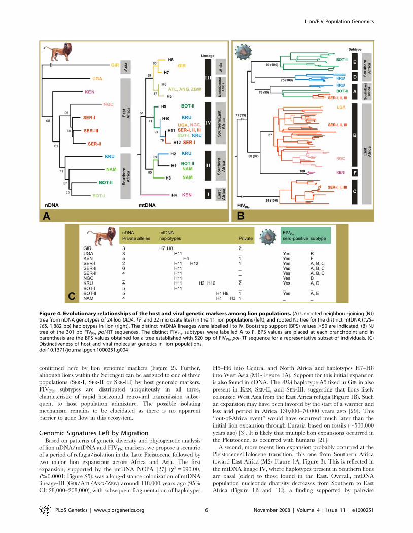
Figure 1. Geographic location of the lion samples and the variability of host and viral genetic markers among lion populations. (A)Historical and current geographic distribution of lion, Panthera leo. A three-letter code pointing to a white dotted circle represents the geographiclocation of the 11 lion populations determined by Bayesian analyses [22] and factorial correspondence analyses [23] of the genetic distinctiveness of357 lion samples (see text): GIR, Gir Forest, India; UGA, Uganda (Queen Elizabeth National Park); KEN, Kenya (Laikipia), SER, Serengeti National Park,Tanzania; NGC, Ngorongoro Crater, Tanzania; KRU, Kruger National Park, South Africa; BOT-I, southern Botswana and Kalahari, South Africa; BOT-II,northern Botswana; and NAM, Namibia. Green squares represent captive individual samples to explore the relationship of lions from more isolated/endangered/depleted areas: ATL, Morocco Atlas lions (n = 4); ANG, Angola (n = 2); and ZBW, Zimbabwe (n = 1). Deduced historical expansions (M1 andM2) are represented by red arrows (see text). (B) Haplotype frequencies observed in the 11 lion populations for nDNA (ADA and TF), and mtDNA (12S–16S) genes, paralleled with the FIVPle serum-prevalence frequencies (black – sero-positive; gray – indeterminate; white – sero-negative). Populationsample sizes are indicated within parenthesis. (C) Statistical parsimony networks of lion ADA, TF, and 12S–16S haplotypes. Circle size is proportional tothe haplotype frequency and crossbars represent the number of step mutations connecting haplotypes. The mtDNA haplotypes H5 and H6 areshaded gray as they were detected only in the individual samples from ANG, ATL, and ZBW, which do not group in unique population clusters (seetext).doi:10.1371/journal.pgen.1000251.g001
Lion/FIV Population Genomics
PLoS Genetics | www.plosgenetics.org 2 November 2008 | Volume 4 | Issue 11 | e1000251
yield similar inferences of population history, as coalescent times
vary as a function of their pattern of inheritance [21]. There is also
a large variance in coalescent times across loci sharing a common
pattern of inheritance especially in complex demographical
histories (Table 1). However, the accurate interpretation of the
differences among loci can provide a more resolved and coherent
population history, affording more-nuanced insights on past
demographic processes, levels of admixture, taxonomic issues,
and on the most appropriate steps for effective conservation and
management of remaining populations.
The goal of this study was to assess the evolutionary history of lion
by (1) characterizing lion population structure relative to patterns of
FIVPle genetic variation, (2) detect signatures of migration using both
host and viral population genomics, and (3) reconstruct lion
demographic history and discuss its implication for lion conservation.
We assess genetic variation from 357 lions from most of its current
distribution, including mitochondrial (mtDNA; 12S–16S, 1,882 bp),
nuclear (nDNA) Y-chromosome (SRY, 1,322 bp) and autosomal
(ADA, 427 bp; TF, 169 bp) sequences, and 22 microsatellites
markers. We further document patterns of FIVPle variation in lions
(FIVPle pol-RT gene, up to 520 bp).
Results/Discussion
Population Structure of LionGenetic analyses of 357 lions from throughout the extant species
range showed that paternally inherited nDNA (SRY) and maternal
inherited (mtDNA) sequence variation was generally low (only one
paternal SRY-haplotype and 12 mtDNA haplotypes; p= 0.0066)
(Figure 1; Figure S1; Tables S1 and S2). The most common
mtDNA haplotype H11 was ubiquitous in Uganda/Tanzania and
parts of Botswana/South Africa, H1 was common in Southern
Africa, and H7 and H8 were unique to Asian lions. The autosomal
nDNA sequences showed fairly distinct patterns of variation
(Figure 1; Figure S1). Of the five ADA haplotypes, A2 was the most
common and most-widely distributed. The other four haplotypes,
which are derived and much less common, included one (A5) that
was fixed in Asian lions. The three TF haplotypes were more
widely and evenly distributed.
Levels of population subdivision among lions were assessed
using microsatellite and sequencing data. Eleven groups were
identified using Bayesian analyses [22] and three-dimensional
factorial correspondence analyses [23] (Figure 2; Table S3). Most
clusters represented geographically circumscribed populations:
Namibia (NAM), Kruger National Park (KRU), Ngorongoro Crater
(NGC), Kenya (KEN), Uganda (UGA), and Gir (GIR). Two distinct
clusters were found in Botswana, BOT-I that included lions from
southern Botswana and Kalahari (South Africa) (Fk = 0.24) and
BOT-II found exclusively in northern Botswana (Fk = 0.18).
Surprisingly, three distinct clusters were found in a single
geographical locale (approximately 60640 km square) in the large
panmyctic population of the Serengeti National Park (SER-I/SER-
II/SER-III) (Fk = 0.18, 0.21, and 0.15, respectively).
Two captive lions from Angola (ANG), one from Zimbabwe (ZBW)
and four Morocco Zoo Atlas lions (ATL; presently extinct from the
wild) (Figure 1A) were included in the analyses to explore the
relationship of lions from more isolated, endangered, or depleted
areas. ANG and ZBW lions were assigned to BOT-II (q = 0.90 and 0.87;
90%CI: 0.47–1.00) and KRU (q = 0.85; 90%CI: 0.52–1.00) (Bayesian
analyses [22]) populations, respectively, as expected based on their
geographical proximity. However, these lions differed from BOT-II
and KRU by up to 8 mtDNA mutations, sharing haplotypes with the
ATL lions (H5 in ANG and H6 in ZBW) (Figure 1B and 1C). The ATL
lions did not group in a unique cluster.
Both nDNA and mtDNA pairwise genetic distances among the
11 lion populations showed a significant relationship with
geographic distance (R2 = 0.75; Mantel’s test, P = 0.0097; and
R2 = 0.15; Mantel’s test, P = 0.0369; respectively) (Figure 3). The
significant positive and monotonic correlation across all the
scatterplot pairwise comparisons for the nDNA markers (bi-
parental) was consistent with isolation-by-distance across the
sampled region. However, the correlation between nDNA FST
and geographic distance considerably decreased when the Asian
GIR population was removed (R2 = 0.19; Mantel’s test, P = 0.0065)
suggesting that caution should be taken in interpreting the pattern
of isolation-by-distance in lions. We further compared linearized
FST estimates [24] plotted both against the geographic distance
(model assuming habitat to be arrayed in an infinite one-
dimensional lattice) and the log geographic distance (model
assuming an infinite two-dimensional lattice). The broad distribu-
tion of lions might suggest a priori that a two-dimensional isolation-
by-distance model would provide the best fit for the nDNA data
Table 1. Expected and observed coalescent times for the different markers studied in lions according to their pattern ofinheritance.
DNA regionPattern ofinheritance
Expected coalescencetime in generations Size (bp)
Number ofhaplotypes FST
DivergencePpa/Ple (H)
Substitutionrate (site/yr)
TMRCA (Myrago)
mtDNA Maternal haploid 2Neff<Nef
12S and 16S 1.882 12 0.92 0.0161 8.161028 0.324
Y-Chromosomal DNA Paternal haploid 2Nefm<Nef
SRY-39 1.322 1 _ 0.0005 0.2561028 _
Autosomal DNA Bisexual diploid 4Nef
ADA 427 5 0.20 0.0023 1.261028 4
TF 169* 3 0.09 0.0030* 1.561028 4.8
microsatellites na na 0.18 na 2.161023 0.027
TMRCA - Time to the most recent common ancestral haplotype.Nef - long-term inbreeding effective size of the population; Neff - inbreeding effective size of females; and Nefm - inbreeding effective size of males.The approximations to the expected coalescence time are made under the assumption that Neff = Nefm = Nef/2.*The TF gene segment used to estimate the divergence Ppa/Ple was 450 bp long.na - not aplicable.doi:10.1371/journal.pgen.1000251.t001
Lion/FIV Population Genomics
PLoS Genetics | www.plosgenetics.org 3 November 2008 | Volume 4 | Issue 11 | e1000251
(R2 = 0.25; Mantel’s test, P = 0.0022), but instead the one-
dimensional isolation-by-distance model performed better
(R2 = 0.71; Mantel’s test, P = 0.0476) (Figure S2).
The pattern observed for the mtDNA (maternal) was more
complex. While there was a significant relationship between
mtDNA FST and geographic distance, there was an inconsistent
pattern across broader geographic distances (Figure 3). This is
partly due to the fixation or near fixation of haplotype H11 in six
populations and the fixation of a very divergent haplotype H4 in
KEN population (Figure 1B and 1C). The removal of the KEN
population considerably increased the correlation between
mtDNA FST and geographic distance (R2 = 0.27; Mantel’s test,
P = 0.0035). Thus, the null hypothesis of regional equilibrium for
mtDNA across the entire sampled region is rejected despite the
possibility that isolation-by-distance may occur regionally.
These contrasting nDNA and mtDNA results may be indicative
of differences in dispersal patterns between males and females,
which would be consistent with evidence that females are more
phylopatric than males. Alternatively, selection for matrilineally
transmitted traits upon which neutral mtDNA alleles hitchhike is
possible, given the low values of nucleotide diversity of the mtDNA
(p= 0.0066). A similar process has been suggested in whales
(p= 0.0007) [25] and African savannah elephants (p= 0.0200)
[26], where both species have female phylopatry and like lions, a
matriarchal social structure. However, genetic drift tends to
overwhelm selection in small isolated populations, predominantly
affecting haploid elements due to its lower effective population size
(Table 1). Therefore, we suggest that the contrasting results
obtained for nDNA and mtDNA are more likely further evidence
that lion populations underwent severe bottlenecks. The highly
structured lion matrilines comprise four monophyletic mtDNA
haplo-groups (Figure 4A; Figure S3). Lineage I consisted of a
divergent haplotype H4 from KEN, lineage II was observed in most
Southern Africa populations, lineage III was widely distributed
from Central and Northern Africa to Asia, and lineage IV
occurred in Southern and Eastern Africa.
Population Structure of FIVPle
Seroprevalence studies indicate that FIVPle is endemic in eight
of the 11 populations but absent from the Asian GIR lions in India
and in Namibia and southern Botswana/Kalahari regions (NAM/
BOT-I) in Southwest Africa (Figure 1B). Phylogenetic analysis of
the conserved pol-RT region in 117 FIV-infected lions depicted
monophyletic lineages [15,18] that affirm six distinct subtypes (A–
F) that are distributed geographically in three distinct patterns
(Figure 4B; Figure S4). First, multiple subtypes may circulate
within the same population as exemplified by subtypes A, B and C
all ubiquitous within the three Serengeti lion populations (SER-I,
SER-II and SER-III) and subtypes A and E within lions of Botswana
(BOT-II) (Figure 4B and 4C and Figure S4). Second, unique FIVPle
subtypes may be restricted to one location as subtype F in Kenya,
subtype E in Botswana (BOT-II), subtype C in Serengeti, and
subtype D in Krugar Park (Figure 4B and 4C and Figure S4).
Third, intra-subtype strains cluster geographically, as shown by
distinct clades within subtype A that were restricted to lions within
Krugar Park, Botswana and Serengeti and within subtype B that
corresponded to Uganda, Serengeti and Ngorongoro Crater lions
(Figure 4B and 4C and Figure S4).
Not unexpectedly, FIVPle pairwise genetic distances, represent-
ed as population FST among the eight lion FIV-infected
Figure 2. Population structure analyses in lions. (A) Bayesian population assignment test [22] of the 357 lions using 24 nDNA loci (ADA, TF, and22 microsatellites) and mtDNA data, and considering K = 11 (11 populations). (B) Three-dimensional factorial correspondence analysis [23] (FCA)based on the 24 nDNA loci genotypes in the 357 lions. Axe 1, 2, and 3 represent 49.90% of the genetic variation observed. (C) FCA representationexcluding the GIR lions. Axe 1, 2, and 3 represent 51.35% of the genetic variation observed. (D) FCA representation considering only the SER lionssupportive of a three distinct population clusters subdivision (SER-I, SER-II, and SER-III).doi:10.1371/journal.pgen.1000251.g002
Lion/FIV Population Genomics
PLoS Genetics | www.plosgenetics.org 4 November 2008 | Volume 4 | Issue 11 | e1000251
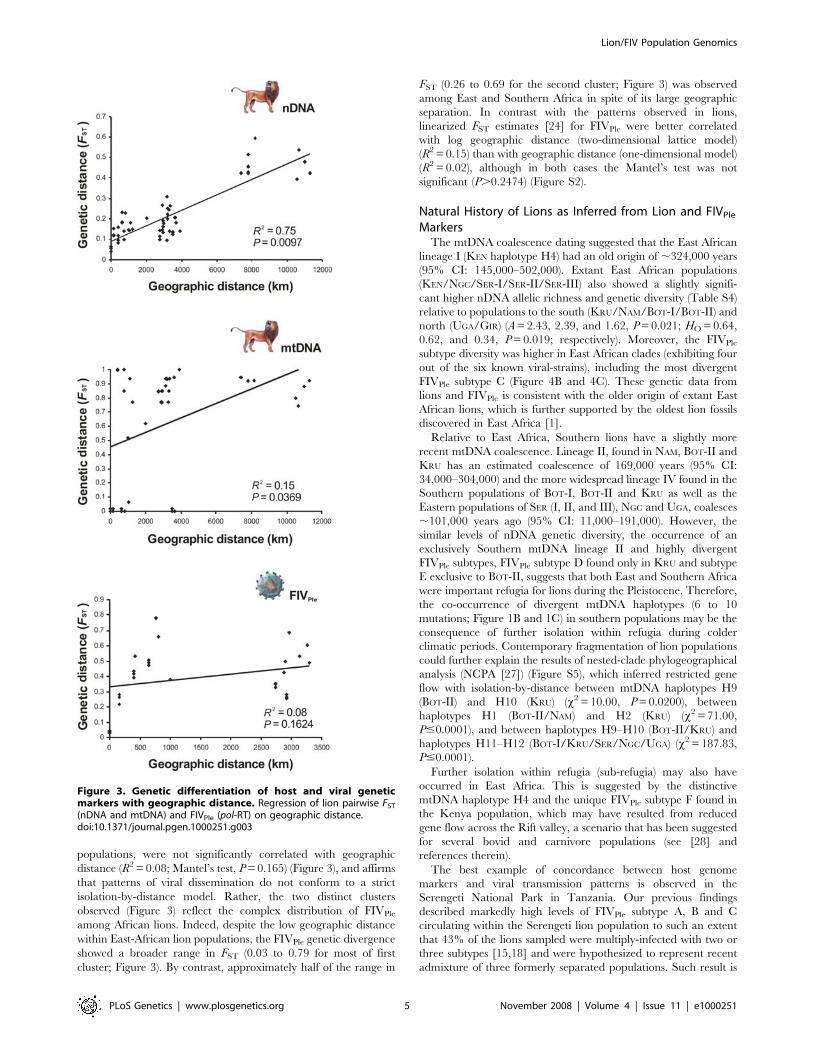
populations, were not significantly correlated with geographic
distance (R2 = 0.08; Mantel’s test, P = 0.165) (Figure 3), and affirms
that patterns of viral dissemination do not conform to a strict
isolation-by-distance model. Rather, the two distinct clusters
observed (Figure 3) reflect the complex distribution of FIVPle
among African lions. Indeed, despite the low geographic distance
within East-African lion populations, the FIVPle genetic divergence
showed a broader range in FST (0.03 to 0.79 for most of first
cluster; Figure 3). By contrast, approximately half of the range in
FST (0.26 to 0.69 for the second cluster; Figure 3) was observed
among East and Southern Africa in spite of its large geographic
separation. In contrast with the patterns observed in lions,
linearized FST estimates [24] for FIVPle were better correlated
with log geographic distance (two-dimensional lattice model)
(R2 = 0.15) than with geographic distance (one-dimensional model)
(R2 = 0.02), although in both cases the Mantel’s test was not
significant (P.0.2474) (Figure S2).
Natural History of Lions as Inferred from Lion and FIVPle
MarkersThe mtDNA coalescence dating suggested that the East African
lineage I (KEN haplotype H4) had an old origin of ,324,000 years
(95% CI: 145,000–502,000). Extant East African populations
(KEN/NGC/SER-I/SER-II/SER-III) also showed a slightly signifi-
cant higher nDNA allelic richness and genetic diversity (Table S4)
relative to populations to the south (KRU/NAM/BOT-I/BOT-II) and
north (UGA/GIR) (A = 2.43, 2.39, and 1.62, P = 0.021; HO = 0.64,
0.62, and 0.34, P = 0.019; respectively). Moreover, the FIVPle
subtype diversity was higher in East African clades (exhibiting four
out of the six known viral-strains), including the most divergent
FIVPle subtype C (Figure 4B and 4C). These genetic data from
lions and FIVPle is consistent with the older origin of extant East
African lions, which is further supported by the oldest lion fossils
discovered in East Africa [1].
Relative to East Africa, Southern lions have a slightly more
recent mtDNA coalescence. Lineage II, found in NAM, BOT-II and
KRU has an estimated coalescence of 169,000 years (95% CI:
34,000–304,000) and the more widespread lineage IV found in the
Southern populations of BOT-I, BOT-II and KRU as well as the
Eastern populations of SER (I, II, and III), NGC and UGA, coalesces
,101,000 years ago (95% CI: 11,000–191,000). However, the
similar levels of nDNA genetic diversity, the occurrence of an
exclusively Southern mtDNA lineage II and highly divergent
FIVPle subtypes, FIVPle subtype D found only in KRU and subtype
E exclusive to BOT-II, suggests that both East and Southern Africa
were important refugia for lions during the Pleistocene. Therefore,
the co-occurrence of divergent mtDNA haplotypes (6 to 10
mutations; Figure 1B and 1C) in southern populations may be the
consequence of further isolation within refugia during colder
climatic periods. Contemporary fragmentation of lion populations
could further explain the results of nested-clade phylogeographical
analysis (NCPA [27]) (Figure S5), which inferred restricted gene
flow with isolation-by-distance between mtDNA haplotypes H9
(BOT-II) and H10 (KRU) (x2 = 10.00, P = 0.0200), between
haplotypes H1 (BOT-II/NAM) and H2 (KRU) (x2 = 71.00,
P#0.0001), and between haplotypes H9–H10 (BOT-II/KRU) and
haplotypes H11–H12 (BOT-I/KRU/SER/NGC/UGA) (x2 = 187.83,
P#0.0001).
Further isolation within refugia (sub-refugia) may also have
occurred in East Africa. This is suggested by the distinctive
mtDNA haplotype H4 and the unique FIVPle subtype F found in
the Kenya population, which may have resulted from reduced
gene flow across the Rift valley, a scenario that has been suggested
for several bovid and carnivore populations (see [28] and
references therein).
The best example of concordance between host genome
markers and viral transmission patterns is observed in the
Serengeti National Park in Tanzania. Our previous findings
described markedly high levels of FIVPle subtype A, B and C
circulating within the Serengeti lion population to such an extent
that 43% of the lions sampled were multiply-infected with two or
three subtypes [15,18] and were hypothesized to represent recent
admixture of three formerly separated populations. Such result is
Figure 3. Genetic differentiation of host and viral geneticmarkers with geographic distance. Regression of lion pairwise FST
(nDNA and mtDNA) and FIVPle (pol-RT) on geographic distance.doi:10.1371/journal.pgen.1000251.g003
Lion/FIV Population Genomics
PLoS Genetics | www.plosgenetics.org 5 November 2008 | Volume 4 | Issue 11 | e1000251
confirmed here by lion genomic markers (Figure 2). Further,
although lions within the Serengeti can be assigned to one of three
populations (SER-I, SER-II or SER-III) by host genomic markers,
FIVPle subtypes are distributed ubiquitously in all three,
characteristic of rapid horizontal retroviral transmission subse-
quent to host population admixture. The possible isolating
mechanism remains to be elucidated as there is no apparent
barrier to gene flow in this ecosystem.
Genomic Signatures Left by MigrationBased on patterns of genetic diversity and phylogenetic analysis
of lion nDNA/mtDNA and FIVPle markers, we propose a scenario
of a period of refugia/isolation in the Late Pleistocene followed by
two major lion expansions across Africa and Asia. The first
expansion, supported by the mtDNA NCPA [27] (x2 = 690.00,
P#0.0001; Figure S5), was a long-distance colonization of mtDNA
lineage-III (GIR/ATL/ANG/ZBW) around 118,000 years ago (95%
CI: 28,000–208,000), with subsequent fragmentation of haplotypes
H5–H6 into Central and North Africa and haplotypes H7–H8
into West Asia (M1- Figure 1A). Support for this initial expansion
is also found in nDNA. The ADA haplotype A5 fixed in GIR in also
present in KEN, SER-II, and SER-III, suggesting that lions likely
colonized West Asia from the East Africa refugia (Figure 1B). Such
an expansion may have been favored by the start of a warmer and
less arid period in Africa 130,000–70,000 years ago [29]. This
‘‘out-of-Africa event’’ would have occurred much later than the
initial lion expansion through Eurasia based on fossils (,500,000
years ago) [3]. It is likely that multiple lion expansions occurred in
the Pleistocene, as occurred with humans [21].
A second, more recent lion expansion probably occurred at the
Pleistocene/Holocene transition, this one from Southern Africa
toward East Africa (M2- Figure 1A, Figure 3). This is reflected in
the mtDNA linage IV, where haplotypes present in Southern lions
are basal (older) to those found in the East. Overall, mtDNA
population nucleotide diversity decreases from Southern to East
Africa (Figure 1B and 1C), a finding supported by pairwise
Figure 4. Evolutionary relationships of the host and viral genetic markers among lion populations. (A) Unrooted neighbour-joining (NJ)tree from nDNA genotypes of 24 loci (ADA, TF, and 22 microsatellites) in the 11 lion populations (left), and rooted NJ tree for the distinct mtDNA (12S–16S, 1,882 bp) haplotypes in lion (right). The distinct mtDNA lineages were labelled I to IV. Bootstrap support (BPS) values .50 are indicated. (B) NJtree of the 301 bp FIVPle pol-RT sequences. The distinct FIVPle subtypes were labelled A to F. BPS values are placed at each branchpoint and inparenthesis are the BPS values obtained for a tree established with 520 bp of FIVPle pol-RT sequence for a representative subset of individuals. (C)Distinctiveness of host and viral molecular genetics in lion populations.doi:10.1371/journal.pgen.1000251.g004
Lion/FIV Population Genomics
PLoS Genetics | www.plosgenetics.org 6 November 2008 | Volume 4 | Issue 11 | e1000251
mismatch analysis [30] (raggedness, r = 0.086; P,0.001). The
fixation of mtDNA haplotype H11 in BOT-I (otherwise fixed only
in East Africa populations) suggests that the colonizing lions
expanded northwards from the Kalahari Desert, which included
bush, woodland and savannah habitats during the climatic
fluctuations of the Pleistocene [31]. This expansion would have
occurred relatively recently as the single rare tip mtDNA
haplotype H12, found only in SER-I, is derived from the interior
widespread haplotype H11 (,14,000–7,000 years; given one
mtDNA substitution every 7,000 years; Table 1). This expansion is
also supported by FIVPle subtype A where haplotypes present in
Southern lions (KRU and BOT-I) are basal to those found in the
East (SER-I, SER-II and SER-III) and a decrease of nucleotide-
diversity of this FIVPle subtype is observed from Southern
(p= 0.15) to Eastern Africa (p= 0.03) (Figure 3B). Interestingly,
a similar northward colonization process from Southern Africa has
been suggested for some of the lion preys, namely the impala,
greater kudu, and wildebeest [32,33].
Utility of Population Genomic DatasetsIf we had restricted our inferences to mtDNA, we might have
concluded that East African lion populations, which are fixed or
nearly fixed for haplotype H11, went extinct during the
Pleistocene/Holocene transition (similar to the well known
mega-fauna extinctions of the Late Pleistocene [34]) and were
then colonized by Southern populations. However, our population
genomics data better fit a scenario of lion population expansion
and interbreeding rather than simple replacement. First, genetic
diversity and allelic richness at nDNA are slightly higher in East
Africa populations relatively to those in Southern Africa. This is
contrary to the expected pattern of population expansion in which
there is usually a progressive decline in genetic diversity and allelic
richness. Second, SER lions carry two diverse FIVPle subtypes
found only in East Africa (B–C), and not only FIVPle subtype A,
which was presumably introduced in East Africa coincidently with
the mtDNA expansion event northwards from South. Third, the
East African FIVPle subtype B found in UGA/SER-I/SER-II/SER-
III/NGC showed evidence of a population expansion (raggedness,
r = 0.004; P,0.01; Fs = 220.37; P,0.00001) and the highest
nucleotide diversity observed within FIVPle subtypes (p= 0.09).
Four, the FIVPle subtype diversity is higher in East African clades
(four out of the six viral strains).
The utility of FIVPle pol-RT as a marker of lion population
structure and natural history is that it can be informative on a
contemporaneous time scale, though it may be less useful at
capturing more ancient demographic events. The extreme diver-
gence among FIVPle subtypes, considered with high sero-prevalence
in eight of the 11 lion populations, and combined with patterns of
geographic concordance, support the hypothesis that FIVPle is not a
recent emergence within modern lions [35]. Populations that harbor
one private FIVPle subtype such KEN (subtype F), BOT-II (subtype E),
and KRU (subtype D) must have been sufficiently isolated for enough
time for the virus to evolve into unique subtypes, a result
corroborated by the high nDNA and mtDNA genetic structure
present in these lion populations (Figure 4). Thus, it is possible that
the initial emergence of FIVPle pre-dates the Late-Pleistocene
expansions of contemporary lion populations [36], but present day
distributions are more useful indicators of very recent host
population dynamics, a result also observed with FIVPco in a
panmictic population of pumas in western North America [19].
Conservation ImplicationsAccurate interpretation of past and contemporary population
demographic scenarios is a primary goal for the effective
conservation of endangered species. In this study, we found
substantial population subdivision, reduced gene flow, and large
differences in FIVPle sequence and sero-prevalence among lion
populations, as well as evidence of historic secondary contact
between populations (Figure 3C; Table S4 to S9). The very low
population level of mtDNA nucleotide diversity, the number of
haplotypes private to a single population (Figure 1), and probably
also the lack of SRY genetic variation across all male lions
(haplotype S1, n = 183) suggests that lion numbers diminished
considerably following the Late Pleistocene. The last century
reduction in lion distribution further eroded its genomic diversity,
and microsatellite variation suggested recent population bottle-
necks in seven out of the 11 populations (standardized differences
test, P,0.05; Table S5) [37].
Although we did not explicitly try to address the adequacy of
lion subspecies designations (currently only one African subspecies
is widely recognized) [38,39], we provided strong evidence that
there is no evidence of substantial genetic exchange of matrilines
among existing populations as the AMOVA [40] within-
population component was uniformly high in all distinct
subdivision scenarios (WST<0.920; P,0.0001; three-six groups;
Table S6). Similarly, significant population structure was detected
from nDNA (FST = 0.18), with low levels of admixture evident
from Bayesian analysis [22] (a= 0.033). Therefore, employing a
bottom-up perspective that prioritizes populations, rather than
large-scale units (e.g. all African lions), might preserve and
maintain lion diversity and evolutionary processes most efficiently
[41].
Material and Methods
Study Site, Sampling, and Molecular Genetic Analyses ofLions
A total of 357 individuals were obtained across most of the lion
range in Africa and Asia (Figure 1A; Table S1). Genetic variation
among lion specimens was assessed using maternal (12S and 16S
genes), paternal (SRY gene) and bi-parental autosomal (22
microsatelite loci, and the ADA and TF genes) markers (GenBank
accession numbers: FJ151632–FJ151652). Analyses of mtDNA in
Panthera species are complicated by the presence of a 12.5 kb
mtDNA integration into chromosome F2 [42]. Accordingly,
mtDNA specific primers were designed for the 12S and 16S genes
(Table S2) and we used long-range PCR amplification. We
designed primers to amplify segments of the ADA (exon 10 and
intron 10) and the TF (intron 3) genes (Table S2), two of the most
variable protein loci in lion populations [5], localized on the
domestic cat Felis catus chromosome A3p and C2q, respectively.
The Y-chromosome SRY-39UTR gene was also amplified [43].
PCR products were amplified from 50 ng of genomic DNA in a
25 mL reaction system containing 1.5 mM MgCl2, 1.0 mM
dNTPs, 0.25 units of AmpliTaq Gold DNA polymerase (Applied
Biosystems), and 16 PCR buffer II; the amplification protocol
was: denaturation 10 min at 95uC, a touch-down cycle of 95uC for
30 s, 52uC for 60 s decreased by 1uC in the next cycle for 10
cycles, 72uC for 120 s, then 35 amplification cycles of 95uC for
30 s, 52uC for 60 s, and 72uC for 120 s, followed by an extension
of 10 min at 72uC. PCR products were sequenced on an ABI 377.
Sequences were aligned and cleaned using SEQUENCHER
(Gene Codes).
Twenty two polymorphic microsatellite loci (20 dinucleotide
repeats: FCA006, FCA008, FCA014, FCA069, FCA077,
FCA085, FCA091, FCA098, FCA105, FCA126, FCA129,
FCA139, FCA205, FCA208, FCA211, FCA224, FCA229,
FCA230, FCA247, and FCA281; and two tetranucleotide repeats:
Lion/FIV Population Genomics
PLoS Genetics | www.plosgenetics.org 7 November 2008 | Volume 4 | Issue 11 | e1000251
FCA391 and FCA441) were amplified [44]. Microsatellites were
scored in an ABI 377 and analyzed using GENESCAN 2.1 and
GENOTYPER 2.5. These loci are located on 11 of the 19 F. catus
chromosomes, occurring in different linkage groups or at least 12
centimorgans apart [44,45].
Sero-Prevalence and Molecular Genetics of FIVPle
Western blots using domestic cat and lion FIV as antigen were
performed as previously described [46,47]. The supernatant from
virus-infected cells was centrifuged at 200 g for 10 min at 5uC.
The resultant supernatant was centrifuged at 150,000 g at 4uC for
2 hours. Pelleted viral proteins were resuspended in 1/20th of the
original volume and total protein content was assayed using the
Biorad Protein Assay. Twenty mg of viral protein were run on 4–
20% Tris-Glycine gels and transferred to PDVF membranes
(BioRad). Membrane strips were exposed 2–12 h to a 1:25 or
1:200 dilution of serum or plasma. After washing, samples were
labeled with goat anti-cat HRP or phosphate conjugated antibody
(KPL laboratories) at a 1:2000 dilution, washed, and incubated in
ECL Western Blotting detection reagents (Amersham Biosciences)
for 2 min, then exposed to Lumi-Film Chemiluminescent
Detection Film (Boehringer Mannheim) or incubated in BCIP/
NBP phosphatase substrate (KPL laboratories) for 15 min [46–
48]. Results were visualized and scored manually based on the
presence and intensity of antibody binding to the p24 gag capsid
protein.
Nested PCR amplification of partial FIVPle pol-RT was
performed [18,46]. Briefly, first round PCR reactions used
100 ng of genomic DNA, 2.5 mM MgCl2 and an annealing
temperature of 52uC. Second round PCR reactions used identical
conditions with 1–5 ml of first-round product as template. All PCR
products were sequenced as described above for lion genetic
analyses (GenBank accession numbers: AY549217–AY552683;
AY878208–AY878235; FJ225347–FJ225382).
Statistical AnalysesWe used the GENETIX 4.02 [49], GENEPOP 3.3 [50], MICROSAT
[51], and DNASP 4.10 [52] to calculate the following descriptive
statistics: (i) percentage of polymorphic loci (P95), number of alleles
per locus (A), observed and expected heterozygosity (HE and HO),
and number of unique alleles (AU); (ii) assess deviations from
HWE; (iii) estimate the coefficient of differentiation (FST), and (iv)
nucleotide (p) and haplotype (h) diversity.
We tested the hypothesis that all loci are evolving under
neutrality for both the lion and the FIVPle data. For frequency
data, we used the method described by Beaumont and Nichols
[53] and implemented in FDIST (http://www.rubic.rdg.ac.uk/
~mab/software.html). The FST values estimated from microsatel-
lite loci plotted against heterozygosity showed that all values fall
within the expected 95% confidence limit and consequently no
outlier locus were identified. For sequence data (lion nDNA/
mtDNA and FIVPle pol-RT), we ruled out any significant evidence
for genetic hitchhiking and background selection by assessing Fu
and Li’s D* and F* tests [54] and Fu’s FS statistics [55].
A Bayesian clustering method implemented in the program
STRUCTURE [22] was used to infer number of populations and
assign individual lions to populations based on multilocus genotype
(microsatellites) and sequence data (ADA, TF, and mtDNA genes)
and without incorporating sample origin. For haploid mtDNA
data, each observed haplotype was coded with a unique integer
(e.g. 100, 110) for the first allele and missing data for the second
(STRUCTURE [22] analyses with or without the mtDNA data were
essentially identical). For K population clusters, the program
estimates the probability of the data, Pr(X|K), and the probability
of individual membership in each cluster using a Markov chain
Monte Carlo method under the assumption of Hardy-Weinberg
equilibrium (HWE) within each cluster. Initial testing of the HWE
in each of the populations defined by the geographic origin of
sampling revealed no significant deviation from HW expectations
with the exception of SER and BOT population (later subdivided by
STRUCTURE [22] in 3 and 2 clusters, respectively; such deviations
from HW expectations were interpreted as evidence of further
population structuring). We conducted six independent runs with
K = 1–20 to guide an empirical estimate of the number of
identifiable populations, assuming an admixture model with
correlated allele frequencies and with burn-in and replication
values set at 30,000 and 106, respectively. STRUCTURE also
estimates allele frequencies of a hypothetical ancestral population
and an alpha value that measures admixed individuals in the data
set. The assignment of admixed individuals to populations using
STRUCTURE [22] has been considered in subsequent population
analyses. For each population cluster k, the program estimates Fk,
a quantity analogous to Wright’s FST, but describing the degree of
genetic differentiation of population k from the ancestral
population.
Patterns of gene flow and divergence among populations were
described using a variety of tests. First, to visualize subtle
relationships among individual autosomal genotypes, three-
dimensional factorial correspondence analyses [23] (FCA) were
performed in GENETIX [49], which graphically projects the
individuals on the factor space defined by the similarity of their
allelic states. Second, neighbor-joining (NJ) analyses implementing
the Cavalli-Sforza & Edwards’ chord genetic distance [56] (DCE)
were estimated in PHYLIP 3.6 [57], and the tree topology support
was assessed by 100 bootstraps. Third, the difference in average
HO and A was compared among population groups using a two-
sided test in FSTAT 2.9.3.2 [58], which allows to assess the
significance of the statistic OSx using 1,000 randomizations. Four,
the equilibrium between drift and gene flow was tested using a
regression of pairwise FST on geographic distance matrix among
all populations for host nDNA(microsatellites)/mtDNA and FIVPle
data. A Mantel test [59] was used to estimate the 95% upper
probability for each matrix correlation. Assuming a stepping stone
model of migration where gene flow is more likely between
adjacent populations, one can reject the null hypothesis that
populations in a region are at equilibrium if (1) there is a non-
significant association between genetic and geographic distances,
and/or (2) a scatterplot of the genetic and geographic distances
fails to reveal a positive and monotonic relationship over all
distance values of a region [60]. We also evaluated linearized FST
[i.e. FST/(12FST)] [24] among populations. We tested two
competing models of isolation-by-distance, one assuming the
habitat to be arrayed in an infinite one-dimensional lattice and
another assuming an infinite two-dimensional lattice. Both models
showed that genetic differentiation increased with raw and log-
transformed Euclidean distances, respectively [24]. We deter-
mined the confidence interval value of the slope of the regression
for the nDNA data using a non parametric ABC bootstrap [61] in
GENEPOP 4.0 [62].
The demographic history of populations was compared using a
variety of estimators based on the coalescence theory. First,
signatures of old demographic population expansion were
investigated for mtDNA and FIVPle pol-RT haplotypes using
pairwise mismatch distributions [63] in DNASP [52]. The
goodness-of-fit of the observed data to a simulated model of
expansion was tested with the raggedness (r) index [64].
Second, the occurrence of recent bottlenecks was evaluated for
microsatellite data using the method of Cornuet & Luikart [37] in
Lion/FIV Population Genomics
PLoS Genetics | www.plosgenetics.org 8 November 2008 | Volume 4 | Issue 11 | e1000251
BOTTLENECK [65] and using 10,000 iterations. This approach,
which exploits the fact that rare alleles are generally lost first
through genetic drift after reduction in population size, employs
the standardize differences test, which is the most appropriate and
powerful when using 20 or more polymorphic loci [37]. Tests were
carried out using the stepwise mutation model (SMM), which is a
conservative mutation model for the detection of bottleneck
signatures with microsatellites [66].
Third, to discriminate between recurrent gene flow and
historical events we used the nested-clade phylogeographical
analysis [27,67] (NCPA) for the mtDNA data. When the null-
hypothesis of no correlation between genealogy and geography is
rejected, biological inferences are drawn using a priori criteria.
The NCPA started with the estimation of a 95% statistical
parsimony [68] mtDNA network in TCS 1.20 [69]. Tree
ambiguities were further resolved using a coalescence criteria
[70]. The network was converted into a series of nested branches
(clades) [71,72], which were then tested against their geographical
locations through a permutational contingency analysis in GEODIS
2.2 [73]. The inferences obtained were also corroborated with the
automated implementation of the NCPA in ANECA [74]. To
address potential weaknesses in some aspects of the NCPA analysis
[75,76], we further validated the NCPA inferences with indepen-
dent methods for detecting restricted gene-flow/isolation-by-
distance (using matrix correlation of pairwise FST and geographic
distance) and population expansion (using pairwise mismatch
distributions).
Four, to test the significance of the total mtDNA genetic
variance, we conducted hierarchical analyses of molecular
variance [40] (AMOVA) using ARLEQUIN 2.0 [77]. Total genetic
variation was partitioned to compare the amount of difference
among population groups, among populations within each groups,
and within populations.
Phylogenetic relationships among mtDNA and FIVPle pol-RT
sequences were assessed using Minimum evolution (ME), Maxi-
mum parsimony (MP), and Maximum likelihood (ML) approaches
implemented in PAUP [78]. The ME analysis for mtDNA consisted
of NJ trees constructed from Kimura two-parameter distances
followed by a branch-swapping procedure and for FIVPle data
employed the same parameter estimates as were used in the ML
analysis. The MP analysis was conducted using a heuristic search,
with random additions of taxa and tree-bisection-reconnection
branch swapping. The ML analysis was done after selecting the
best evolutionary model fitting the data using MODELTEST 3.7 [79].
Tree topologies reliability was assessed by 100 bootstraps. For the
FIVPle data, the reliability of the tree topology was further assessed
through additional analyses using 520 bp of FIVPle pol-RT
sequences in a representative subset of individuals.
The time to the most recent common ancestor (TMRCA) for
the ADA and TF haplotypes was estimated following Takahata et
al. [80], where we calculate the ratio of the average nucleotide
differences within the lion sample to one-half the average
nucleotide difference between leopards (P. pardus) and lions and
multiplying the ratio by an estimate of the divergence time
between lions and leopards (2 million years based on undisputed
lion fossils in Africa) [81,82]. The mtDNA TMRCA was estimated
with a linearized tree method in LINTREE [83] and using the
equation H = 2mT, where H was the branch height (correlated to
the average pairwise distance among haplotypes), m the substitu-
tion rate, and T the divergence time. Leopard and snow leopard
(P. uncia) sequences were used as outgroups. Inference of the
TMRCA for microsatellite loci followed Driscoll et al. [6] where
the estimate of microsatellite variance in average allele repeat-size
was used as a surrogate for evolutionary time based on the rate of
allele range reconstitution subsequent to a severe founder effect.
Microsatellite allele variance has been shown to be a reliable
estimator for microsatellite evolution and demographic inference
in felid species [6].
Supporting Information
Figure S1 Genetic variation of 12S–16S (mtDNA) and ADA and
TF (nDNA) genes in lions. (A) Haplotypes and variable sites for the
12S–16S mtDNA region surveyed in lions (total length 1,882 bp).
Position 1 corresponds to position 1441 of the domestic cat (Felis
catus) mtDNA genome (GenBank U20753). The ‘‘-’’ represents a
gap and ‘‘.’’ matches the nucleotide in the first sequence. Shading
indicates a fixed difference in the mtDNA lineage. (B) Haplotypes
and variable sites for the ADA gene segment surveyed in lions (total
length 427 bp). (C) Haplotypes and variable sites for the TF gene
segment surveyed in lions (total length 427 bp).
Found at: doi:10.1371/journal.pgen.1000251.s001 (0.12 MB PDF)
Figure S2 Linearized genetic differentiation of host and viral
genetic markers with geographic distance. Regression of linearized
FST estimates [24] for lion (nDNA and mtDNA) and FIVPle (pol-RT)
genetic data plotted both against the geographic distance (model
assuming habitat to be arrayed in an infinite one-dimensional lattice;
one-dimension isolation-by-distance [IBD]) and the log geographic
distance (model assuming an infinite two-dimensional lattice; two-
dimension isolation-by-distance) on geographic distance.
Found at: doi:10.1371/journal.pgen.1000251.s002 (0.13 MB PDF)
Figure S3 Phylogenetic relationships of the 12S–16S mtDNA lion
haplotypes. Neighbour-joining tree of the 1,882 bp 12S–16S
mtDNA sequences. Bootstrap values are placed at each branchpoint
for the minimum evolution/maximum parsimony/maximum like-
lihood analyses, respectively (ME/MP/ML). Outgroups: Ppa –
leopard, Panthera pardus; Pun – snow-leopard, Panthera uncia. The
symbol (N) represents nodes with bootstrap support ,50 or an
inferred polytomy in the bootstrap 50% majority-rule consensus tree.
Found at: doi:10.1371/journal.pgen.1000251.s003 (0.10 MB PDF)
Figure S4 Phylogenetic relationships of the FIVPle pol-RT
sequences. Neighbour-joining tree of the 301 bp FIVPle pol-RT
sequences. The distinct FIVPle subtypes were labelled A to F.
Bootstrap (BPS) values are placed at each branchpoint (ME/MP/
ML) and in parenthesis are the BPS values obtained for a tree
established with 520 bp of FIVPle pol-RT sequence for a
representative subset of individuals.
Found at: doi:10.1371/journal.pgen.1000251.s004 (0.13 MB PDF)
Figure S5 Nested design and summary results of the nested
clade phylogeographic analysis (NCPA) for lion mtDNA data. (A)
Nested design of the mtDNA haplotype network used for the
NCPA. (B) Summary results of the NCPA. RGF/IBD - Restricted
gene flow/isolation by distance. LDC/FR – long distance
colonization/fragmentation.
Found at: doi:10.1371/journal.pgen.1000251.s005 (0.10 MB PDF)
Table S1 List of the lion samples used in this study.
Found at: doi:10.1371/journal.pgen.1000251.s006 (0.11 MB PDF)
Table S2 Primers used to amplify the mtDNA (12S–16S) and
nDNA (ADA and TF) portions surveyed in this study.
Found at: doi:10.1371/journal.pgen.1000251.s007 (0.06 MB PDF)
Table S3 Structure cluster assignment results of 357 lions based
on nDNA (ADA, TF, and 22 microsatellites) and mtDNA markers.
Burn-in and replication values set at 30,000 and 1,000,000,
respectively.
Found at: doi:10.1371/journal.pgen.1000251.s008 (0.06 MB PDF)
Lion/FIV Population Genomics
PLoS Genetics | www.plosgenetics.org 9 November 2008 | Volume 4 | Issue 11 | e1000251
Table S4 Gene diversity and frequency values in lion popula-
tions.
Found at: doi:10.1371/journal.pgen.1000251.s009 (0.07 MB PDF)
Table S5 Bottleneck analysis in lion populations using the
standardized differences test and the stepwise mutation model
(SMM).
Found at: doi:10.1371/journal.pgen.1000251.s010 (0.05 MB PDF)
Table S6 Results of the hierarchical AMOVA in lions for four
different geographical scenarios.
Found at: doi:10.1371/journal.pgen.1000251.s011 (0.06 MB PDF)
Table S7 Lion population pairwise FST estimates. Below the
diagonal mtDNA data (12S–16S) and above the diagonal
microsatellite data (22 loci).
Found at: doi:10.1371/journal.pgen.1000251.s012 (0.06 MB PDF)
Table S8 Taxon specific unique nDNA alleles in lion popula-
tions (FCA-microsatellites and ADA locus).
Found at: doi:10.1371/journal.pgen.1000251.s013 (0.06 MB PDF)
Table S9 Summary statistics for FIVPle data.
Found at: doi:10.1371/journal.pgen.1000251.s014 (0.00 MB PDF)
Acknowledgments
Tissues were obtained in full compliance with specific Federal Fish and
Wildlife Permits. The content of this publication does not necessarily reflect
the views or policies of the Department of Health and Human Services, nor
does mention of trade names, commercial products, or organizations imply
endorsement by the U.S. Government. We would like to thank A. Beja-
Pereira, M. Branco and J. Martenson for suggestions and technical
assistance. Comments made by the Associate Editor A. Estoup and three
anonymous referees improved a previous version of this manuscript.
Author Contributions
Conceived and designed the experiments: AA JLT SJO WEJ. Performed
the experiments: AA JLT MER WEJ. Analyzed the data: AA JLT MER
SJO WEJ. Contributed reagents/materials/analysis tools: AA JLT MER
JPS CP CW HW GH LF PS LS MD PJF KAA KCP GM DW MB SJO
WEJ. Wrote the paper: AA JLT JPS CP SJO WEJ.
References
1. Werdelin L, Lewis ME (2005) Plio-Pleistocene Carnivora of eastern Africa:species richness and turnover patterns. Zool J Linn Soc 144: 121–144.
2. Petter G (1973) Carnivores Pleistocenes du Ravin d’Olduvai (Tanzanie). InLeakey LSB, Savage RJG, Coryndon SC, eds. Fossil vertebrates of Africa.
London: Academic Press 3: 43–100.
3. Hemmer H (1974) Untersuchungen zur Stammesgeschichte der Pantherkatzen(Pantherinae) Teil 3. Zur Artgeschichte de Lowen Panthera (Panthera) leo
(Linnaeus, 1758). Veroffentlichungen der Zoologischen Staatssammlung 17:167–280.
4. Bauer H, Nowell K (2006) Panthera leo. In: IUCN 2006, 2006 IUCN Red List of
Threatened Species (http://www.iucnredlist.org/search/details.php/15951/all).
5. O’Brien SJ, Martenson JS, Packer C, Herbst L, De Vos V, Joslin P, Ott-Joslin J,Wildt DE, Bush M (1987) Biochemical genetic variation in geographic isolates of
African and Asiatic lions. National Geographic Research 3: 114–124.
6. Driscoll CA, Menotti-Raymond M, Nelson G, Goldstein D, O’Brien SJ (2002)
Genomic microsatellites as evolutionary chronometers: a test in wild cats.
Genome Res 12: 414–423.
7. Burger J, Rosendahl W, Loreille O, Hemmer H, Eriksson T, et al. (2004)
Molecular phylogeny of the extinct cave lion Panthera leo spelaea. Mol Phylogenet
Evol 30: 841–849.
8. Dubach J, Patterson B, Briggs M, Venzke K, Flamand J, et al. (2005) Molecular
genetic variation across the southern and eastern geographic ranges of theAfrican lion, Panthera leo. Cons Genet 6: 15–24.
9. Barnett R, Yamaguchi N, Barnes I, Cooper A (2006) Lost populations and
preserving genetic diversity in the lion Panthera leo: implications for its ex situ
conservation. Cons Genet 7: 507–514.
10. Luo SJ, Kim JH, Johnson WE, van der Walt J, Martenson J, et al. (2004)
Phylogeography and genetic ancestry of tigers (Panthera tigris). PLoS Biol 2(12):e442.
11. Johnson WE, Eizirik E, Pecon-Slattery J, Murphy WJ, Antunes A, et al. (2006)
The late Miocene radiation of modern Felidae: a genetic assessment. Science311: 73–77.
12. Packer C, Gilbert DA, Pusey AE, O’Brien SJ (1991) A molecular genetic analysisof kinship and cooperation in Africa lions. Nature 351: 562–565.
13. Packer C, Hilborn R, Mosser A, Kissui B, Borner M, et al. (2005) Ecological
change, group territoriality, and population dynamics in Serengeti lions. Science307: 390–393.
14. Spong G, Stone J, Creel S, Bjorklund M (2002) Genetic structure of lions
(Panthera leo L.) in the Selous Game Reserve: implications for the evolution ofsociality. J Evol Biol 15: 945–953.
15. Brown EW, Yuhki N, Packer C, O’Brien SJ (1994) A lion lentivirus related to
feline immunodeficiency virus: epidemiologic and phylogenetic aspects. J Virol68: 5953–5968.
16. Roelke-Parker ME, Munson L, Packer C, Kock R, Cleaveland S, et al. (1996) A
canine distemper virus epidemic in Serengeti lions (Panthera leo). Nature 379:441–445.
17. Packer C, Altizer S, Appel M, Brown E, Martenson J, et al. (1999) Viruses of theSerengeti: patterns of infection and mortality in African lions. J Anim Ecol 68:
1161–1178.
18. Troyer JL, Pecon-Slattery J, Roelke ME, Black L, Packer C, et al. (2004)Patterns of feline immunodeficiency virus multiple infection and genome
divergence in a free-ranging population of African lions. J Virol 78: 3777–3791.
19. Biek R, Drummond AJ, Poss M (2006) A virus reveals population structure andrecent demographic history of its carnivore host. Science 311: 538–541.
20. Falush D, Wirth T, Linz B, Pritchard JK, Stephens M, et al. (2003) Traces of
human migrations in Helicobacter pylori populations. Science 299: 1582–1585.
21. Templeton AR (2002) Out of Africa again and again. Nature 416: 45–51.
22. Pritchard JK, Stephens M, Donnelly P (2000) Inference of population structure
using multilocus genotype data. Genetics 155: 945–959.
23. Benzecri JP (1982) L’analyse des donnees, vol II. L’ analyse des correspon-
dances. Paris: Dunod.
24. Rousset F (1997) Genetic differentiation and estimation of gene flow from F-
statistics under isolation by distance. Genetics 145: 1219–1228.
25. Whitehead H (1998) Cultural selection and genetic diversity in matrilineal
whales. Science 282: 1708–1711.
26. Nyakaana S, Arctander P, Siegismund HR (2002) Population structure of the
African savannah elephant inferred from mitochondrial control region
sequences and nuclear microsatellite loci. Heredity 89: 90–98.
27. Templeton AR, Routman E, Phillips CA (1995) Separating population structure
from population history: a cladistic analysis of the geographical distribution of
mitochondrial DNA haplotypes in the tiger salamander, Ambystoma tigrinum.
Genetics 140: 767–782.
28. Pitra C, Hansen AJ, Lieckfeldt D, Arctander P (2002) An exceptional case of
historical outbreeding in African sable antelope populations. Mol Ecol 11: 1197–208.
29. van Andel TH, Tzedakis PC (1996) Palaeolithic landscapes of Europe and
environs, 150,000–25,000 years ago: an overview. Quat Sci Rev 15: 481–500.
30. Rogers AR, Harpending H (1992) Population growth makes waves in the
distribution of pairwise genetic differences. Mol Biol Evol 9: 552–569.
31. Partridge T (1993) Warming phases in Southern Africa during the last 150,000
years; an overview. Palaeogeography, Palaeoclimatology, Palaeoecology 101:
237–244.
32. Arctander P, Johansen C, Coutellec-Vreto MA (1999) Phylogeography of three
closely related African bovids (tribe Alcelaphini). Mol Biol Evol 16: 1724–39.
33. Nersting LG, Arctander P (2001) Phylogeography and conservation of impala
and greater kudu. Mol Ecol 10: 711–719.
34. Barnosky AD, Koch PL, Feranec RS, Wing SL, Shabel AB (2004) Assessing the
causes of late Pleistocene extinctions on the continents. Science 306: 70–75.
35. Carpenter MA, Brown EW, Culver M, Johnson WE, Pecon-Slattery J, et al.
(1996) Genetic and phylogenetic divergence of feline immunodeficiency virus in
the puma (Puma concolor). J Virol 70: 6682–6693.
36. Pecon-Slattery J, Troyer JL, Johnson WE, O’Brien SJ (2008) Evolution of Feline
Immunodeficiency Virus in Felidae: Implications For Human Health And
Wildlife Ecology. Vet Immunol Immunopathol 123: 32–44.
37. Cornuet JM, Luikart G (1996) Description and power analysis of two tests for
detecting recent population bottlenecks from allele frequency data. Genetics 144:
2001–2014.
38. Neff N (1983) The Big Cats: the Paintings of Guy Cheleach. New York: Abrams.
39. Nowak RM, Paradiso JL (1983) Walker’s Mammals of the World, 4th ed.
Baltimore: Johns Hopkins University Press.
40. Excoffier L, Smouse PE, Quattro JM (1992) Analysis of molecular variance
inferred from metric distances among DNA haplotypes: application to human
mitochondrial DNA restriction data. Genetics 131: 479–491.
41. Antunes A, Faria R, Weiss S, Alexandrino P (2001) Complex evolutionary
history in the brown trout: insights on the recognition of conservation units.
Cons Genet 2: 337–347.
42. Kim JH, Antunes A, Luo SJ, Menninger J, Nash WG, et al. (2006) Evolutionary
analysis of a large mtDNA translocation (numt) into the nuclear genome of the
Panthera genus species. Gene 366: 292–302.
43. King V, Goodfellow PN, Pearks Wilkerson AJ, Johnson WE, O’Brien SJ, et al.
(2007) Evolution of the male determining gene SRY within the cat family
Felidae. Genetics 175: 1855–1867.
Lion/FIV Population Genomics
PLoS Genetics | www.plosgenetics.org 10 November 2008 | Volume 4 | Issue 11 | e1000251
44. Menotti-Raymond M, David VA, Lyons LA, Schaffer AA, Tomlin JF, et al.
(1999) A genetic linkage map of microsatellites in the domestic cat (Felis catus).Genomics 57: 9–23.
45. Menotti-Raymond M, David VA, Roelke ME, Chen ZQ, Menotti KA, et al.
(2003) Second-generation integrated genetic linkage/radiation hybrid maps ofthe domestic cat (Felis catus). J Hered 94: 95–106.
46. Troyer JL, Pecon-Slattery J, Roelke ME, Johnson W, VandeWoude S, et al.(2005) Seroprevalence and genomic divergence of circulating strains of feline
immunodeficiency virus among Felidae and Hyaenidae species. J Virol 79:
8282–8294.47. VandeWoude S, O’Brien SJ, Langelier K, Hardy WD, Pecon-Slattery J, et al.
(1997) Growth of lion and puma lentiviruses in domestic cat cells andcomparisons with FIV. Virology 233: 185–192.
48. Brown EW, Miththapala S, O’Brien SJ (1993) Prevalence of exposure to felineimmunodeficiency virus is exotic felid species. J Zool Wildl Med 20: 265–272.
49. Belkir K, Borsa P, Goudet J, Chikhi L, Bonhomme F (1999) Genetix, logiciel
sous Windows TM pour la genetique des populations. Montpellier (France):Laboratoire Genome et Populations, CNRS UPR 9060, Universite de
Montpellier II.50. Raymond M, Rousset F (1995) Population genetics software for exact tests and
ecumenicism. J Hered 86: 248–249.
51. Minch E, Ruiz-Linares A, Goldstein D, Feldman M, Cavalli-Sforza LL (1995)MICROSAT Version 1.4d: a computer program for calculating various statistics
on microsatellite allele data.52. Rozas J, Sanchez-DelBarrio JC, Messeguer X, Rozas R (2003) DnaSP, DNA
polymorphism analyses by the coalescent and other methods. Bioinformatics 19:2496–2497.
53. Beaumont MA, Nichols RA (1996) Evaluating loci for use in the genetic analysis
of population structure. Proc R Soc Lond B 263: 1619–1626.54. Fu Y-X, Li W-H (1993) Statistical tests of neutrality of mutations. Genetics 133:
693–709.55. Fu Y-X (1997) Statistical tests of neutrality against population growth,
hitchhiking and background selection. Genetics 147: 915–925.
56. Cavalli-Sforza LL, Edwards AWF (1967) Phylogenetic analysis: models andestimation procedures. Evolution 32: 550–570.
57. Felsenstein J (1993) PHYLIP (Phylogeny inference package). Version 3.5c.Seattle: Distributed by the author, Department of Genetics, University of
Washington.58. Goudet J (1995) FSTAT. Vers. 1.2. A computer program to calculate F-statistics.
J Hered 86: 485–486.
59. Mantel N (1967) The detection of disease clustering and a generalized regressionapproach. Cancer Res 27: 209–220.
60. Hutchison DW, Templeton AR (1999) Correlation of pairwise genetic andgeographic distance measures: inferring the relative influences of gene flow and
drift on the distribution of genetic variability. Evolution 53: 1898–1914.
61. Leblois R, Estoup A, Rousset F (2003) Influence of mutational and samplingfactors on the estimation of demographic parameters in a ‘‘continuous’’
population under isolation by distance. Mol Biol Evol 20: 491–502.62. Rousset F (2008) Genepop’007: a complete re-implementation of the Genepop
software for Windows and Linux. Mol Ecol Res 8: 103–106.63. Rogers AR, Harpending H (1992) Population growth makes waves in the
distribution of pairwise genetic differences. Mol Biol Evol 9: 552–569.
64. Harpending HC (1994) Signature of ancient population growth in a low-resolution mitochondrial DNA mismatch distribution. Hum Biol 66: 591–600.
65. Piry S, Luikart G, Cornuet JM (1999) BOTTLENECK: a computer program for
detecting recent effective population size reductions from allele frequency data.J Hered 90: 502–503.
66. Leblois R, Estoup A, Streiff R (2006) Genetics of recent habitat contraction and
reduction in population size: does isolation by distance matter? Mol Ecol 15:3601–3615.
67. Templeton AR (1998) Nested clade analysis of phylogeographic data: Testing
hypotheses about gene flow and population history. Mol Ecol 7: 381–397.
68. Templeton AR, Crandall KA, Sing CF (1992) A cladistic analysis of phenotypic
associations with haplotypes inferred from restriction endonuclease mapping and
DNA sequence data. III. Cladogram estimation. Genetics 132: 619–633.
69. Clement M, Posada D, Crandall KA (2000) TCS: a computer program to
estimate gene genealogies. Mol Ecol 9: 1657–1660.
70. Crandall KA, Templeton AR (1993) Empirical tests of some predictions fromcoalescent theory with applications to intraspecific phylogeny reconstruction.
Genetics 134: 959–969.
71. Templeton AR, Boerwinkle E, Sing CF (1987) A cladistic analysis of phenotypicassociations with haplotypes inferred from restriction endonuclease mapping. I.
Basic theory and an analysis of alcohol dehydrogenase activity in Drosophila.
Genetics 117: 343–351.
72. Templeton AR, Sing CF (1993) A cladistic analysis of phenotypic associations
with haplotypes inferred from restriction endonuclease mapping. IV. Nested
analyses with cladogram uncertainty and recombination. Genetics 134:659–669.
73. Posada D, Crandall KA, Templeton AR (2000) GeoDis: a program for the
cladistic nested analysis of the geographical distribution of genetic haplotypes.
Mol Ecol 9: 487–488.
74. Panchal M (2007) The automation of Nested Clade Phylogeographic Analysis.
Bioinformatics 23: 509–10.
75. Knowles LL, Maddison WP (2002) Statistical phylogeography. Mol Ecol 11:
2623–2635.
76. Panchal M, Beaumont MA (2007) The automation and evaluation of nestedclade phylogeographic analysis. Evolution 61: 1466–1480.
77. Schneider S, Kueffer JM, Roesslie D, Excoffier L (1997) Arlequin Version 1.1.
Switzerland: Genetics and Biometry Laboratory, Dept. of Anthropology,University of Geneva.
78. Swofford DL (2001) PAUP* Phylogenetic Analysis Using Parsimony and Other
Methods’ Computer Program. Sunderland, MA: Sinauer.
79. Posada D, Crandall KA (1998) MODELTEST: Testing the model of DNA
substitution. Bioinformatics 14: 817–818.
80. Takahata N, Lee SH, Satta Y (2001) Testing multiregionality of modern humanorigins. Mol Biol Evol 18: 172–183.
81. Petter G (1973) Carnivores Pleistocenes du Ravin d’Olduvai (Tanzanie). In
Leakey LSB, Savage RJG, Coryndon SC, eds. Fossil vertebrates of Africa.London: Academic Press 3: 43–100.
82. Werdelin L, Lewis ME (2005) Plio-Pleistocene Carnivora of eastern Africa:
species richness and turnover patterns. Zool J Linn Soc 144: 121–144.
83. Takezaki N, Rzhetsky A, Nei M (1995) Phylogenetic test of the molecular clock
and linearized trees. Mol Biol Evol 12: 823–833.
Lion/FIV Population Genomics
PLoS Genetics | www.plosgenetics.org 11 November 2008 | Volume 4 | Issue 11 | e1000251