Embed Size (px)
Citation preview

ADVERTISEMENT
THE MELANOCORTIN PATHWAY: A NEW TARGET FOR OCULAR DISEASE THERAPY
Authors: John Dodd, PhD,1 Carl Spana, PhD,1 Andrew W. Taylor, PhD,2 Paul S. Kayne, PhD,1 Robert Jordan, BS,1 Jason Winters, MBA,1 Alison Obr, PhD,1 Eric D. Donnenfeld, MD3
1Palatin Technologies, Inc., Cranbury, NJ 2Boston University School of Medicine, Boston, MA3Ophthalmic Consultants of Long Island, Garden City, NY
Corresponding author:John Dodd, PhDPalatin Technologies, Inc.4C Cedar Brook DriveCranbury, NJ 08512
Financial support: Palatin Technologies, Inc. (Cranbury, NJ). The sponsor participated in the design of the study, conducting the study, data collection, data management, data analysis, interpretation of the data, preparation, review and approval of the manuscript.
Conflicts of Interest/Competing InterestsJohn Dodd, Carl Spana, Paul S. Kayne, Robert Jordan, Jason Winters, and Alison Obr are employees of Palatin Technologies, Inc. Andrew W. Taylor has received consulting fees and a sponsored research agreement from Palatin Technologies. Eric D. Donnenfeld is a consultant for Acufocus, Allergan, Alcon, AMO, Aquesys, Bausch & Lomb, CRST, Beaver-Visitec Elenza, Glaukos, Icon Biosciences, Kala, Katena Lacripen, Mati Pharmaceuticals, Merck, Mimetogen, Novabay, Novaliq, Odyssey, Omega Ophthalmics, Pfizer, Ocuhub, Omeros, PRN, RPS, Shire, Strathspey Crown, Tearlab, TLC Laser Centers, TrueVision, Versant Ventures, and Zeiss.

3
ADVERTISEMENT
2
ADVERTISEMENT
endothelial growth factor production in the diabetic retina, suggesting that PL8331 treatment may block vascular leakage and neovascularization. In a mouse model of age-related macular degeneration, PL8331 and PL9654 reduced retinal leakage, angiogenesis, and fibrosis compared with the control, supporting continued development of PL8331 and PL9654 for retinopathy treatment.
Efficacy and tolerability of PL9643, a MCr pan-agonist (except for MC2r), was investigated in adults (n=160) with mild, moderate, or severe dry eye disease. Topical PL9643 led to benefits in signs and symptoms by 2 weeks, many of them maintained at 12 weeks. PL9643 was well tolerated, with no treatment-related ocular adverse events and a safety profile comparable to vehicle. These positive results support the continued development of PL9643 for dry eye disease. A phase 3 trial of PL9643 in patients with dry eye disease is planned.
Therapeutically targeting the melanocortin pathway represents a novel approach to the treatment of ocular diseases. Synthetic MCr receptor agonists show promise in the treatment of uveitis and retinopathy in preclinical animal models and PL9643 has demonstrated efficacy in treating signs and symptoms of dry eye disease in humans.
Melanocortins are hormone agonists that include several melanocyte-stimulating hormones (MSHs) and adrenocorticotropin hormones. These exert their effect through binding to melanocortin receptors (MCrs). Melanocortin agonism contributes to the resolution of inflammation through multiple pathways. Many MCr agonists are being investigated as potential therapies to treat various diseases and a number have been approved for clinical use.
The MC1r selective agonist, PL8177, was investigated in a mouse model of uveitis, where it significantly reduced inflammation scores and preserved photoreceptors and integrity of the retinal pigmented epithelial cell monolayer vs untreated mice, similar to the positive control (α-MSH). PL8177s novel structure reduces its metabolic breakdown and there is no oral absorption or systemic exposure which, together with its high potency, offer promise for clinical use.
The MCr pan-agonists, PL8331 and PL9654 have been evaluated in 2 preclinical studies as potential treatments for diabetic retinopathy. In a mouse model of diabetic retinopathy, PL8331 treatment preserved retinal thickness, mitigated optic nerve cupping, promoted preservation of retinal structure, and suppressed vascular
ABSTRACT

5
ADVERTISEMENT
4
ADVERTISEMENT
suppressing the production of proinflammatory cytokines interleukin 1 (IL-1), IL-6, and IF-8.5,9 There is instead an increase in the production of the anti-inflammatory cytokine IL-10, possibly through ERK and AP-1 activation within the leukocytes and monocytes.
MCr Agonists and TherapyMCr agonists have an important role in resolving inflammation that has been demonstrated in many preclinical models of disease both in vivo and in vitro.5,6 The preclinical evidence makes the melanocortin pathway a potentially attractive target for new therapies. Endogenous α-MSH is produced at the site of inflammation, where it mediates the physiological effects of inflammation. However, when endogenous α-MSH is administered via injection, its half-life is too short to be of practical therapeutic use. Oral administration is also problematic because endogenous MSHs are rapidly digested unless special measures are taken, such as bond modification, D-amino acid or unnatural amino acid substitutions.10 There are many
activated by the binding of an MCr agonist, initiates an intracellular signaling cascade starting with cyclic AMP (cAMP). cAMP then activates protein kinase C, leading to the activation of a variety of secondary messenger pathways (including mitogen-activated protein [MAP] kinase, JAK-STAT, and extracellular signal regulated kinases [ERKs]), which in turn mediate the downstream anti-inflammatory effects (Figure 1). The MC2r receptor binds only ACTH, whereas MCrs 1, 3, 4, and 5 bind the other endogenous melanocortins with varying degrees of affinity and specificity.3
Immunological Role of the Melanocortin PathwayMany immune cells (eg, T cells, monocytes, macrophages, neutrophils, lymphocytes, and mast cells) express MCrs on their surface, and MCrs are also found on dendritic cells, podocytes, and fibroblasts among others. Melanocortin agonism contributes to the resolution of inflammation through multiple mechanistic pathways. Endotoxin-stimulated neutrophils or macrophages binding endogenous α-MSH or a synthetic MCr agonist result in inhibition of transcription factor nuclear factor kappa B
Melanocortins are a family of neuropeptide hormone agonists that includes several melanocyte-stimulating hormones (MSH) and adrenocorticotropin hormone (ACTH). They are derived from the posttranslational processing of the prohormone pro-opiomelanocortin (POMC), which is synthesized in the pituitary gland and various peripheral tissues.1 First discovered in pituitary tumor cells, melanocortins are present in numerous body tissues including the eye.2 The melanocortin pathway plays an important role in resolving inflammatory tissue healing processes throughout the body and maintaining immunological homeostasis.3
Melanocortin Hormones and ReceptorsThere are 4 endogenous peptide melanocortin hormones: α-, β-, and γ-MSH and ACTH, and these exert their effect through binding to melanocortin receptors (MCrs).4-6 There are 5 types of MCrs, types 1 through 5.3,7,8 (Table 1) see p.20. The MCrs are G-protein–coupled receptors with 7 transmembrane domains. Each receptor is coupled to adenylate cyclase, which, when
INTRODUCTION
Figure 1. Melanocortin receptor pathway and inflammation inhibition in macrophages The binding of α-MSH to MC1r activates adenyl cyclase which then generates cAMP, activating protein kinase C. Activation of protein kinase C leads to elevation of Ca2+ into the cell which produces IP3. IP3 activates the MAPK and JAK-STAT pathways which inhibit the degradation of IkB and activate CREB. CREB produces downstream anti-inflammatory effects. α-MSH, α-melanocyte-stimulating hormone; CREB, cAMP response element-binding protein; IkB, inhibitor of nuclear factor kappa B; IP3, inositol triphosphate; JAK-STAT, Janus kinase-signal transducer and activator of transcription proteins; MAPK, mitogen-activated kinase; MC1r, melanocortin 1 receptor. From Moscowitz AE, Asif H, Lindenmaier LB, Calzadilla A, Zhang C, Mirsaeidi M. The importance of melanocortin receptors and their agonists in pulmonary disease. Front Med (Lausanne). 2019;6:145.
MCr agonists that are being investigated as potential therapies to treat various diseases and conditions, and a number of these agonists have been approved, including bremelanotide, afemelanotide, and setmelanotide (Table 2) see p.21. Many synthetic agonists have greater
potencies than their natural equivalents, along with improved pharmacokinetics and distinct, selective receptor affinities.
MCr Agonists and Ocular Therapy
In the human eye, MC3r and MC4r receptors are found in the retinal
ganglion cells (RGCs); MC3r, MC4r, and MC5r are located in the inner retina layers; and MC1r and MC5r receptors are expressed in retinal pigment epithelial cells. MC5r is found only in the neural outer plexiform layers.11 In addition, MCr receptors are found on resident and infiltrating immune cells.12

7
ADVERTISEMENT
6
ADVERTISEMENT
examination of non-anesthetized mice whose pupils were dilated with 1.0% tropicamide. There was also visual evaluation of retinal histology at the end of the study using conventional hematoxylin and eosin staining.6 The 2 intraperitoneal injections of PL8177 given 48 hours apart significantly reduced EAU
inflammation scores vs the untreated mice, over a period of greater than 30 days (Figure 2). The results were similar to those observed with the positive control (α-MSH) administered in the same fashion. Retinas from the untreated mice showed cellular infiltration, uneven nuclear layers with folding, loss of outer limiting membrane, some loss of the
plexiform layer, thinning of the photoreceptor layer, and vasculitis in the central vessels. In contrast, the stratified tissue architecture of the retina was restored in mice treated with PL8177. PL8177 also preserved photoreceptors and the integrity of the retinal pigmented
Glucocorticoids are the usual method of treating ocular inflammatory conditions because they suppress proinflammatory signals and suppress immune cell activity.13 Endogenous glucocorticoids are produced by the binding of ACTH to MC2r receptors on the adrenal cortex in response to the release of corticotropin-releasing hormone due to stress or infection. Long-term treatment of the eye with glucocorticoids has a number of adverse effects, including apoptosis in the trabecular meshwork, optic nerve damage due to an increase in intraocular pressure, and glaucoma.14-18 In addition, glucocorticoids have been linked to cataract formation.19,20
Owing to the role of the melanocortin system in resolving inflammation, melanocortin agonists have potential therapeutic benefits in ocular inflammatory diseases as well as other ocular pathologies. By targeting MC1r and MC5r receptors in particular, melanocortin agonists have the potential to treat ocular conditions and pathologies from dry eye to diabetic retinopathy with at least the same efficacy as glucocorticoids but with a better safety profile.21,22 α-MSH, for example, is naturally present within the eye, where it acts to
prevent damaging inflammation by suppressing the activation of inflammatory responses by monocytes.13 It also decreases the production of proinflammatory interferon-g and tumor necrosis factor alpha (TNF-α) by antigen-stimulated effector T cells, such as T cells that would mediate autoimmune disease, while promoting the activation of regulatory T cells α-MSH has been shown to protect against breakdown of the blood-retinal barrier, to reduce retinal vascular leakage, to improve choroidal and retinal microvessel ultrastructure, and to improve electrophysiological functions in diabetic rat retinas.21 It plays an important role in maintaining the normal immunosuppressive microenvironment of the eye.23
To emulate the activity of endogenous α-MSH, Palatin Technologies has designed 2 classes of peptide agonists: pan agonists, which bind to multiple MCr receptors (but not MC2r), and selective agonists of MC1r. In vivo studies have demonstrated that compounds from both classes of agonists can reverse the pathology in inflammatory and autoimmune diseases, including rheumatoid arthritis, inflammatory bowel disease, dry eye, uveitis, and diabetic retinopathy, in preclinical animal models. Additionally, these compounds have high potency and a good safety profile.6
MCr Agonist Therapy for UveitisThe US Food and Drug Administration has granted orphan drug designation for the selective MC1r agonist PL8177 for the treatment of noninfectious intermediate, posterior, pan and chronic anterior uveitis. Noninfectious uveitis describes a group of inflammatory diseases that produces swelling, destroys eye tissue, and can result in vision loss.24 The effect of PL8177 was investigated in a mouse model of uveitis, experimental autoimmune uveitis (EAU), which was induced by injecting an antigen emulsion of complete Freund’s adjuvant, with 5 mg/mL of desiccated Mycobacterium tuberculosis, and 2 mg/mL of interphotoreceptor retinoid binding protein peptide amino acids 1–20 into 15 C57BL/6 mice. After establishment of clinically positive uveitis, each mouse (n=5) was given 2 intraperitoneal injections of PL8177 0.3 mg/kg/mouse, with doses separated by 48 hours. A positive control group consisted of EAU mice injected with 2 doses of native α-MSH 50 µg/mouse also on day 1 and 48 hours later. Untreated EAU mice served as negative controls (n=5). Uveitis inflammation scoring (0–5 scale) was conducted every 2 or 3 days by fundus
Figure 2. Effect of PL8177 treatment versus controls on inflammation scores of mice with induced EAU. *P=0.0001 by analysis of variance. α-MSH, alpha-melanocyte-stimulating hormone; EAU, experimental autoimmune uveitis. From Spana C, Taylor AW, Yee DG, Makhlina M, Yang W, Dodd J. Probing the role of melanocortin type 1 receptor agonists in diverse immunological diseases. Front Pharmacol. 2018;9:1535.

9
ADVERTISEMENT
8
ADVERTISEMENT
diabetic retinopathy is anti-VEGF therapy via intravitreal injection (e.g. pegaptanib, ranibizumab, bevacizumab, and aflibercept). Limitations and disadvantages of anti-VEGF treatment include poor compliance and the requirement for monthly or bimonthly intravitreal injections for efficacy.26,28 Anti-VEGF intravitreal injections have been associated with the severe adverse event of endophthalmitis, with a reported incidence range of 0.019% to 1.6%.29
Preclinical studies have looked at α-MSH’s role in protecting the diabetic retina against hypervascular permeability, electrophysiological dysfunction, and morphological deterioration in a rat streptozotocin (STZ) model of diabetes. They have shown that α-MSH inhibits blood-retina barrier breakdown and vascular leakage and improves retinal morphology and electrophysiological functions, as well as normalizing oxidative stress and reducing apoptosis, among other effects.21,30 The MCr pan-agonists PL8331 and PL9654 have been evaluated in 2 preclinical studies as potential treatments for various ocular diseases including diabetic retinopathy.
PL8331 Study in a Mouse Model of Diabetic RetinopathyThe effect of monthly administered PL8331 was investigated in a mouse model of diabetic retinopathy using C57BL/6 mice (n=16). The mice received an intraperitoneal injection of STZ (40 mg/kg) each day for a week,
and blood glucose was checked 1 week after the final injection, and once a week thereafter. STZ induces and replicates the early signs of nonproliferative diabetic retinopathy, such as thickening of vascular basement membrane, increased vascular permeability, and loss of retinal pericytes and capillaries.22,31 There were 3 groups of mice: untreated diabetic mice; PL8331-treated diabetic mice; and healthy mice. PL8331 was administered as a 1-μL 3.3-μM intravitreal injection. One eye of each diabetic mouse was treated and the other used as a matched untreated control. The injections were performed 1, 4, 8, 12, and 16 weeks after onset of diabetes. The eyes were collected on week 17 and processed for analysis. Some eyes were fixed, sectioned, and stained for analysis of retinal thickness and RGC density (counted RGCs were counted in
1 section of each eye per mm length). Other eyes were dissected to collect the neuroretina or the RPE eyecups. The neuroretinas were homogenized and assayed by enzyme-linked immunosorbent assay for VEGF. The RPE eyecups were incubated in media for 24 hours and the conditioned media collected. The conditioned media was used to treat in vitro macrophages activated with lipopolysaccharide endotoxin, and the macrophage culture supernatant was collected and assayed for TNF-α and IL-10 after 24 hours of incubation. Analysis of retinal sections showed that the untreated diabetic mice had thinner retinas compared with both the healthy and PL8331-treated mice, as well as optic nerve cupping (Figure 4).32 There was also significant survival of RGCs in the eyes of diabetic mice treated with PL8331 compared with those
epithelial (RPE) cell monolayer (Figure 3). An interesting result of this study was the ability the MC1r selective agonist PL8177 had to replicate the anti-inflammatory and protective functions that were seen with the positive control α-MSH. This is important because PL8177’s high potency suggests that low doses can achieve good efficacy, and its selective MC1r binding presents low off-target effects in a clinical context. Its novel structure reduces its metabolic breakdown in the
gastrointestinal tract, and there is no oral absorption or systemic exposure.6
MCr Agonist Therapy for Retinopathy Diabetic retinopathy, the most common type of retinopathy, has a complex pathophysiology and is usually considered a microvascular disease. The pathophysiological mechanisms include genetic and epigenetic factors, glycosylation, increased production of free radicals and
inflammatory factors, and the stimulation to neovascularize by vascular endothelial growth factor (VEGF).25-27 Inflammation and retinal neurodegeneration may be independent pathways of pathogenesis. Most commonly, vision loss in diabetic retinopathy is due to diabetic macular edema, which is the thickening of the macula due to the accumulation of fluid after breakdown of the blood-retina barrier.26 The most common pharmaceutical treatment for
Figure 3. Effect of PL8177 treatment on retinal histology in mice with induced experimental autoimmune uveitis. (A) Histology of retinas from healthy mice (top), untreated EAU mice (lower left), and EAU mice treated with PL8177 0.3 mg/kg/mouse (lower right). In contrast with the healthy retinas, the retinas from the untreated mice showed: cellular infiltration; uneven nuclear layers with folding; loss of the outer limiting membrane; loss of the intervening plexiform layer between the inner and outer nuclear layers in some places; a thinner photoreceptor layer, suggesting photoreceptor dropout; and signs of vasculitis in the central retinal vessels. (B) At higher magnification (40x), disruption of the retinal pigment epithelial monolayer and presence of immune cells in the photoreceptor layer were detectable. In contrast to the untreated retinas (lower left), EAU retinas treated with PL8177 (lower right) retained the even layers of the retina with little evidence of photoreceptor loss (similar thickness of photoreceptor layer as in healthy eyes [top]), and some of the outer limiting membrane. The retinal pigment epithelial monolayer was intact and there was a clear outer plexiform layer between the inner and outer nuclear layers. One detectable difference between the healthy retinas (top) and the PL8177-treated EAU retinas (lower right) is that photoreceptor nuclei were not lined up in the latter as they were in the healthy eyes. EAU, experimental autoimmune uveitis. From Spana C, Taylor AW, Yee DG, Makhlina M, Yang W, Dodd J. Probing the role of melanocortin type 1 receptor agonists in diverse immunological diseases. Front Pharmacol. 2018;9:1535.
Figure 4. Effect of PL8331 on the retina of diabetic retinopathy model mice. Retinal sections from healthy, PL8331-treated diabetic mice, and untreated diabetic mice. NFL, nerve fiber layer; GCL, ganglion cells layer; IPL, inner plexiform layer; INL, inner nuclear layer; OPL, outer plexiform layer; ONL, outer nuclear layer. Sections were hematoxylin and eosin (H&E) stained and labeled for the layers of the retina. Scale bar = 50 μm.
NFLGCLIPLINLOPL
ONL
HealthyLens
NFLGCLIPLINLOPL
ONL
UntreatedDiabetic Retinopathy
PL8331 Treated
Optic nerve head Optic nerve head Optic nerve head
11
ADVERTISEMENT
10
ADVERTISEMENT
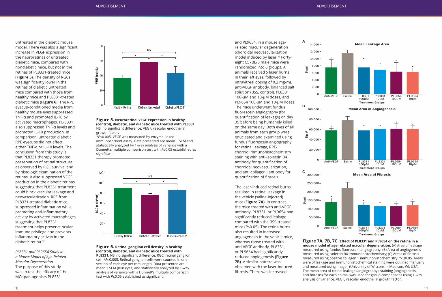
and PL9654, in a mouse age-related macular degeneration (choroidal neovascularization) model induced by laser.32 Forty-eight C57BL/6 male mice were randomized into 6 groups. All animals received 5 laser burns in their left eyes, followed by intravitreal dosing of 0.2 mg/mL anti-VEGF antibody, balanced salt solution (BSS, control), PL8331 100-µM and 10-µM doses, and PL9654 100-µM and 10-µM doses. The mice underwent fundus fluorescein angiography (for quantification of leakage) on day 35 before being humanely killed on the same day. Both eyes of all animals from each group were enucleated and examined using fundus fluorescein angiography for retinal leakage, RPE/choroid immunohistochemistry staining with anti-isolectin B4 antibody for quantification of choroidal neovascularization, and anti-collagen I antibody for quantification of fibrosis.
The laser-induced retinal burns resulted in retinal leakage in the vehicle (saline-injected) mice (Figure 7A). In contrast, the mice treated with anti-VEGF antibody, PL8331, or PL9654 had significantly reduced leakage compared with the BSS-treated mice (P<0.05). The retina burns also resulted in increased angiogenesis in the vehicle mice, whereas those treated with anti-VEGF antibody, PL8331, or PL9654 had significantly reduced angiogenesis (Figure 7B). A similar pattern was observed with the laser-induced fibrosis. There was increased
untreated in the diabetic mouse model. There was also a significant increase in VEGF expression in the neuroretinas of untreated diabetic mice, compared with nondiabetic mice, but not in the retinas of PL8331-treated mice (Figure 5). The density of RGCs was significantly lower in the retinas of diabetic untreated mice compared with those from healthy mice and PL8331-treated diabetic mice (Figure 6). The RPE eyecup-conditioned media from healthy mouse eyes suppressed TNF-α and promoted IL-10 by activated macrophages. PL-8331 also suppressed TNF-α levels and promoted IL-10 production. In comparison, untreated diabetic RPE eyecups did not affect either TNF-α or IL-10 levels. The conclusion from this study is that PL8331 therapy promoted preservation of retinal structure as observed by RGC survival and by histologic examination of the retinas. It also suppressed VEGF production in the diabetic retinas, suggesting that PL8331 treatment could block vascular leakage and neovascularization. RPE from PL8331-treated diabetic mice suppressed inflammation while promoting anti-inflammatory activity by activated macrophages, suggesting that PL8331 treatment helps preserve ocular immune privilege and prevents inflammatory activity in the diabetic retina.32
PL8331 and PL9654 Study in a Mouse Model of Age-Related Macular DegenerationThe purpose of this study was to test the efficacy of the MCr pan-agonists PL8331
Figure 7A, 7B, 7C. Effect of PL8331 and PL9654 on the retina in a mouse model of age-related macular degeneration. (A) Area of leakage measured using fundus fluorescein angiography; (B) Area of angiogenesis measured using isolectin B4 immunohistochemistry; (C) Areas of fibrosis measured using positive collagen 1 immunohistochemistry. *P≤0.05. Areas were of leakage and immunohistochemical staining were outlined manually and measured using Image J (University of Wisconsin, Madison, WI, USA). The mean area of retinal leakage (angiography), staining (angiogenesis and fibrosis) for each animal was used for group comparisons using 1-way analysis of variance. VEGF, vascular endothelial growth factor.
Figure 5. Neuroretinal VEGF expression in healthy (control), diabetic, and diabetic mice treated with PL8331. NS, no significant difference; VEGF, vascular endothelial growth factor.*P≤0.005. VEGF was measured by enzyme-linked immunosorbent assay. Data presented are mean ± SEM and statistically analyzed by 1-way analysis of variance with a Dunnett’s multiple comparison test with P≤0.05 established as significant.
Figure 6. Retinal ganglion cell density in healthy (control), diabetic, and diabetic mice treated with PL8331. NS, no significant difference; RGC, retinal ganglion cell. *P≤0.005. Retinal ganglion cells were counted in one section of each eye per mm length. Data presented are mean ± SEM (n=8 eyes) and statistically analyzed by 1-way analysis of variance with a Dunnett’s multiple comparison test with P≤0.05 established as significant.
14,000
12,000
10,000
8000
6000
4000
2000
0
*
Anti-VEGF Saline PL8331 PL8331 PL9654 PL9654 100µM 10µM 100µM 10µM
Pixels
2
*
**
*
Mean Leakage AreaA
B
C
Treatment Groups
100,000
80,000
60,000
40,000
20,000
0
*
Anti-VEGF Saline PL8331 PL8331 PL9654 PL9654 100µM 10µM 100µM 10µM
Pixels
2
* **
*
Mean Area of Angiogenesis
Treatment Groups
300,000
250,000
200,000
150,000
100,000
50,000
0
*
Anti-VEGF Saline PL8331 PL8331 PL9654 PL9654 100µM 10µM 100µM 10µM
Pixels
2
** *
*
Mean Area of Fibrosis
Treatment Groups

13
ADVERTISEMENT
12
ADVERTISEMENT
warming masks/goggles, and massages. Likewise the use of punctual plugs is effective in aqueous-deficient dry eye disease.35,36 A new treatment is lifitegrast, an antagonist of lymphocyte function–associate antigen 1 (LFA-1), a heterodimeric integrin found in large granular lymphocytes, as well as B and T lymphocytes. By binding to LFA-1, lifitegrast prevents the activation and proliferation of lymphocytes and therefore the production of inflammatory cytokines, which lead to inflammation and cell destruction.36
Despite all these therapeutic approaches, many patients find that current treatments do not provide sufficient improvement.35,37,38 α-MSH has been shown to improve corneal dysfunction and to correct overexpression of proinflammatory factors as well has having cryoprotective and anti-apoptotic activity in a rat model of scopolamine-induced dry eye syndrome.39 α-MSH also promoted healing of corneal epithelial lesions and reduced ocular irritation after corneal abrasion in rats.40 These protective effects were deemed to be due to
the action of α-MSH on the PKA-CREB and MEK-ERK pathways.39
PL9643, a MCr pan-agonist (not active at MC2r), is currently being investigated for anti-inflammatory ocular indications, including dry eye disease. This was a phase 2 study (ClinicalTrials.gov NCT04268069) that evaluated the efficacy and tolerability of PL9643 in adults with mild, moderate, or severe dry eye disease.41 This study consisted of a 12-week treatment period in 160 patients who were randomized 1:1 to either PL9643 or vehicle. They received PL9643 1 µg/mL or vehicle (ophthalmic solution) topically 3 times daily. Patients were assessed in the clinic at 2, 4, 8, and 12 weeks postrandomization. Although this was an exploratory study, the sponsor (Palatin Technologies) did designate coprimary endpoints of inferior corneal fluorescein staining and ocular discomfort after 12 weeks. Fluorescein staining was used for the cornea and Lissamine green staining for the conjunctiva. The staining was graded according to the Ora Calibra® Cornea and Conjunctiva Staining Scale (Ora Calibra, Andover, MA, USA). Ocular discomfort was measured using the single-question Ora Calibra Ocular Discomfort Scale (0 = no discomfort to 4 = constant
discomfort). Secondary endpoints included dry eye disease signs (fluorescein and Lissamine staining, tear film break-up time, conjunctival redness) and patient-reported symptoms (burning, dryness, eye discomfort, eye dryness, foreign body sensation, grittiness, itching, stinging, ocular discomfort) measured by the Ora Calibra Ocular Discomfort and 4-Symptom Questionnaire and visual analog scales.
Although positive results were not found when all patients (mild, moderate, and severe dry eye) were assessed, evaluation of the moderate and severe subgroup demonstrated utility of PL9643 in dry eye disease. Baseline demographics of the moderate or severe subset were balanced across the groups with a median age of 69 (range, 51–84) years for the vehicle group and 69.5 (range, 51–80) years for the PL9643 group; 71% and 70% of patients, respectively, were female. For the coprimary endpoint of inferior corneal fluorescein staining in patients in the moderate or severe subgroup, significant improvement at week 12 was observed for the PL9643 group compared with the vehicle group for the (LS mean difference [SEM], –0.5 [0.2]; P<0.05). Significant improvement at week 12 was also observed in total corneal
fibrosis in the vehicle group and significantly less fibrosis with anti-VEGF antibody, PL8331, and PL9654 treatment. This study demonstrated that in this model system both PL8331 and PL9654 used at the tested dosage had an inhibitory effect on leakage, fibrosis, and neovascularization caused by laser injury. Their effects are comparable to that of the traditionally used anti-VEGF antibody treatment for choroidal neovascularization in wet age-related macular degeneration.
Summary of Effect of Melanocortin Agonists PL8331 and PL9654 on Agonists on the Preclinical Retinopathy Model SystemsPL8331 treatment preserved retinal thickness and mitigated optic nerve cupping, promoted preservation of retinal structure observed by RGC survival and histology of the retinas in a mouse model of diabetic retinopathy, promoted preservation of retinal structure observed by RGC survival and histology of the retinas, and suppressed VEGF production in the diabetic retina, suggesting that PL8331 treatment may block vascular leakage and neovascularization. In a mouse model of age-related
macular degeneration, PL8331 and PL9654 reduced retinal leakage, angiogenesis, and fibrosis compared with control to an extent equivalent to anti-VEGF treatment. In conclusion, intravitreal delivery of PL8331 and PL9654 significantly reduced several markers of retinal damage in mouse models of eye injury, supporting the continued development of PL8331 and PL9654 for the treatment of retinopathy.
Dry Eye DiseaseDry eye disease is an inflammatory disease involving the ocular surface, meibomian glands, the main lacrimal gland, and their innervation. Infection, environmental factors, stress, antigen stimulation, and genetic factors are among the factors that may trigger the pathogenesis. T-helper cells penetrate the lacrimal glands and the ocular surface owing to the production of proinflammatory cytokines, matrix metalloproteinases, and chemokines, leading to inflammation and damage to the surface of the eye. It can be classified into aqueous-deficient dry eye (insufficient tears) and hyper-evaporative dry eye (in which the tear film evaporates more rapidly than usual).33,34
First-line treatments for dry eye disease include lifestyle changes (such as minimizing alcohol, ensuring adequate fluid intake, using humidifiers, getting adequate sleep) and artificial tear eyedrop solutions.33,35 The topical fungal antimetabolite cyclosporine A is also commonly used. Unfortunately, it is associated with a number of adverse effects, including ocular burning, conjunctival hyperemia, discharge, epiphora, eye pain, foreign body sensation, visual disturbance, and pruritis, and it is also more expensive than most commonly prescribed ophthalmic medications.35 Anti-inflammatory agents such as corticosteroid eyedrops have been shown to improve the signs and symptoms of moderate to severe dry eye, but are recommended only for short-term use. Tetracycline and macrolides such as azithromycin have been used for their additional anti-inflammatory effects but are recommended only in low doses. Omega-3 fatty acids, which block proinflammatory cytokines and inflammatory processes in the eyes, are also commonly used. Nonpharmacological approaches include eyelid hygiene with hot compresses, infrared heaters,

15
ADVERTISEMENT
14
ADVERTISEMENT
staining (inferior + superior + central), and superior corneal staining (Figure 8A). Efficacy was also observed in the conjunctiva using Lissamine green staining. The regions showing efficacy were the total conjunctival score at week 12 and the temporal region at both weeks 2 and 12 (Figure 8B). Tear film break-up time improved significantly from baseline at week 12 (P<0.05), and signs of conjunctival redness also
Figure 8A, 8B. Differences between PL9643 and vehicle in the subset of patients with moderate or severe dry eye disease in (A) fluorescein and (B) Lissamine green staining. LS, least squares.
0.2
0
–0.2
–0.4
–0.6
–0.8
–1.0
–1.2 Total Sum Total Corneal Total Nasal Temporal Inferior Superior Central (corneal+ (inferior+ Conjunctival conjunctival) superior+ (temporal+ central) nasal)
Diffe
renc
e in
LS
Mea
n Ch
ange
Fro
m B
asel
ine
A
0.2
0
–0.2
–0.4
–0.6
–0.8
–1.0 Total Sum Total Corneal Total Nasal Temporal Inferior Superior Central (corneal+ (inferior+ Conjunctival conjunctival) superior+ (temporal+ central) nasal)
Diffe
renc
e in
LS
Mea
n Ch
ange
Fro
m B
asel
ine
B
*P≤0.05
*P≤0.05
� Week 2 � Week 12
*
**
*
*
*
Figure 9. Differences between PL9643 and vehicle in conjunctival redness and tear film break-up time in the subset of patients with moderate or severe dry eye disease. Conjunctival redness was graded on a 0–4 scale in 0.5 increments (0 = normal, without vasodilation), 4 = broad ciliary and prominent, horizontal conjunctival vasodilation). Tear film break-up time; 2 measurements were taken and averaged unless the 2 measurements are >2 seconds apart and are each <10 seconds, in which case, a third measurement would be taken and the 2 closest of the 3 would be averaged. LS, least squares.
0
–0.02
–0.04
–0.06
–0.08
–0.10
–0.12
–0.14
–0.16
–0.18
–0.20
Diffe
renc
e in
LS
Mea
n Ch
ange
From
Bas
elin
e
Conjunctival Redness
*P≤0.05� Week 2 � Week 12
*
0.35
0.30
0.25
0.20
0.15
0.10
0.05
0
Diffe
renc
e in
LS
Mea
n Ch
ange
From
Bas
elin
e
Tear Film Break-up Time
showed numeric improvements from baseline, although it did not reach significance at week 2 or 12 (Figure 9). For the other primary endpoint of ocular discomfort using the Ora Calibra Ocular Discomfort Scale, patients showed numeric improvement over vehicle (LS mean change from baseline, –0.2 at week 2 and –0.1 at week 12), but this was not statistically significant. For the secondary endpoints,
there was consistent numeric improvement at week 2 with PL9643 compared with vehicle in ocular discomfort, burning, grittiness, and stinging as measured by the Ora Calibra Ocular Discomfort and 4-Symptom Questionnaire. The 2-week improvement in ocular discomfort from the scale was significant (P<0.05). Using the visual analog scale, PL9643 demonstrated numeric improvement over

17
ADVERTISEMENT
16
ADVERTISEMENT
Therapeutically targeting the melanocortin pathway represents a
novel approach to the treatment of inflammatory and immune-
mediated diseases. Preclinical evidence has demonstrated the broad
range of biological activity of melanocortin agonists in numerous
inflammatory diseases, including ophthalmic diseases. The role of
synthetic MCr receptor agonists shows promise in the treatment of
uveitis and retinopathy in preclinical animal models. Additionally,
in a phase 2 study treatment with PL9643 improved the signs and
symptoms in patients with moderate to severe dry eye disease.
vehicle at 2 weeks and 12 weeks for several endpoints including itching, foreign body sensation, eye discomfort, and eye dryness (Figure 10).
PL9643 demonstrated a good safety and tolerability profile. Among 39 treatment-emergent adverse events (TEAEs), 16 occurred in patients receiving PL9643 compared with 23 events in patients receiving vehicle. There were 9 ocular TEAEs were reported in the study, chalazion on the upper lid (n=1), stinging upon instillation (n=1), burning upon instillation (n=1), intermittent burning upon instillation (n=3), possible Herpes zoster (n=1), worsening of visual acuity (n=1), and intermittent itching upon instillation (n=1), all of which were considered mild in severity (1 event with PL9643 and 8 events with vehicle). The ocular adverse event reported in a subject receiving PL9643, chalazion on upper lid, was not considered related to study drug.41
This phase 2 study showed that in patients with moderate or severe dry eye disease, topical PL9643 solution led to benefits in some signs and symptoms by the first evaluation at 2 weeks, many of which were maintained at 12 weeks. PL9643 was well tolerated, with no treatment-related ocular-related adverse events and a safety profile comparable to that of vehicle. These positive results support the continued development of PL9643 for dry eye disease. A phase 3 trial comparing PL9643 to vehicle in ≥240 patients with moderate to severe dry eye disease is planned.
Coprimary sign endpoints assessed at week 12 will be inferior fluorescein staining and total conjunctival Lissamine green staining. The coprimary symptom endpoint will be ocular discomfort at week 2. Key secondary endpoints assessed at week 2
will include total conjunctival Lissamine green staining and the symptom endpoints of burning and eye discomfort. Other studies (PL9643-302 and PL9643-303) are also planned.
Figure 10A, 10B. Differences between PL9643 and vehicle in the subset of patients with moderate or severe disease. (A) Ora Calibra® Ocular Discomfort and 4-Symptom Questionnaire scores; (B) Components of the visual analog scale. Ocular Discomfort and 4-Symptom Questionnaire: patients rated each of the symptoms on a 0–5 scale where 0 = none to 5 = worst. Visual analog scale: patients rated each symptom on a 0%–100% continuous scale. 0% corresponds to “no discomfort” and 100% corresponds to “maximal discomfort.” LS, least squares.
0.3
0.2
0.1
0
–0.1
–0.2
–0.3
–0.4
–0.5 Ocular Burning Dryness Grittiness Stinging Discomfort
LS M
ean
Chan
ge F
rom
Bas
elin
e
A. Ora Calibra® Ocular Discomfort and 4-Symptom Questionnaire
4
2
0
–2
–4
–6
–8 Burning/ Itching Foreign Body Eye Eye Photophobia Pain Stinging Sensation Discomfort Dryness
LS M
ean
Chan
ge F
rom
Bas
elin
e
B. Visual Analog Scale
*P≤0.05
� Week 2 � Week 12
*
CONCLUSION

19
ADVERTISEMENT
18
ADVERTISEMENT
21. Cai S, Yang Q, Hou M, et al. Alpha-melanocyte-stimulating hormone protects early diabetic retina from blood-retinal barrier breakdown and vascular leakage via MC4R. Cell Physiol Biochem. 2018;45:505-522.
22. Rossi S, Maisto R, Gesualdo C, et al. Activation of melanocortin receptors MC 1 and MC 5 attenuates retinal damage in experimental diabetic retinopathy. Mediators Inflamm. 2016;2016:7368389.
23. Taylor AW, Kitaichi N, Biros D. Melanocortin 5 receptor and ocular immunity. Cell Mol Biol (Noisy-le-grand). 2006;52:53-59.
24. Krishna U, Ajanaku D, Denniston AK, Gkika T. Uveitis: a sight-threatening disease which can impact all systems. Postgrad Med J. 2017;93:766-773.
25. Lechner J, O’Leary OE, Stitt AW. The pathology associated with diabetic retinopathy. Vision Res. 2017;139:7-14.
26. Wang W, Lo ACY. Diabetic retinopathy: pathophysiology and treatments. Int J Mol Sci. 2018;19:1816.
27. Wong TY, Cheung CM, Larsen M, et al. Diabetic retinopathy. Nat Rev Dis Primers. 2016;2:16012.
28. Tah V, Orlans HO, Hyer J, et al. Anti-VEGF therapy and the retina: an update. J Ophthalmol. 2015;2015:627674.
29. Falavarjani KG, Nguyen QD. Adverse events and complications associated with intravitreal injection of anti-VEGF agents: a review of literature. Eye (Lond). 2013;27:787-794.
30. Zhang L, Dong L, Liu X, et al. Alpha-melanocyte-stimulating hormone protects retinal vascular endothelial cells from oxidative stress and apoptosis in a rat model of diabetes. PLoS One. 2014;9:e93433.
31. Zeng XX, Ng YK, Ling EA. Neuronal and microglial response in the retina of streptozotocin-induced diabetic rats. Vis Neurosci. 2000;17:463-471.
32. Dodd J, Makhlina M, Yang WH, et al. Protective effects of 2 melanocortin agonists delivered by intravitreal injection in mouse models of retinopathy (ARVO Annual Meeting abstract). Invest Ophthalmol Vis Sci. 2021;62:3291.
33. Messmer EM. The pathophysiology, diagnosis, and treatment of dry eye disease. Dtsch Arztebl Int. 2015;112:71-81; quiz 82.
34. The definition and classification of dry eye disease: report of the Definition and Classification Subcommittee of the International Dry Eye WorkShop (2007). Ocul Surf. 2007;5:75-92.
35. O’Neil EC, Henderson M, Massaro-Giordano M, Bunya VY. Advances in dry eye disease treatment. Curr Opin Ophthalmol. 2019;30:166-178.
36. Haber SL, Benson V, Buckway CJ, et al. Lifitegrast: a novel drug for patients with dry eye disease. Ther Adv Ophthalmol. 2019;11:2515841419870366.
37. Mah F, Milner M, Yiu S, et al. PERSIST: Physician’s Evaluation of Restasis® Satisfaction in Second Trial of topical cyclosporine ophthalmic emulsion 0.05% for dry eye: a retrospective review. Clin Ophthalmol. 2012;6:1971-1976.
38. Tauber J, Karpecki P, Latkany R, et al. Lifitegrast ophthalmic solution 5.0% versus vehicle for treatment of dry eye disease: results of the randomized phase III OPUS-2 study. Ophthalmology. 2015;122:2423-2431.
39. Ru Y, Huang Y, Liu H, et al. alpha-Melanocyte-stimulating hormone ameliorates ocular surface dysfunctions and lesions in a scopolamine-induced dry eye model via PKA-CREB and MEK-Erk pathways. Sci Rep. 2015;5:18619.
40. Pavan J, Lukenda A, Stambuk N, et al. Effects of alpha-MSH on corneal epithelial lesions in rats. Coll Antropol. 2012;36:1407-1411.
41. Kenyon K, Ousler GW, Watson M, et al. Efficacy and safety of the melanocortin agonist PL9643 in a phase 2 study of subjects with dry eye disease (ARVO Annual Meeting abstract). Invest Ophthalmol Vis Sci. 2021;62:1333.
8. Mountjoy KG, Robbins LS, Mortrud MT, Cone RD. The cloning of a family of genes that encode the melanocortin receptors. Science. 1992;257:1248-1251.
9. Wang W, Guo DY, Lin YJ, Tao YX. Melanocortin regulation of inflammation. Front Endocrinol (Lausanne). 2019;10:683.
10. Cai M, Hruby VJ. The melanocortin receptor system: a target for multiple degenerative diseases. Curr Protein Pept Sci. 2016;17:488-496.
11. Taylor AW, Lee D. Applications of the role of alpha-MSH in ocular immune privilege. Adv Exp Med Biol. 2010;681:143-149.
12. Moscowitz AE, Asif H, Lindenmaier LB, et al. The importance of melanocortin receptors and their agonists in pulmonary disease. Front Med (Lausanne). 2019;6:145.
13. Clemson CM, Yost J, Taylor AW. The role of alpha-MSH as a modulator of ocular immunobiology exemplifies mechanistic differences between melanocortins and steroids. Ocul Immunol Inflamm. 2017;25:179-189.
14. Friedman DS, Holbrook JT, Ansari H, et al. Risk of elevated intraocular pressure and glaucoma in patients with uveitis: results of the multicenter uveitis steroid treatment trial. Ophthalmology. 2013;120:1571-1579.
15. Clark AF, Wordinger RJ. The role of steroids in outflow resistance. Exp Eye Res. 2009;88:752-759.
16. Multicenter Uveitis Steroid Treatment Trial Research Group, Kempen JH, Altaweel MM, et al. Randomized comparison of systemic anti-inflammatory therapy versus fluocinolone acetonide implant for intermediate, posterior, and panuveitis: the multicenter uveitis steroid treatment trial. Ophthalmology. 2011;118:1916-1926.
17. Lewis JM, Priddy T, Judd J, et al. Intraocular pressure response to topical dexamethasone as a predictor for the development of primary open-angle glaucoma. Am J Ophthalmol. 1988;106:607-612.
18. Sen HN, Vitale S, Gangaputra SS, et al. Periocular corticosteroid injections in uveitis: effects and complications. Ophthalmology. 2014;121:2275-2286.
19. Butcher JM, Austin M, McGalliard J, Bourke RD. Bilateral cataracts and glaucoma induced by long term use of steroid eye drops. BMJ. 1994;309:43.
20. Klein BE, Klein R, Lee KE, Danforth LG. Drug use and five-year incidence of age-related cataracts: The Beaver Dam Eye Study. Ophthalmology. 2001;108:1670-1674.
1. Bicknell AB. The tissue-specific processing of pro-opiomelanocortin. J Neuroendocrinol. 2008;20:692-699.
2. Cawley NX, Li Z, Loh YP. 60 years of PMC: biosynthesis, trafficking, and secretion of pro-opiomelanocortin-derived peptides. J Mol Endocrinol. 2016;56:T77-97.
3. Ahmed TJ, Montero-Melendez T, Perretti M, Pitzalis C. Curbing inflammation through endogenous pathways: focus on melanocortin peptides. Int J Inflam. 2013;2013:985815.
4. Brzoska T, Luger TA, Maaser C, et al. Alpha-melanocyte-stimulating hormone and related tripeptides: biochemistry, antiinflammatory and protective effects in vitro and in vivo, and future perspectives for the treatment of immune-mediated inflammatory diseases. Endocr Rev. 2008;29:581-602.
5. Catania A, Lonati C, Sordi A, et al. The melanocortin system in control of inflammation. ScientificWorldJournal. 2010;10:1840-1853.
6. Spana C, Taylor AW, Yee DG, et al. Probing the role of melanocortin type 1 receptor agonists in diverse immunological diseases. Front Pharmacol. 2018;9:1535.
7. Catania A, Gatti S, Colombo G, Lipton JM. Targeting melanocortin receptors as a novel strategy to control inflammation. Pharmacol Rev. 2004;56:1-29.
REFERENCES

21
ADVERTISEMENT
20
ADVERTISEMENT
TABLE 1. Melanocortin Receptors3,5,9
Receptors Hormone/endogenous agonist (binding affinity)
Distribution* Signaling Pathway
Function Role in Disease/Potential Therapeutic Use
MC1r α-MSH=ACTH>β-MSH>γ-MSH
Skin (keratinocytes, melanocytes, fibro-blasts) periaqueductal gray matter macro-phages, monocytes, lymphocytes, neutro-phils, astrocytes, glial cells, endothelial cells
cAMP
ERK1/ERK2
Skin pigmenta-tion Anti-inflam-matory
Wound healing
Inflammation
Skin cancer
Vitiligo
Alopecia areata
MC2r ACTHAdrenal glands, adi-pocytes, osteoblasts, chondrocytes
cAMP Steroidogenesis
Familial
glucocorticoid deficiency
MC3r γ-MSH>α-MSH=ACTH=β-MSH
Brain (cortex, thala-mus, hypothalamus, hippocampus, septal area), placenta, heart, gut,
macrophages, mono-cytes, B-lymphocytes
cAMP
Intracellular [Ca2+]
Energy homeo-stasis
Cardiovascular function
Anti-inflamma-tory
Inflammation
Gouty arthritis
Obesity
Tuberculosis
MC4r α-MSH=ACTH>β-MSH>γ-MSH
Brain (brainstem, cortex, thalamus, hy-pothalamus, dentate gyrus, cortex, amyg-dala, hippocampus, septal reg corpus stri-atum, midbrain, pons, medulla oblongata)
cAMP
Energy homeo-stasis
Cardiovascular function
Food intake
Erectile function
Obesity
Cachexia
Sexual dysfunction
MC5r α-MSH>ACTH=β-MSH>>γ-MSH
Ubiquitous in periph-eral tissues. skeletal muscle, brain, lung, spleen, thymus, bone marrow. adrenal gland, testis, ovary, uterus
B-, T-, and splenic lymphocytes
cAMP
Intracellular [Ca2+] JAK/STAT
Exocrine gland function
Anti-inflamma-tory Defensive behavior
Inflammation
Seborrheic dermatitis
Acne vulgaris
*Distribution investigated in humans, mice and rats. Not all distributions have been investigated in all species.
ACTH, adrenocorticotrophic hormone; cAMP, cyclic AMP; ERK, extracellular signal regulated kinases; JAK-STAT, Janus kinase-signal transducer and activator of transcription proteins; MCr, melanocortin receptor; MSH, melanocyte-stimulating hormone.
TABLE 2. Selected MCr Agonists Available or in Commercial Development
Agonist Receptor Target/Activity
Administration Status Disease/Condition Company
Vyleesi®
(bremelanotide) MC4r SC injection (FDA approved) Approved (US) Hypoactive sexual
desire disorderPalatin Technologies
PL8177 Selective MC1r agonist Oral In development Inflammatory bowel
diseasesPalatin Technologies
PL9643 MC1r, MC3r, MC4r, MC5r Topical In development
Dry eye disease, uveitis, diabetic retinopathy, diabetic macular edema
Palatin Technologies
PL9654 MCr pan- agonist Intravitreal In development Diabetic retinopathy,
macular degenerationPalatin Technologies
PL8331 MCr pan- agonist
Topical, intravitreal In development Dry eye disease,
diabetic retinopathyPalatin Technologies
PL8905 Selective MC4r agonist SC injection In development Obesity, diabetes
Palatin Technologies
Scenesse®
(afemelanotide)
Selective MC1r agonist (α-MSH analog)
Sc implantApproved
(US/EU)
Erythropoietic protoporphyria
Clinuvel Pharmaceuticals
Scenesse®
(afemelanotide)
Selective MC1r agonist (α-MSH analog)
- In developmentVariegate porphyria, vitiligo, arterial ischemic stroke
Clinuvel Pharmaceuticals
CUV9900Selective MC1r agonist (α-MSH analog)
- In development Skin protectant Clinuvel Pharmaceuticals
VLRX001 (parvysmela-notide)
Topical/ transdermal In development Vitiligo Clinuvel
Pharmaceuticals
Imcivree (setmelanotide)
MC4r receptor agonist SC injection
Approved
(US/EU)
Chronic weight man-agement in adult/pediatric patients with obesity due to POMC, proprotein convertase subtilisin/kexin type 1 (PCSK1), or leptin receptor (LEPR) deficiency
Rhythm Pharmaceuticals
Setmelanotide MC4r receptor agonist - In development
Bardet-Biedl syndrome, Alström syndrome, additional obesity conditions
Rhythm Pharmaceuticals
FDA, US Food and Drug Administration; MCr, melanocortin receptor; MSH, melanocyte-stimulating hormone; POMC, pro-opiomelanocortin; SC, subcutaneous.

23
ADVERTISEMENT
22
ADVERTISEMENT
ACKNOWLEDGMENTS
Editorial assistance was provided by Robin Smith, PhD,
of The Curry Rockefeller Group, LLC (Tarrytown, NY), which
was funded by Palatin Technologies, Inc. (Cranbury, NJ).

ADVERTISEMENT
Palatin.com