Embed Size (px)
Citation preview

The pathophysiology and retention of gadolinium
Brent Wagner, MD
South Texas Veterans Health Care System, Department of Medicine/Nephrology University of Texas Health Science Center at San Antonio, San Antonio, Texas
Associate Professor of Medicine

Objectives
Elucidation of the mechanisms of gadolinium-based contrast agent-induced toxicity is an active area of investigation
A model has been established in rodents
The focus of this presentation is the work in my laboratory concerning the mechanisms of gadolinium-based contrast agent toxicity and how this is manifested systemically
One gadolinium-based contrast agent has been used in these experiments, Omniscan (gadodiamide/caldiamide), but the findings may be applicable for the other gadolinium-based contrast agents
2
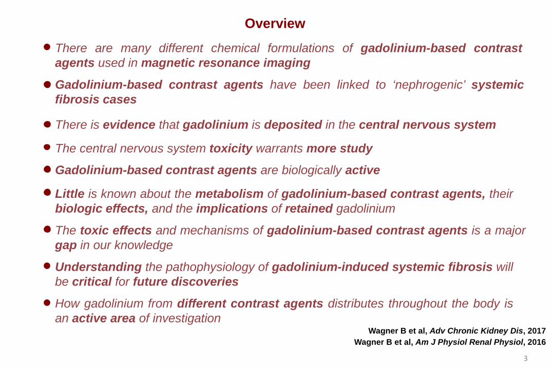
Gadolinium-based contrast agents have been linked to ‘nephrogenic’ systemic fibrosis cases
There are many different chemical formulations of gadolinium-based contrast agents used in magnetic resonance imaging
There is evidence that gadolinium is deposited in the central nervous system
The toxic effects and mechanisms of gadolinium-based contrast agents is a major gap in our knowledge
Overview
Gadolinium-based contrast agents are biologically active
Wagner B et al, Am J Physiol Renal Physiol, 2016 Wagner B et al, Adv Chronic Kidney Dis, 2017
The central nervous system toxicity warrants more study
Understanding the pathophysiology of gadolinium-induced systemic fibrosis will be critical for future discoveries
How gadolinium from different contrast agents distributes throughout the body is an active area of investigation
Little is known about the metabolism of gadolinium-based contrast agents, their biologic effects, and the implications of retained gadolinium
3

The periodic table of elements
K V Ti Ca Sc Cr Co Mn Fe Ni Ge Ga Cu Zn As Kr Se Br
Rb Nb Zr Sr Y Mo Rh Tc Ru Pd Sn In Ag Cd Sb Xe Te I
Cs Ta Hf Ba W Ir Re Os Pt Pb Tl Au Hg Bi Rn Po At
Fr Db Rf Ra Sg Mt Bh Hs Ds Fl Uut Rg Cn Uup Uuo Lv Uus
Ce La Pr Sm Nd Pm Eu Ho Dy Gd Tb Er Lu Tm Yb
Th Ac Pa Pu U Np Am Es Cf Cm Bk Fm Lr Md No
H
Li Be
Na Mg
He
C B N Ne O F
Si Al P Ar S Cl
4

‘Nephrogenic’ systemic fibrosis, a man-made disease caused by magnetic resonance imaging contrast agents
Dural thickening
Diaphragm Heart
Extremities
Trunk
Buttocks
Face
Joint contractures
Psoas Rete testes
Kidney
Extremities
Lung
Liver
Girardi M et al, J Am Acad Dermatol, 2011
5

Gadolinium-based contrast agents can be acutely nephrotoxic in humans
Degeneration of tubular epithelial cells
Glomerulosclerosis
Toluidine blue stain - Kidney
Flattening of tubular
epithelial cells Calcium phosphate
H&E stain - Kidney
Cellular proliferation
Immunostain - Kidney Electron microscopy - Kidney
Akgun H et al, Archives of Pathology & Laboratory Medicine, 2006
6
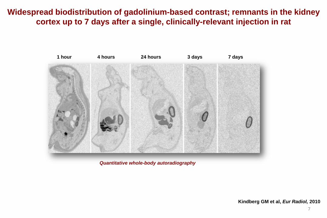
Quantitative whole-body autoradiography
1 hour 4 hours 24 hours 3 days 7 days
Widespread biodistribution of gadolinium-based contrast; remnants in the kidney cortex up to 7 days after a single, clinically-relevant injection in rat
Kindberg GM et al, Eur Radiol, 2010 7

Differential effects of gadolinium-based contrast agents in rats
H&E staining - Skin
Control Gadodiamide Gadoteridol
Control Gadodiamide Gadoteridol
Immunohistochemical staining – Skin fibronectin (marker of fibrosis)
Control Gadodiamide Gadoteridol
Fibronectin
GAPDH
Immunoblot - Skin
N
C
N
C
O-
O-
O
O
N -O
O
C
Gd3+ Omniscan (gadodiamide >>
caldiamide) O
C N
O
C N ProHance
(gadoteridol)
N
Gd3+
N
N N
Do C, Barnes JL, Tan C, Wagner B, Am J Physiol Renal Physiol, 2014 8

Gadodiamide administration in mice with normal renal function
Control
(2.5 mmol/kg body weight, i.p.)
Sacrifice
Mice (C57BL6)
Gadodiamide Gadodiamide (20-25 doses)
Kidney Skin
0
500
1000
Gad
olin
ium
(µ
g/g
tissu
e)
Gadolinium content, kidney
***
0
20
40
60
Gad
olin
ium
(µ
g/g
tissu
e)
Gadolinium content, skin
***
0
3000
5000
Gad
olin
ium
(n
g/g
tissu
e)
Gadolinium content, cerebellum
***
0
2000
4000
Gad
olin
ium
(n
g/g
tissu
e)
Gadolinium content, cerebrum
***
1000
4-5 weeks
9

Gadodiamide Control Gadodiamide Control
Transmission electron microscopy - Glomeruli Transmission electron microscopy - Tubules
100 nm
2 µm
Electron microscopy shows electron-dense deposits in the kidneys of gadodiamide-treated mice with normal renal function
Wagner laboratory, unpublished
10

100 nm
Li R et al, ACS Nano, 2014
Gd2O3 Gadodiamide
Transmission electron microscopy - Tubule
Transmission electron microscopy - Mesangium
Transmission electron microscopy – Phagolysosomal-
simulated fluid
Transmission electron microscopy - Water
The renal deposits resemble Gd2O3 disordered mesh-like nanowire/nanoparticle aggregates in vitro
Wagner laboratory, unpublished
11

Gadodiamide induces renal fibrosis in mice
PAS staining
Control Gadodiamide
Fibronectin
Collagen IV
220 kDa Fibronectin
GAPDH
Control Gadodiamide
Gadodiamide
PAS staining - Kidney
Gadodiamide
PAS staining - Kidney
Immunoblot - Skin Wagner laboratory, unpublished
12
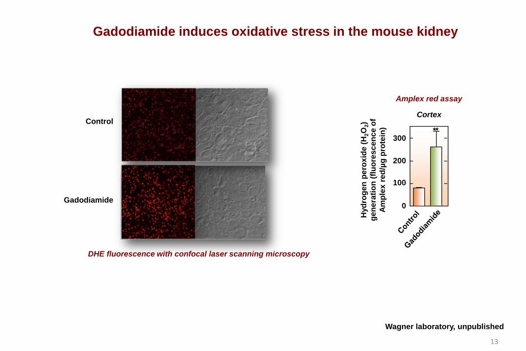
Control
Gadodiamide
DHE fluorescence with confocal laser scanning microscopy
Gadodiamide induces oxidative stress in the mouse kidney
Hyd
roge
n pe
roxi
de (H
2O2)
ge
nera
tion
(fluo
resc
ence
of
Ampl
ex re
d/µg
pro
tein
)
0
100
200
300 * *
Amplex red assay
Cortex
Wagner laboratory, unpublished
13
0
20
40
Der
mal
nuc
lei
(per
HPF
)
Cellularity
***
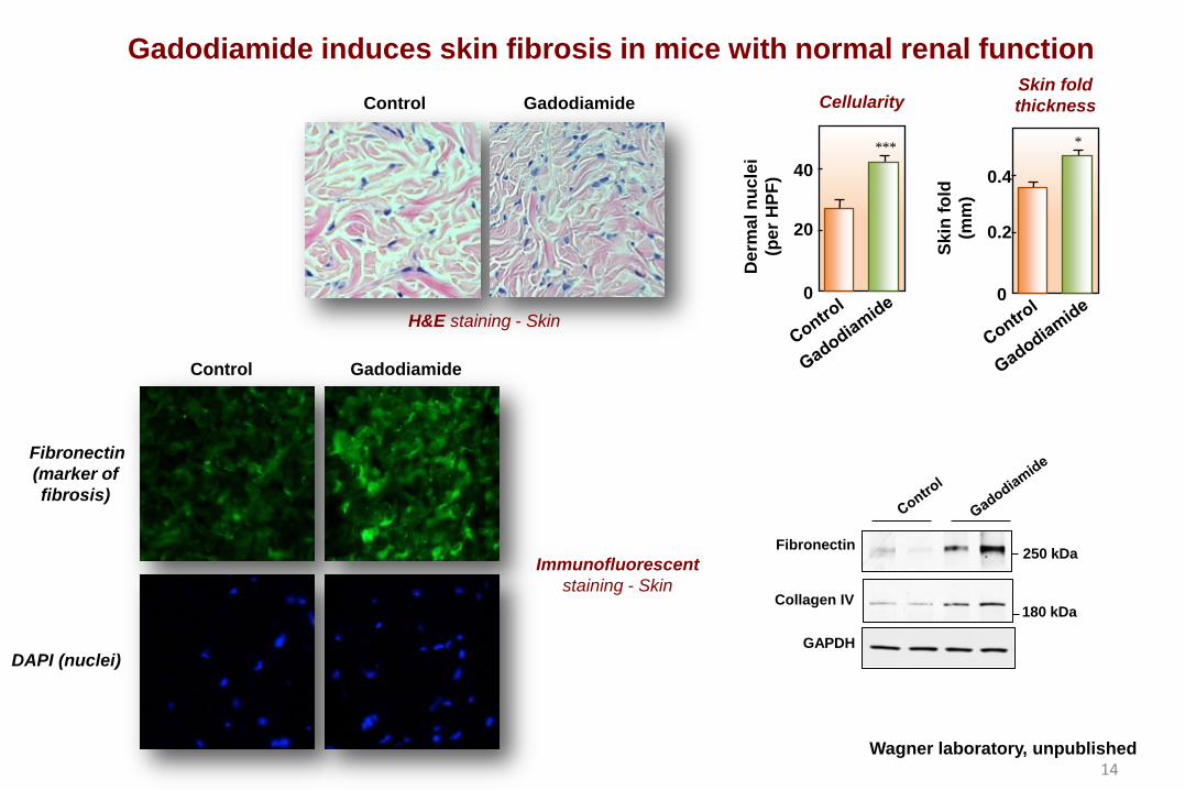
Gadodiamide induces skin fibrosis in mice with normal renal function
H&E staining - Skin
Control Gadodiamide
Immunofluorescent staining - Skin
Control Gadodiamide
DAPI (nuclei)
Fibronectin (marker of fibrosis)
180 kDa
250 kDa Fibronectin
GAPDH
Collagen IV
0
0.2
0.4
Skin
fold
(m
m)
Skin fold thickness
*
Wagner laboratory, unpublished 14

Gadodiamide treatment leads to inflammation and bone marrow-derived cells to the dermis in mice with normal renal function
DAPI (nuclei)
CD45RO (fibrocyte marker)
Immunofluorescent staining - Skin
Control Gadodiamide
DAPI (nuclei)
CD163 (inflammation)
Immunofluorescent staining - Skin
Control Gadodiamide
Wagner laboratory, unpublished 15

Gadodiamide increases oxidative stress in the skin of mice with normal renal function
In situ DHE staining and confocal microscopy - Skin
Control Gadodiamide
DAPI (nuclei)
3-Nitrotyrosine
Immunofluorescent staining - Skin
Control Gadodiamide
Wagner laboratory, unpublished 16
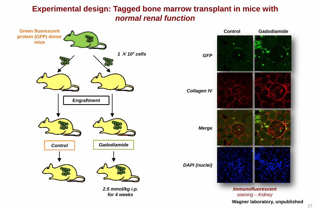
Control Gadodiamide
Green fluorescent protein (GFP) donor
mice
Engraftment
2.5 mmol/kg i.p. for 4 weeks
1 ✕ 107 cells
Experimental design: Tagged bone marrow transplant in mice with normal renal function
Collagen IV
GFP
Control Gadodiamide
DAPI (nuclei)
Merge
Immunofluorescent staining – Kidney
Wagner laboratory, unpublished 17

GFP
CD34
Merge
DAPI (nuclei)
Control Gadodiamide
Immunofluorescent staining - Skin
Control Gadodiamide
GFP
CD45RO
Merge
DAPI (nuclei)
Gadodiamide induces the recruitment of bone marrow-derived fibroblasts to the skin in mice
Immunofluorescent staining - Skin Wagner laboratory, unpublished 18

Biopsies of patients with NSF demonstrate significant expression of the hematopoietic progenitor marker CD34
Dermal hyper-cellularity H&E staining - Skin Immunostaining for CD34 - Skin
Clinical photographs of a patient showing skin lesions
Kroshinsky D et al, N Eng J Med, 2009 19

Conclusions
Our experiments show that renal insufficiency is not requisite for fibrosis
Mechanistically, our experiments demonstrate that it is the recruitment of bone marrow-derived cells that mediate the deleterious actions
The pathologic effects of gadolinium-based contrast agents are not well-characterized
Dechelation of gadolinium is a hypothetical pathologic mechanism.
Studies concerning the biologic effects of rare earth metals in general and their retention in human organs are in the nascent stage
Gadolinium retention can be detected in humans and in our models; This allows the mechanistic study of gadolinium-induced organ injury
The science on this topic is at ground zero
We provide examples of important avenues to understanding the mechanisms of disease (lending itself to the discovery of biomarkers)
20

Working hypothesis
Patient with normal kidney function
MRI Gadolinium-based contrast exposure
Gadolinium retention
Organ injury
Impaired function
Gadolinium-based contrast exposure MRI
Gadolinium-induced disease
Biomarkers Precision/Personalized Medicine
21

Working hypothesis
Patient with normal kidney function
MRI Gadolinium-based contrast exposure
Gadolinium retention
Organ injury
Impaired function
Gadolinium-based contrast exposure MRI
Pre-existing conditions
(obesity, diabetes, pregnancy,
inflammation, etc.)
Gadolinium-induced disease
Biomarkers Precision/Personalized Medicine
22

Acknowledgments
Hanna E. Abboud, M.D.
Viktor Drel, Ph.D.
Jeffrey L. Barnes, Ph.D.
UTHSCSA
Catherine Do, M.D.
Denis Féliers, Ph.D.
Chunyan Tan
Wagner’s Laboratory
Yves Gorin, Ph.D.
Doug-Yoon Lee, Ph.D.
NIH RO1DK102085 (PI)
Supported by : Veterans Administration Merit Award I01BX001958 (PI) Veterans Administration Career Development Award (PI)
Veterans Administration VISN 17 New Investigator Award (PI)
Northwestern
Keith MacRenaris, Ph.D. Nephrology
Jochen Reiser, M.D., Ph.D.
Rush University Medical Center Internal Medicine Department
Seema Ahuja, M.D.
UNC
Michael Jay, Ph.D. John Prybylski, Ph.D.
UTSA Miguel Yacaman, Ph.D. Josefina Arellano-Jimenez, Ph.D.
23


![Am J Physiol Heart Circ Physiol 2011[1]](https://img.pdfslide.net/doc/110x75/577ce0031a28ab9e78b28109/am-j-physiol-heart-circ-physiol-20111.jpg)