Embed Size (px)
Citation preview

SUMMARY
Coscinodiscus wailesii Gran et Angst is a large centricdiatom (280–500 µm diameter) from marine phyto-plankton, characterized by a cylindrical frustule withflat valvar surface, two marginal rings of rimoportulaeon the mantle, and two macrorimoportulae. Cells fromcultured and natural populations collected in ParanaguáBay, Paraná, southern Brazil were observed under lightand scanning electron microscopes to verify the popu-lations’ correct identity and morphology. In both popu-lations, a typical central rosette or a hyaline area was found in the valvar center. The species’ distributionin Brazilian waters was revised, and a discussion onpossible vectors of transport was made. Blooms of thespecies occur sporadically in the coast of Paraná,seeming to affect the local trophic chain.
Key words: Coscinodiscus wailesii, diatom, distribution,harmful algae, Southern Brazil, taxonomy.
INTRODUCTION
In recent decades, blooms of marine phytoplanktonhave attracted the attention of public organizations in many countries due to their negative consequenceson human health, seafood safety and marine trophicchains (Hallegraeff 1993; Hallegraeff 1995). As aresult, an international Harmful Algal Bloom Pro-gramme was implemented in 1992 through the initia-tive of Intergovernmental Oceanographic Commission ofUNESCO, aiming to ‘understand causes of algal blooms,predict their occurrences, and mitigate their effects’.
Among the diverse algal groups (dinoflagellates,diatoms, cyanophyceans, rhaphidophytes) composingthe 3400–4100 microalgae species, it was estimatedthat 300 of them could produce blooms (Smayda1997). Diatoms, when growing intensively, may causemass mortality of marine organisms through (i) the pro-duction of toxins, as domoic acid (Pseudonitzschiaspp.); (ii) respiratory diseases in fishes due to gill
damage caused by setae or spines present on the frus-tules of Chaetoceros spp.; (iii) decreasing of otherphytoplankton species as a result of competition fornutrients; and (iv) oxygen depletion in the watercolumn, from intense algal respiration and the incom-plete diatom decomposition by bacteria near the seabottom (Hasle and Fryxell 1995).
Since 1980, the centric diatom Coscinodiscuswailesii Gran et Angst has been recorded worldwide,developing blooms and damaging shellfish and macro-algae cultivations, and commercial fishery areas (Nagaiet al. 1995a; Nehring 1998). Its distribution, firstrestricted to the tropical Pacific and west Atlanticoceans, has extended to Europe, the USA and Japan inrecent years (Lange et al. 1992; Rick and Dürselen1995; Nehring 1998). Indeed, in some areas, it hasbecome an important component of the phytoplank-tonic community. In Brazil, despite the various taxo-nomic works published since 1960, the species wasonly reported recently in the southern region (Valente-Moreira 1987; Moreira-Filho et al. 1990; Fernandes1992; Souza-Mosimann et al. 1993).
In this work, we analyze the morphology of C. wailesiifrom cultured and natural populations sampled inParanaguá Bay, Paraná State as well as discussing its distribution and potential vectors of transport toBrazilian waters.
MATERIALS AND METHODS
Material for analyses was obtained in May 1999 fromthe waters of Paranaguá Bay (25°25′S to 25°35′S and48°20′W to 48°45′W), Paraná State, Southern Brazil(Fig. 1), an estuarine complex of 117 km2 with anaverage depth of 4.3 m. The Bay is influenced by thesubtropical climate (Cfa) with two well-defined seasons:rainy in summer and dry in winter, with annual average
Phycological Research 2001; 49: 89–96
The recently established diatom Coscinodiscus wailesii(Coscinodiscales, Bacillariophyta) in Brazilian waters. I: Remarks on morphology and distributionLuciano F. Fernandes,1* Leticia Zehnder-Alves1 and Jackson C. Bassfeld2
1Universidade Federal do Paraná, Departamento de Botânica, Setor de Ciências Biológicas, Centro Politécnico, CP 19031, Jardim das Américas, Curitiba, Paraná, Brazil, CEP 81531-990, 2Centro de Estudos do Mar, Universidade Federal do Paraná, Av. Beira-mar, s.n., CEP 83255-000, Pontal do Paraná, PR, Brazil
*To whom correspondence should be addressed.Email: [email protected] editor: T. Motomura.Received 12 June 2000; accepted 7 November 2000.

rainfall of 1988 mm. Salinity varies from 12 psu to34 psu, range of water temperature is 18–30°C, andthe annual average of semidiurnal tides is 2.2 m. Man-groves, salt marshes of Spartina spp., and tidal flats arethe main environments bordering the estuary.
Field samples were collected with a plankton net(200 µm mesh size), and separated into two aliquots;one for the study of natural populations, and another forthe preparation of clonal cell cultures in Guillard F/2medium (Guillard 1975), where salinity was set at30 psu, temperature at 25°C and a photon flux ofapproximately 100 µmol photons m–2 s–1in 8:16 LDcycle.
Permanent slides were prepared for light microscopy(LM) according to the method of Hasle and Fryxell(1970), using Naphrax (Northern Biological Supplies,Ipswich, UK) as mounting medium. An Olympus BX40light microscope (Tokyo, Japan) was used for the frus-tule observations and photomicrographs. Some cleanedsamples were prepared for observations in a PhillipsLX30 scanning electron microscope (SEM) (Amster-dam, Netherlands), under 20–25 kV acceleratingvoltage. Descriptive terminology of valve structuresfollows that recommended by Ross et al. (1979) andRound et al. (1990).
RESULTS
Species morphology has already been investigated byothers (Cupp 1943; Schmid and Volcani 1983; Rincéand Paulmier 1986; Schmid 1990; Hasle and Lange1992; Nagai and Manabe 1994; 1995b; Hasle andFryxell 1995; Nagai et al. 1995a; Hasle and Syvertsen1996; Nagai and Imai 1997), and only diagnostic features and complementary information are describedhere.
The cells cultivated in this work showed 268–306 µmvalvar diameters, and a 112–210 µm pervalvar axis(Figs 2–4). Natural populations had 195–385 µmvalvar diameters and a 96–420 µm pervalvar axis. The valve dimensions found in the literature were230–500 µm valvar in diameter and a 44–350 µm pervalvar axis (Cupp 1943; Schmid and Volcani 1983;Schmid 1990; Hasle and Lange 1992).
The valvar surface had 32–35 hexagonal areolae in10 µm, with sharp radial disposition (Figs 5,6). Theareolae had a complex structure, with 10–13 cribralpores surrounded by larger pores (each one crossed bya silica bar), and a large internal circular foramen(Figs 13–16). Regarding the external central region,three basic variations could be recognized: (i) a well-developed hyaline area; (ii) a central rosette surroundinga small circular hyaline area; and (iii) a typical centralrosette composed by 9–11 larger elongated areolae(Figs 7–12). These features are commonly observed inthe valves during their successive size reduction afterthe cell division (Schmid 1990). It is important toemphasize that in some valves a small hyaline area alsooccurs on the inner side, and that a rosette may appearon its external side (Fig. 13) or a circular hyaline areareplacing the rosette (Figs 14,15). Many labiateprocesses (rimoportulae) are scattered on the surface(Figs 16,20). In internal view, each rimoportula is pro-jected through a tube (i.e. not sessile), and the labiatestructure (semicircular and slightly concave) is turned ina 60–90° angle (Figs 19,20). Externally, there is onesimple circular opening (Hasle and Lange 1992).
The mantle is orthogonal to the surface and straight,showing two marginal rings of rimoportulae (Figs 17–19).One is located on the connection between the mantleand the surface, and the other at the mantle edge (Figs 17,18). The latter has two macrorimoportulae,
90 L. F. Fernandes et al.
Fig. 1. Maps showing the sam-
pling locations in Paranaguá Bay
and the historical distribution
records of Coscinodiscus wailesii
in Brazil, as well as the sampling
periods in which the species has
been found.

120° apart, with a characteristic morphology (Fig. 17).That is, the inner opening is composed of two silicaspiral-like projections. Many hyaline interstriae occuron the mantle, arising from the edge rimoportulae andreaching the mantle corner (Figs 17,18).
The cingulum is areolated, and is composed of twoto three bands in each theca, ligulate.
DISCUSSION
The precise identification of a phytoplankton species isa crucial step in the investigation of harmful algalblooms, as the species may potentially produce toxinsor affect the functioning of plankton communities. Inthis work, we found cells bearing well-developedrosettes or lacking that structure in both natural andcultivated populations from Paranaguá Bay. The studieson harmful algae published up to now describe C. wailesii as not having rosette, besides a series of diagnostic characteristics (Hasle and Lange 1992;Hasle and Fryxell 1995; Hasle and Syvertsen 1996).Such findings emphasize the necessity for high cautionin identifying C. wailesii, as it could be easily mistakenwith Coscinodiscus concinnus W. M. Smith and Coscino-discus centralis Ehrenberg. The valve size, the numberof areolae in 10 µm, the distributional pattern ofareolae and the presence or absence of a central rosette are features that overlap in the three mentionedspecies. Additionally, their macrorimoportulae aresimilar to each other when viewed in SEM. When arosette is present, C. wailesii becomes more related to C. centralis. In her comprehensive work on frustuleintraclonal variation of cultivated C. wailesii, Schmid(1990) pointed out that the central rosette graduallyreplaces the hyaline area, following the successive valvereduction. However, this author and colleagues there-after reported the phenomenon only for cultivated cells,but not for natural populations, as observed in thepresent work.
Despite this confusion, some characteristics whichare easily viewed under LM allow the species’ distinc-tion (Hasle and Lange 1992; Hasle and Fryxell 1995).The valvar surface of C. wailesii is flat, and the mantleis at a right angle, while in C. concinnus and C. centralisthe valve is convex, including the mantle. The marginalregion of the valve in C. wailesii shows two rows of rimoportulae: one at the limit between the mantle andthe surface; and another in the edge of the mantle. Inthe last two species, there is only one row, located atthe mantle edge. Small rimoportulae also occur scat-tered on the valvar surface of C. wailesii, but are lackingin C. concinnus and C. centralis (Hasle and Lange1992; Hasle and Fryxell 1995; Hasle and Syvertsen1996).
The species has been observed in estuarine or neriticwaters of many localities from temperate to tropicalPacific zones (Rincé and Paulmier 1986; Rick andDürselen 1995). Its distribution in Brazil is showed in Fig. 1. The species was recorded for the first time by Valente-Moreira (1987), but this author did notsupply measures or photographs, hampering any further comparison or confirmation of its identity. In the same sample, Valente-Moreira found C. concinnus andC. centralis, closely related to C. wailesii. Souza-Mosimann et al. (1993; figs 10,11 as ‘Coscinodiscusasteromphalus’ ) also registered it in the coasts of SantaCatarina State, near estuaries. In Paranaguá Bay, thespecies has been reported in low abundance since1991 in all months of the year, and sporadically producing blooms (Fernandes 1992; F. P. Brandini and H. L. Spach, unpubl. data). In these bloom periods,a sharp decrease of phytoplankton, zooplankton andichthyoplankton organisms were observed (R. M. Lopesand H. L. Spach, unpubl. data). Though the relation-ship between the two events has not been satisfactorilyexplained, the simple possibility of negative effects onthe pelagic chain motivates taxonomic and auto-ecological studies of the diatom. A similar effect was
91Coscinodiscus wailesii from Brazil
Figs 2–4. Light microscopy of Coscinodiscus wailesii collected in Paranaguá Bay, Paraná in May 1999. 2. Valve view of a living cell.
3,4. Lateral views. Note flat valvar surface, orthogonal mantle, discoid chloroplasts, and nucleus suspended in the center by cytoplasm
strands.

observed when the species was cultivated with thecopepods Temora longicornis Müller and Calanushelgolandicus Claus, which showed inefficient grazingand seemed to avoid feeding on C. wailesii (Roy et al.1989). Such a reduced grazing by zooplankton may
also explain the success of C. wailesii after the invasionof a new environment.
Some reports associate periods of intensive growthof C. wailesii with the plumes formed from the con-fluence of the shelf currents with the lower-salinity,
92 L. F. Fernandes et al.
Figs 5–12. Light microscopy of Coscinodiscus wailesii, Paranaguá Bay, Paraná. Acid cleaned valves. 5,6. General valve views showing
the central region with hyaline areas and interstriae, or a typical central rosette. 7–12. Close-up of central regions, illustrating
different kinds of rosettes. Note the small hyaline area surrounded by rosette areolae in Figs 7–9.

nutrient-rich waters of estuaries and bays (Rincé andPaulmier 1986). In these environments, abundances ofC. wailesii are low (3.0 × 10² cells L–1 to 18.2 × 10²
cells L–1), sometimes reaching 1.3 × 10⁴ cells L–1. InParanaguá Bay and adjacencies, the diatom alsoappeared in low densities, except in periods of moreintensive growth from September to March, reaching4.7 × 103 cells L–1 (Fernandes 1992; L. F. Fernandesunpubl. data). In other months, its importance in theplankton was discrete, lower than 1% of phytoplanktondensities.
In a synthesis of the global distribution of C. wailesii,Rincé and Paulmier (1986) recorded its higher fre-quencies in waters with salinities between 30 psu and36 psu, and temperatures from 1°C to 13°C. InParanaguá Bay, the species has been found in less salinewaters (8–35 psu), but with temperatures ranging from 18.5°C to 28.5°C (Fernandes 1992; H. L. Spachunpubl. data). The cells found in the bay were healthy,despite the low salinities. In contrast to our field obser-vations, Nagai and Imai (1999a) recorded few viablecells in laboratory cultures at low salinity (15 psu).
93Coscinodiscus wailesii from Brazil
Figs 13–16. Scanning electron microscope images of central regions of Coscinodiscus wailesii, Paranaguá Bay, Paraná. 13,14. External
views, showing the presence and the absence of central rosettes. 15,16. Internal views of hyaline areas, from which many hyaline
interstriae arise. Some labiate processes (rimoportulae) in the valvar surface also are indicated (arrowheads).

These authors also suggested the salinity is an impor-tant environmental factor in regulating the cell sizerestoration (by means of pseudoauxospore production)of C. wailesii cells. Other reports of C. wailesii in south-west Atlantic waters were not found in the literaturebut, judging from its presumable eurithermy, eurihalobyand cosmopolite distribution (except for cold regions),the species has the potential to be introduced eithernorthwards or southwards in the next years.
Some hypotheses have been proposed to explain theworldwide spread of C. wailesii in recent years (Langeet al. 1992; Rick and Dürselen 1995; Nehring 1998).It seems more probable the species has been trans-ported to other latitudes through ship ballast water, ashas been pointed out for other nocive phytoplankton(Hallegraeff 1993, 1995). Another alternative is thecontamination resulting from the introduction and cultivation of exotic commercial invertebrates which are
94 L. F. Fernandes et al.
Figs 17–20. Scanning electron microscopy images of Coscinodiscus wailesii, Paranaguá Bay, Paraná. Internal views of valve edge
and mantle, and of valvar surface. 17,18. Rings of small rimoportulae in the corner of the mantle (white arrowheads) and in the mantle
edge (black arrowheads). A microrimoportula is also indicated (MR) in Fig. 17. Note also interstriae arising from the rimoportulae.
19. Detail of three rimoportulae on the mantle edge. Note their direction to the same side. 20. Detail of two rimoportulae located on
the valvar surface.
maintained in waters bearing a non-indigenous phyto-plankton community (Rincé and Paulmier 1986). Thecapacity of C. wailesii to produce benthic resting cellsstill viable after being maintained in the dark for over3 months (Nagai and Manabe 1994; Nagai et al. 1995a;Nagai et al. 1996; Nagai and Imai 1999b) supports thehypothesis of transportation via ballast water. Such anadaptation could allow for its survival in ballast tanksduring the 8–20 days trip under adverse conditions.Paranaguá Port, in Paranaguá Bay, receives ships frommany countries in different latitudes, making thehypothesis of ballast water translocation more attrac-tive. Moreover, there is no cultivation of exotic marineorganisms in the region, but only native ones, such asshrimps, oysters and mussels.
One could question the absence of previous recordsof C. wailesii in other Brazilian portuary regions withinternational routes. Perhaps this is because there areno long-term programs of phytoplankton monitoring inthe embayments and in the ship ballast waters, whichwould prevent a permanent potential contaminationsource of local waters. Of course, in some regions theenvironmental conditions are not yet suitable for thespecies’ growth.
Public health organizations should pay more atten-tion to the problem of the introduction of exotic speciesin Brazilian marine waters, with the purpose of takingpreventive measurements, and to mitigate their effects,as has occurred in several countries. In some instances,the species could be nocive or toxic, with negative con-sequences on public health, and to the commercialfishery and aquiculture.
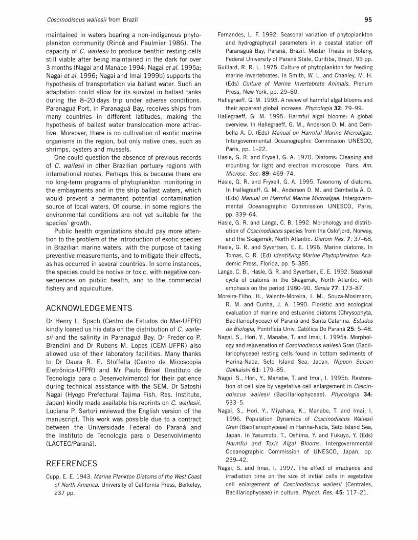
ACKNOWLEDGEMENTS
Dr Henry L. Spach (Centro de Estudos do Mar-UFPR)kindly loaned us his data on the distribution of C. waile-sii and the salinity in Paranaguá Bay. Dr Frederico P.Brandini and Dr Rubens M. Lopes (CEM-UFPR) alsoallowed use of their laboratory facilities. Many thanks to Dr Daura R. E. Stoffella (Centro de MicoscopiaEletrônica-UFPR) and Mr Paulo Brixel (Instituto de Tecnologia para o Desenvolvimento) for their patienceduring technical assistance with the SEM. Dr SatoshiNagai (Hyogo Prefectural Tajima Fish. Res. Institute,Japan) kindly made available his reprints on C. wailesii.Luciana P. Sartori reviewed the English version of themanuscript. This work was possible due to a contractbetween the Universidade Federal do Paraná and the Instituto de Tecnologia para o Desenvolvimento(LACTEC/Paraná).
REFERENCES
Cupp, E. E. 1943. Marine Plankton Diatoms of the West Coastof North America. University of California Press, Berkeley,237 pp.
Fernandes, L. F. 1992. Seasonal variation of phytoplanktonand hydrographycal parameters in a coastal station offParanaguá Bay, Paraná, Brazil. Master Thesis in Botany,Federal University of Paraná State, Curitiba, Brazil, 93 pp.
Guillard, R. R. L. 1975. Culture of phytoplankton for feedingmarine invertebrates. In Smith, W. L. and Chanley, M. H.(Eds) Culture of Marine Invertebrate Animals. PlenumPress, New York, pp. 29–60.
Hallegraeff, G. M. 1993. A review of harmful algal blooms andtheir apparent global increase. Phycologia 32: 79–99.
Hallegraeff, G. M. 1995. Harmful algal blooms: A globaloverview. In Hallegraeff, G. M., Anderson D. M. and Cem-bella A. D. (Eds) Manual on Harmful Marine Microalgae.Intergovernmental Oceanographic Commission UNESCO,Paris, pp. 1–22.
Hasle, G. R. and Fryxell, G. A. 1970. Diatoms: Cleaning andmounting for light and electron microscope. Trans. Am.Microsc. Soc. 89: 469–74.
Hasle, G. R. and Fryxell, G. A. 1995. Taxonomy of diatoms.In Hallegraeff, G. M., Anderson D. M. and Cembella A. D.(Eds) Manual on Harmful Marine Microalgae. Intergovern-mental Oceanographic Commission UNESCO, Paris,pp. 339–64.
Hasle, G. R. and Lange, C. B. 1992. Morphology and distrib-ution of Coscinodiscus species from the Oslofjord, Norway,and the Skagerrak, North Atlantic. Diatom Res. 7: 37–68.
Hasle, G. R. and Syvertsen, E. E. 1996. Marine diatoms. InTomas, C. R. (Ed) Identifying Marine Phytoplankton. Aca-demic Press, Florida, pp. 5–385.
Lange, C. B., Hasle, G. R. and Syvertsen, E. E. 1992. Seasonalcycle of diatoms in the Skagerrak, North Atlantic, withemphasis on the period 1980–90. Sarsia 77: 173–87.
Moreira-Filho, H., Valente-Moreira, I. M., Souza-Mosimann, R. M. and Cunha, J. A. 1990. Floristic and ecological evaluation of marine and estuarine diatoms (Chrysophyta,Bacillariophyceae) of Paraná and Santa Catarina. Estudosde Biologia, Pontifícia Univ. Católica Do Paraná 25: 5–48.
Nagai, S., Hori, Y., Manabe, T. and Imai, I. 1995a. Morphol-ogy and rejuvenation of Coscinodiscus wailesii Gran (Bacil-lariophyceae) resting cells found in bottom sediments ofHarina-Nada, Seto Island Sea, Japan. Nippon SuisanGakkaishi 61: 179–85.
Nagai, S., Hori, Y., Manabe, T. and Imai, I. 1995b. Restora-tion of cell size by vegetative cell enlargement in Coscin-odiscus wailesii (Bacillariophyceae). Phycologia 34:533–5.
Nagai, S., Hori, Y., Miyahara, K., Manabe, T. and Imai, I.1996. Population Dynamics of Coscinodiscus WailesiiGran (Bacillariophyceae) in Harina-Nada, Seto Island Sea,Japan. In Yasumoto, T., Oshima, Y. and Fukuyo, Y. (Eds)Harmful and Toxic Algal Blooms. IntergovernmentalOceanographic Commission of UNESCO, Japan, pp.239–42.
Nagai, S. and Imai, I. 1997. The effect of irradiance and irradiation time on the size of initial cells in vegetative cell enlargement of Coscinodiscus wailesii (Centrales, Bacillariophyceae) in culture. Phycol. Res. 45: 117–21.
95Coscinodiscus wailesii from Brazil

Nagai, S. and Imai, I. 1999a. The effect of salinity on the size of initial cells during vegetative cell enlargement ofCoscinodiscus wailesii (Bacillariophyceae) in culture.Diatom Res. 14: 337–42.
Nagai, S. and Imai, I. 1999b. Factors inducing resting-cellformation of Coscinodiscus wailesii Gran (Bacillario-phyceae) in culture. Plankton Biol. Ecol. 46: 94–103.
Nagai, S. and Manabe, T. 1994. Auxospore formation of agiant diatom Coscinodiscus wailesii (Bacillariophyceae), inculture. Bull. Plankton Soc. Jpn. 40: 151–67.
Nehring, S. 1998. Non-indigenous phytoplankton species inthe North Sea: Supposed region of origin and possibletransport vector. Arch. Fish. Mar. Res. 46: 181–94.
Rick, H. J. and Dürselen, C. D. 1995. Importance and abun-dance of the recently established species Coscinodiscuswailesii Gran et Angst in the German Bight. HelgolanderMeeresun. 49: 355–74.
Rincé, Y. and Paulmier, G. 1986. Données nouvelles sur ladistribution de la diatomée marine Coscinodiscus wailesiiGran & Angst (Bacillariophyceae). Phycologia 25: 73–9.
Ross, R., Cox, E. J., Karayeva, N. I. et al. 1979. An amendedterminology for the siliceous components of the diatomcell. Nova Hedwigia Beih. 64: 513–33.
Round, F. E., Crawford, R. M. and Mann, D. G. 1990. TheDiatoms. Biology and Morphology of the Genera. Cam-bridge University Press, Cambridge, pp. 747.
Roy, S., Harris, R. P. and Pulet, S. A. 1989. Inefficientfeeding by Calanus helgolandicus and Temora longicornison Coscinodiscus wailesii: Quantitative estimation usingchlorophyll-type pigment and effects on dissolved freeamino acids. Mar. Ecol. Prog. Ser. 52: 145–53.
Schmid, A. M. 1990. Intraclonal variation in the valve structure of Coscinodiscus wailesii Gran et Angst. NovaHedwigia Beih. 100: 101–19.
Schmid, A. M. and Volcani, B. E. 1983. Wall morphogenesisin Coscinodiscus wailesii Gran and Angst. J. Phycol. 19:387–402.
Smayda, T. J. 1997. Harmful algal blooms: Their ecophysiol-ogy and general relevance to phytoplankton blooms in thesea. Limnol. Oceanogr. 42: 1137–53.
Souza-Mosimann, R. M., Felício-Fernandes, G., Silva, R. L.and Fernandes, L. F. 1993. Diatoms from stomach con-tents of three shrimp species of artesanal marine fishery in Santa Catarina State, Brazil. Insula 22: 83–106.
Valente-Moreira, I. M. 1987. The marine and estuarine diatomflora in the balnearies of Canoas and Ipanema, ParanáState, Brazil. Estudos Biologia, Pontifícia Univ. Católica DoParaná 17: 26–48.
96 L. F. Fernandes et al.