Embed Size (px)
Citation preview
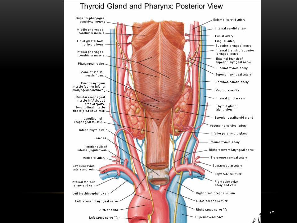
THYROID GLAND
1
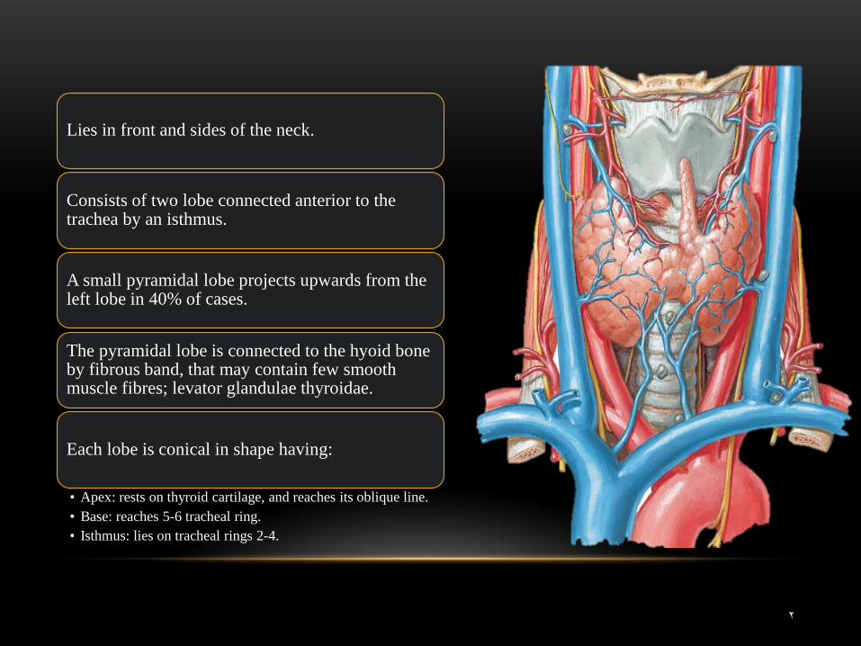
Lies in front and sides of the neck.
Consists of two lobe connected anterior to the trachea by an isthmus.
A small pyramidal lobe projects upwards from the left lobe in 40% of cases.
The pyramidal lobe is connected to the hyoid bone by fibrous band, that may contain few smooth muscle fibres; levator glandulae thyroidae.
Each lobe is conical in shape having:
• Apex: rests on thyroid cartilage, and reaches its oblique line. • Base: reaches 5-6 tracheal ring. • Isthmus: lies on tracheal rings 2-4.
2
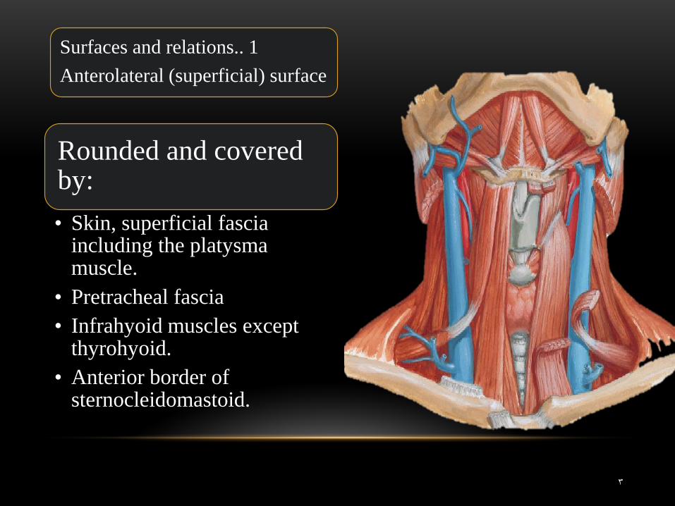
Rounded and covered by:
• Skin, superficial fascia including the platysma muscle.
• Pretracheal fascia • Infrahyoid muscles except
thyrohyoid. • Anterior border of
sternocleidomastoid.
Surfaces and relations.. 1
Anterolateral (superficial) surface
3
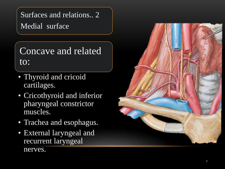
Concave and related to:
• Thyroid and cricoid cartilages.
• Cricothyroid and inferior pharyngeal constrictor muscles.
• Trachea and esophagus. • External laryngeal and
recurrent laryngeal nerves.
Surfaces and relations.. 2
Medial surface
4
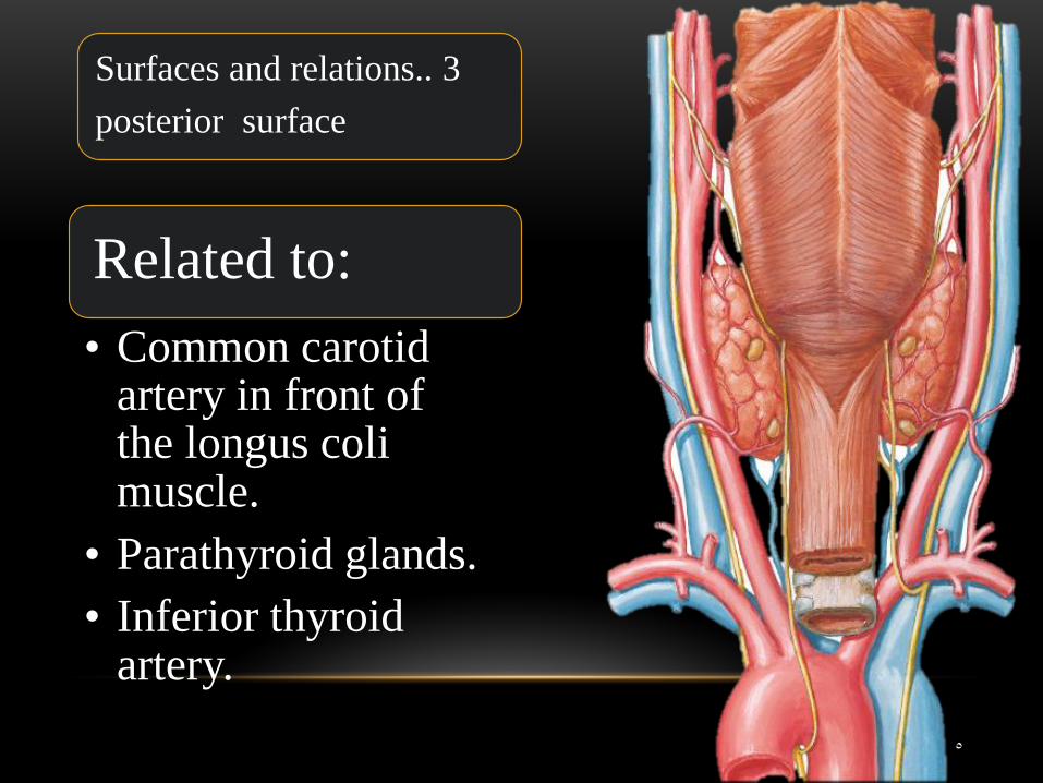
Related to:
• Common carotid artery in front of the longus coli muscle.
• Parathyroid glands. • Inferior thyroid
artery.
Surfaces and relations.. 3
posterior surface
5
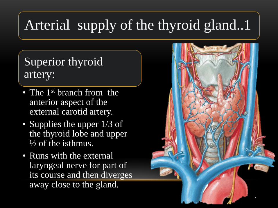
Superior thyroid artery:
• The 1st branch from the anterior aspect of the external carotid artery.
• Supplies the upper 1/3 of the thyroid lobe and upper ½ of the isthmus.
• Runs with the external laryngeal nerve for part of its course and then diverges away close to the gland.
Arterial supply of the thyroid gland..1
6
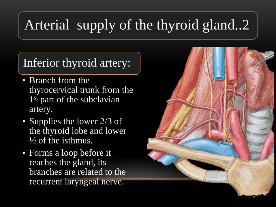
Inferior thyroid artery:
• Branch from the thyrocervical trunk from the 1st part of the subclavian artery.
• Supplies the lower 2/3 of the thyroid lobe and lower ½ of the isthmus.
• Forms a loop before it reaches the gland, its branches are related to the recurrent laryngeal nerve.
Arterial supply of the thyroid gland..2
7
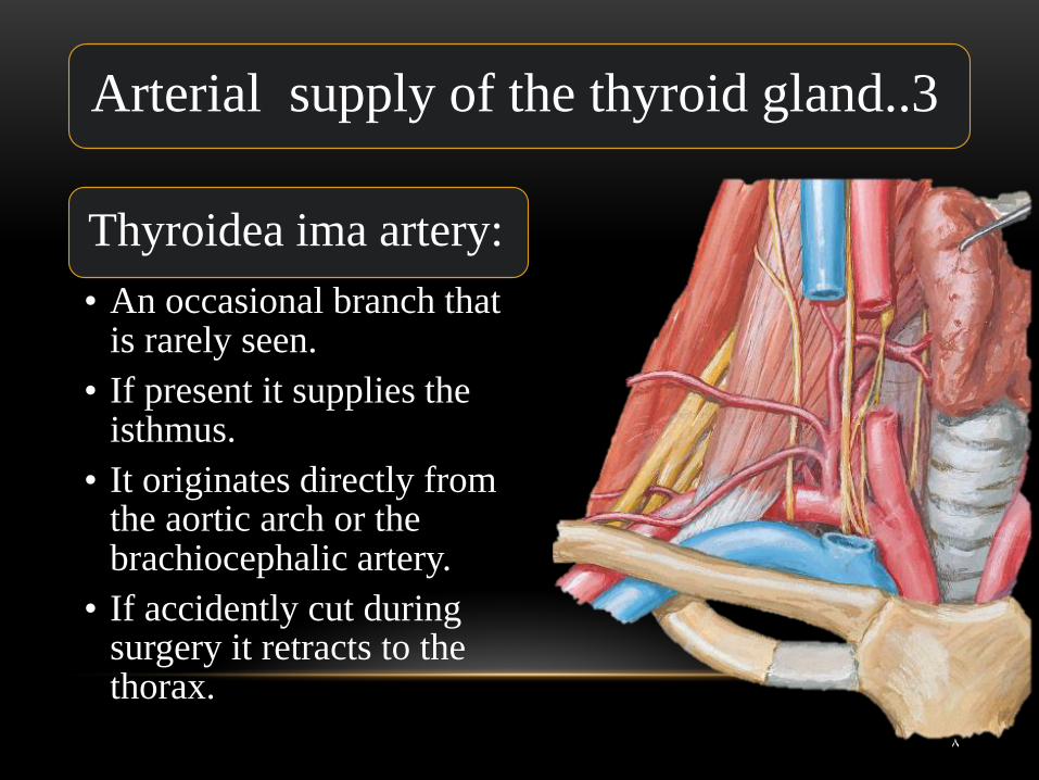
Thyroidea ima artery:
• An occasional branch that is rarely seen.
• If present it supplies the isthmus.
• It originates directly from the aortic arch or the brachiocephalic artery.
• If accidently cut during surgery it retracts to the thorax.
Arterial supply of the thyroid gland..3
8
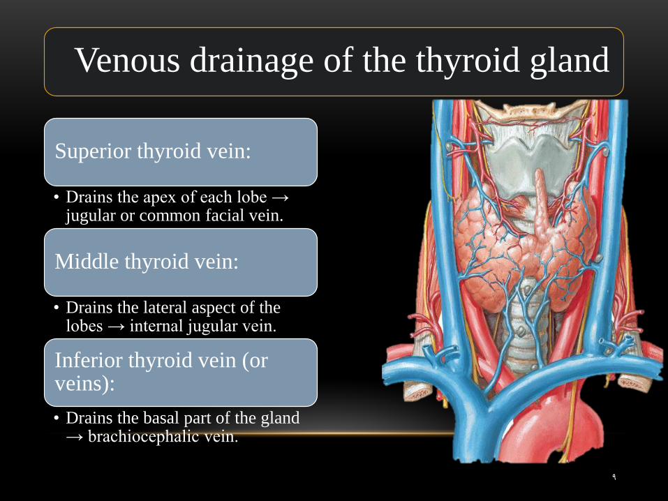
Superior thyroid vein:
• Drains the apex of each lobe → jugular or common facial vein.
Middle thyroid vein:
• Drains the lateral aspect of the lobes → internal jugular vein.
Inferior thyroid vein (or veins):
• Drains the basal part of the gland → brachiocephalic vein.
Venous drainage of the thyroid gland
9
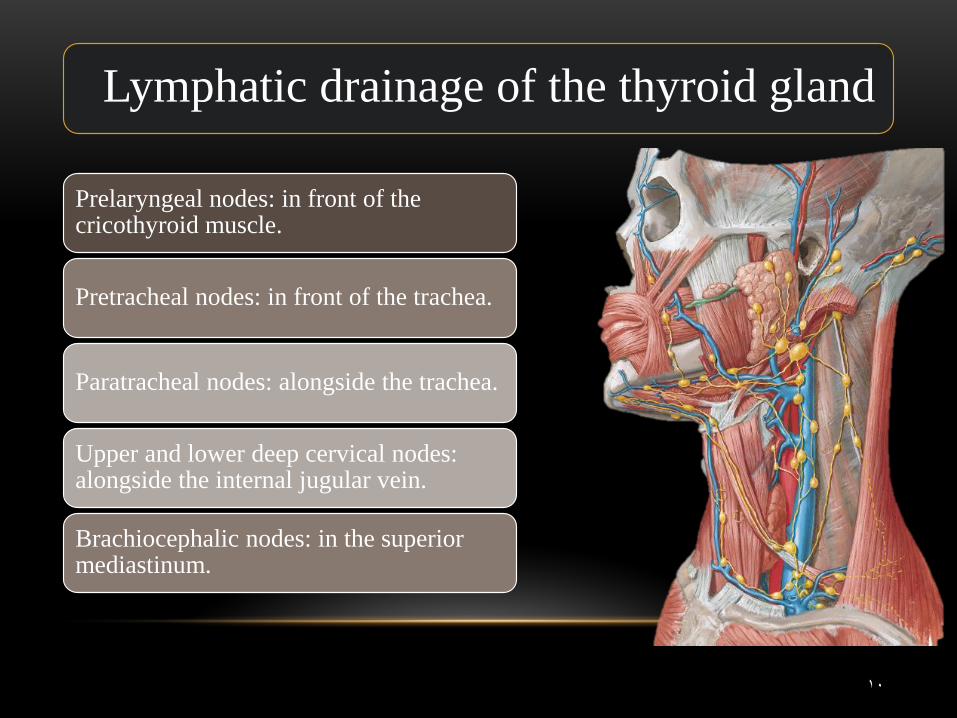
Prelaryngeal nodes: in front of the cricothyroid muscle.
Pretracheal nodes: in front of the trachea.
Paratracheal nodes: alongside the trachea.
Upper and lower deep cervical nodes: alongside the internal jugular vein.
Brachiocephalic nodes: in the superior mediastinum.
Lymphatic drainage of the thyroid gland
10
11
12
13
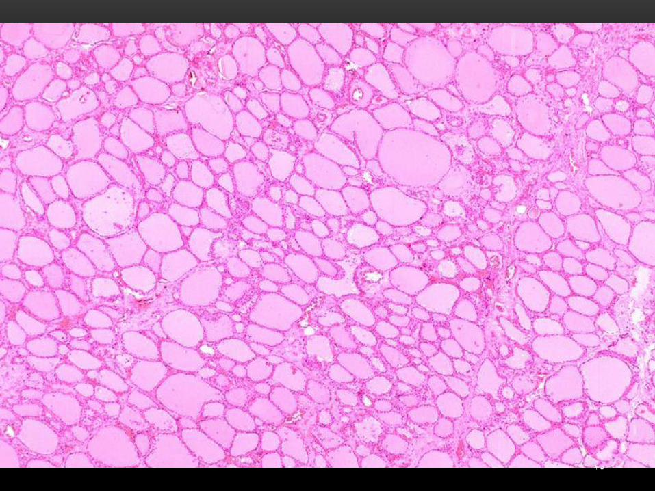
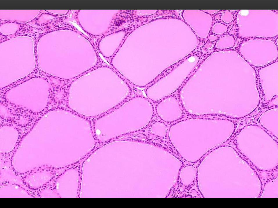
Thyroid follicle:
• The structural and functional unit of the thyroid gland.
• Consists of a group of cells resting on the same basal lamina surrounding a lumen filled with colloid.
• The follicles are variable in size. • Hormones are stored in the follicles. • Each follicle is surrounded by variable
amount of connective tissue.
14
15
16
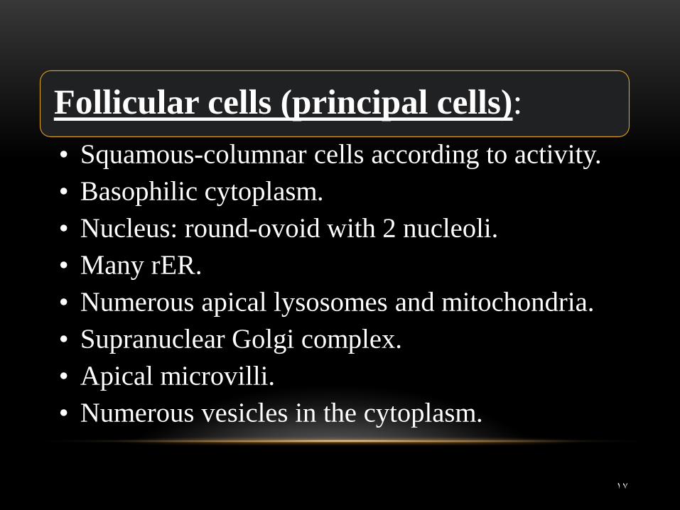
Follicular cells (principal cells):
• Squamous-columnar cells according to activity. • Basophilic cytoplasm. • Nucleus: round-ovoid with 2 nucleoli. • Many rER. • Numerous apical lysosomes and mitochondria. • Supranuclear Golgi complex. • Apical microvilli. • Numerous vesicles in the cytoplasm.
17
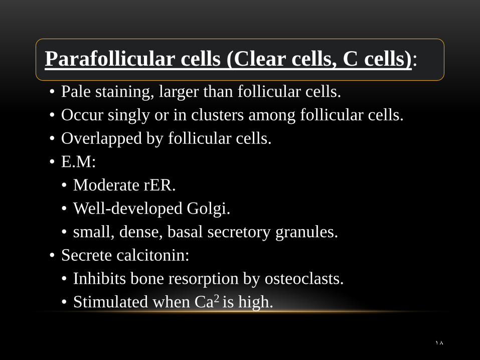
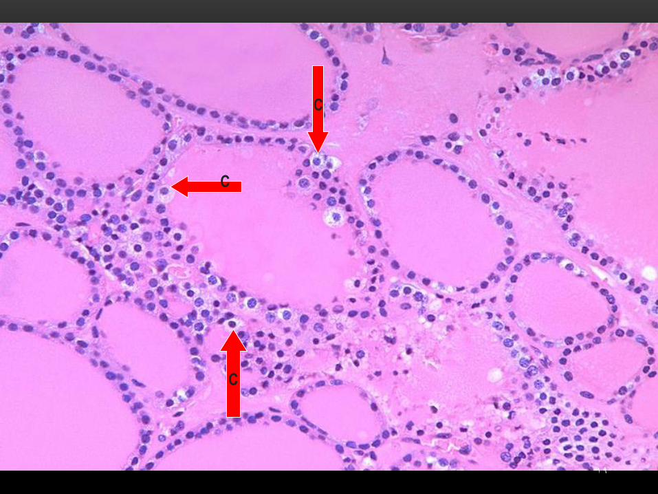
Parafollicular cells (Clear cells, C cells):
• Pale staining, larger than follicular cells. • Occur singly or in clusters among follicular cells. • Overlapped by follicular cells. • E.M:
• Moderate rER. • Well-developed Golgi. • small, dense, basal secretory granules.
• Secrete calcitonin: • Inhibits bone resorption by osteoclasts. • Stimulated when Ca2 is high.
18
C
C
C
19
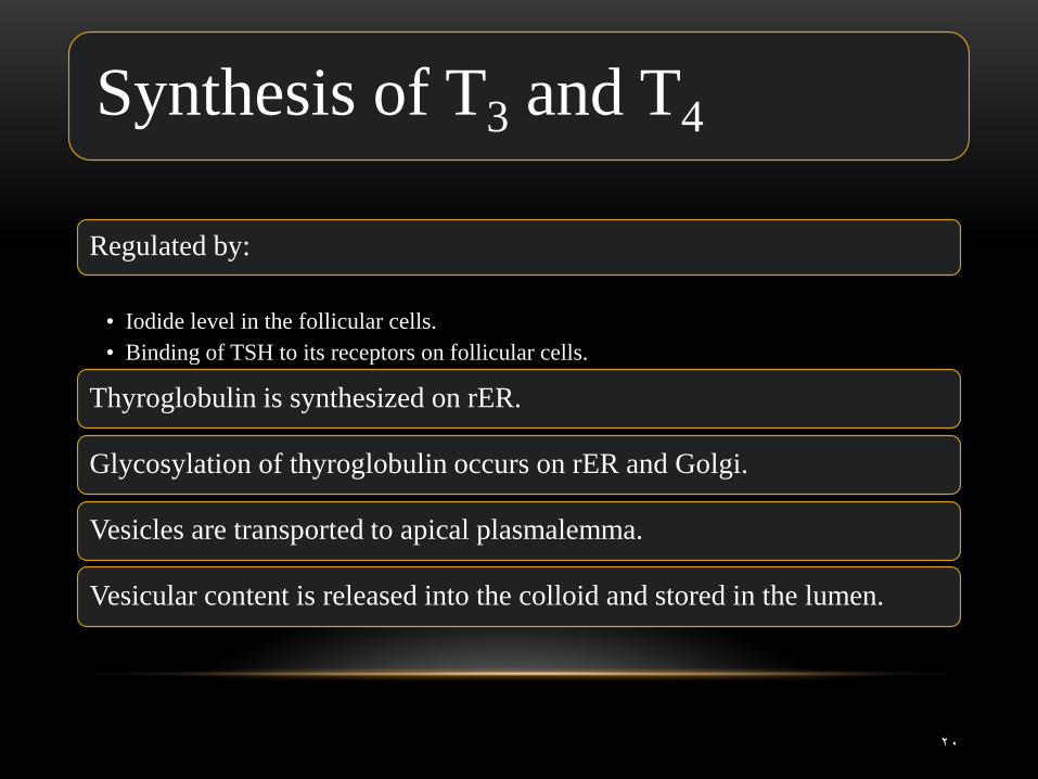
Synthesis of T3 and T4
Regulated by:
• Iodide level in the follicular cells. • Binding of TSH to its receptors on follicular cells.
Thyroglobulin is synthesized on rER.
Glycosylation of thyroglobulin occurs on rER and Golgi.
Vesicles are transported to apical plasmalemma.
Vesicular content is released into the colloid and stored in the lumen.
20
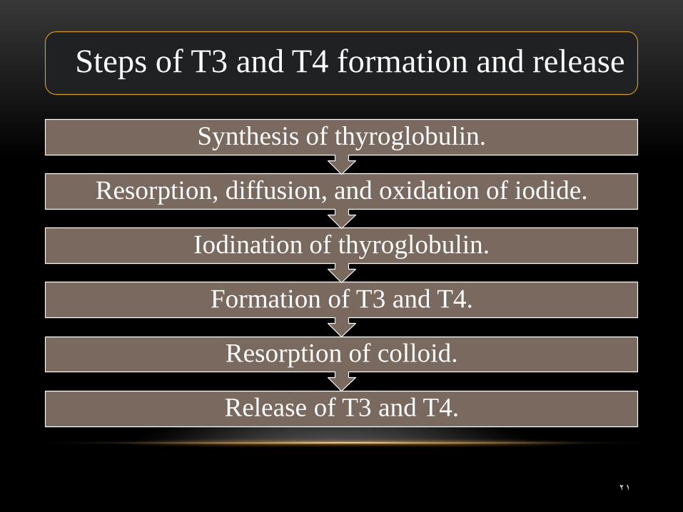
Steps of T3 and T4 formation and release
Release of T3 and T4.
Resorption of colloid.
Formation of T3 and T4.
Iodination of thyroglobulin.
Resorption, diffusion, and oxidation of iodide.
Synthesis of thyroglobulin.
21
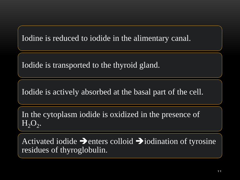
Iodine is reduced to iodide in the alimentary canal.
Iodide is transported to the thyroid gland.
Iodide is actively absorbed at the basal part of the cell.
In the cytoplasm iodide is oxidized in the presence of H2O2.
Activated iodide enters colloid iodination of tyrosine residues of thyroglobulin.
22
23
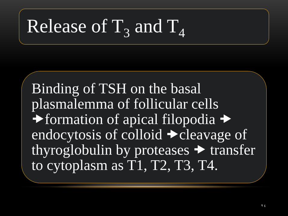
Release of T3 and T4
Binding of TSH on the basal plasmalemma of follicular cells formation of apical filopodia endocytosis of colloid cleavage of thyroglobulin by proteases transfer to cytoplasm as T1, T2, T3, T4.
24
Clinical Applications
25
26
27
28
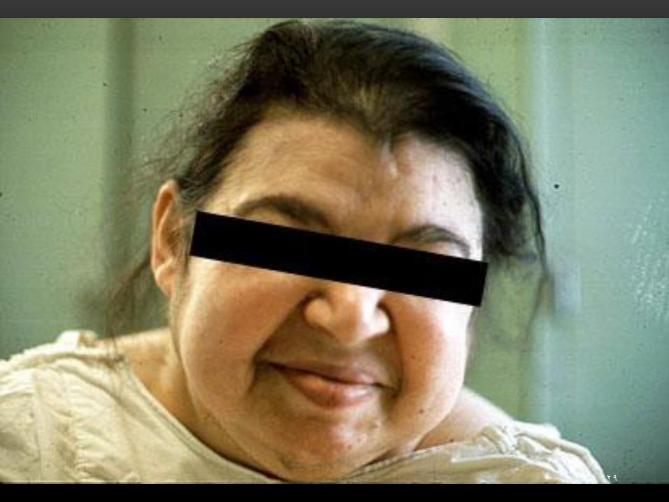
29
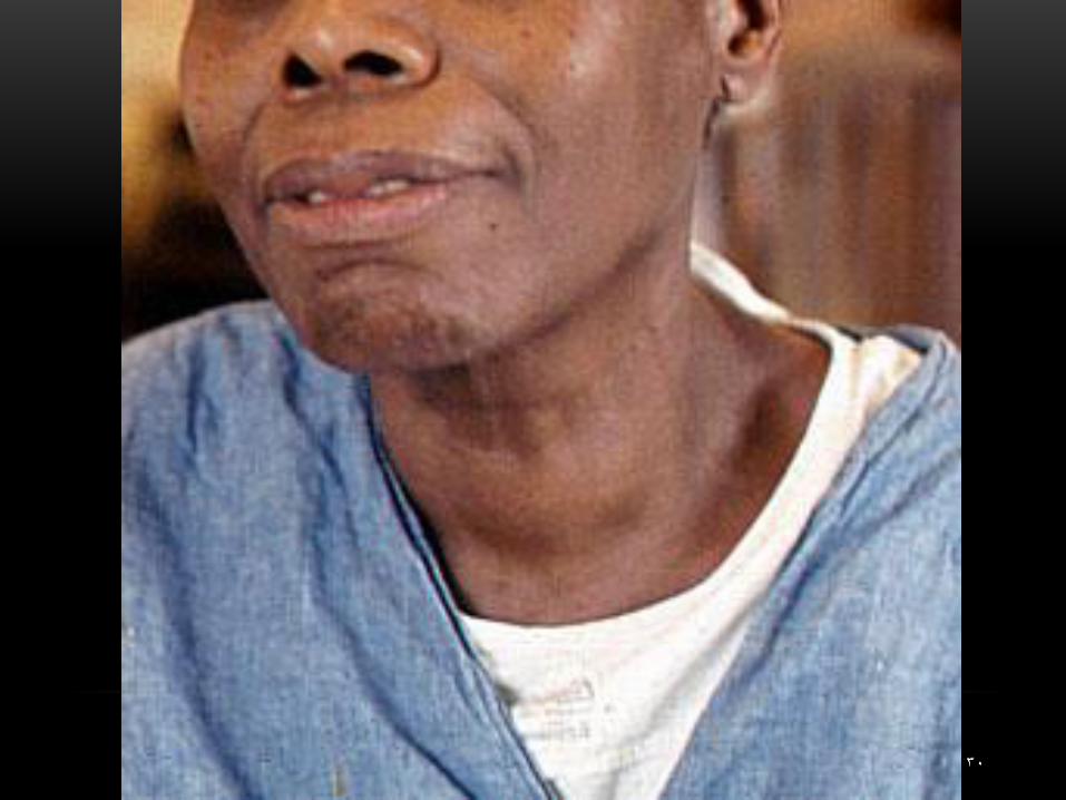
30
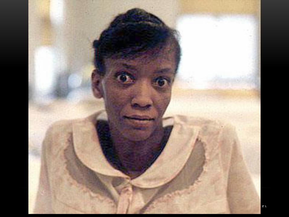
31