Embed Size (px)
DESCRIPTION
Toxoplasmosis in pregnancy. Introduction . A zoonoziz , caused by T.gondii , an intracellular protozoan parasite Its more common in tropical & coastal regions is less common in regions that are either cold ,warm or at high elevation 1 of every 900 pregnancies in the USA. Transmission. - PowerPoint PPT Presentation
Citation preview

TOXOPLASMOSIS IN PREGNANCY

Introduction A zoonoziz , caused by
T.gondii , an intracellular protozoan parasite
Its more common in tropical & coastal regions
is less common in regions that are either cold ,warm or at high elevation
1 of every 900 pregnancies in the USA

Transmission Cats
host for T. gondii They acquire infection by
eating infected wild rodents and birds
A week after infection, the cat begins to shed oocysts in its feces
Shedding of the oocysts persists for about 2 weeks
within days to weeks these oocysts sporulate and become extremely infectious

Transmission Food
ingestion of contaminated food is an important cause of toxoplasmosis
Meat is the most common infected food
unpasteurized milk and unfiltered water sources also are at risk
Organ transplantation


Pathophysiology Acute toxoplasmosis generally is well tolerated
in immunocompetent adults may result in vertical infection to the fetus and
lead to potentially serious consequences In immunocompetent adult, symptoms usually
are mild or inapparent In about 10%:
fever fatigue Malaise headache myalgias lymphadenopathy
These symptoms will resolve in weeks to months without specific therapy

Pathophysiology In immunosuppressed :
signs and symptoms often will be more pronounced can result in significant ocular and CNS abnormalities
Reactivation infection in immunosuppressed pregnant women also can cause fetal infection

T. gondii three forms
trophozoites or proliferative tissue cysts Oocytes
Trophozoite is seen in the acute phase of the infection in the human
Mulitiplies every 4-6 hours cells rupture
Releasing organisms to
invade other cells


Utero Transmission Newborns become infected in utero by
transplacental passage of the parasite when the mother has acute infection
Chronic infections (onset precedes pregnancy) do not lead to congenital infection except in the rare circumstance of an immunocompromised
host with reactivation likelihood of fetal infection increases with each
trimester of pregnancy Fetal infection is :
15% in the first trimester, 25% second, 60% in third trimester

Utero Transmission The severity of damage associated with
timing of maternal infection the risks decrease toward term
Severe fetal disease or fetal death: occurs in about 10% of cases when infection
occurs during the first trimester extremely rare with infection during the third
trimester Mild damage is more frequent in the
second and third trimesters (about 5%)

Utero Transmission Subclinical infections increase from about
2% with first-trimester infections to 50% with third-trimester infections
acute infection could be associated with preterm delivery and stillbirth but not with spontaneous abortion

TRANSPLACENTAL TRANSMISSION
Time of infection Likelihood of transmission in
untreated mothers
Disease in infant
>6 months before conception
No Risk ---
<6 months before conception
Very Low Risk
---
First trimester 10-25% Most severeSecond trimester 30-54% Less severeThird trimester 60-65% Usually
Asymptomatic

Diagnosis in Pregnancy1. Maternal Infection
usually is asymptomatic 10% to 20% of infected mothers have
lymphadenopathy (Posterior cervical is the most frequent )
The infection also can result in a mononucleosis like syndrome with:
fatigue assitude and rarely, can cause encephalitis The clinical picture can be much more severe in
immunocompromised adults.

1. Maternal Infection clinicians are forced to rely on serologic
tools for the diagnosis of toxoplasmosis in pregnancy
diagnosis of primary infection : demonstration of a seroconversion to this
organism significant rise in antibody titer obtained from
maternal sera taken at two different times detection of toxoplasma-specific IgM antibody

Maternal Infection Adults with primary infection develop IgG and
IgM antibody to toxoplasma rapidly
Toxoplasma-specific IgG antibody: develops within after infection peaks in 6 to 8 weeks2 weeks drops down over the subsequent several months then persists for life
Toxoplasma-specific IgM develops within 10 days after infection remains elevated for 6 months to more than 6 years

Maternal Infection IgM titers may not provide useful
information to document recent primary infection in pregnant women
The IFA test frequently is more useful than ELISA in differentiating remote from recent primary infection of a pregnant woman
In any case, the presence of IgG and the absence of IgM suggest an infection that is probably at least a year old

Maternal Infection Up to 40% of positive toxoplasma-specific IgM
are false positives
Avidity testing is a newer type of testing
Approximately 50% of placentas of congenitally infected infants will show T. gondii cysts on histologic slides
Additional cases can be detected by the presence of parasites in the cord blood

Maternal Infection The organism also has been isolated from
placental tissue of acutely infected mothers in 2% to 25% of cases
Isolation of organisms from tissue specimens, buffy coat heparinized blood body fluids

2. Prenatal Diagnosis Antenatal diagnosis of fetal toxoplasmosis
culture of amniotic fluid fetal blood
The main difficulties with culture techniques: some assays may take up to several weeks few laboratories are able to perform the assay
amniocentesis performed too early in gestation occasionally can be falsely negative

Prenatal Diagnosis Toxoplasma-specific IgM, when present
in fetal blood from cordocentesis, also has been used to diagnose fetal infection prenatally
fetal-specific IgM antibody frequently does not develop until after 21 to
24 weeks gestation is positive in only about 50% of infected
cases
Additionally, cordocentesis is a procedure that entails some risk.

Prenatal Diagnosis the PCR has been used to detect T. gondii in amniotic
fluid and has been shown to be useful in the detection of in utero infections
Prenatal ultrasound also may demonstrate abnormalities
Ventriculomegaly and hydrocephalus as well as microcephaly will be poor prognosticators
Intracranial calcifications, placentomegaly, hepatomegaly cataracts, and hydrops may be other signs

3. Neonatal Infection Most congenitally infected newborns are asymptomatic at
birth
Literature has shown that detection of toxoplasma-specific immunoglobulin A (IgA) may be a reliable method for the diagnosis of toxoplasmosis in the newborn
Demonstration of toxoplasma-specific IgM infection may be diagnostic, although in newborns approximately 20% of infections are not detectable by
toxoplasma-specific IgM at birth
A number of these asymptomatic, untreated infants will go on to have delayed and potentially serious manifestations
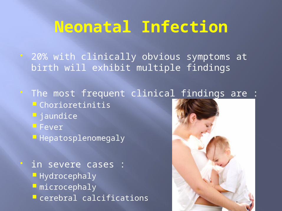
Neonatal Infection 20% with clinically obvious symptoms at birth will
exhibit multiple findings
The most frequent clinical findings are : Chorioretinitis jaundice Fever Hepatosplenomegaly
in severe cases : Hydrocephaly microcephaly cerebral calcifications


Treatment Treatment of acute toxoplasmosis in
immunocompetent, nonpregnant adults is primarily supportive
the prognosis following acute infection is good, except in cases of profound immunosuppression
The treatment in pregnancy is a bit more complex

Treatment In Europe spiramycin is the first-line agent
used agent generally does not cross the
placenta, and if fetal infection is detected, women also are treated with a combination of pyrimethamine folinic acid sulfonamide

Treatment Although not definitive, treatment with these regimens
may prevent maternal-to-fetal transmission of the infection or improve the outcome among infected fetuses
The standard dosage is : 25 mg of pyrimethamine by mouth given daily 1 g of sulfadiazine by mouth four times daily for 1 year Folinic acid, 6 mg given intramuscularly or by mouth every
other day
***Pyrimethamine is a folic acid antagonist and therefore may have teratogenic effects when given in the first
trimester

Prevention
avoid eating raw or undercooked meat Fruits and vegetables should be peeled and
washed before eating proper hand hygiene Gloves should be used for gardening and during
any contact with soil or sand Pregnant women should avoid close contact
with cat feces need for obstetricians to educate pregnant
patients about these important preventive steps

Prevention routine serologic screening programs screening of newborns and the institution
of treatment during the neonatal period to minimize the morbidity
Many infections in children that otherwise would be missed on routine clinical examination can be detected with IgM assays
Treatment of these infected infants has been associated with very low rates of subsequent neurologic or retinal disease


Many thanks . . .